Báo cáo khoa học: The chloroplast ClpP complex in Chlamydomonas reinhardtii contains an unusual high molecular mass subunit with a large apical domain doc
Bạn đang xem bản rút gọn của tài liệu. Xem và tải ngay bản đầy đủ của tài liệu tại đây (597.18 KB, 14 trang )
The chloroplast ClpP complex in Chlamydomonas
reinhardtii contains an unusual high molecular mass
subunit with a large apical domain
Wojciech Majeran
1
, Giulia Friso
2
, Klaas Jan van Wijk
2
and Olivier Vallon
1
1 Institut de Biologie Physico-Chimique, Paris, France
2 Department of Plant Biology Cornell University, Ithaca, New York, USA
Most intracellular proteolysis is carried out by
large self-compartmentalized ATP-dependent proteases,
combining a chaperone and a peptidase activity [1].
The chaperone activity is necessary to unfold protein
substrates, and feed them into a proteolytic chamber
where peptidolysis occurs. In the eukaryotic cell, mito-
chondria and chloroplasts harbor three major types
of ATP-dependent proteases, all inherited from their
eubacterial ancestors: FtsH, Lon, and Clp [2–4].
Clp proteases are composed of two components, a
chaperone of the Hsp100 type and a peptidase of the
ClpP family. In Escherichia coli and other bacteria, the
chaperone is either ClpA or ClpX, resulting in the for-
mation of ClpAP, ClpXP or mixed ClpAXP complexes
[3,5–7]. ClpP is a serine-type endopeptidase, with the
three catalytic residues, Ser, His and Asp appearing in
this order in the sequence [8]. The X-ray crystallo-
graphic structure of the E. coli ClpP complex [9] shows
two stacked rings of seven identical ClpP subunits,
delineating an internal proteolytic chamber where the
14 catalytic sites are exposed. By itself, this tetradeca-
meric complex is only capable of hydrolyzing short
peptides. Degradation of proteins requires association
with ClpA or ClpX (which also govern substrate spe-
cificity) and ATP hydrolysis [10,11]. The chaperones
can function on their own to remodel or refold dena-
tured proteins, but their association with ClpP pro-
motes a different mechanism, whereby the extended
polypeptide chain of the substrate is fed into the pro-
teolytic ClpP chamber via a narrow axial opening. In
E. coli, the ClpAP and ClpXP protease complexes
function in the degradation of denatured proteins,
especially under heat stress, but also of specific sub-
strates recognized via N- or C-terminal sequence
Keywords
mass spectroscopy; native gel
electrophoresis; protein complex;
proteolysis; Volvocale
Correspondence
O. Vallon, UMR 7141, Institut de Biologie
Physico-Chimique, 13 rue Pierre et Marie
Curie, 75005 Paris, France
Fax: +33 15841 5022
Tel: +33 15841 5058
E-mail:
(Received 15 July 2005, revised 23 August
2005, accepted 31 August 2005)
doi:10.1111/j.1742-4658.2005.04951.x
The composition of the chloroplast-localized protease complex, ClpP, from
the green alga Chlamydomonas reinhardtii was characterized by nondena-
turing electrophoresis, immunoblotting and MS. The detected ClpP
complex has a native mass of 540 kDa, which is 200 kDa higher than
ClpP complexes in higher plant chloroplasts, mitochondria or bacteria. The
540-kDa ClpP complex contains two nuclear-encoded ClpP proteins (ClpP3
and P5) and five ClpR (R1, R2, R3, R4 and R6) proteins, as well two pro-
teins, ClpP1
L
and ClpP1
H
, both probably derived from the plastid clpP1
gene. ClpP1
H
is 59 kDa and contains a 30-kDa insertion sequence (IS1)
not found in other ClpP proteins, responsible for the high MW of the com-
plex. Based on comparison with other sequences, IS1 protrudes as an addi-
tional domain on the apical surface of the ClpP ⁄ R complex, probably
preventing interaction with the HSP100 chaperone. ClpP1
L
is a 25-kDa
protein similar in size to other ClpP proteins and could arise by post-trans-
lational processing of ClpP1
H
. Chloramphenicol-chase experiments show
that ClpP1
L
and ClpP1
H
have a similar half-life, indicating that both are
stable components of the complex. The structure of the ClpP complex is
further discussed in conjunction with a phylogenetic analysis of the ClpP ⁄ R
genes. A model is proposed for the evolution of the algal and plant
complex from its cyanobac terial ancestor.
5558 FEBS Journal 272 (2005) 5558–5571 ª 2005 FEBS
motifs. The diversity of these motifs was revealed when
a tagged and inactive variant of E. coli ClpP was used
to trap substrates of ClpXP in vivo [12]. In addition,
ClpXP participates in the degradation of nascent pro-
teins stalled on ribosomes, after they are C-terminally
tagged and released by the ssrA trans-termination sys-
tem. Substrate binding is influenced by helper and
modulator proteins, such as SspB [13] or ClpS [14].
Much less is known about the Clp proteases of
eukaryotes. The mitochondrial ClpP complex appears
highly similar to the bacterial enzyme [15]. It interacts
with a ClpX chaperone to form an ATP-dependent
protease [15–17]. Plant mitochondrial ClpP2 has been
shown to form a homo-oligomeric complex of
320 kDa [18]. In plastids, the most likely partners of
ClpP are the ClpC chaperone [19] and ClpD (Erd1)
[20]. The ClpP complex in plastids of Arabidopsis
thaliana and other Brassicaceae has been examined in
detail. It is a hetero-oligomer, slightly larger than its
mitochondrial counterpart (350 kDa), associating
nucleus- and plastid-encoded subunits. In all plastid
types examined, it associates five different ClpP pro-
teins (ClpP1, ClpP3, ClpP4, ClpP5 and ClpP6, of which
ClpP1 is chloroplast-encoded), and four nonproteolytic
ClpR proteins (ClpR1, ClpR2, ClpR3 and ClpR4)
[18,21]. ClpR proteins are homologous to ClpP, but
lack one or several of the catalytic site residues, and are
therefore supposed to play a structural, rather than a
catalytic, role in the complex. In addition, the plastid
ClpP ⁄ R complex contains two additional subunits
unique to land plants, ClpS1 and ClpS2. The functions
of ClpS1,2 (which are not homologous to the E. coli
ClpS) are unknown, but molecular modeling based on
their homology to the N-terminal domain of ClpA sug-
gests that they dock onto the apical surface of the (pre-
sumably tetradecameric) ClpP ⁄ R complex to regulate
association with the chaperone [18].
All of the plant Clp proteins are encoded in the
nucleus, except for ClpP1 which is plastid-encoded in
green algae and vascular plants, i.e. the green lineage
of plants. ClpP genes are also found in Cyanobacteria
[22] and in the genome of the Cyanophora cyanelle, an
ancestral chloroplast. In the green alga Chlamydomonas
reinhardtii and in vascular plants, the plastid clpP1
gene is essential and cannot be disrupted [23,24].
Reducing its expression level by mutating its initiation
codon leads to a reduction in degradation rate for
several thylakoid membrane proteins under stress con-
ditions or in the presence of destabilizing mutations
[25,26]. Other than that, little is known about the sub-
strates and functions of the plastid Clp protease.
ClpP1 proteins in the Chlamydomonas genus are
unusual in that they contain insertion sequences, not
found in other ClpP proteins, which have been pro-
posed to behave as protein introns [23]. One of them,
IS2, is found only in the species C. eugametos, and
possesses characteristics of the well-known self-splicing
protein elements called inteins [27–29]. Inteins are
autonomously folding protein domains capable of self-
excision, and are present in many essential proteins
throughout the three kingdoms of life. In contrast, the
other insertion sequence, IS1, which is found in both
C. eugametos and C. reinhardtii, lacks most of the
typical features of an intein, in particular the
LAGLI ⁄ DADG motif and C-terminal HN dipeptide
[23]. In a previous study, we have shown that antibodies
raised to the entire C. reinhardtii ClpP1 reading frame
recognize two proteins of 25 kDa (referred to here as
ClpP1
L
) and 59 kDa (ClpP1
H
). As splicing of the clpP1
mRNA has been ruled out [23]; unpublished data), this
supports the protein splicing hypothesis. Presumably,
ClpP1
H
is the primary translation product of clpP1,
while ClpP1
L
is derived from ClpP1
H
by splicing or
some other form of post-translational processing. Here,
we analyze the ClpP ⁄ R complex of C. reinhardtii by
gel electrophoresis followed by MS and show that it
contains both ClpP1
L
and ClpP1
H
.
Results
Identification of nuclear ClpP ⁄ R genes
The C. reinhardtii EST databases contained sequences
from eight nuclear-encoded ClpP ⁄ R homologs
(Table 1). By combining EST data and partial sequen-
cing of selected cDNAs, complete cDNA sequences
were obtained for all of them except for CLPR3 and
CLPP2. All the corresponding CLPP ⁄ R genes were
found in the C. reinhardtii draft genomic sequence,
version 2.0, some still containing sequence gaps. Mod-
els for four of the genes were corrected based on EST
data, in-house cDNA sequencing, comparison with
version 1.0 of the genome, or alignment with plant or-
thologs (see Table 1; the proposed changes have been
deposited as model notes in the JGI gene models). The
C. reinhardtii ClpP ⁄ R proteins and genes were named
on the basis of their closest Arabidopsis homologs [2],
as judged from the phylogenetic tree (Fig. 1) deduced
from a clustalw alignment of ClpP proteases
(Fig. S1). Only the central domain for each gene was
used for tree building, avoiding N-terminal and C-ter-
minal extensions where alignment was not meaningful.
Only three of the C. reinhardtii proteins (ClpP2,
ClpP4 and ClpP5) are predicted to contain the three
conserved catalytic site residues, and thus to be enzy-
matically active. The other five were missing either
W. Majeran et al. Chlamydomonas ClpP complex analysed by MS
FEBS Journal 272 (2005) 5558–5571 ª 2005 FEBS 5559
Table 1. ClpP ⁄ R genes in Chlamydomonas reinhardtii. For each gene are indicated the name, best contig in EST assembly (Chlre2), gene model(s) in version 2.0 of the Chlamydomonas
genome, predicted molecular mass and length of precursor and mature protein, N-terminal sequence, length of C-terminal extension and peptides found in the ESI ⁄ MS ⁄ MS analysis.
Protein
Best contig in
EST assembly
a
Additional
sequencing
Gene
model
Precursor
(kDa);
length (AA)
Mature:
(kDa);
length (AA)
Precursor
N-terminal
sequence
Length of
C-terminal
extension
b
MS sequence tags Notes on protein
ClpP1 – – CLPP_CHLRE
(chloroplast)
59.3 [523] 26.7 [237]? MPIGVPRII 41 ()2) LDVAEIYSLSTYRASLVGDSTQTQESNS Probable post-translational
processing or splicing
ClpP2 16.56.2.51 – C_160152
c
25,0 [231] 21,3 [194] MQRLSALAA 7 (18) None
ClpP4 37.11.2.11 AV387965 C_370132
d
38.4 [345] 32.8 [293] MVAAALLGG 105 (38–49) TAEFQGDPMGLLLRYGLIDHIIGGEEA
VFNVKRNSMPNSR
Long C-terminal extension
ClpP5 162.16.2.11 AV387186 C_1620010 27.8 [256] 22.0 [201]
e
MAQLLLQNK 11 (9) FQGVVSQLFQQRGPPPPNLPVIER
ClpR1 24.46.1.0 AV619744 C_240023
f
46.2 [411] 38.9 [345]? MLLNRPKLG 31 (31) FGNPPDLPSLLLQQRAPLYTGVTWK
AVDAQLQANELDYATKPLFLPEAER
Long N-terminal extension
ClpR2 56.23.1.51 – C_560017 31.6 [282] 25.0 [220] MQALNQRPS 22 (15) None
ClpR3 105.11.2.11 – (C_1050047,
C_17380001,
C_24440001)
g
46.5 [415] 43.4 [386] MRVHHAMTG 178 (23) SLPHSLAMIQQPRDGVKLAILNAE Long C-terminal extension
ClpR4 12.4.2.11 – C_120194 32.7 [293] 28.9 [257] MAALGCLSR. . . 47 (15) LGGMQASDIDIYRFGNEHEAIAVYSMM
KEAGPPPDLATR
TEEQIMTDFTRPRHEAIAVYSMMK
ClpR6 19.134.4.11 AV622513 C_190082
h
31.2 [283] 25.6 [231] MATLLQHGR 32 (0) EVGLVDDLTPGPFLKIIYINDKLEGLIDEIIR
LMMTQPMGGSQGDIYQIK
a
In bold if believed to be correct.
b
Compared to E. coli ClpP; value for the Arabidopsis orthologue(s) is indicated in parentheses.
c
An additional 5¢ exon and intron must be introduced to
generate the full-length protein; the unique, truncated EST from this gene is from an incompletely matured mRNA.
d
Sequence gap; exon 5 wrong.
e
Based on MS ⁄ MS data; 21 g and C9
nucleotide sequences differ but produce the same protein.
f
Sequence gap; exon 4 wrong.
g
Sequence gaps and misassembly; corrected based on cDNA data and genome version
1.0 : 5¢ must be extended, and two consecutive frameshifts corrected, restoring conserved residues in helix 5; 10th and 11th exons wrong.
h
The splice sites of the eighth intron must be
shifted seven nucleotides downstream, restoring two conserved residues.
Chlamydomonas ClpP complex analysed by MS W. Majeran et al.
5560 FEBS Journal 272 (2005) 5558–5571 ª 2005 FEBS
P2 At
0.1
P Ec
P Gv
R2 Ot
R2 Cr
R2 At
P1 Cr
P1 Cv
P1 Ot
P1 Nt
P1 At
P1 Tp
P1 Cm
P Gt NM
P3 Te
P3 N7
slr0165
P3 Se
R2 Cm
R2 Tp
P4 Cm
P4B Tp
P4A Tp
P5 Ot
P5 At
P5 Cr
P3 At
P4 At
P4 Cr
P4 Ot
P6 At
R6 Cr
R6 Ot
P Pf
P Cp
sll0534
P2 N7
P2 Te
P2 Se
slr0542
P1 N7
P1 Se
R Cp
R Gv
R Pf
R4 At
R4 Ot
R4 Cr
R1 At
R1 Ot
R1 Cr
R1 Cm
R Gt NM
R1 Tp
R3 Cr
R3 Ot
R3 At
R N7
R Te
slr0164
R Se
P Hs
P2 Tp
P2 Cr
P2 Cm
P2 Ot
mitochondria
Cyanobacteria
ClpR
"plastid" group
green lineage
"red" lineage
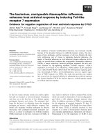
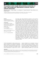
Fig. 1. Phylogenetic tree, obtained by the Neighbor-Joining method (with Kimura’s distance correction) of ClpP ⁄ R proteins from Chlamydomon-
as (boxed in red) and other photosynthetic eukaryotes and prokaryotes. Blue color indicates Cyanobacteria: Gleobacter violaceus (Gv), Nostoc
sp. PCC 7120 (N7), Synechocystis sp. PCC 6803 (S6), Synecococcus elongatus (Se) and Thermosynechococcus elongatus (Te). Greenish blue
indicates Cyanophora paradoxa (Cp, a Glaucocystophyte) and red Cyanidioschyzon merolae (Cm, a Rhodophyte). The brown color indicates
Guillardia theta (Gt, a Cryptophyte) and Thalassiosira pseudonana (Tp, a Diatom). The Viridiplantae (green, of a lighter shade for chloroplast
genes) are Arabidopsis thaliana (At), Chlamydomonas reinhardtii (Cr), Chlorella vulgaris (Cv), Nicotiana tabaccum (Nt), Ostreococcus tauri (Ot).
Non photosynthetic organisms (gray) are E. coli (Ec), Plasmodium falciparum (Pf) and Homo sapiens (Hs). Genes that have undergone two
migration events due to secondary endosymbiosis are shown with shading. The branches leading to ClpR proteins, where loss of catalytic activ-
ity is presumed to have occurred, are marked by a cross. The alignment used (Fig. S1) was excerpted from a large-scale alignment of ClpP from
plants and selected bacteria (available at EMBL as ALIGN_000912), after truncation to the ClpP domain. E. coli ClpP was used as the outgroup.
W. Majeran et al. Chlamydomonas ClpP complex analysed by MS
FEBS Journal 272 (2005) 5558–5571 ª 2005 FEBS 5561
one, two, or three of the catalytic residues S, H and D
and were named ClpR1, ClpR2, ClpR3, ClpR4 and
ClpR6, respectively. The latter is closest to the
A. thaliana ClpP6 protein, but the catalytic H is
missing in Chlamydomonas ClpR6, as well as in ortho-
logs found in two other green algae, Volvox carteri
and Ostreococcus tauri. Compared to E. coli ClpP, sev-
eral of the C. reinhardtii proteins showed long C-ter-
minal extensions, usually longer than the A. thaliana
homologs (Table 1): C-terminal extensions have been
proposed to fold back onto the apical surface of the
complex, potentially blocking the chaperone binding
site [15,18]. For all of the nuclear-encoded ClpP ⁄ R
proteins, the presence of an N-terminal extension when
compared to bacterial homologs suggests that they are
targeted to an organelle. The targetp and predotar
programs which are used to predict intracellular sort-
ing in vascular plants confirmed targeting to plastids
or mitochondria; however, it is difficult to predict
which, as the programs do not discriminate well
between Chlamydomonas plastid and mitochondrial
targeting sequences. In addition, the Chlamydomonas
ClpR1 contains a particularly long N-terminal exten-
sion (168 residues before the part conserved with other
ClpP), predicted to be retained in the mature protein.
A similar extension is also found in Arabidopsis but
no significant similarity can be identified between the
Chlamydomonas and Arabidopsis ClpR1 proteins in
this region.
Importantly, none of the nuclear-encoded ClpP ⁄ R
proteins of Chlamydomonas showed extended similarity
to the chloroplast-encoded ClpP1. No stretch of simi-
larity longer than five residues was observed, making it
unlikely that the ClpP1
L
band recognized by the anti-
bodies corresponds to the product of one of these
nuclear genes.
Experimental identification of the
Chlamydomonas ClpP ⁄ R complex
As a first step towards the characterization of the
C. reinhardtii ClpP ⁄ R complex, we analyzed the subcel-
lular localization of clpP1 gene products. Purified chlo-
roplasts from the cell wall-less mutant CW15 were
fractionated into a stroma and a crude membrane frac-
tion (thylakoids plus envelope). As expected, Western
blotting identified ClpP1
L
mostly in the stromal frac-
tion, with a small proportion (5–10%) associated with
the membranes (Fig. 2). A similar proportion was
found in membranes purified by floatation on a sucrose
gradient (not shown). When chloroplast membranes
were further treated with the Yeda press (Fig. 2, lane
4), the bound ClpP1 was efficiently released, indicating
a loose association similar to that observed for higher
plant ClpP [18,21]. A similar behavior was found for
ClpP1
H
and ClpC (data not shown), as well as for the
chloroplast GrpE homolog CGE1 and for RbcL, both
of which can be taken as examples of stromal proteins.
Total soluble proteins, prepared by French press
lysis of wild-type cells were separated by nondenatur-
ing electrophoresis (colorless-native; CN ⁄ PAGE) on a
4–13% gradient gel and blotted onto nitrocellulose for
immunodetection of ClpP1. In these experiments, a
high molecular mass ClpP complex could be identified,
migrating just below the 550-kDa Rubisco complex
(Fig. 3). Based on comparison with molecular mass
standards and the chloroplast proteins CF
1
(390 kDa)
and Rubisco (550 kDa), the apparent molecular mass
of the ClpP complex is 540 kDa. This is markedly
higher than the 350-kDa complex identified in higher
plants. No ClpP1 was detected in the low molecular
mass region of the gel. After electrophoresis in the sec-
ond dimension, many proteins could be resolved but,
due the complexity of the protein mixture, no partic-
ular silver-stained spot could be identified as candidate
components of the ClpP1-immunoreactive complex.
When we used, instead of a whole cell lysate, chloro-
plast stromal proteins or proteins released from the
membranes by Yeda press treatment, we encountered
the same caveat. The only MS ⁄ MS peptides that could
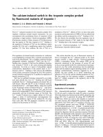
Fig. 2. Chloroplast localization of ClpP1. Immunoblots were reacted
with antibodies to ClpP1, CGE1 (chloroplast GrpE homolog), cyto-
chrome f,theb subunit of the ATP-synthase CF
1
, and RbcL, the
large subunit of Rubisco. 1: Chloroplasts isolated from strain
CW15; 2: soluble fraction and 3: membrane fraction after mechan-
ical lysis of chloroplasts; 4: soluble fraction after Yeda press treat-
ment of fraction 3 (overloaded approximately 10-fold).
Chlamydomonas ClpP complex analysed by MS W. Majeran et al.
5562 FEBS Journal 272 (2005) 5558–5571 ª 2005 FEBS
be identified in these experiments were from degrada-
tion products of the very abundant Rubisco large sub-
unit migrating approximately at the same position in
the first dimension.
We therefore resorted to using a Rubisco-less
mutant as our starting material for chloroplast prepar-
ation. Figure 4 shows 2D electrophoresis of a soluble
fraction prepared by Yeda press treatment of chloro-
plasts purified from a double mutant rbcL-18-5b cw15.
In the first dimension (CN ⁄ PAGE, Fig. 4A, B), the
ClpP1 antibody again recognizes the high molecular
mass complex of 540 kDa described above. Its sub-
units are separated according to their size in the
second, denaturing, dimension. As can be seen by com-
parison of Fig. 4B and C, both the ClpP1
H
and
ClpP1
L
forms of ClpP1 are present in the complex,
indicating that both are bona fide constituents of the
ClpP complex. In addition to these major spots, two
weaker immunoreactive spots (labeled *) can be seen
on Fig. 4C. These spots have been detected occasion-
ally with antibodies raised against different prepara-
tions of ClpP1, both in 2D gels and, less frequently, in
1D denaturing gels. We suspect that they represent
degradation products of ClpP1
H
.
In a Coomassie blue stained gel, several protein spots
(Fig. 4D, labeled 1–7) appeared to comigrate with the
ClpP complex detected by immunoblot. The lower
spots migrate approximately in the position of ClpP1
L
in this type of gel, just below the LHCII protein P17.
A
B
C
D
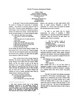
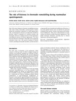
Fig. 4. Identification of the subunits of the ClpP ⁄ R complex. Chloro-
plasts from the cw15 rbcL18–5b strain were ruptured by Yeda
press treatment, and soluble proteins separated by 2D-electrophor-
esis. (A) Coomassie blue staining, and (B) immunoblot with ClpP1
antibody, of the first dimension (CN ⁄ PAGE, 4–18% acrylamide gra-
dient). (C) Immunoblot with ClpP1 antibody of the second dimen-
sion (SDS ⁄ urea PAGE, 12–18% acrylamide gradient); on the right is
an immunoblot of the same fraction separated by SDS ⁄ urea PAGE,
with arrowheads showing the positions of ClpP1
H
and ClpP1
L
(filled
and open, respectively; * indicates putative degradation products of
ClpP1
H
). (D) Coomassie blue staining of the 2D gel used to cut out
the spots for MS ⁄ MS. They are labeled 1–7 and an enlargement of
the ClpP region after bands were cut out is shown on the right.
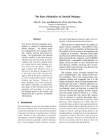
Fig. 3. Identification of the ClpP ⁄ R complex. For clarity, native gels
are shown horizontally, migration left to right, and denaturing gels
(SDS or SDS-urea) are shown vertically, migration top to bottom.
Top panel: nondenaturing 4–13% gradient CN ⁄ PAGE (Coomassie
blue staining), showing molecular mass markers (bovine thyroglo-
bulin, 670 kDa; E. coli b-galactosidase, 465 kDa; bovine catalase,
220 kDa; BSA, 120 and 60 kDa; apyrase, 50 kDa) and soluble pro-
teins from wild-type cells ruptured in a French press. Middle panel:
immunoblot of the CN ⁄ PAGE with antibodies to b-CF
1
and ClpP1.
Lower panel: second dimension SDS ⁄ PAGE (7.5–15% acrylamide
gradient, silver staining; the size of the molecular mass markers is
indicated on the right, the position of Rubisco is shown by an
arrowhead).
W. Majeran et al. Chlamydomonas ClpP complex analysed by MS
FEBS Journal 272 (2005) 5558–5571 ª 2005 FEBS 5563
The seven spots were excised from the gel and subjected
to in-gel trypsin digestion. The resulting peptides were
eluted and analyzed by MALDI-TOF and ESI-Q-TOF
MS ⁄ MS. All of these spots were found to contain
ClpP ⁄ R proteins (Table 1), as well as unrelated pro-
teins. Each ClpP ⁄ R protein was identified by two to five
peptides The only ambiguous peptide was one of those
attributed to ClpR3 (DGVKLAILNAE ): although
the sequence determined matches ClpR3, the remaining
C-terminal peptide mass, 195.14 Da, was not compat-
ible with that predicted from the cDNA or genomic
data (.CYER). It may represent a post-translationally
processed form of the protein. Spots 1 and 2 contained
peptides derived from ClpR3 and ClpR4, respectively.
Spots 3–7 contained peptides from ClpR1, ClpR6,
ClpP4 and ClpP5, respectively. For ClpP5, one of the
sequence tags started after a Q55, not after an R or K.
It probably corresponds to the original N-terminal end
of the mature protein. Importantly, two peptides
derived from ClpP1 were found in spots 4 and 6, whose
position corresponds approximately to that of the im-
munoreactive band ClpP1
L
in denaturing gel. Both of
them originated from the last 90 C-terminal residues of
the protein, one of them from the very C terminus.
In the MS analysis, no peptides were found corres-
ponding to ClpP2. This was expected, based on the
well-established mitochondrial localization of the
ClpP2 homolog in higher plants. More surprising was
the absence of detectable ClpR2, since its Arabidopsis
homolog has been found consistently in the chloroplast
ClpP complex [18,21]. Using antibodies raised against
a synthetic peptide from the C terminus of Chlamydo-
monas ClpR2, we found that this protein was present
in reduced amounts in the clpP1-AUU mutant
(Fig. 5A), where the accumulation of the complex is
reduced due to a mutation in the start codon [25]. In
CN ⁄ PAGE, immunoreactivity comigrated exactly with
the chloroplast ClpP complex detected with the ClpP1
antibody (Fig. 5B).
IS1 is unique to ClpP1 in Chlamydomonas spp.
and related organisms
ClpP1
H
is the highest molecular mass subunit ever
found in a ClpP complex, and one of the most peculiar
as its large size is due to the presence of an intervening
sequence IS1. IS1 has been reported in several
Chlamydomonas species, and its sequence has been
published for both C. reinhardtii and C. eugametos
[23]. We asked whether other organisms than
Chlamydomonas contain sequences similar to IS1 in
one of their ClpP genes. blast searches in the NR and
other databases with the two available IS1 sequences
failed to detect any other homolog, confirming its very
narrow taxonomic range. In particular, clpP1 from
other green algae (Nephroselmis olivacea, Ostreococcus
tauri) were found to contain no IS1-like sequence. To
determine the sequence of the clpP1 gene from the
related alga V. carteri, a 4.2 kbp sequence contig was
assembled from 52 whole genome sequencing reads
(available from the JGI website). It encodes a protein
of 530 residues with an IS1 sequence in frame with the
rest of the protein. The N- and C-terminal regions are
virtually identical to those of Chlamydomonas. The
Volvox IS1 is less conserved, but aligns well with its
Chlamydomonas counterparts (Fig. 6). The best-con-
served regions are the N-terminal and C-terminal bor-
ders, rich in K and E residues, and two internal
regions rich in aromatic residues. The secondary struc-
ture prediction algorithms predator [30] and gor4
[31] propose a mostly a-helical structure, in particular
in the regions that are best conserved (Fig. 6). Efforts
to compute a structural model for IS1 (in collabor-
ation with D. Ripoll, Cornell University) have failed
AB
Fig. 5. ClpR2 is part of the ClpP complex.
(A) immunoblots of wild-type and clpP1-
AUU mutant cells, reacted with the ClpR2
antibody. (B) 1D and 2D immunoblots
(CN ⁄ PAGE, 6–13% acrylamide gradient and
SDS ⁄ urea ⁄ PAGE) of the chloroplast stroma
of wild-type cells; on the right, for align-
ment, wild-type cells have been deposited
for electrophoresis in the second dimension.
Chlamydomonas ClpP complex analysed by MS W. Majeran et al.
5564 FEBS Journal 272 (2005) 5558–5571 ª 2005 FEBS
to generate a reliable prediction, due to the lack of
homology with proteins of known structure.
Stability of ClpP1
H
If ClpP1
H
is the precursor of ClpP1
L
, then its presence
in the ClpP ⁄ R complex implies either that the conver-
sion to ClpP1
L
is a slow process, or that it is rapid
but limited to a fraction of the ClpP1
H
produced. To
address this question, we examined the stability of
the two immunoreactive forms after addition of the
chloroplast translation inhibitor chloramphenicol,
which will instantaneously block production of
ClpP1
H
. As can be seen in Fig. 7, both ClpP1
H
and
ClpP1
L
were very stable, and only started to decline
after a 26-h incubation. This probably reflects the
natural turnover of the complex. No evidence was
obtained for a conversion of ClpP1
H
to ClpP1
L
.We
conclude that the bulk of accumulated ClpP1
H
is
stable, and that if ClpP1
L
is produced from ClpP1
H
(by protein splicing or otherwise), this process is lim-
ited to a fraction of ClpP1
H
, probably during or imme-
diately after the formation of the ClpP ⁄ R complex.
Discussion
The Chlamydomonas ClpP complex is substantially
larger than its higher plant counterpart
Studies with Arabidopsis and other vascular plants
have shown that eight nucleus-encoded ClpP ⁄ R
proteins associate with the plastid-encoded ClpP1 to
form an hetero-oligomeric complex which appears
identical in composition between various plastid types
Fig. 7. Chloramphenicol chase experiment. Wild-type cells were
treated at t ¼ 0 with chloramphenicol (100 lgÆmL
)1
) to block trans-
lation of chloroplast-encoded proteins. Samples collected at various
times were separated by electrophoresis and immunoblotted with
the ClpP1 antibody. The ClpP1
H
and ClpP1
L
bands decrease slowly
and concomitantly after 24 h, indicating that both are stable in the
cell. As a loading control, a duplicate blot was reacted with an anti-
body to the Photosystem II protein OEE3.
Fig. 6. Alignment of IS1 sequences in C. reinhardtii, V. carteri and C. eugametos. Conserved residues are shaded. The top lines are the sec-
ondary structure predictions of the programs GOR4 and PSI-PRED for the Volvox IS1.
W. Majeran et al. Chlamydomonas ClpP complex analysed by MS
FEBS Journal 272 (2005) 5558–5571 ª 2005 FEBS 5565
[18]. Using 2D electrophoresis, ESI ⁄ MS ⁄ MS and speci-
fic antibodies, we now have identified the components
of the chloroplast ClpP complex in the green alga
C. reinhardtii. We show that it is a hetero-oligomeric
assembly of nine ClpP ⁄ R proteins, of which seven are
nucleus-encoded and two are encoded by the plastid
clpP1 gene. Obviously, some of these subunits must be
present in more than one copy, in order to build a
tetradecameric complex. We also note that the two
halves of the complex cannot be identical, as the total
number of gene products is larger than seven.
The most striking difference between the Chlamydo-
monas Clp complex and that of vascular plants is its
size (540 kDa vs. 350 kDa), which itself is due at least
in part to the presence of the high molecular mass sub-
unit ClpP1
H
. We had shown before that ClpP1
H
is rel-
atively abundant in the cell [25], we now show that it
is a stable constitutive subunit of the ClpP ⁄ R complex.
Because our detection relies entirely on the reaction
with ClpP1 and ClpR2 antibodies, we cannot rule out
that other ClpP ⁄ R complexes lacking ClpP1 and
ClpR2 are also present as a smaller complex. However,
the narrow band in CN ⁄ PAGE (see Figs 3 and 4) does
suggest a unique stoichiometry for the Chlamydomonas
ClpP complex, similarly as in plastids of the Brassica-
ceae. Since one of the subunits, ClpP1
H
, is two to three
times larger than the others, any variation in its stoi-
chiometry is unlikely as it would lead to a hetero-
geneity in electrophoretic mobility. If no other subunit
is present than those reported here, the increase in
molecular mass compared to vascular plants must be
explained by the presence of the high molecular mass
ClpP1
H
, and to a lesser extend by the larger size of
some of the other subunits (ClpP4, ClpR1, ClpR3).
Based on these considerations, we propose a stoichio-
metry of at least three or four copies of ClpP1
H
per
complex. At this stage, we cannot exclude comigration
of different complexes of approximately the same
mass, differing only in the stochiometry of some of the
lower molecular mass subunits. In plants, there is indi-
rect evidence for variations in the composition of the
complex [32].
Structural and functional consequences of the
presence of the high Mr ClpP1
H
subunit
The presence of ClpP1
H
in the complex imposes strong
structural constraints on the interaction with chaper-
ones, hence on proteolysis. Assuming that the N- and
C-terminal domains of ClpP1
H
fold similarly as the
corresponding region in other ClpP proteins, then its
IS1 domain, inserted between helix 2 and strand 2,
must protrude from the apical surface very close to the
presumed site of interaction with ClpC. The disrupted
loop contains some of the hydrophobic residues that
Kim et al. proposed to dock the IGF loop in the
chaperone [33]. Because of the large size of IS1
(30 kDa), interaction with a Hsp100 chaperone is prob-
ably impossible on the apical surface of a ClpP1
H
-con-
taining heptameric ring. Thus, if the Chlamydomonas
ClpP complex is to carry out ATP-dependent proteo-
lysis in combination with ClpC, we must hypothesize
that ClpP1
H
is found only in one of the heptamers, and
that only the other one can dock the chaperone.
This must be brought in register with the observa-
tion that the Chlamydomonas ClpP complex shows no
trace of ClpS1 or ClpS2, which in vascular plants are
tightly bound subunits of the complex [18]. Extensive
search in the Chlamydomonas genome and EST dat-
abases failed to identify a homolog for these proteins,
and similar results were obtained when the Ostreo-
coccus and red algal genomes were queried. Thus, the
ClpS1 and ClpS2 proteins seem to be restricted to land
plants, as suggested before [18]. These proteins, highly
similar to the N-terminal domain of HSP100 chaper-
ones, are believed to associate with the apical surface
of the complex, making it unable to dock the chaper-
one. We propose that IS1 in Chlamydomonas and Vol-
vox plays a similar role, prohibiting access of one side
of the ClpP ⁄ R complex to the chaperones. Access to
the proteolytic chamber of the complex would be pos-
sible through only one of its axial pores.
Other models of course are possible, but they seem
less likely. For example IS1 itself may be able to bind
ClpC, or another chaperone, thus allowing, albeit
through a markedly different mechanism, coupling of
ATP-dependent protein unfolding with protein degra-
dation. Alternatively, IS1 could by itself carry out the
functions normally devoted to the chaperone: substrate
recognition and unfolding. Still, it is difficult to recon-
cile such elaborate functions with the narrow taxo-
nomic distribution of IS1 and its relatively fast
evolution rate.
Biogenesis of ClpP1
L
Our results show that a polypeptide of 25 kDa,
which we call ClpP1
L
, is part of the Chlamydomonas
ClpP ⁄ R complex. Its is recognized by an antibody
raised against ClpP1
H
. Two peptides (totaling 27 resi-
dues, including the bordering Arg) were identified by
MS in the region of the 2D gel where ClpP1
L
migrates,
that are absolutely identical to sequences deduced from
clpP1. No extended similarity exists between ClpP1
and other ClpP ⁄ R proteins, so that these peptides must
be derived from a product of clpP1 itself. Extensive
Chlamydomonas ClpP complex analysed by MS W. Majeran et al.
5566 FEBS Journal 272 (2005) 5558–5571 ª 2005 FEBS
searches, including nonassembled reads from the nuc-
lear genome and EST databases failed to identify
sequences with high similarity to ClpP1. We conclude
that both ClpP1
H
and ClpP1
L
are products of the
chloroplast clpP1 gene. Experiments are underway to
test the protein splicing hypothesis and determine if
and how ClpP1
L
is generated from ClpP1
H
.
Origin and evolution of ClpP ⁄ R proteins
Except for the mitochondrial ClpP2, the diversity of
plant ClpP ⁄ R genes does not seem to correspond to a
diversity of peptidases, as originally proposed [34], but
to a diversity of structural and catalytic roles within
the hetero-oligomeric plastidial ClpP complex. The
clear orthology between the Chlamydomonas and Ara-
bidopsis clpP1, CLPP5, CLPR1, CLPR2, CLPR3 and
CLPR4 genes, as well between the algal CLPR6 and
the CLPP6 of higher plants, indicates that the hetero-
oligomeric organization of the ClpP complex was
established early in the green lineage, and maintained
by strong functional constraints. The only change
occurred when the ancestral CLPP4 duplicated to give
CLPP3 and CLPP4.
A general evolutionary trend has been to render
more and more of the ClpP subunits inactive (Fig. 1).
This occurred first when a ClpR gene appeared in
Cyanobacteria (and independently in other bacterial
lineages, see supplemental Fig. 1). Then ClpR2 arose,
and ClpR6 in green algae. Noncatalytic subunits are
also found in the eukaryotic proteasome. Inactive iso-
forms of FtsH are also found in the Arabidopsis gen-
ome [4], although the proteins have not been identified
yet. It is possible that they participate in hetero-oligo-
meric complexes similar to those identified for active
FtsH isoforms [35].
The diversification of ClpP itself began in Cyanobac-
teria, with one (Gleobacter), two (Thermosynechococcus )
or three (the general case) isoforms. Our phylo-
genetic analysis suggests that the cyanobacterial ClpP1
and ClpP2 (sensu Synechococcus) have no descendant
in plants: large scale alignments including more bacter-
ial sequences (available at the EMBL-Align database
as ALIGN_000912) place the origin of the eukaryotic
ClpP2 within Proteobacteria rather than Cyanobac-
teria. This suggests that it was inherited, together with
the upstream clpX gene, from the ancestral mitochond-
rial, rather than plastidial, endosymbiont. The third
cyanobacterial ClpP (called ClpP3 in Synechococcus)
is undoubtedly at the origin of ClpP1, which is plastid-
encoded in the green lineage and nucleus-encoded in
the red lineage and in secondary endosymbionts. All of
these sequences start with MPIGVP, in the case of
nuclear genes preceded by an N-terminal targeting
peptide. In the mitochondrial and bacterial enzymes,
the N-terminal end of ClpP has been found to lie in
the internal chamber, and the flexible N-terminal loop
proposed to play a role in substrate translocation
[15,36]. The conservation of the N-terminal sequence
in ClpP1 suggests a similar role. A similar sequence is
also found in the nuclear-encoded ClpR2 proteins,
although less well conserved (Fig. S1). We propose
that it also lies at the mature N terminus. Based on
the phylogenetic analysis (Fig. 1), ClpR2 proteins cer-
tainly derive from the same cyanobacterial ClpP3
ancestor as ClpP1, but have incurred mutations in two
or three of the catalytic site residues. The various trees
that can be constructed are ambiguous as to whether
this duplication occurred prior to the separation of the
green and red lineages, or afterwards.
Similarly, the ClpR1, ClpR3 and ClpR4 proteins
originate from the cyanobacterial ClpR. This branch is
characterized by the presence of a stretch of two to
four proline residues just before the first helix and by
an eight to nine-residue extension of the loop between
strand 2 and helix 3 (L1 loop). This extension has been
hypothesized to influence access to the substrate bind-
ing pocket [18]. Another interesting feature of these
proteins is the mutation into a bulkier residue of the
second of two G residues found just before helix 3,
which form the top of the substrate binding pocket
and are highly conserved in ClpP-type subunits. Thus,
this type of ClpR proteins not only have accumulated
mutations in active site residues, but also appear to
have evolved ways to prevent binding of the substrate
in the vicinity of their active site. The gene duplica-
tions that gave ClpR1, R3 and R4 are specific to the
green lineage, as only one ClpR is found in the red
algal, diatom and Plasmodium genomes.
In Cyanobacteria, the ClpP3 and ClpR genes are
always found in tandem and cotranscribed. Both are
essential in Synechocystis sp. PCC 6803 and in
Synechococcus sp. PCC 7942 [4,37]. In the latter, their
protein levels vary in a coordinated fashion during
stress or in a ClpP2 deletion strain [37]. This, plus their
ancestrality to at least some of the subunits of the
plant ClpP ⁄ R complex, leads us to propose that ClpP3
and ClpR together form a complex in Cyanobacteria,
the ancestor of that in plants. In this view, the simplest
evolutionary scenario suggests that ClpP1 and ClpR2
subunits have taken on the positions occupied by
ClpP3 in the cyanobacterial complex, while ClpR1, R3
and R4 occupy those of ClpR subunits. As the origin
of the remaining ClpP proteins (P3, P4, P5, P6 ⁄ R6) is
unclear, their position in the complex is not predicted
by this model. Note that ClpP4 proteins in the red
W. Majeran et al. Chlamydomonas ClpP complex analysed by MS
FEBS Journal 272 (2005) 5558–5571 ª 2005 FEBS 5567
lineage are not strictly speaking orthologous to that in
green organisms.
A simpler situation prevails in Apicomplexan para-
sites such as Plasmodium spp. (three genomes) and
Toxoplasma gondii which contain only one ClpP and
one ClpR. As the exact position of the primary endo-
symbiont is not known, this could reflect either an
ancestral organization or secondary gene losses. These
proteins are probably targeted to the apicoplast, as
Cryptosporidium parvum, an Apicomplexan that has lost
its apicoplast, also has lost both ClpP and ClpR [38,39].
In any event, apicomplexan ClpP appears as an interes-
ting target for the design of antiparasitic drugs, should
it turn out to be essential as its plant homolog is.
In summary, we show that the Chlamydomonas
ClpP ⁄ R complex differs from that in other organisms,
in containing several copies of a high molecular mass
subunit, derived from an unusually large clpP1 gene.
This protein (clpP1
H
) is predicted to expose a large
IS1 domain on the apical surface of the ClpP barrel,
probably interfering with the docking of HSP100
chaperones.
Experimental procedures
Database searches and sequence alignment
The cDNA sequences of Chlamydomonas nuclear CLPP ⁄ R
genes were collected from the Chlamydomonas EST project
(www.chlamy.org) or determined through assembly by CAP3
[40] of ESTs and additional internal sequences obtained from
clones of the Kazusa EST project (www.kazusa.or.jp/en/
plant/chlamy/EST/). Chlamydomonas genomic sequences and
V. carteri whole genome shotgun sequences were collected
from the JGI website ( />chlre2.home.html and />chlre1.home.html). Ostreococcus tauri sequence contigs were
searched by blast courtesy of Herve
´
Moreau (UMR 7628,
Banuyls, France).
Protein sequences were aligned with clustalw, and the
alignment was edited with the program bioedit. Protein
distances were calculated using the pam matrix, and a
Neighbor phylogenic tree (randomized: 65; 5) was derived
with the phylip package and visualized with treeview.
Predictions for chloroplast localization were made using
targetp [41] and predotar [42].
MS
Stained protein spots were excised from the gel, washed,
reduced with dithiothreitol, alkylated with iodoacetamide,
and digested with modified trypsin (Promega, Charbon-
nie
`
res, France) as described in [43]. The peptides were extrac-
ted and dissolved in 20 lL 5% formic acid and applied to the
MALDI-TOF target plate by the dried droplet method using
a-cyano-4-hydrocynnamic acid as matrix. When necessary,
the samples were concentrated using microcolumns [44] and
eluted directly onto the MALDI target. The mass spectra
were obtained using a MALDI-TOF mass spectrometer
(Voyager-DE-STR, Perseptive Biosystems Inc., Framing-
ham, MA). The spectra were annotated with the program
m ⁄ z from Proteometrics and internally calibrated using tryp-
tic peptides from autodigestion. The resulting peptide mass
lists were searched against the latest version of the NCBI
nonredundant database using the search engine ProFound
and a database of Clp proteins from Chlamydomonas made
in-house, using the PS1 solution engine from Perseptive Bio-
systems. The search strategy was in principle as described
previously [43]. To analyze the samples further, the remain-
der of the extracted peptides were desalted and concentrated
on microcolumns (Poros R2, PE Biosystems, Foster City,
CA) and eluted directly into nanoelectrospray needles (Pro-
tana A ⁄ S, Odense, Denmark) with 1.2 lL 50% MeOH and
1% formic acid [45]. The spectra were acquired on an electro-
spray tandem mass spectrometer (Q-TOF, Micromass, Mill-
ford, MA). The instrument was calibrated with 1 lgÆlL
)1
NaI in 50% isopropanol. The spectra were used to search the
public databases with the program mascot. Alternatively,
the MS ⁄ MS spectra were interpreted using masslynx and
pepseq (Micromass) and were used to search different public
databases and the in-house’ Clp database using fasta3.
Growth of Chlamydomonas and subcellular
fractionation
Chlamydomonas reinhardtii wild-type and cell-wall less
mutant (cw15) strains were grown on Tris-Acetate medium
[46] at 25 °C under 30 lEÆm
)2
s
)1
continuous illumination.
The rbcL mutant (rbcL-18-5b, kindly provided by R.L.
Spreitzer) and a double mutant cw15 rbcL-18-5b (kindly
provided by Katia Wostrikoff) were grown in the dark.
Wild-type cells were broken by a single passage through a
French press operated at 6000 psi. The soluble (stroma)
fraction was recovered after centrifugation at 100 000 g for
20 min. Chloroplasts were purified from cell wall-less
strains essentially as in [47], washed twice in medium I and
broken by resuspension in 10 mm Hepes ⁄ KOH pH 7.5,
5mm MgCl
2
and repeated passage through a fine gauge
needle. Membranes were removed by ultracentrifugation
(20 min, 100 000 g). In some experiments, chloroplasts or
membrane fractions were treated by passage through a
Yeda press (Yeda, Rehovot, Israel) at 10
7
Pa.
Denaturing electrophoresis
Denaturing electrophoresis (SDS ⁄ PAGE) was carried out
in 7.5–15% acrylamide gradient gels, or in 12–18% gels
Chlamydomonas ClpP complex analysed by MS W. Majeran et al.
5568 FEBS Journal 272 (2005) 5558–5571 ª 2005 FEBS
containing 8 m urea [48]. Proteins were electroblotted onto
nitrocellulose membrane [49]. Immunoblots were revealed
either with [
125
I]-labeled protein-A [50] and scanned using
a phosphorimager scanner (Molecular Dynamics, Sunny-
vale, CA), or with the ECL system (Amersham, Louisville,
CO). The ClpR2 antiserum was raised against the peptide
KMPSTGPSFKFERQNDE, corresponding to residues
214–236, after C-terminal coupling to keyhole-limpet hemo-
cyanin. The anti-ClpP1 serum has been raised against the
entire ORF of C. reinhardtii clpP1 [25].
Colorless-native PAGE
Chloroplast soluble protein fractions were supplemented
with 500 mm amino-6-caproic acid, 50 mm Bistris HCl
(pH 7.4), 15% glycerol and 0.004% Ponceau Red. Protein
extracts were loaded onto a 0.75-mm thick, 18-cm long
polyacrylamide gradient gel [51]. Electrophoresis was car-
ried out overnight at 4 °C, at 350 V. Molecular weight
markers were BSA (60 kDa, 120 kDa), bovine liver cata-
lase (220 kDa), E. coli b-galactosidase (465 kDa) and
bovine thyroglobulin (670 kDa). First dimension gel lanes
were cut out and equilibrated in 1% SDS, 10 mm dithio-
threitol, 2 mm EDTA and 100 mm Na
2
CO
3
for 15 min at
65 °C. Gel strips were then washed five times at room
temperature in Laemmli ⁄ SDS buffer. Gel slices were
loaded on a second dimension 1.5-mm thick 12–18% poly-
acrylamide ⁄ urea gels, immobilized with Laemmli stacking
gel and migrated overnight. Gels were stained using
Coomassie blue G-250 (Biosafe, Biorad, Marnes-la-
Coquette, France) or transferred onto a nitrocellulose mem-
brane (Hybond-ECL, Amersham) for immunoblotting.
Acknowledgements
This work was supported by the CNRS UPR1261, by
a grant from the Novartis Foundation to W.M and by
the National Science Foundation – MCB #343444- to
KJvW. We thank Katia Wostrikoff for the construc-
tion of the cw15; rbcl-18–5b double mutant. The
Chlamydomonas, Volvox and Thalassiosira genome
sequence data appear courtesy of the US Department
of Energy Joint Genome Institute (.
gov/). We thank the Kazusa Institute and the
Chlamydomonas Genome Project for EST data and
cDNA material, and H. Moreau (Observatoire Oce
´
a-
nologique de Banyuls, France) for granting us access
to the Ostreococcus blast analysis.
References
1 Lupas A, Flanagan JM, Tamura T & Baumeister W
(1997) Self-compartmentalizing proteases, Trends
Biochem Sci. 22, 99–404.
2 Adam Z, Adamska I, Nakabayashi K, Ostersetzer O,
Haussuhl K, Manuell A, Zheng B, Vallon O, Rodermel
SR, Shinozaki K & Clarke AK (2001) Chloroplast and
mitochondrial proteases in arabidopsis. a proposed
nomenclature. Plant Physiol 125, 1912–1918.
3 Gottesman S, Squires C, Pichersky E, Carrington M,
Hobbs M, Mattick JS, Dalrymple B, Kuramitsu H,
Shiroza T & Foster T (1990) Conservation of the regu-
latory subunit for the Clp ATP-dependent protease in
prokaryotes and eukaryotes. Proc Natl Acad Sci USA
87, 3513–3517.
4 Sokolenko A, Pojidaeva E, Zinchenko V, Panichkin V,
Glaser VM, Herrmann RG & Shestakov SV (2002) The
gene complement for proteolysis in the cyanobacterium
Synechocystis sp. PCC 6803 and Arabidopsis thaliana
chloroplasts. Curr Genet 41, 291–310.
5 Woo KM, Chung WJ, Ha DB, Goldberg AL & Chung
CH (1989) Protease Ti from Escherichia coli requires
ATP hydrolysis for protein breakdown but not for
hydrolysis of small peptides. J Biol Chem 264, 2088–
2091.
6 Maurizi MR, Thompson MW, Singh SK & Kim SH
(1994) Endopeptidase Clp: ATP-dependent Clp protease
from Escherichia coli. Methods Enzymol 244, 314–331.
7 Grimaud R, Kessel M, Beuron F, Steven AC & Maurizi
MR (1998) Enzymatic and structural similarities
between the Escherichia coli ATP-dependent proteases,
ClpXP and ClpAP. J Biol Chem 273, 12476–12481.
8 Maurizi MR, Clark WP, Katayama Y, Rudikoff S,
Pumphrey J, Bowers B & Gottesman S (1990) Sequence
and structure of Clp P, the proteolytic component of
the ATP- dependent Clp protease of Escherichia coli.
J Biol Chem 265, 12536–12545.
9 Wang J, Hartling JA & Flanagan JM (1997) The struc-
ture of ClpP at 2.3 A resolution suggests a model for
ATP-dependent proteolysis. Cell 91, 447–456.
10 Wojtkowiak D, Georgopoulos C & Zylicz M (1993)
Isolation and characterization of ClpX, a new ATP-
dependent specificity component of the Clp protease of
Escherichia coli. J Biol Chem 268, 22609–22617.
11 Gottesman S, Clark WP & Maurizi MR (1990) The
ATP-dependent Clp protease of Escherichia coli.
Sequence of clpA and identification of a Clp-specific
substrate. J Biol Chem 265, 7886–7893.
12 Flynn JM, Neher SB, Kim YI, Sauer RT & Baker TA
(2003) Proteomic discovery of cellular substrates of the
ClpXP protease reveals five classes of ClpX-recognition
signals. Mol Cell 11, 671–683.
13 Levchenko I, Seidel M, Sauer RT & Baker TA (2000) A
specificity-enhancing factor for the ClpXP degradation
machine. Science 289, 2354–2356.
14 Guo F, Esser L, Singh SK, Maurizi MR & Xia D
(2002) Crystal structure of the heterodimeric complex of
the adaptor, ClpS, with the N-domain of the AAA+
chaperone, ClpA. J Biol Chem 277, 46753–46762.
W. Majeran et al. Chlamydomonas ClpP complex analysed by MS
FEBS Journal 272 (2005) 5558–5571 ª 2005 FEBS 5569
15 Kang SG, Maurizi MR, Thompson M, Mueser T &
Ahvazi B (2004) Crystallography and mutagenesis point
to an essential role for the N-terminus of human mito-
chondrial ClpP. J Struct Biol 148, 338–352.
16 Halperin T, Zheng B, Itzhaki H, Clarke AK & Adam Z
(2001) Plant mitochondria contain proteolytic and regu-
latory subunits of the ATP-dependent Clp protease.
Plant Mol Biol 45, 461–468.
17 Kang SG, Ortega J, Singh SK, Wang N, Huang NN,
Steven AC & Maurizi MR (2002) Functional proteolytic
complexes of the human mitochondrial ATP-dependent
protease, hClpXP. J Biol Chem 277, 21095–21102.
18 Peltier JB, Ripoll DR, Friso G, Rudella A, Cai Y,
Ytterberg J, Giacomelli L, Pillardy J & van Wijk KJ
(2004) Clp protease complexes from photosynthetic and
non-photosynthetic plastids and mitochondria of plants,
their predicted three-dimensional structures, and func-
tional implications. J Biol Chem 279, 4768–4781.
19 Halperin T, Ostersetzer O & Adam Z (2001) ATP–
dependent association between subunits of Clp protease
in pea chloroplasts. Planta 213, 614–619.
20 Weaver LM, Froehlich JE & Amasino RM (1999)
Chloroplast-targeted ERD1 protein declines but its
mRNA increases during senescence in Arabidopsis. Plant
Physiol 119, 1209–1216.
21 Peltier JB, Ytterberg J, Liberles DA, Roepstorff P &
van Wijk KJ (2001) Identification of a 350 kDa ClpP
protease complex with 10 different Clp isoforms in
chloroplasts of Arabidopsis thaliana. J Biol Chem 276,
16318–16327.
22 Clarke AK, Schelin J & Porankiewicz J (1998) Inactiva-
tion of the clpP1 gene for the proteolytic subunit of the
ATP-dependent Clp protease in the cyanobacterium
Synechococcus limits growth and light acclimation. Plant
Mol Biol 37, 791–801.
23 Huang C, Wang S, Chen L, Lemieux C, Otis C, Turmel
M & Liu XQ (1994) The Chlamydomonas chloroplast
clpP gene contains translated large insertion sequences
and is essential for cell growth. Mol Gen Genet 244,
151–159.
24 Shikanai T, Shimizu K, Ueda K, Nishimura Y,
Kuroiwa T & Hashimoto T (2001) The chloroplast clpP
gene, encoding a proteolytic subunit of ATP- dependent
protease, is indispensable for chloroplast development
in tobacco. Plant Cell Physiol 42, 264–273.
25 Majeran W, Wollman FA & Vallon O (2000) Evidence
for a role of ClpP in the degradation of the chloroplast
cytochrome b
6
f complex. Plant Cell 12, 137–150.
26 Majeran W, Olive J, Drapier D, Vallon O & Wollman
FA (2001) The light sensitivity of ATP synthase
mutants of Chlamydomonas reinhardtii. Plant Physiol
126, 421–433.
27 Wang S & Liu XQ (1997) Identification of an unusual
intein in chloroplast ClpP protease of Chlamydomonas
eugametos. J Biol Chem 272, 11869–11873.
28 Hodges RA, Perler FB, Noren CJ & Jack WE (1992)
Protein splicing removes intervening sequences in an
archaea DNA polymerase. Nucl Acids Res 20, 6153–
6157.
29 Paulus H (2000) Protein splicing and related forms
of protein autoprocessing. Annu Rev Biochem 69,
447–496.
30 Frishman D & Argos P (1996) Incorporation of non-
local interactions in protein secondary structure predic-
tion from the amino acid sequence. Protein Eng 9,
133–142.
31 Garnier J, Gibrat JF & Robson B (1996) GOR method
for predicting protein secondary structure from amino
acid sequence. Methods Enzymol 266, 540–553.
32 Sjogren LL, Macdonald TM, Sutinen S & Clarke AK
(2004) Inactivation of the clpC1 gene encoding a chloro-
plast Hsp100 molecular chaperone causes growth retar-
dation, leaf chlorosis, lower photosynthetic activity,
and a specific reduction in photosystem content. Plant
Physiol 136, 4114–4126.
33 Kim YI, Levchenko I, Fraczkowska K, Woodruff RV,
Sauer RT & Baker TA (2001) Molecular determinants
of complex formation between Clp ⁄ Hsp100 ATPases
and the ClpP peptidase. Nat Struct Biol 8, 230–233.
34 Porankiewicz J, Wang J & Clarke AK (1999) New
insights into the ATP-dependent Clp protease: Escheri-
chia coli and beyond. Mol Microbiol 32, 449–458.
35 YuF, Park S & Rodermel SR (2004) The Arabidopsis
FtsH metalloprotease gene family: interchangeability of
subunits in chloroplast oligomeric complexes. Plant J
37, 864–876.
36 Gribun A, Kimber MS, Ching R, Sprangers R, Fiebig
KM & Houry WA (2005) The ClpP double-ring tetra-
decameric protease exhibits plastic ring–ring interactions
and the N-termini of its subunits form flexible loops
that are essential for ClpXP and ClpAP complex forma-
tion. J Biol Chem 280, 16185–16196.
37 Schelin J, Lindmark F & Clarke AK (2002) The clpP
multigene family for the ATP-dependent Clp protease in
the cyanobacterium Synechococcus. Microbiology 148,
2255–2265.
38 Abrahamsen MS, Templeton TJ, Enomoto S, Abrahan-
te JE, Zhu G, Lancto CA, Deng M, Liu C, Widmer G,
Tzipori S, Buck GA, Xu P, Bankier AT, Dear PH,
Konfortov BA, Spriggs HF, Iyer L, Anantharaman V,
Aravind L & Kapur V (2004) Complete genome
sequence of the apicomplexan, Cryptosporidium parvum.
Science 304, 441–445.
39 Huang J, Mullapudi N, Lancto CA, Scott M, Abraham-
sen MS & Kissinger JC (2004) Phylogenomic evidence
supports past endosymbiosis, intracellular and horizon-
tal gene transfer in Cryptosporidium parvum. Genome
Biol 5, R88.
40 Huang X & Madan A (1999) CAP3: a DNA Sequence
Assembly program. Genome Res 9, 868–877.
Chlamydomonas ClpP complex analysed by MS W. Majeran et al.
5570 FEBS Journal 272 (2005) 5558–5571 ª 2005 FEBS
41 Emanuelsson O, Nielsen H, Brunak S & von Heijne G
(2000) Predicting subcellular localization of proteins
based on their N-terminal amino acid sequence. J Mol
Biol 300, 1005–1016.
42 Small I, Peeters N, Legeai F & Lurin C (2004) Predotar:
a tool for rapidly screening proteomes for N-terminal
targeting sequences. Proteomics 4, 1581–1590.
43 Shevchenko A, Wilm M, Vorm O & Mann M (1996)
Mass spectrometric sequencing of proteins silver-stained
polyacrylamide gels. Anal Chem 68, 850–858.
44 Gobom J, Nordhoff E, Mirgorodskaya E, Ekman R &
Roepstorff P (1999) Sample purification and prepara-
tion technique based on nano-scale reversed-phase
columns for the sensitive analysis of complex peptide
mixtures by matrix-assisted laser desorption ⁄ ionization
mass spectrometry. J Mass Spectrom 34, 105–116.
45 Wilm M, Shevchenko A, Houthaeve T, Breit S,
Schweigerer L, Fotsis T & Mann M (1996) Femtomole
sequencing of proteins from polyacrylamide gels by
nano-electrospray mass spectrometry. Nature 379,
466–469.
46 Harris EH (1989) The Chlamydomonas Source Book: a
Comprehensive Guide to Biology and Laboratory Use.
Academic Press, San Diego.
47 Schroda M, Vallon O, Whitelegge JP, Beck CF & Woll-
man FA (2001) The chloroplastic GrpE homolog of
Chlamydomonas: two isoforms generated by differential
splicing. Plant Cell 13, 2823–2839.
48 Piccioni RG, Bennoun P & Chua NH (1981) A nuclear
mutant of Chlamydomonas reinhardtii defective in
photosynthetic photophosphorylation. Characterization
of the algal coupling factor ATPase. Eur J Biochem 117,
93–102.
49 Towbin H, Staehelin T & Gordon J (1979) Electro-
phoretic transfer of proteins from polyacrylamide gels
to nitrocellulose sheets: procedure and some applica-
tions. Proc Natl Acad Sci USA 76, 4350–4354.
50 Burnette WN (1981) ‘Western blotting’: electrophoretic
transfer of proteins from sodium dodecyl sulfate–poly-
acrylamide gels to unmodified nitrocellulose and radio-
graphic detection with antibody and radioiodinated
protein A. Anal Biochem 112, 195–203.
51 Schagger H, Cramer WA & von Jagow G (1994) Analy-
sis of molecular masses and oligomeric states of protein
complexes by blue native electrophoresis and isolation
of membrane protein complexes by two-dimensional
native electrophoresis. Anal Biochem 217, 220–230.
Supplementary material
The following supplementary material is available for
this article online:
Figure S1. Alignment of ClpP ⁄ R proteins, generated
by clustalw and edited manually. It includes proteins
from photosynthetic and nonphotosynthetic eukaryotes
and eubacteria, as depicted in the table. Sequences
have been trimmed at the N and C termini to leave
only the ClpP domain. Top: structure of E. coli ClpP
(1TYF). Residues involved in catalysis are marked by
*; those involved in binding the IGF loop of the chap-
erone are marked by ! (hydrophobic) or X (the D resi-
due). The location of IS1 in Chlamydomonas is marked
by an arrow. The loop insertion in ClpR proteins is
boxed.
FEBS Journal 272 (2005) 5558–5571 ª 2005 FEBS 5571
W. Majeran et al. Chlamydomonas ClpP complex analysed by MS