CURRENT PERSPECTIVES IN HIV INFECTION pdf
Bạn đang xem bản rút gọn của tài liệu. Xem và tải ngay bản đầy đủ của tài liệu tại đây (11.1 MB, 480 trang )
CURRENT PERSPECTIVES
IN HIV INFECTION
Edited by Shailendra K. Saxena
Current Perspectives in HIV Infection
/>Edited by Shailendra K. Saxena
Contributors
Wan Majdiah Wan Mohamad, Rehana Basri, Osaro Erhabor, TEDDY ADIAS, Cagla Akay, Jennifer King, Brigid Jensen,
Patrick Gannon, Claudia Colomba, Raffaella Rubino, Robert Muga, Arantza Sanvisens, Ferran Bolao, Daniel Fuster,
Santiago Pérez-Hoyos, Jordi Tor, Marta Torrens, Gabriel Vallecillo, Inmaculada Rivas, José Miguel Azevedo-Pereira,
Bakari Adamu Girei, Sani-Bello Fatima, Jose Castro, Maria Alcaide, Paula Freitas, Doris Wilflingseder, Wilfried Posch,
Enrique Valdes, Joseph Ongrádi, Balázs Stercz, Károly Nagy, Mauro Pistello, Abdulkarim Alhetheel, Mahmoud Aly,
Marko Kryworuchko, Gbemisola Agbelusi, Chi Dola, Amanda Johnson, Olivia Chang, Maga Martinez, Peter J. Jay
Chipimo, Nitya Nathwani, Shailendra K. Saxena
Published by InTech
Janeza Trdine 9, 51000 Rijeka, Croatia
Copyright © 2013 InTech
All chapters are Open Access distributed under the Creative Commons Attribution 3.0 license, which allows users to
download, copy and build upon published articles even for commercial purposes, as long as the author and publisher
are properly credited, which ensures maximum dissemination and a wider impact of our publications. After this work
has been published by InTech, authors have the right to republish it, in whole or part, in any publication of which they
are the author, and to make other personal use of the work. Any republication, referencing or personal use of the
work must explicitly identify the original source.
Notice
Statements and opinions expressed in the chapters are these of the individual contributors and not necessarily those
of the editors or publisher. No responsibility is accepted for the accuracy of information contained in the published
chapters. The publisher assumes no responsibility for any damage or injury to persons or property arising out of the
use of any materials, instructions, methods or ideas contained in the book.
Publishing Process Manager Iva Simcic
Technical Editor InTech DTP team
Cover InTech Design team
First published April, 2013
Printed in Croatia
A free online edition of this book is available at www.intechopen.com
Additional hard copies can be obtained from
Current Perspectives in HIV Infection, Edited by Shailendra K. Saxena
p. cm.
ISBN 978-953-51-1057-6
free online editions of InTech
Books and Journals can be found at
www.intechopen.com
Contents
Preface IX
Section 1 HIV and Altered Immune Responses 1
Chapter 1 Immune Responses and Cell Signaling During Chronic HIV
Infection 3
Abdulkarim Alhetheel, Mahmoud Aly and Marko Kryworuchko
Chapter 2 Role of Dendritic Cell Subsets on HIV-Specific Immunity 31
Wilfried Posch, Cornelia Lass-Flörl and Doris Wilflingseder
Chapter 3 Hematopoietic Stem Cell Transplantation in HIV Infected
Patients 57
Nitya Nathwani
Section 2 HIV Screening 75
Chapter 4 Screening for HIV Infection in Pregnancy 77
Chi Dola, Maga Martinez, Olivia Chang and Amanda Johnson
Chapter 5 Human Immunodeficiency Virus Testing Algorithm in Resource
Limiting Settings 95
Teddy Charles Adias and Osaro Erhabor
Section 3 HIV and NeuroAIDS 107
Chapter 6 NeuroAIDS: Mechanisms, Causes, Prevalence, Diagnostics and
Social Issues 109
Shailendra K. Saxena, Sneham Tiwari and Madhavan P.N. Nair
Chapter 7 Human Immunodeficiency Virus Infection and Co-Morbid
Mental Distress 125
Peter J. Chipimo and Knut Fylkesnes
Chapter 8 Neurological Manifestations of HIV-1 Infection and Markers
for HIV Progression 137
Rehana Basri and Wan Mohamad Wan Majdiah
Chapter 9 Persistence of HIV-Associated Neurocognitive Disorders in the
Era of Antiretroviral Therapy 161
Jennifer M. King, Brigid K. Jensen, Patrick J. Gannon and Cagla Akay
Section 4 Manifestations of HIV Infection 207
Chapter 10 Oral Manifestations of HIV 209
G.A. Agbelusi, O.M. Eweka, K.A. Ùmeizudike and M. Okoh
Chapter 11 Endocrine Manifestations of HIV Infection 243
Bakari Adamu Girei and Sani-Bello Fatima
Chapter 12 Lipodystrophy: The Metabolic Link of HIV Infection with
Insulin-Resistance Syndrome 261
Paula Freitas, Davide Carvalho, Selma Souto, António Sarmento and
José Luís Medina
Chapter 13 HIV/AIDS: Vertical Transmission 301
Enrique Valdés Rubio
Chapter 14 Reproductive Health Challenges of Living with HIV-Infection in
Sub Saharan Africa 325
O. Erhabor, T.C. Adias and C.I. Akani
Section 5 Prevention and Treatment of HIV Infection 349
Chapter 15 The Downside of an Effective cART: The Immune
Restoration Disease 351
Claudia Colomba and Raffaella Rubino
Chapter 16 HIV Infection and Viral Hepatitis in Drug Abusers 367
Arantza Sanvisens, Ferran Bolao, Gabriel Vallecillo, Marta Torrens,
Daniel Fuster, Santiago Pérez-Hoyos, Jordi Tor, Inmaculada Rivas
and Robert Muga
ContentsVI
Chapter 17 Prevention of Sexually Transmitted HIV Infection 385
Jose G. Castro and Maria L. Alcaide
Section 6 Recent Advances 409
Chapter 18 HIV-2 Interaction with Target Cell Receptors, or Why HIV-2 is
Less Pathogenic than HIV-1 411
José Miguel Azevedo-Pereira
Chapter 19 Interaction of FIV with Heterologous Microbes in the Feline
AIDS Model 447
Joseph Ongrádi, Stercz Balázs, Kövesdi Valéria, Nagy Károly and
Pistello Mauro
Contents VII
Preface
During the past three decades, the world scientific community has witnessed major achieve‐
ments understanding the pathogenesis of Human immunodeficiency virus (HIV) which
leads to a deadly catastrophic disease acquired immune deficiency syndrome (AIDS). As per
recent UNAIDS reports currently ~34 million adults and children are estimated to be living
with HIV. Ever since the discovery of HIV, it has been an ultimate challenge to the health
and scientific authorities. There is a constant research being done by scientists worldwide to
find ways to combat with HIV. HIV has occupied place as a topmost health and social disas‐
ter. It is affecting several developing economies. Thus it becomes an urgency to find ways of
management against HIV infection. To device a way, basic and thorough knowledge about
HIV, stands as a priority. We need to understand viral morphology, functions, and mecha‐
nisms of viral replication, budding, cell signaling, pathogenesis, interaction with host fac‐
tors, and various other important aspects. However many aspects of HIV infection are still
poorly understood.
HIV-1, a retrovirus, attacks the T-lymphocytes of the hosts, and causes several multifaceted
altered immune responses and finally leads to fatality. HIV-1 displays extraordinary genetic
variation, leading to the classification of the viral strains into phylogenetically distinct
groups and subtypes. Amongst the various subtype/clade (A to K) of HIV-1, subtype C is
linked to ~48% of the infections globally and is associated with rapidly growing epidemics
in Sub-Saharan Africa and parts of Asia, including India and China. In addition to genetic
and demographic factors, biological properties unique to the subtype of HIV may also play
a role in their exponential proliferation.
HIV is capable of being latent and hidden in various reservoirs in the body where drug tar‐
geting becomes impossible. HIV can enter brain and attack neuronal cells and deregulate
there functioning which leads to neuropathogenesis. Hence drug targeting to viral reser‐
voirs like brain stands as a big issue. Drugs capable of travelling across the Blood Brain Bar‐
rier (BBB) are an urgent need. Along with these genes specific targeting drugs are also
important. These drugs can focus on one particular gene or a part of gene that is motif,
which is conserved and is most stable. This stable part can be very well targeted by the de‐
signed drugs.
Henceforth, keeping in mind all the issues, this book gives a comprehensive overview of
HIV and AIDS including NeuroAIDS. The book is divided into several parts which cover
various topics deeply, explaining HIV and related pathology, immunity and immunopathol‐
ogy, altered immune responses, screening, diagnosis, manifestations, prevention, treatment,
epidemiology and etiology to current clinical recommendations in management of HIV/
AIDS including NeuroAIDS, It also highlights the ongoing issues, recent advances and fu‐
ture directions in diagnostic approaches and therapeutic strategies.
The authors and editors of the book hope that this work might increase the interest in this
field of research and that the readers will find it useful for their investigations, management
and clinical usage. Also I would like to thank Council of Scientific and Industrial Research
(CSIR-CCMB), Director CCMB Dr CM Rao, colleagues, family, and parents who gave me a
lot of encouragement and support during the work on this book.
Shailendra K. Saxena, PhD, DCAP, FAEB,
CSIR-Centre for Cellular and Molecular Biology,
Hyderabad, India
Preface
X
Section 1
HIV and Altered Immune Responses
Chapter 1
Immune Responses and
Cell Signaling During Chronic HIV Infection
Abdulkarim Alhetheel, Mahmoud Aly and
Marko Kryworuchko
Additional information is available at the end of the chapter
/>1. Introduction
The immune response can be defined by the reaction of the immune system to a particular
antigen to which it is exposed. In order to understand immune responses against an infectious
agent such as human immunodeficiency virus (HIV) and their regulation during the course of
chronic HIV infection, we will provide a brief overview of HIV and its proteins and attempt
to shed light on this disease process. We will also review the immune system, its components
and describe how these components interact at the molecular levels to fight an invading
pathogen such as HIV.
2. Human immunodeficiency virus (HIV)
AIDS (Acquired Immuno-Deficiency Syndrome) in patients was discovered in 1981 and
characterized by the appearance symptoms including persistent lymphadenopathy and
opportunistic infections such as Kaposi sarcoma, Pneumocystis carinii pneumonia. In addition,
it was found that all of these patients shared a common defect in cell-mediated immunity
characterized by a significant decrease in CD4+T lymphocytes, later revealed to be a principal
target of infection [1-3]. Three years later, the causative agent of AIDS was identified as HIV
[4, 5]. HIV was classified under the lentivirus genus and the Retroviridae family. It is an
enveloped virus with a size of about 100 nm in diameter. Its genome consists of two identical
copies of positive-sense single stranded RNA (ssRNA) that are reverse transcribed into cDNA
in infected cells [2, 5]. Each ssRNA is about 9,500 nucleotides in length, and encodes three
structural genes called gag, pol, env, and a complex of several other nonstructural regulatory
© 2013 Alhetheel et al.; licensee InTech. This is an open access article distributed under the terms of the
Creative Commons Attribution License ( which permits
unrestricted use, distribution, and reproduction in any medium, provided the original work is properly cited.
genes known as tat, rev, nef, vif, vpr, and vpu [2, 5]. The gag gene encodes the viral structural
proteins including p24 (capsid), p17 (matrix), p7 (nucleocapsid). The pol gene, on the other
hand, encodes viral enzymes including p32 (integrase), p66 and p51 (reverse transcriptase),
and p10 (protease). The env gene encodes the coat glycoproteins gp120 (surface) and gp41
(transmembrane), which play a major role in viral attachment and fusion with host target cell
membranes. The nonstructural genes including transactivator of transcription (Tat), regulator
of virion protein expression (Rev), negative regulatory factor (Nef), viral infectivity factor (Vif),
viral protein R (Vpr), and viral protein U (Vpu) proteins, respectively, are also essential for
viral replication and pathogenesis [2, 5].
3. The immune system and its cellular components
The immune system is a very complex and dynamic network, which can be broadly divided
into innate and adaptive components [4,6,7]. The cellular components of innate immunity
include dendritic cells, natural killer (NK) cells, NK T cells, macrophages, and granulocytes,
whereas, the adaptive immunity is mediated by B and T lymphocytes [4,6-8]. The components
of both branches act in conjunction and are regulated by soluble mediator proteins known as
cytokines and chemokines in order to fight, clear, and protect the host from a wide variety of
pathogens [4,6-8].
3.1. The innate immune system
The innate immune system is the first line of defense against invading pathogens. Viral
infections including HIV induce the interferon (IFN) response that is characterized by the
production and secretion of pro-inflammatory cytokines including type-I IFN (IFN-α/β). These
cytokines have antimicrobial and anti-proliferative properties and serve to propagate the
adaptive immune responses [9]. In humans, cellular RNA molecules are short stem secondary
structures. In contrast, RNA viruses produce long dsRNA molecules in the infected cells as a
part of their life cycle. Thus, the long dsRNA can be recognized as a foreign molecule and
triggers both cellular and humoral innate immune responses [10]. There are two well charac‐
terized ways in which a cell can recognize pathogens. Distinct extracellular pathogen compo‐
nents are recognized by different Toll- like receptors (TLR) expressed on the cell surface or in
the endosome such as TLR2, TLR3, TLR4, TLR7, TLR8, and TLR9 [11]. Intracellular replicating
pathogens however, are recognized by RNA helicases, which are encoded by the retinoic acid-
inducible gene I (RIG-I) and/or melanoma differentiation-associated gene 5 (MDA5) [12].
Following viral recognition, the activation and translocation of the transcription factor nuclear
factor κB (NFκB) and interferon-regulatory factor (IRF)-3 to the nucleus occurs and promotes
the transcription of IFN type I [13]. Production of type-I IFN stimulates the surrounding cells
to produce a wide range of antiviral proteins including protein kinase R (PKR), myxovirus
resistance factor, 2'-5' oligoadenylate synthase/RNaseL and dsRNA adenosine deaminase 1,
which subsequently leads to the activation of eukaryotic initiation factor (eIF)-2, and transla‐
tion inhibition of both host and viral mRNAs [14].
Current Perspectives in HIV Infection
4
Monocytes, which are the precursors of macrophages, as a part of the innate immune system,
play a major role in controlling and clearing pathogens. They exhibit antimicrobial, antifungal,
and antiparasitic properties [4,6-8]. They possess phagocytic and endocytic activity. In
addition, they act as antigen presenting cells by uptaking, processing, and presenting antigen
in the context of major histocompatibility complex (MHC) class II to CD4+ T cells. Moreover,
they secrete inflammatory cytokines such as IFN type-I (IFN-α/β), interleukin (IL)-1, IL-6,
IL-12, and chemokines such as IL-8 [4,6-8]. This stimulates the adaptive immune system and
leads to the activation and differentiation of B and T lymphocyte populations. These important
monocyte/macrophage (M/M) functions are largely driven and regulated by the responsive‐
ness of these cells to numerous cytokines such as IFN-γ, IL-10, and Tumor Necrosis Factor
(TNF)-α, and signals delivered to them via the TLR family through recognition of different
microbial products such as bacterial lipopolysaccharide (LPS) and viral proteins and nucleic
acids including those of HIV [4,6-8].
3.2. The adaptive immune system
B and T lymphocytes form the arm of the adaptive and antigen-specific immune response. B
lymphocytes are antigen presenting cells, upon antigenic and cytokine stimulation they
differentiate into plasma cells which produce antigen-specific antibodies. While T lympho‐
cytes are divided into two distinct populations: helper and cytotoxic cells which are differ in
their function T helper lymphocytes express the CD4 surface receptor, recognize antigens
presented as peptide epitopes bound to MHC class II molecules expressed on the surface of
antigen presenting cells, and function mainly as cytokine producing cells to ‘help’ the devel‐
opment of the immune response. Activated CD4+ T cells differentiate into T helper (Th)-1 and
Th-2 effectors, and memory cell sub-populations. The Th-1 and Th-2 subsets of CD4+ T cells
were originally defined by their polarized cytokine production patterns [15,16]. Th-1 cells
produce IFN-γ, IL-2, IL-12 and lymphotoxin-α, which enhance antigen presentation, phago‐
cytosis, and cell-mediated cytotoxicity. On the other hand, Th-2 cells secrete IL-4, IL-5, IL-9,
IL-10, and IL-13, promoting more of an antibody response [16-18]. Cytotoxic T lymphocytes
however, express the CD8 surface receptor, and recognize antigenic peptide epitopes present‐
ed on cell surface MHC class I molecules. Antigen-activated CD8+ T cells also proliferate and
differentiate into effectors and memory cell populations, largely in response to cytokines that
share the common γc receptor, such as IL-2, IL-15, and IL-7. Cytotoxic T cells secrete IFN-γ,
which inhibits virus replication, as well as perforin, and granzymes in order to kill virus-
infected cells.
3.3. HIV and the cellular immune response
HIV is commonly transmitted by sexual contact, and thus it initially interacts with and activates
the innate immune system and antigen presenting cells including macrophages and dendritic
cells at the mucosal surfaces [5,19,20]. Importantly, these cells then migrate to the lymphoid
tissues and thereby also deliver the virus to other susceptible cells located at these sites. In the
lymphoid tissues, HIV interacts and infects other cells such as CD4+ T cells and is able to
disseminate to other areas such as the brain and gut [5,21]. Subsequently, inflammatory cells
Immune Responses and Cell Signaling During Chronic HIV Infection
/>5
and cytokines accumulate during chronic infection and immune activation causing severe
reactions and tissue pathology. This includes destruction of regulatory immune cells, mainly
CD4+ T cells, and overall impairment of immune functions, which are the hallmarks of chronic
HIV infection [5,22-24]. Studies have shown that M/M and T lymphocyte functions are
impaired over the course of HIV infection, thus contributing to the overall immune dysfunction
and appearance of the opportunistic infections observed in HIV-infected patients. Several ex
vivo and in vitro studies have reported that many M/M defects arise during chronic HIV
infection including poor phagocytic activity [25-27], altered cytokine and chemokine secretion
[24,28-31], impaired antigen uptake and MHC class II molecule expression [32,33]. Other
studies have shown defects in T lymphocyte effector functions including impairment of CD4
T lymphocytes to produce IL-2 and to proliferate in response to recall antigens (influenza,
tetanus toxoid), alloantigens (mixed lymphocytes reaction), or exogenous mitogens (phyto‐
hemagglutinin) [34,35]. Also, CD8 T lymphocytes exhibit an altered differentiation and
proliferative phenotype and impaired capacity to kill virus-infected cells and clear the virus
[36]. However, the molecular mechanism by which HIV impairs these cellular functions
remains unclear. One possible mechanism by which chronic HIV infection may adversely
affect immune cell function is through the modulation of cell signaling molecules, as observed
in several cell types including M/M, CD4+ and CD8+ T cells, and neuronal cells [37-42]. This
may occur by the direct action of HIV and its different immunomodulatory proteins such as
Gp120, Nef, Tat, and Vpr, or indirectly via its effects on the cytokine secretion profile induced
during the course of the disease as discussed in more detail below [43-46].
4. Cytokines
As mentioned above, cytokines are small secreted proteins with molecular weights of about
10-40 kDa [18,47,48]. These proteins function as mediators to regulate both the innate and
adaptive immune responses [4,6,7]. They transmit the biochemical message from the extrac‐
ellular environment to the nucleus of the targeted cell via cytokine-cytokine receptor interac‐
tion and subsequent triggering of complex intracellular signal transduction [49,50]. They can
affect cell function in a paracrine as well as an autocrine manner. There are many cytokines
produced by the immune system. Certain cytokines are associated with the initial response to
an infection or inflammation and are referred to as inflammatory cytokines. Other cytokines
are induced according to the nature of the infectious agent and the type of immune responses
produced against them. For instance, infection with Influenza virus, Vaccinia virus, or Listeria
monocytogenes is known to induce a Th-1 immune response [51]. This type of immune response
is associated with the production of cytokines such as IL-2, IFN-γ, and IL-12, which regulate
cell-mediated immunity including delayed hypersensitivity reactions, activation of macro‐
phages and leukocyte cytolytic processes, and result in the protection and elimination of
intracellular pathogens [16,50,52]. On the other hand, infection with Nippostrongylus barsilien‐
sis or Leishmania major is known to induce a Th-2 response [51]. This immune response is
characterized by secretion of cytokines such as IL-4, IL-5, IL-9, IL-10, and IL-13 that predom‐
inantly regulate antibody-mediated immunity and generally lead to the protection and
Current Perspectives in HIV Infection
6
clearance of extracellular antigens/pathogens [16,50,52]. During chronic HIV infection, both
types of immune response and their associated cytokines are dysregulated, which may result
in altered M/M and lymphocyte functions and increased susceptibility to programmed cell
death (PCD) [53-56].
The following section will focus on cytokines that play an important role in regulating M/M
as well as T lymphocytes effector functions and cell survival. These cytokines include IFN-γ,
granulocyte-macrophage colony-stimulating factor (GM-CSF), IL-10, IL-4, IL-2, IL-7, and IL-15
(summarized in Table 1).
Cytokine Producer cells Effects on M/M, T cells
STAT signaling in
viremic patient
IFN-γ
Th1 lymphocytes, activated NK
cells, and CD8 T cells
Upregulates the activation of MHC class
I and II, and activates pathogen killing.
Increased STAT1
activation
IFN-α
Leukocytes, and virus-infected
cells
Upregulates the activation of MHC class
I.
Decreased STAT1
activation
GM-CSF T cells, Macrophages
Stimulates growth and differentiation
of myelomonocytic lineage cells.
Enhances phagocytosis.
Not significantly
affected
IL-10 T cells, Macrophages
Potent suppressor of monocytes/
macrophage function (e.g. inhibits MHC
class II activation, antigen presentation,
and phagocytosis).
Not significantly
affected
IL-4 Th2 lymphocytes
Induces activation of MHC class II,
induces endocytosis, and mannose
receptor activation.
Not significantly
affected
IL-2
Activated T lymphocytes and
dendritic cells
Promotes T cell proliferation and T reg
development
Decreased STAT5
activation
IL-7
Bone marrow and stromal cells
in lymphoid organs
Maintains thymocytes survival.
Decreased STAT5
activation
IL-15
M/M, dendritic cells, mast cells,
epithelial cells, and fibroblast
Induces survival and proliferation of
CD8 T cells, NK cells and NK T cells.
Not significantly
affected
Table 1. Cytokines and their effects on monocyte/macrophage and T lymphocyte functions
4.1. Cytokines that affect monocytes
Cytokines such as IFN-γ and GM-CSF affect mainly M/M, while, IL-10 and IL-4 act on both
M/M and lymphocytes. IFN-γ is an 18-kDa potent pleiotropic cytokine produced by NK cells,
NK T cells, Th-1, and CD8+ T cells. It has a critical role in the regulation of both innate and
adaptive immunity [57,58]. It inhibits Th-2 and promotes Th-1 cell polarization and differen‐
Immune Responses and Cell Signaling During Chronic HIV Infection
/>7
tiation. Also, it inhibits viral replication and regulates cell death [57,58]. Moreover, it activates
monocytes and macrophages, increases MHC class II expression, promotes antigen processing
and presentation, and enhances their phagocytic, antimicrobial, and tumoricidal activities
[59-64]. For instance, it has been shown that treatment of M/M with IFN-γ enhanced phagocytic
activity against many pathogens including Aspergillus fumigatus, Cryptococcus neoformans,
Listeria monocytogenes, Mycobacterium avium, Toxoplama cruzi and gondii [26,61,65]. Other studies
have revealed that the lack of IFN-γ responses, such as in IFN- γ, IFN-γ receptor (IFN-γR), or
STAT1-deficient mice, or in patients with mutations in the IFN-γ-R gene, lead to impaired
immunity and increased susceptibility to infection [66-70]. GM-CSF is a 22-kDa protein
secreted by macrophages and T cells. It facilitates growth and differentiation of monocyte and
granulocyte lineages. It also enhances M/M effector functions including phagocytic, antimi‐
crobial and antiparasitic activities [71,72].
IL-10 is a potent immunosuppressive and anti-inflammatory cytokine produced by macro‐
phages and T cells. It downregulates MHC class II molecule expression and antigen presen‐
tation to CD4+ T cells [73,74]. It also inhibits the expression of co-stimulatory molecules, B7.1/
B7.2, on monocytes and macrophages as well as the production of various cytokines such as
TNF-α, IL-1, IL-2, IFN-γ, IL-3, and GM-CSF [73,75,76]. In addition, it suppresses macrophage
nitric oxide production, and anti-fungal activity [77]. Moreover, it stimulates proliferation and
differentiation of B cells, and polarizes T cells towards a Th-2 type response [17,78].
IL-4 is a 20-kDa cytokine secreted by Th-2 lymphocytes that promotes a Th-2 immune response.
It has dual immunoregulatory functions [18]. It activates B cell differentiation and antibody
production. Also, it enhances macrophage cytotoxicity and their expression of MHC class II
and mannose receptor [79-84]. On the other hand, it inhibits cytokine secretion such as TNF-
α, IL-1, IL-6, IL-18, GM-CSF and granulocyte colony-stimulating factor (G-CSF) [85-94]. It also
suppresses cytokine-induced macrophage activation, oxidative burst, and intracellular killing
[62,95]. Moreover, it downregulates monocyte adhesion and CD14 expression [96,97], mono‐
cyte-mediated cytotoxicity, nitric oxide production, and anti-fungal activity [77,98].
4.2. Cytokines that affect lymphocytes
Cytokines that share the γ-chain receptor, such as IL-2, IL-7, and IL-15, play a critical role in
lymphocyte growth and differentiation [36,99]. IL-2 is a protein produced mainly by activated
CD4 but also CD8 T lymphocytes and dendritic cells. It is a T cell growth factor and plays a
critical role in regulating the immune response. It plays a major role in activating the immune
system in the presence of antigenic stimulation, but also in downregulating this response
following pathogen clearance. IL-2 stimulates T cell proliferation and is essential for develop‐
ing regulatory T cells. In addition, IL-2 has been shown to upregulate expression of Tumor
Necrosis Family death receptor ligand, FasL, in activated T cells thereby enhancing their
susceptibility to activation-induced cell death [100,101].
IL-7 is a pleiotropic cytokine secreted by bone marrow and stromal cells of lymphoid organs.
It stimulates the growth and maintains the survival of thymocytes (B and T lymphocyte
progenitor cells) by increasing the expression of the anti-apoptotic molecule Bcl-2 and down-
Current Perspectives in HIV Infection
8
regulating the expression of the pro-apoptotic molecule Bax [102-105]. Thus, it is an essential
element for T cell survival, proliferation, and optimal effector function.
IL-15 is a cytokine that is produced by different cell types including M/M, dendritic cells, mast
cells, epithelial cells, and fibroblasts. It plays an important role in growth and homeostasis. It
provokes adaptive and innate immune responses. For example, it shares several biological
effects with IL-2 such as mediating survival and proliferation of naïve and memory CD8 T
cells. It also stimulates NK T cell expansion and regulates the development of NK cells and its
cytotoxicity [36,99,106].
It has been reported that during the course of chronic HIV infection, many inflammatory and
anti-inflammatory cytokines such as TNF-α, IFN-β, IFN-γ, IL-18, IL-2, IL-10, and IL-4 are
increased in patients serum [77,107-115], and thus may play a role in the alteration of M/M and
T lymphocyte functions and signaling pathways (Table 1) [38-42]. Several studies have also
proposed and used cytokines such as IFN-γ, GM-CSF, IL-4, IL-2, IL-7 and IL-15 as therapeutics
in clinical trials for diseases including HIV and myeloma in an attempt to compensate for
impairments in the cytokine network [36,99,116-118].
4.3. Cytokine signaling pathways
Cytokine signaling pathways can be defined as biochemical signaling cascades that are
triggered within minutes to relay the information required to mediate various cytokine-
dependent cellular functions [119-123]. Most cytokines share general mechanisms of sig‐
nal transduction in which cytokine-cytokine receptor binding causes the assembly of the
specific receptor subunits. Subsequently, a number of tyrosine kinases from the Src and
Syk families are activated leading to signal transduction through mainly three major sig‐
naling pathways: (i) Janus Kinase (JAK)/Signal Transducer and Activator of Transcription
(STAT), (ii) Phosphoinositide 3-kinase (PI3K), and (iii) Mitogen-activated protein kinase
(MAPK) [124-126]. These signaling pathways form a very complex and evolutionarily
conserved network.
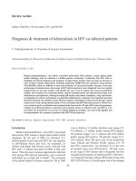
A general overview of these cascades is illustrated in Figure 1. Briefly, when the ligand-
receptor interaction occurs, subsequent events are activated based on the nature of these
ligands and receptors. For example, a receptor with intrinsic kinase activity (e.g. epidermal
growth factor receptor) is usually autophosphorylated directly leading to the creation of a
docking site for an adapter protein complex called Grb2/SOS (son of sevenless) [36]. As a result,
SOS is recruited to the plasma membrane where it encounters and activates a small G protein
named Ras [36,127,128]. Activated Ras induces the activation of several downstream signaling
molecules, including a serine/threonine kinase called Raf, which in turn activates the MAPK
and PI3K signaling pathways [36,127,129]. PI3K signaling molecules can also be activated
directly via the p110α catalytic subunit of the PI3K [127]. A receptor with no intrinsic kinase
activity (e.g. cytokine receptors) generally requires activation of receptor-associated kinases
such as JAKs for its phosphorylation. Subsequently, activated JAKs can activate the STAT
signaling pathway directly and also interact with and activate Grb2/SOS, which in turn
activates PI3K and MAPK signaling [36,122,130,131].
Immune Responses and Cell Signaling During Chronic HIV Infection
/>9
Figure 1. Overview of the major intracellular signaling pathways Upon ligand-receptor binding, signal transduc‐
tion triggers takes place based on the type and nature of the receptor. If the receptor has intrinsic tyrosine kinase ac‐
tivity, autophosphorylation of the tyrosine residues of the receptor will occur and thus creates docking sites for a
variety of different signaling molecules that have SH2 and PTB domains. Grb2/SOS complexes bind to docking sites
and lead to recruitment of SOS (son of sevenless) to the plasma membrane where they interact with Ras. Subsequent‐
ly, activated Ras molecules activate several downstream molecules including Raf, MAPKK, and MAPK. The PI3K signal‐
ing pathway can be activated directly via the p110α catalytic subunit of the PI3K. Phosphorylated receptors also
Current Perspectives in HIV Infection
10
activate phospholipase Cγ (PLCγ), which activate Protein Kinase C (PKC) and calcium-dependent signaling pathways. If
the receptor has no intrinsic kinase activity, activation of the Janus Kinase (Jak) or other receptor-associated kinase
occurs. Subsequently, activated Jaks phosphorylate the receptor and thus create docking sites for various signaling
molecules including members of the Signal Transducers and Activators of Transcription (STAT) family. Signal transduc‐
tion culminates in the transcriptional activation of STAT responsive genes that influence cellular proliferation, differen‐
tiation, cytokine production, mobility, phagocytosis, and survival [modified from [187]].
Evidence has also demonstrated the presence of a complex crosstalk between these pathways.
For instance, it has been shown that Jak2 is responsible for the activation of STAT, Erk MAPK,
and Akt signaling pathways in response to growth hormone in hepatoma and preadipocyte
cells [132]. Another report has demonstrated a role for Akt in serine phosphorylation of the
STAT1 transcription factor and upregulation of gene expression in response to IFN-γ [133].
HIV-induced perturbation of the JAK/STAT, PI3K, and MAPK signaling pathways in immune
cells including M/M and T lymphocytes has been documented (summarized in Table 1, 4)
[41,134-146]. These effects appear to be to the advantage of the virus. On one hand, it may help
the virus to replicate and establish infection. On the other hand, it may also help the virus to
escape the immune system. In the following subsections, we will provide a brief overview of
cytokine signaling and where HIV infection appears to target these cascades.
4.3.1. JAK/STAT signaling pathway
The JAK/STAT pathway is one of the major signaling pathways involved in cytokine responses.
Studies have shown that many ligands such as epidermal growth factor (EGF), receptor
tyrosine kinases (RTK), G protein-coupled receptors (GPCR) and several cytokine families
including interferons and interleukins are the main triggers of the JAK/STAT signaling cascade
[147-149]. An overview of the JAK/STAT signal transduction pathway is illustrated in Figure
1. Initially, cytokine-receptor interaction triggers tyrosine transphosphorylation of receptor-
associated JAKs. This is followed by phosphorylation of receptor cytoplasmic domains by JAKs
and recruitment of latent STAT proteins via their Src homology 2 (SH2) domains to the
activated (tyrosine phosphorylated) receptor. This is followed by STAT tyrosine phosphory‐
lation. Activated STATs form dimers via their SH2 domains and are translocated into the
nucleus where they bind STAT responsive elements [119,120,123], and thus promote tran‐
scription of STAT responsive genes such as cytokine-inducible SH2-containing protein (CIS),
members of the IRF family, and numerous other genes [150-153].
In mammalian cells, four JAKs (Jak1, Jak2, Jak3 and Tyk2) and seven STAT proteins
(STAT1, 2, 3, 4, 5a, 5b, and 6) with their different isoforms have been identified.
[147,154]. Through IL-6-induced signaling, Jak1 is the principal kinase in the downstream
signaling cascade. It has been shown in many cell lines that down regulation of Jak1
would lead to impaired signal transduction. Activated JAKs lead to phosphorylation of
STAT proteins. However, JAK kinases do not appear to show specificity for a particular
STAT protein [147,154]. STAT proteins play an important role in regulating and main‐
taining both innate and adaptive immune responses (summarized in Table 2)
[119-121,123]. For instance, studies have suggested that impairment of JAK/STAT signal‐
ing may increase susceptibility to many infections including HIV [65,67,70,155].
Immune Responses and Cell Signaling During Chronic HIV Infection
/>11
STAT
gene
Activating cytokines
Examples of STAT
responsive genes
Phenotype of knockout mice
STAT1 IFNs,IL-6,IL-10
IRF-1, ISG54, MIG, GBP,
CIITA
Impaired IFN and innate immune
responses, increase susceptibility to
tumors, opportunistic and viral
infections
STAT2 IFNs IRF-1, ISG54 Impaired Type-1 IFN responses
STAT3 IL-2,IL-6,IL-10
JunB, SAA3, JAB, C-
reactive protein, Bcl-xL
Embryonic lethal
STAT4 IL-12
IFN-γ, IRF-1, MHC class
II, CD23, Fc-γRI
Defect in IL-4 and IL-12 responses, and
impaired Th1 differentiation.
STAT5 a, b
Numerous (e.g. IL-2,IL-7,IL-15, GM-
CSF)
CIS, IL-2R-α, β-casein,
osm, pim1, p21
Impaired proliferation, growth and
survival, defect in IL-2 responses,
impaired growth.
STAT6 IL-4,IL-13 IL-4R-α, C-γ-1, C-γ-4
Defect in IL-4 responses, and impaired
Th1 differentiation.
Table 2. STATs proteins and their role in the immune system
A number of reports have suggested that defects in cytokine responsiveness arise in different
cell types during chronic HIV infection and these defects could be due to the direct effects of
HIV and/or its proteins, or due to indirect effects associated with alterations of the host cytokine
profile [38-42,139,141-143,156]. In M/M, it has been revealed that GM-CSF-induced STAT5
activation in monocyte-derived macrophages (MDM) is inhibited by in vitro HIV-1 infection
[156]. Other in vitro reports have suggested that HIV and its Gp120 and Nef proteins are capable
of activating STAT1 and STAT3 in monocytic cell lines and MDM [141-143]. Recently, the HIV
matrix protein p17 has been shown to induce STAT1 and pro-inflammatory cytokines in
macrophages [139]. Moreover, in ex vivo studies, we found that among the responses to
cytokines tested (IFN-γ, IFN-α, IL-10, IL-4, and GM-CSF) in terms of STAT induction in
monocytes, only IFN-γ showed a significant upregulation of STAT1 activation in HIV+ patients
that were off antiretroviral therapy (ART) compared to HIV- controls and patients on ART [39].
Furthermore, this potentiation of IFN-γ-induced STAT1 activation was associated with
increased total STAT1 expression levels and monocyte cell death [39]. Another ex vivo study
has shown a defect in IFN-α induced STAT1 activation in monocytes obtained from a similar
set of HIV patients, and this defect was due to the decreased IFN-α receptor expression levels
on these cells [42].
In lymphocytes, we and others have shown that both IL-7Rα expression and IL-7-induced
STAT5 activation was impaired in CD8 T cells from HIV+ patients [36,40,41]. STAT activation in
response to IL-4 and IL-10 did not appear to be similarly impaired [40]. We also found that IL-2-
induced STAT5 activation was inhibited in CD8+ T cells from a subset of HIV-infected patients
naive to therapy, but was restored, at least in part, after ART [38]. Somewhat similar results have
been observed in other in vitro studies in which activation of STAT5 in response to IL-2 was in‐
hibited by HIV-1 infection through prior Gp120-CD4 interactions in CD4+ T cells [37,144].
Current Perspectives in HIV Infection
12
4.3.2. PI3K signaling pathway
Phosphoinositide 3-kinases or phosphatidylinositol-3-kinases (PI3Ks) belong to a family of
enzymes that have serine/threonine kinase activity. These enzymes can be activated by various
stimuli including growth factors, antigens, cytokines [157,158], and are capable of phosphor‐
ylating the third position hydroxyl group of the inositol ring of phosphatidylinositol (PtdIns)
[157,159]. This family is composed of four classes, which differ in their structure and functions
(known as Ia, Ib, II, and III). However, all of them contain at least one catalytic domain and
one regulatory domain [157,159]. Many PI3K cellular functions rely on the ability of PI3Ks to
activate protein kinase B (PKB, also known as Akt) (Figure 1). In humans, three Akt genes have
been identified named akt1, akt2, and akt3.
PI3-kinases have been shown to play a major role in diverse cellular functions, including cell
growth, proliferation, differentiation, survival, and migration [160-163]. Thus, dysregulation
of this pathway may influence different cellular responses that are associated with immunity
as well as carcinogenesis (Table 3) [157,164]. It has also been reported that there is a basal
activation of the PI3K/Akt pathways in macrophages that is required for their survival [165].
Certain reports have suggested a critical role for PI3K signaling in chronic immune activation
by promoting cell survival [166]. For instance, an in vitro study has revealed that HIV infection
and its protein Tat was sufficient to activate the PI3K/Akt pathway in macrophages [166].
Interestingly, PI3K/Akt inhibitors including Miltefosine, an antiprotozoal drug known to
inhibit PI3K/Akt pathway, significantly reduced HIV-1 production from infected macrophages
and increased susceptibility to cell death in response to extracellular stress, as compared to
uninfected cells [166]. Another study has shown that inhibition of Akt phosphorylation is
required for TNF related apoptosis inducing ligand (TRAIL)-induced cell death in HIV
infected macrophages [167].
Target Gene
Phenotype
p85
α
Decreased B cell development and activation, increased antiviral responses
p85
β
Increased insulin sensitivity
p110
α
Embryonic lethal and defective proliferation
P110
β
Embryonic lethal
P110
γ
Decreased T cell development and activation, decreased inflammation, chemotaxis,
and oxidative burst
PTEN
Embryonic lethal, autoimmune disease, decreased T cell development, increased T cell
activation, and chemotaxis
SHIP1
Increased myeloid cell proliferation and survival, increased B cell activation,
chemotaxis, and mast cell degranulation
SHIP2 Perinatal lethal
Table 3. Characteristics of PI3K knockout mice
Immune Responses and Cell Signaling During Chronic HIV Infection
/>13
Viral protein Effects on M/M Effects on lymphocytes
gp120 Stimulates STAT1 activation Stimulates STAT1 activation
p17 Stimulates STAT1 activation No report
Tat Stimulates MAPK, Akt activation Stimulates Akt, MAPK activation
Nef Stimulates STAT1 & 3, MAPK activation Stimulates Erk & p38 MAPK activation
Vpr Stimulates MAPK activation No report
HIV infection
Inhibits STAT5 activation, Stimulates STAT1, Akt
activation
Inhibits STAT5 activation, Stimulates STAT1,
MAPK activation
Table 4. HIV viral proteins and their effects on monocytes/macrophages and lymphocytes
4.3.3. MAPK signaling pathway
Mitogen-activated protein kinases (MAPKs) are also a family of enzymes that have serine/
threonine kinase activity [168]. This family of kinases is generally activated in response to vari‐
ous extracellular stimuli such as growth factors and inflammatory signals, as well as cellular
stress. They regulate different cellular processes including mitosis, proliferation, differentia‐
tion, and cell death [168]. The MAPK family is composed of three major subfamilies of kinases
known as the extracellular receptor kinases (ERKs), the c-Jun N-terminal kinases/stress-activat‐
ed protein kinases (JNK/SAPK) and the p38 MAP kinases [169]. Activation of a specific MAP
kinase requires activation of a small GTP binding protein (e.g. Ras) which results in the phos‐
phorylation of a series of downstream kinases (Figure 1) [128]. Activation of the MAPK kinase
kinase (MAPKKK) (e.g. Raf) leads to the activation of downstream MAPK kinase (MAPKK),
and finally, specific MAPK (p38, Erk or JNK) [170,171]. The Erk MAPK family is found in two
isoforms called Erk1 and Erk2. Both isoforms are phosphorylated by members of the MEK fami‐
ly, which are often activated by extracellular stimuli such as growth factors, LPS and chemo‐
therapeutic agents [129,172,173]. The JNK family is found in three isoforms named JNK1, JNK2,
and JNK3 [174], while the P38 family is found in five different isoforms called p38 (SAPK2),
p38β, p38β2, p38γ (SAPK3), and p38δ [175,176]. Both JNK and p38 MAPKs are phosphorylated
by SAPK/Erk kinases (SEKs) and mitogen-activated protein kinase kinases (MKKs), which are
usually induced by inflammatory cytokines as well as other stressors such as endotoxins, reac‐
tive oxygen species, protein synthesis inhibitors, and ultraviolet (UV) irradiation [174,177-179].
MAPKs have been shown to activate various downstream transcription factors such as activa‐
tor transcription factor (ATF)-2, SP-1 (a member of Specificity Protein/Krüppel-like Factor fami‐
ly) and activator protein (AP)-1, and even STAT3 [178,180-182].
Several reports have shown that activation of the MAPKs resulted in phosphorylation of HIV
Rev, Tat, Nef, and p17 proteins and enhanced viral replication [140,183]. Other studies have
demonstrated a role for MAPK in regulating monocyte and lymphocyte functions and cell
death during HIV infection. For example, in monocytes, it has been shown that the HIV Tat
protein stimulates IL-10 production via activation of calcium/MAPK signaling pathways in
human monocytes [134,135,184]. Another report has suggested that HIV Vpr is capable of
inducing programmed cell death in primary monocytes and the monocytic cell line THP-1 cells
[185]. Further, it has been shown that HIV and its protein nef induced FasL, Programmed
Death-1 expression and apoptosis in peripheral blood mononuclear cells (PBMCs) and the
Jurkat T cell line through activation of the p38 MAPK signaling pathway [138,186].
Current Perspectives in HIV Infection
14
Figure 2. A model for the effect of chronic HIV infection on cellular signal transduction Cell signaling molecules
may be regulated directly or indirectly during chronic HIV infection. In the direct setting, HIV and its proteins (Gp120,
Nef, Tat, Vpr), through the binding of cellular receptors or internalization by endocytosis, alter signaling pathways in‐
cluding JAK/STAT, PI3K, and MAPK. In the indirect scenario, HIV infection may adversely affects the host cytokine net‐
work, which may in turn affect signal transduction. Both scenarios may thus promote viral replication and defective
host immune effector functions and reduce immune cell survival [modified from [187].
5. Conclusion
It is well established that HIV targets the immune system and mainly immune cells that express
the CD4 surface receptor, but the virus is not exclusive to these cells. Thus, through the course
Immune Responses and Cell Signaling During Chronic HIV Infection
/>15