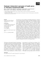
Báo cáo khoa học: Nitric oxide-induced epidermal growth factor-dependent phosphorylations in A431 tumour cells pot
Bạn đang xem bản rút gọn của tài liệu. Xem và tải ngay bản đầy đủ của tài liệu tại đây (434.67 KB, 10 trang )
Nitric oxide-induced epidermal growth factor-dependent
phosphorylations in A431 tumour cells
Marı
´
a J. Ruano
1
, Silvia Herna
´
ndez-Hernando
1
, Amparo Jime
´
nez
1
, Carmen Estrada
2
and Antonio Villalobo
1
1
Instituto de Investigaciones Biome
´
dicas, Consejo Superior de Investigaciones Cientı
´
ficas and Universidad Auto
´
noma de Madrid,
Spain;
2
A
´
rea de Fisiologı
´
a, Facultad de Medicina, Universidad de Ca
´
diz, Spain
Nitric oxide (NO
•
) strongly inhibits the proliferation of
human A431 tumour cells. It also inhibits tyrosine phos-
phorylation of a 170-kDa band corresponding to the
epidermal growth factor receptor (EGFR) and induces the
phosphorylation at tyrosine residue(s) of a 58-kDa protein
which we have denoted NOIPP-58 (nitric oxide-induced
58-kDa phosphoprotein). The NO
•
-induced phosphoryla-
tion of NOIPP-58 is strictly dependent on the presence of
EGF. Phosphorylation of NOIPP-58 and inhibition of the
phosphorylation of the band corresponding to EGFR are
both cGMP-independent processes. We also demonstrate
that the p38 mitogen-activated protein kinase (p38MAPK)
pathway is activated by NO
•
in the absence and presence of
EGF, whereas the activity of the extracellular signal-regula-
ted protein kinase 1/2 (ERK1/2) and the c-Jun N-terminal
kinase 1/2 (JNK1/2) pathways are not significantly affected
or are slightly decreased, respectively, on addition of this
agent. Moreover, we show that the p38MAPK inhibitor,
SB202190, induces rapid vanadate/peroxovanadate-sensi-
tive dephosphorylation of prephosphorylated EGFR and
NOIPP-58. We propose that the dephosphorylation of both
NOIPP-58 and EGFR are mediated by a p38MAPK-
controlled phosphotyrosine-protein phosphatase (PYPP).
Activation of the p38MAPK pathway during nitrosative
stress probably prevents the operation of this PYPP, allow-
ing NOIPP-58, and in part EGFR, to remain phosphoryl-
ated and therefore capable of generating signalling events.
Keywords: cell proliferation; p38MAPK; phosphotyrosine
phosphatase; tyrosine kinase.
Nitric oxide (NO
•
), a highly reactive gas synthesized in
mammalian cells from
L
-arginine by a family of related
enzymes denoted NOS (nitric oxide synthase), is involved in
multiple physiological processes, such as control of the blood
pressure, regulation of neuronal activities, and immune
response [1]. In addition, NO
•
participates in the control of
cell proliferation in a great variety of cell types [2–12].
The relevance of NO
•
in the control of cell proliferation
in vivo has been demonstrated during development in
Drosophila. Inhibition of NOS from embryonic imaginal
discs produces hypertrophy of organs, and, conversely, the
ectopic expression of NOS has a hypotrophic effect [7]. NO,
however, has a complex mode of action, as it can exert
opposite effects on cell proliferation. In this context, NO
•
has been reported to stimulate cell proliferation by cGMP-
dependent mechanisms associated with activation of the
AP-1 transcription complex [5,9] and, on the other hand, to
inhibit cell proliferation by cGMP-dependent [2,4,6] and
cGMP-independent [3,8–12] mechanisms. However, these
apparently contradictory actions of NO
•
depend on, among
other factors, the type of cells under study.
Activation of a cAMP-dependent protein kinase, but not
a cGMP-dependent protein kinase, appears to be respon-
sible in part for the NO
•
-mediated inhibition of cell
proliferation mediated by the cGMP-dependent pathway
in smooth muscle cells [6]. On the other hand, the
concomitant inhibition of both the ribonucleotide reductase
[9] and the intrinsic tyrosine kinase activity of epidermal
growth factor receptor (EGFR) [10,12] by NO
•
may
contribute to the inhibition of cell proliferation through
the cGMP-independent pathway. The inhibition of the cell
cycle that takes place in NO
•
-exposed cells has been
reported to occur at either the early G
2
plus M phases [13]
or the early and late G
1
phase [9,14]. Cell growth arrest at
Correspondence to A. Villalobo, Instituto de Investigaciones Bio-
me
´
dicas, Consejo Superior de Investigaciones Cientı
´
ficas and Uni-
versidad Auto
´
noma de Madrid c/Arturo Duperier 4, E-28029 Madrid,
Spain. Fax: + 34 91 585 4401, E-mail:
Abbreviations: DEA-NO, 1,1-diethyl-2-hydroxy-2-nitroso-hydrazine
sodium; DETA-NO, 2,2¢-(hydroxynitrosohydrazono)bis-ethanamine;
DMEM, Dulbecco’s modified Eagle’s medium; ECL, enhanced
chemiluminescence; EGF, epidermal growth factor; EGFR, epider-
mal growth factor receptor; ERK1/2, extracellular signal-regulated
protein kinases 1 and 2; JNK1/2, c-Jun N-terminal kinases 1 and 2;
MAPK, mitogen-activated protein kinase; MEK, MAP/ERK kinase;
NOIPP-58, nitric oxide-induced 58 kDa phosphoprotein; NOS, nitric
oxide synthase; ODQ, 1-H-[1,2,4]oxadiazolo[4,3-a]quinoxalin-1-one;
PD153035, 4-[(3-bromophenyl)amino]-6,7-dimethoxyquinazoline;
P-ERK1/2, phosphorylated form of ERK1/2; P-JNK1/2, phospho-
rylated form of JNK1/2; P-p38MAPK, phosphorylated form of
p38MAPK; PVDF, poly(vinylidene difluoride); PYPP, phospho-
tyrosine-protein phosphatase; SB202190, 4-(4-fluorophenyl)-2-
(4-hydroxyphenyl)-5-(4-pyridyl)1H-imidazole; SPER-NO, N-(2-ami-
noethyl)-N-(2-hydroxy-2-nitrosohydrazino)-1,2-ethylenediamine.
Enzymes: Nitric oxide synthase (EC 1.14.13.39); phosphotyrosine-
specific phosphatase (EC 3.1.3.48); protein-tyrosine kinase
(EC 2.7.1.112); protein kinase (EC 2.7.1.37); ribonucleotide reductase
(EC 1.17.4.1 and EC 1.17.4.2).
(Received 8 October 2002, revised 20 January 2003,
accepted 27 February 2003)
Eur. J. Biochem. 270, 1828–1837 (2003) Ó FEBS 2003 doi:10.1046/j.1432-1033.2003.03546.x
the G
1
phase appears to be associated with the induction of
p21
Waf1/Cip1
, a cyclin-dependent kinase inhibitor [14].
The transduction of extracellular signals into cellular
responses is mediated in many instances by an array of
different mitogen-activated protein kinase (MAPK) path-
ways [15–18]. Among these kinases is the family of
p38MAPKs [19–23], which are activated by dual tyrosine/
threonine kinases responsive to pro-inflammatory cytokines
and environmental stress [24]. However, there is increasing
evidence that the p38MAPK pathways are involved in
important physiological functions besides the stress
response [18,22]. Of special interest is the fact that
p38MAPK is activated by NO
•
([25–28] and this work)
and its derived metabolites [29,30]. This process appears to
be mediated by a cGMP-dependent protein kinase [28].
In this paper, we demonstrate that NO
•
inhibits tyrosine
phosphorylation of the 170-kDa band corresponding to
EGFR and induces reversible phosphorylation at tyrosine
residue(s) of a newly identified 58-kDa protein which we have
named NOIPP-58 (nitric oxide-induced 58-kDa phospho-
protein) in the presence, but not in the absence, of EGF. Both
of these processes are mediated by cGMP-independent
mechanisms. We also show that the phosphorylation/
dephosphorylation cycle of NOIPP-58 appears to be under
the control of EGFR and a p38MAPK-regulated phospho-
tyrosine-protein phosphatase (PYPP). Moreover, this phos-
phatase also dephosphorylates EGFR with great efficiency.
Therefore, activation of the p38MAPK pathway by nitro-
sative stress probably prevents operation of this PYPP,
allowing NOIPP-58, andinpartEGFR,togeneratesignalling
events.
Experimental procedures
Reagents
Dulbecco’s modified Eagle’s medium (DMEM), fetal
bovine serum and
L
-glutamine were obtained from Gibco,
[methyl-
3
H]thymidine (46 CiÆmmol
)1
) and enhanced chemi-
luminescence (ECL) reagents were from Amersham, and
OptiPhase HiSafe 2 scintillation fluid was from Wallac,
Turku, Finland
1
. The nitric oxide donors 1,1-diethyl-2-
hydroxy-2-nitrosohydrazine sodium (DEA-NO), 2,2¢-
(hydroxynitrosohydrazono)bis-ethanamine (DETA-NO)
and N-(2-aminoethyl)-N-(2-hydroxy-2-nitrosohydrazino)-
1,2-ethylenediamine (SPER-NO) were from Research Bio-
chemicals International, St Louis, MO, USA
2
. EGF (from
male mouse submaxillary glands) and the antibody to nitro-
tyrosine were from Upstate Biotechnology, Lake Placid, NY,
USA
3
. The recombinant monoclonal antibody to phospho-
tyrosine (RC20) conjugated to horseradish peroxidase was
fromTransduction Laboratories,Heidelberg, Germany
4
.Fast
Green FCF, Trypan blue, catalase (from bovine liver), and
peroxidase-conjugated anti-mouse IgGs (Fc-specific) were
from Sigma. Polyclonal antibody to phospho-specific
p38MAPK (developed in rabbit using a phosphopeptide
corresponding to residues 172–186 of human p38MAPK),
anti-(total p38MAPK) (developed in rabbit against residues
341–360 of the human protein), 4-[(3-bromophenyl)amino]-
6,7-dimethoxyquinazoline (PD153035), and 4-(4-fluoro-
phenyl)-2-(4-hydroxyphenyl)-5-(4-pyridyl)1H-imidazole
(SB202190) were obtained from Calbiochem. Monoclonal
antibodies to phospho-specific extracellular signal-regulated
protein kinases 1 and 2 (ERK1/2) (E-4) (developed in mouse
against a segment of the human ERK1 protein that contains
phosphorylated Tyr204) and to phospho-specific c-Jun
N-terminal kinases 1 and 2 (JNK1/2) (G-7) (developed in
mouse against a conserved segment of the human proteins
containingphosphorylated Thr183andTyr185residues) were
obtained from Santa Cruz Biotechnology. Horseradish
peroxidase-conjugated goat anti-rabbit IgGs were provided
by Zymed, San Francisco, CA, USA
5
. Poly(vinylidene
difluoride) (PVDF) membranes were from Millipore, and
PP1 was obtained from Biomol
6
, Plymouth Meeting, PA,
USA. Gentamicin was obtained from Normon, Madrid,
Spain
7
, and Tween 20 was from Bio-Rad. AX X-ray films
were purchased from Konica, and 1-H-[1,2,4]oxadiazolo-
[4,3-a]quinoxalin-1-one (ODQ) was obtained from Tocris,
London, UK
8
.
Cell cultures
Human epidermoid carcinoma A431 cells, a cell line that
overexpresses both the wild-type EGFR and aberrant
extracellular forms of this receptor [31], and the different
fibroblast cell lines used were grown in DMEM sup-
plemented with 10% (v/v) fetal bovine serum, 2 m
M
L
-glutamine and 40 lgÆmL
)1
gentamicin in a humidified
atmosphere of 5% (v/v) CO
2
in air at 37 °C. Cells were
counted using a Neubauer chamber after detachment from
the culture dishes.
Cell viability
Living and dead cells were counted by the Trypan blue
exclusion method after control and DEA-NO-treated cells
had been detached from the culture dishes by trypsinization.
The viability of A431 tumour cells was not affected by
DEA-NO treatment in the conditions used in this study.
Untreated cells and cells treated with 5 m
M
DEA-NO for
15 min had a viability of 85 ± 9% (n ¼ 4) and 93 ± 3%
(n ¼ 3), respectively. We observed no significant cell
detachment from the culture dishes on overnight treatment
with 1 m
M
DEA-NO.
Incorporation of [
methyl
-
3
H]thymidine
Incorporation of [methyl-
3
H]thymidine into DNA was
carried out in confluent cell cultures essentially as described
[32], but in the absence of EGF to attain maximum
proliferation, as this growth factor has an antimitogenic
effect on A431 tumour cells [33]. Cells grown to confluence
in 24-well culture dishes and deprived of fetal bovine serum
overnight, were washed twice with 130 m
M
NaCl/2.7 m
M
KCl/11.5 m
M
sodium/potassium phosphate (pH 7.4)
(NaCl/P
i
) and incubated for 14–16 h in 0.5 mL DMEM
supplemented with 1.2 l
M
(2 lCiÆmL
)1
)[methyl-
3
H]thymi-
dine in the absence and presence of 50 l
M
ODQ and the
concentrations of DEA-NO indicated in the legends of the
figures. Thereafter, cells were treated with ice-cold 10%
(w/v) trichloroacetic acid for 10 min, solubilized with 0.2
M
NaOH for 24 h, and neutralized with 0.2
M
HCl. The
radioactivity incorporated into the acid-insoluble material
was measured using a scintillation counter.
Ó FEBS 2003 NO
•
-induced EGF-dependent phosphorylations (Eur. J. Biochem. 270) 1829
Detection of phosphotyrosine-containing proteins
Cells grown to confluence in 6-well culture dishes were
deprived of fetal bovine serum overnight, washed twice with
NaCl/P
i
, and incubated, unless indicated otherwise, at
37 °C for 15 min in 1.5 mL fetal bovine serum-free DMEM
in the absence and presence of the concentrations of DEA-
NO indicated in the legends of the figures. Thereafter, 10 n
M
EGF was added and the cells were incubated for 1–5 min
under the same conditions. Controls in the absence of EGF
were also performed. Ice-cold 10% (w/v) trichloroacetic
acid was then added, and the fixed cells were scraped from
the plates and processed by slab-gel electrophoresis using
the method of Laemmli [34], at 12 mA in linear 5–20% (w/v)
polyacrylamide gradient gels in the presence of 0.1% (w/v)
SDS at pH 8.3. The proteins were then electrotransferred to
a PVDF membrane for 2–3 h at 300 mA, fixed with 0.2%
(v/v) glutaraldehyde in 25 m
M
Tris/HCl (pH 8)/150 m
M
NaCl/2.7 m
M
KCl (NaCl/Tris), and transiently stained with
Fast Green FCF to ascertain the regularity of the transfer
procedure. The PVDF membrane was blocked with 5%
(w/v) BSA in NaCl/Tris for 2 h at room temperature and
washed with 0.1% (w/v) Tween 20 in NaCl/Tris. The
PVDF membrane was then probed overnight with a
1 : 5000 dilution of the RC20 antibody conjugated to
horseradish peroxidase, and washed with 0.1% (w/v)
Tween 20 in NaCl/Tris. The phosphotyrosine-containing
proteins were visualized on development with the ECL
reagents following instructions from the manufacturer and
exposure of X-ray films for appropriate periods of time. The
intensities of the phosphotyrosine-containing protein bands
of interest were quantified with a computer-assisted scan-
ning densitometer using the NIH Image 1.59 program.
Corrections were made for the amount of protein present in
the electrophoretic tracks as detected by Fast Green FCF
staining followed by densitometric reading. To avoid any
exposure time differences between gels loaded with samples
corresponding to experiments performed in parallel, we
exposed a single film to two different gels at the same time,
or used a fix chronometer-measured exposure time for each
film.
Detection of the active forms of different MAPKs
Cells grown and treated with DEA-NO and/or EGF as
described were scraped from the culture dishes. The
solubilized proteins were processed by SDS/PAGE and
transferred to a PVDF membrane. After blocking of the
membrane as described above, P-ERK1/2, P-JNK1/2 and
P-p38MAPK, which represent the active forms of these
kinases, were probed overnight using 1 : 1000–1 : 2000
dilutions of specific antibodies against the human phos-
phorylated proteins, washed three times with 0.1% (w/v)
Tween 20 in NaCl/Tris, and thereafter incubated for 3 h
with a 1 : 2000 dilution of appropriate secondary IgGs
conjugated to horseradish peroxidase. Development was
carried out by ECL, and band intensities were quantified as
described above. To confirm identical loading in the
electrophoretic wells, protein staining of the PVDF mem-
brane with Fast Green FCF and densitometric reading with
a computer-assisted scanning densitometer using the NIH
Image 1.59 program was routinely performed.
Preparation of peroxovanadate
Peroxovanadate was prepared from orthovanadate essen-
tially as described [35], with the following modifications. A
solution of 10 m
M
sodium orthovanadate was incubated
with 10 m
M
H
2
O
2
for30minin5mLNaCl/P
i
at room
temperature. After completion of the synthesis, 17 UÆmL
)1
catalase was added for 30 min to reduce any trace of
unreacted H
2
O
2
remaining in the sample. One unit of
catalase transforms 1 lmol H
2
O
2
Æmin
)1
at pH 7 and 25 °C.
The resulting peroxovanadate solution was used immedi-
ately without being stored.
ODQ bioassay
To determine the inhibitory action of the ODQ stocks used
in this work, we assayed the effect of this compound on a
well-known cGMP-dependent system using an acetylcho-
line-induced arterial relaxation bioassay as described [36].
We observed that 10 l
M
ODQ prevents 99% of the
relaxation induced by 10 l
M
acetylcholine in noradrenal-
ine-precontracted rat carotid arterial segments. From this
we ascertained that the concentration of 50 l
M
ODQ used
in the treatment of A431 tumour cells was sufficient to
inhibit any endogenous guanylate cyclase activity.
Results
NO
•
inhibits cell proliferation by a cGMP-independent
mechanism
We studied the effect of NO
•
on the proliferation of A431
tumour cells by measuring the incorporation of
[methyl-
3
H]thymidine into DNA. Figure 1 shows that the
NO
•
donor DEA-NO strongly inhibits this process in a
Fig. 1. NO
•
inhibits DNA synthesis by a cGMP-independent mechan-
ism. Incorporation of [methyl-
3
H]thymidine into DNA was determined
as described in Experimental procedures in confluent cells treated with
the indicated concentrations of DEA-NO, in the absence (s)and
presence (d)of50l
M
ODQ. Results are from quadruplicate samples
from two separate experiments, and the error bars represent the SEM.
1830 M. J. Ruano et al.(Eur. J. Biochem. 270) Ó FEBS 2003
concentration-dependent manner in the absence (open
symbols) and presence (filled symbols) of ODQ, a potent
inhibitor of the soluble NO
•
-sensitive guanylate cyclase [37].
Thus, it appears that NO
•
-promoted inhibition of cell
proliferation does not require the synthesis of cGMP.
Moreover, the proliferation of A431 tumour cells appears to
be far more sensitive to DEA-NO than other cell lines
tested. Thus, we determined an apparent inhibition constant
for DEA-NO (K¢
i[DEA-NO]
) in the proliferation process of
50 l
M
in A431 tumour cells (Fig. 1), compared with
3–5 m
M
in EGFR-T17 fibroblasts [10] and 0.75–2 m
M
in
NB69 neuroblastoma cells [12].
NO
•
-induced EGF-dependent phosphorylations
The action of NO
•
on the EGF-dependent phosphorylation
of proteins was assessed in whole cells treated with different
NO
•
donors. Increasing concentrations of DEA-NO pro-
gressively inhibited tyrosine phosphorylation of the 170-
kDa band corresponding to EGFR (Fig. 2A). Although we
cannot exclude the possibility that additional proteins form
part of this band, most of the observed phosphorylation
probably occurred on the EGFR itself, as A431 tumour
cells overexpress this receptor (10–50 times more receptors
per cell than most cell types) [31]. Moreover, PD153035, a
potent and selective inhibitor of EGFR [38], completely
prevented phosphorylation of the 170-kDa band. Therefore,
for simplicity we shall refer to phosphorylation of EGFR
from now on. Quantitative determinations showed that this
process has a K¢
i[DEA-NO]
of 1–2 m
M
. In contrast, similar
concentrations of DEA-NO induce, in the presence of EGF,
phosphorylation at tyrosine residue(s) of a 58-kDa protein
which we have named NOIPP-58 (Fig. 2B,C). The phos-
phorylation of NOIPP-58 has an apparent activation
constant for DEA-NO (K¢
a[DEA-NO]
)of 2m
M
. Phos-
phorylation of NOIPP-58 is not detected, however, in the
presence of increasing concentrations of DEA-NO but in
the absence of EGF (Fig. 2C). The inhibition of EGFR
phosphorylation by PD153035 results in the parallel inhi-
bition of NOIPP-58 phosphorylation (results not shown).
Using other NO
•
donors of the NONOate family that have
different efficiencies in releasing NO
•
[39], such as SPER-
NO and DETA-NO, we found that the inhibition of EGFR
phosphorylation was linear and inversely proportional to
log
10
of the half-life of NO
•
release into the medium (results
not shown). Figure 3 shows that phosphorylation of EGFR
and NOIPP-58 have dissimilar kinetics. The phosphoryla-
tion of EGFR (circles) is progressively inhibited with
increasing exposure to DEA-NO with a t
1/2
of 5min.In
contrast, the phosphorylation of NOIPP-58 (triangles) is a
transient process reaching a maximum at 5 min followed
by dephosphorylation with a t
1/2
of 10 min.
As the molecular mass of NOIPP-58 is close to that of the
nonreceptor tyrosine kinase Src, we investigated whether the
two molecules were identical. We excluded this possibility
by demonstrating that the immunoblot signal from
Fig. 2. NO
•
inhibits the phosphorylation of EGFR and promotes the
phosphorylation of NOIPP-58 in an EGF-dependent manner. Cells were
incubated with the indicated concentrations of DEA-NO for 30 min
before treatment with 10 n
M
EGF for 5 min (A and B, and C only
where indicated). Phosphorylation of EGFR (A) and NOIPP-58
(B and C) were determined using an antibody to phosphotyrosine as
described in Experimental procedures. The arrows indicate the posi-
tion of migration of EGFR (A) and NOIPP-58 (B and C). Typical
experiments of a total of five performed under similar conditions are
presented.
Fig. 3. NO
•
inhibits phosphorylation of EGFR and induces phosphory-
lation of NOIPP-58 with different kinetics. Cells were treated with
5m
M
DEA-NO for the indicated times. Thereafter, 10 n
M
EGF was
added, and 1 min later phosphorylation of EGFR (d) and NOIPP-58
(m) were determined as described in Experimental procedures. Results
are from two separate experiments, and the error bars represent the
range of values obtained.
Ó FEBS 2003 NO
•
-induced EGF-dependent phosphorylations (Eur. J. Biochem. 270) 1831
immunoprecipitated Src in its phosphorylated form does
not match that of NOIPP-58. Moreover, the addition of
PP1, a highly potent inhibitor of the Src tyrosine kinase
family, including Lck, Lyn, Hck, and Src itself [40], did not
significantly affect the phosphorylation of NOIPP-58
(results not shown).
NO
•
appears to also have a small effect on the apparent
activation constant of EGF for its receptor. Thus, we
determined from experiments performed using different
concentrations of EGF and measuring the phosphorylation
of the receptor, that in A431 tumour cells K¢
a[EGF]
varies
from 0.2 n
M
to 1n
M
in the absence and presence of
DEA-NO, respectively. Similarly, in EGFR-T17 fibroblasts,
we found K¢
a[EGF]
values of 0.05 n
M
and 1.5 n
M
in the
absence and presence of DEA-NO, respectively, under
similar experimental conditions.
The NO
•
-promoted inhibition of the phosphorylation
of both EGFR and NOIPP-58 are cGMP-independent
processes
To study whether the actions of NO
•
on the phosphoryla-
tion of EGFR and NOIPP-58 require an increase in
intracellular cGMP, we performed experiments using
different concentrations of the guanylate cyclase inhibitor
ODQ [37]. Figure 4 shows that EGFR phosphorylation in
the absence of DEA-NO was partially inhibited ( 40%) by
ODQ (open circles). However, the residual phosphorylation
of the receptor observed in the presence of DEA-NO
( 30% of the control) did not increase in the presence of
ODQ (filled circles). Moreover, the EGF-dependent NO
•
-
induced phosphorylation of NOIPP-58 was not affected by
ODQ (filled triangles), nor was this guanylate cyclase
inhibitor able to promote any phosphorylation of NOIPP-
58 in the absence of DEA-NO and presence of EGF (open
triangles). These experiments show that both the NO
•
-
elicited inhibition of EGFR phosphorylation and the EGF-
dependent NO
•
-induced phosphorylation of NOIPP-58 are
cGMP-independent processes.
Activation of the p38MAPK pathway by NO
•
As different MAPKs are central to signalling by EGFR, we
tested whether NO
•
regulates the different MAPK path-
ways. Figure 5 shows that addition of DEA-NO to A431
tumour cells does not significantly affect the phosphoryla-
tion level of ERK1/2. The clone of A431 tumour cells used
in this study has an already activated ERK1/2 pathway in
the absence of EGF. This is consistent with the high
proliferation rate of this cell line in the absence of added
growth factors (results not shown). Therefore, the addition
of EGF does not increase the level of ERK1/2 phosphory-
lation. In contrast, DEA-NO somewhat decreases the active
form of JNK1/2 in the absence or presence of EGF, whereas
this NO
•
donor strongly activates p38MAPK both in the
absence and presence of EGF, as determined by measuring
the phosphorylation levels of these MAPKs. Additional
phosphorylated bands of lower molecular mass are recog-
nized by the antibody to P-JNK1/2 in the presence of DEA-
NO. This may represent proteolytic products of these
kinases and/or the cross-detection of the phosphorylated
form of p38MAPK. Control experiments showed that the
level of total p38MAPK was somewhat decreased after
DEA-NO treatment but was not significantly affected by
EGF. Figure 6 shows the time courses of phosphorylation
of EGFR (Fig. 6A), NOIPP-58 (Fig. 6B), and p38MAPK
Fig. 4. NO
•
inhibits phosphorylation of EGFR and induces phospho-
rylation of NOIPP-58 by cGMP-independent mechanisms. Cells were
treated for 15 min with the indicated concentrations of ODQ. There-
after, the cells were incubated in the absence (open symbols) and
presence (filled symbols) of 5 m
M
DEA-NO for another 15 min. Then
10 n
M
EGF was added and 5 min later phosphorylation of EGFR
(circles) and NOIPP-58 (triangles) were determined as described in
Experimental procedures. Results are from four (EGFR) and six
(NOIPP-58) separate experiments, and the error bars represent SD.
Fig. 5. NO
•
activates p38MAPK but does not activate ERK1/2 or
JNK1/2 pathways. Cells were incubated in the absence and presence of
5m
M
DEA-NO for 15 min and then stimulated with 10 n
M
EGF for
5 min as indicated. The active phosphorylated forms of the different
MAPKs (P-ERK1/2, P-JNK1/2 and P-p38MAPK) were determined
as described in Experimental procedures. The arrows indicate the
phosphorylated forms of these kinases. A control showing total
p38MAPK is also presented. Typical experiments from a total of 11
performed under similar conditions are presented.
1832 M. J. Ruano et al.(Eur. J. Biochem. 270) Ó FEBS 2003
(Fig. 6C) in the absence (open symbols) and presence (filled
symbols) of DEA-NO. It is apparent that activation of the
p38MAPK pathway, although very prominent in the
presence of DEA-NO, also occurs to a lesser extent in its
absence, most significantly after 10 min of exposure to
EGF, as previously demonstrated [24].
EGFR and NOIPP-58 are both dephosphorylated
by a p38MAPK-regulated PYPP
To test whether the p38MAPK pathway regulates the
phosphorylation state of both EGFR and NOIPP-58, the
tyrosine phosphorylation levels of these proteins were
monitored before and after addition of SB202190 to
EGF-stimulated cells treated with DEA-NO. Figure 7
shows that addition of SB202190 induces rapid dephospho-
rylation of EGFR (left panel) and NOIPP-58 (right panel).
The dephosphorylation of EGFR induced by SB202190
also ocurrs in the absence of DEA-NO (results not shown).
The effect of SB202190 on the tyrosine phosphorylation
levels of these proteins was also assayed in the absence and
presence of the PYPP inhibitors vanadate and peroxovana-
date [35,41]. As shown in Fig. 7, both inhibitors prevent the
dephosphorylation of EGFR and NOIPP-58 induced by the
addition of SB202190, although peroxovanadate was far
more efficient than vanadate, in accordance with its higher
capacity to permeate cell membranes [35]. Overall, these
results illustrate that the dephosphorylation of EGFR and
NOIPP-58 is under the control of a vanadate/peroxovana-
date-sensitive p38MAPK-regulated PYPP.
Discussion
We have previously shown that NO
•
inhibits the prolifer-
ation of EGFR-T17 fibroblasts and NB69 neuroblastoma
cells by a cGMP-independent pathway [10,12]. The effect of
NO
•
was slightly more pronounced when the cells were
grown in the presence of EGF than when grown in the
presence of fetal bovine serum, suggesting that EGFR may
be a target for NO
•
[10,12]. Moreover, using an in vitro
permeabilized-cell system, we showed that NO
•
targets
EGFR inhibiting its tyrosine kinase activity, a process that
was reversed by dithiothreitol, suggesting S-nitrosylation of
the receptor [10]. We now demonstrate that addition of
DEA-NO also inhibits the proliferation of A431 tumour cells
by a cGMP-independent mechanism, but in a more efficient
fashion than in the other cell lines tested (see Fig. 1 and
[10,12]). In contrast, the sensitivity of the EGFR tyrosine
kinase to NO
•
in whole A431 tumour cells (this work) and
permeabilized EGFR-T17 fibroblasts [10] was within the
same order of magnitude (K¢
i[DEA-NO]
1–2 m
M
).
The concentration of NO
•
donor required to achieve
substantial inhibition of EGFR phosphorylation in both
cell types appears to be rather high. However, although we
did not determine the concentration of free NO
•
in our
experimental system, this is expected to be several orders of
Fig. 6. Time course of EGF-induced phosphorylation of EGFR,
NOIPP-58, and p38MAPK in the absence and presence of NO
•
. Cells
were incubated in the absence (open symbols) and presence (filled
symbols) of 5 m
M
DEA-NO for 15 min. Thereafter, 10 n
M
EGF was
added at time zero, and phosphorylation of EGFR (A), NOIPP-58
(B), and p38MAPK (C) was determined at the indicated times as
described in Experimental procedures. Results are from two separate
experiments, and the error bars represent the range of values obtained.
Ó FEBS 2003 NO
•
-induced EGF-dependent phosphorylations (Eur. J. Biochem. 270) 1833
magnitude lower than the actual concentration of NO
•
donors used. There are several reasons including: (a) the low
solubility of NO
•
in water [42]; (b) the high reactivity of NO
•
with different cellular targets that are S-nitrosylated and
may act as molecular scavengers [43–49]; and (c) the rapid
transformation of NO
•
into peroxynitrite and other meta-
bolites by different cellular systems [50].
We have observed that the t
1/2
for the release of NO
•
from
donors of the NONOate family [39] inversely correlates with
the magnitude of the observed inhibition of the phosphory-
lation of EGFR in intact cells. NONOates, in contrast with
other NO
•
donors such as SNAP (S-nitroso-N-acetylpeni-
cillamine), SIN-1 [3-(morpholinosydnonimine hydrochlo-
ride)] and sodium nitroprussiate, have a simple mechanism
of decomposition in aqueous solution and hence release
NO
•
into the medium without the need of any metabolic
transformation by the cell and/or the formation of any
NO
•
-derived byproduct [39]. Thus, our results suggest that
NO
•
itself, and not NO
•
-derived metabolites, is probably
the active species inhibiting the phosphorylation of EGFR
in whole A431 cells, in agreement with our observations in
permeabilized EGFR-T17 fibroblasts [10] and intact NB69
neuroblastoma cells [12]. We performed Western blots using
an antibody to nitrotyrosine in cells treated with DEA-NO
and SPER-NO. Despite the high background yield by this
antibody, we detected no labelled band at 170 kDa,
suggesting that tyrosine residues in EGFR were not nitrated
(results not shown).
Interestingly, NO
•
does not inhibit the binding of
[
125
I]EGF to its receptor in EGFR-T17 fibroblasts [10].
However, we have demonstrated in both A431 tumour cells
and EGFR-T17 fibroblasts that NO
•
slightly increases the
apparent activation constant of EGF for tyrosine phos-
phorylation of the receptor when monitored in whole cells.
This suggests that the binding of EGF to the receptor is not
impaired by NO
•
, but the bound EGF cannot activate the
NO
•
-modified EGFR with the same efficiency as it does the
native receptor.
Our experiments also show that the NO
•
-promoted
inhibition of EGFR phosphorylation in A431 tumour cells
and other cell lines is a cGMP-independent process (this
work and [10,12]). We have found, however, that phos-
phorylation of EGFR in the absence of NO
•
is partially
sensitive to the guanylate cyclase inhibitor ODQ (Fig. 4).
The inhibition of EGFR phosphorylation by ODQ is a
concentration-dependent process up to 10 l
M
, conditions
under which the guanylate cyclase is fully inhibited [37].
However, higher concentrations of ODQ do not further
affect the phosphorylation of the receptor, suggesting that
only a part ( 40%) of this process is dependent on cGMP.
The interplay between cGMP and EGFR appears to be
quite complex, as it has been shown that cGMP inhibits the
EGF-induced activation of the MAPK pathway via phos-
phorylation of Raf by a cGMP-dependent protein kinase
[51,52] and through the induction of MAPK phosphatase 1
[52]. The effect of ODQ on EGFR phosphorylation
described in this work is a new and unexpected observation
that may underscore a potent activation of the receptor by a
regulatory cGMP-dependent protein kinase or another
cGMP-dependent system.
The NO
•
-dependent phosphorylation of NOIPP-58 is
strictly dependent on the presence of EGF, and therefore
requires a partially active EGFR. As no phosphorylation of
NOIPP-58 was detected in the absence of NO
•
, either in the
absence or presence of EGF, we propose that the partially
active EGFR may be directly responsible for the phos-
phorylation of NOIPP-58. This is supported by the fact
that the K¢
i[DEA-NO]
for EGFR phosphorylation and the
K¢
a[DEA-NO]
for NOIPP-58 phosphorylation have compar-
able values (1–2 m
M
). An NO
•
-modified NOIPP-58 is
probably the actual substrate of EGFR.
We have also shown, as previously reported by others
[25–28], that NO
•
induces the activation of the p38MAPK
pathway, not only in A431 tumour cells (Figs 5 and 6), but
also in several murine fibroblast cell lines such as EGFR-
T17 and N7xHERc, which overexpress human EGFR,
Swiss 3T3 and NIH 3T3, which, respectively, express a
moderate and low number of EGFR molecules, and clone
2.2, which does not express this receptor (results not shown).
This demonstrates that the NO
•
-mediated activation of
Fig. 7. EGFR and NOIPP-58 are dephosphorylated by a p38MAPK-regulated vanadate/peroxovanadate-sensitive PYPP. Cells incubated for 30 min
in the absence (None) and presence of 1 m
M
vanadate (V) or 1 m
M
peroxovanadate (PV) were treated with 5 m
M
DEA-NO for 15 min. The cells
were then stimulated with 10 n
M
EGF for 4 min, and thereafter 100 l
M
SB202190 or the solvent dimethyl sulfoxide was added as indicated.
Phosphorylation of EGFR (left panel) and NOIPP-58 (right panel) were determined 1 min later as described in Experimental procedures. A typical
experiment from a total of three performed in similar conditions is presented.
1834 M. J. Ruano et al.(Eur. J. Biochem. 270) Ó FEBS 2003
p38MAPK is an EGFR-independent process. The NO
•
-
dependent activation of the p38MAPK pathway may
contribute to the arrest of the cell cycle, as it has been
shown in a different system on activation of the activin
receptor pathway [53]. Although our results do not allow us
to establish a direct correlation between the NO
•
-induced
inhibition of cell proliferation and the phosphorylation/
dephosphorylation events under study, as the two processes
are achieved at different concentrations of DEA-NO, we
cannot exclude the possibility that low concentrations of
DEA-NO during long exposure times, such as those
required for the inhibition of [methyl-
3
H]thymidine incor-
poration into DNA, may affect the phosphorylation state of
the relevant proteins during the long period required to
complete a full cell cycle. Nevertheless, it is likely that
distinct systems involved in cell proliferation are affected by
NO
•
.
Inhibition of the p38MAPK pathway activates a vana-
date/peroxovanadate-sensitive PYPP which dephosphory-
lates EGFR. In cells exposed to NO
•
and in the presence of
EGF, conditions in which NOIPP-58 is phosphorylated,
p38MAPK inhibition results in dephosphorylation of both
NOIPP-58 and EGFR by the same mechanism (Fig. 7).
This suggests that, under normal physiological conditions,
when cells are stimulated by EGF, or during nitrosative
stress generated by activation of NOS, the activated
p38MAPK pathway signals to down-regulate the activity
of the PYPP acting on EGFR and NOIPP-58 (see model in
Fig. 8). This system may therefore be a mechanism for
keeping EGFR and the potential signalling capacity of the
phosphorylated form of NOIPP-58 partially operative by
preventing their dephosphorylation.
To the best of our knowledge, this is the first demon-
stration of the existence of a p38MAPK-regulated PYPP
modulating the activity of both EGFR and the phosphory-
lation state of NOIPP-58, a protein substrate of this
receptor. Further studies should uncover the physiological
function of NOIPP-58, as well as the molecular character-
istics of the p38MAPK-regulated phosphatase involved in
dephosphorylation of EGFR and NOIPP-58, and whether
similar dephosphorylation pathways act on other activated
receptors of the ErbB family and/or other unrelated tyrosine
kinase receptors.
Acknowledgements
We appreciate helpful discussions with Dr Jose
´
Martı
´
n-Nieto during
the preparation of this work, and the assistance of Hongbing Li in the
preparation of some figures. We also thank Dr M. C. Gonza
´
lez for
performing ODQ bioassays. M.J.R. was supported by a postdoctoral
fellowship from the Consejerı
´
a de Educacio
´
nyCulturadelaComunidad
de Madrid. This work was supported by grants to A.V. from the
Comisio
´
n Interministerial de Ciencia y Tecnologı
´
a (SAF99-0052 &
SAF2002-03258), the Consejerı
´
a de Educacio
´
n y Cultura de la
Comunidad de Madrid (08.1/0027/2001-1), and the Agencia Espan
˜
ola
de Cooperacio
´
n Internacional (2002 CN0013).
References
1. Gow, A.J. & Ischiropoulos, H. (2001) Nitric oxide chemistry and
cellular signaling. J. Cell Physiol. 187, 277–282.
2. Garg, U.C. & Hassid, A. (1989) Nitric oxide-generating vaso-
lidators and 8-bromo-cyclic guanosine monophosphate inhibit
mitogenesis and proliferation of cultured rat vascular smooth
muscle cells. J. Clin. Invest. 83, 1774–1777.
Fig. 8. Signalling events during nitrosative stress in A431 tumour cells. NO
•
induces partial inactivation of EGFR, probably by S-nitrosylation [10],
and activation of the p38MAPK pathway. The partially active EGFR in the presence of NO
•
favours the phosphorylation of NOIPP-58, perhaps
because of inactivation of a p38MAPK-regulated PYPP that has as substrates both NOIPP-58 and EGFR. The symbols -Y-P and -Y/T-P represent
the phosphorylated form of either EGFR/NOIPP-58 or p38MAPK, respectively, at tyrosine or tyrosine/threonine residues. The active (a) and
inactive (i) forms of the different proteins are indicated. Nitrosative stress in A431 tumour cells inactivates a p38MAPK-regulated PYPP, thereby
favouring phosphorylation of NOIPP-58 by the partially active EGFR and avoiding total dephosphorylation of the receptor.
Ó FEBS 2003 NO
•
-induced EGF-dependent phosphorylations (Eur. J. Biochem. 270) 1835
3. Garg, U.C. & Hassid, A. (1990) Nitric oxide-generating vaso-
lidators inhibit mitogenesis and proliferation of BALB/c 3T3
fibroblasts by a cyclic GMP-independent mechanism. Biochem.
Biophys. Res. Commun. 171, 474–479.
4. Assender, J.W., Southgate, K.M. & Newby, A.C. (1991) Does
nitric oxide inhibit smooth muscle proliferation? J. Cardiovasc.
Pharmacol. 17, S104–S107.
5. O’Connor, K.J., Knowles, R.G. & Patel, K.D. (1991)
Nitrovasodilators have proliferative as well as antiproliferative
effects. J. Cardiovasc. Pharmacol. 17, S100–S103.
6. Cornwell, T.L., Arnold, E., Boerth, N.J. & Lincoln, T.M. (1994)
Inhibition of smooth muscle cell growth by nitric oxide and acti-
vation of cAMP-dependent protein kinase by cGMP. Am. J.
Physiol. 267, C1405–C1413.
7. Kuzin, B., Roberts, I., Peunova, N. & Enikolopov, G. (1996)
Nitric oxide regulates cell proliferation during Drosophila devel-
opment. Cell 87, 639–649.
8. Motohashi, S., Kasai, K., Banba, N., Hattori, Y. & Shimoda, S.
(1996) Nitric oxide inhibits cell growth in cultured human thyro-
cytes. Life Sci. 59, 227–234.
9. Sciorati, C., Nistico, G., Meldolesi, J. & Clementi, E. (1997) Nitric
oxide effects on cell growth: GMP-dependent stimulation of the
AP-1 transcription complex and cyclic GMP-independent slowing
of cell cycling. Br.J.Pharmacol.122, 687–697.
10. Estrada, C., Go
´
mez, C., Martı
´
n-Nieto, J., De Frutos, T.,
Jime
´
nez, A. & Villalobo, A. (1997) Nitric oxide reversibly inhibits
the epidermal growth factor receptor tyrosine kinase. Biochem.
J. 326, 369–376.
11. Buga, G.M., Wei, L.H., Bauer, P.M., Fukuto, J.M. & Ignarro, L.J.
(1998) N
G
-Hydroxy-
L
-arginine and nitric oxide inhibit Caco-2
tumor cell proliferation by distinct mechanisms. Am.J.Physiol.
275, R1256–R1264.
12. Murillo-Caretero, M., Ruano, M.J., Matarredona, E.R., Villa-
lobo, A. & Estrada, C. (2002) Antiproliferative effect of nitric
oxide on epidermal growth factor-responsive human neuro-
blastoma cells. J. Neurochem. 83, 119–131.
13. Takagi, K., Isobe, Y., Yasukawa, K., Okouchi, E. & Suketa, Y.
(1994) Nitric oxide blocks the cell cycle of mouse macrophage-like
cells in the early G
2
+Mphase.FEBS Lett. 340, 159–162.
14. Gansauge, S., Nussler, A.K., Beger, H.G. & Gansauge, F. (1998)
Nitric oxide-induced apoptosis in human pancreatic carcinoma
cell lines is associated with a G
1
-arrest and an increase of the
cyclin-dependent kinase inhibitor p21
WAF1/CIP1
. Cell Growth Dif-
fer. 9, 611–617.
15. Gutkind, J.S. (1998) The pathways connecting G protein-coupled
receptors to the nucleus through divergent mitogen-activated
protein kinase cascades. J. Biol. Chem. 273, 1839–1842.
16. Dhanasekaran, N. & Reddy, E.P. (1998) Signaling by dual spe-
cificity kinases. Oncogene 17, 1447–1455.
17. Cobb, M.H. (1999) MAP kinase pathways. Prog. Biophys. Mol.
Biol. 71, 479–500.
18. Chang, L. & Karin, M. (2001) Mammalian MAP kinase signalling
cascades. Nature (London) 410, 37–40.
19. Han,J.,Lee,J D.,Bibbs,L.&Ulevitch,R.J.(1994)AMAP
kinase targeted by endotoxin and hyperosmolarity in mammalian
cells. Science 265, 808–811.
20. Jiang,Y.,Chen,C.,Li,Z.,Guo,W.,Gegner,J.A.,Lin,S.&
Han, J. (1996) Characterization of the structure and function of a
new mitogen-activated protein kinase (p38b). J. Biol. Chem. 271,
17920–17926.
21. Li, Z., Jiang, Y., Ulevitch, R.J. & Han, J. (1996) The primary
structure of p38c: a new member of p38 group of MAP kinases.
Biochem. Biophys. Res. Commun. 228, 334–340.
22. Nebreda, A.R. & Porras, A. (2000) p38 MAP kinases: beyond the
stress response. Trends Biochem. Sci. 25, 257–260.
23. Ono, K. & Han, J. (2000) The p38 signal transduction pathway
activation and function. Cell. Signal. 12, 1–13.
24. Raingeaud, J., Gupta, S., Rogers, J.S., Dickens, M., Han, J.,
Ulevitch, R.J. & Davis, R.J. (1995) Pro-inflammatory cytokines
and environmental stress cause p38 mitogen-activated protein
kinase activation by dual phosphorylation on tyrosine and
threonine. J. Biol. Chem. 270, 7420–7426.
25. Huwiler, A. & Pfeilschifter, J. (1999) Nitric oxide stimulates the
stress-activated protein kinase p38 in rat renal mesangial cells.
J. Exp. Biol. 202, 655–660.
26. Callsen, D. & Bru
¨
ne, B. (1999) Role of mitogen-activated protein
kinases in S-nitrosoglutathione-induced macrophage apoptosis.
Biochemistry 38, 2279–2286.
27. Browning, D.D., Windes, N.D. & R.D. (1999) Activation of p38
mitogen-activated protein kinase by lipopolysaccharide in human
neutrophils requires nitric oxide-dependent cGMP accumulation.
J. Biol. Chem. 274, 537–542.
28. Browning, D.D., McShane, M.P., Marty, C. & R.D. (2000) Nitric
oxide activation of p38 mitogen-activated protein kinase in 293T
fibroblasts requires cGMP-dependent protein kinase. J. Biol.
Chem. 275, 2811–2816.
29. Lander, H.M., Jacovina, A.T., Davis, R.J. & Tauras, J.M. (1996)
Differential activation of mitogen-activated protein kinases by
nitric oxide-related species. J. Biol. Chem. 271, 19705–19709.
30. Schieke, S.M., Briviba, K., Klotz, L.O. & Sies, H. (1999) Activa-
tion pattern of mitogen-activated protein kinases elicited by per-
oxynitrite: attenuation by selenite supplementation. FEBS Lett.
448, 301–303.
31. Ullrich, A., Coussens, L., Hayflick, J.S., Dull, T.J., Gray, A., Tam,
A.W.,Lee,J.,Yarden,Y.,Libermann,T.A.,Schlessinger,J.,
Downward, J., Mayes, E.L.V., Whittle, N., Waterfield, M.D. &
Seeburg, P.H. (1984) Human epidermal growth factor receptor
cDNA sequence and aberrant expression of the amplified gene in
A431 epidermoid carcinoma cells. Nature (London) 309, 418–425.
32. Dicker, P. & Rozengurt, E. (1980) Phorbol esters and vasopressin
stimulate DNA synthesis by a common mechanism. Nature
(London) 287, 607–612.
33. Bromberg,J.F.,Fan,Z.,Brown,C.,Mendelsohn,J.&Darnell,
J.E. Jr (1998) Epidermal growth factor-induced growth inhibition
requires Stat1 activation. Cell Growth Differ. 9, 505–512.
34. Laemmli, U.K. (1970) Cleavage of structural proteins during the
assembly of the head of bacteriophage T4. Nature (London) 227,
680–685.
35. Ruff, S.J., Chen, K. & Cohen, S. (1997) Peroxovanadate induces
tyrosine phosphorylation of multiple signaling proteins in mouse
liver and kidney. J. Biol. Chem. 272, 1263–1267.
36. Furchgott, R.F. & Zawadzki, J.V. (1980) The obligatory role of
endothelial cells in the relaxation of arterial smooth muscle by
acetylcholine. Nature (London) 288, 373–376.
37. Moro, M.A., Russell, R.J., Cellek, S., Lizasoain, I., Su, Y.,
Darley-Usmar, V.M., Radomski, M.W. & Moncada, S. (1996)
cGMP mediates the vascular and platelet actions of nitric oxide:
confirmation using an inhibitor of the soluble guanylyl cyclase.
Proc.NatlAcad.Sci.USA93, 1480–1485.
38. Fry, D.W., Kraker, A.J., McMichael, A., Ambroso, L.A., Nelson,
J.M.,Leopold,W.R.,Connors,R.W.&Bridges,A.J.(1994)A
specific inhibitor of the epidermal growth factor receptor tyrosine
kinase. Science 265, 1093–1095.
39. Keefer, L.K., Nims, R.W., Davies, K.M. & Wink, D.A. (1996)
ÔNONOatesÕ (1-substituted diazen-1-ium-1,2-diolates) as nitric
oxide donors: convenient nitric oxide dosage forms. Methods
Enzymol. 268, 281–293.
40. Hanke, J.H., Gardner, J.P., Dow, R.L., Changelian, P.S., Bris-
sette, W.H., Weringer, E.J., Pollok, B.A. & Connelly, P.A. (1996)
Discovery of a novel, potent, and Src family-selective tyrosine
1836 M. J. Ruano et al.(Eur. J. Biochem. 270) Ó FEBS 2003
kinase inhibitor: study of Lck- and FynT-dependent T cell acti-
vation. J. Biol. Chem. 271, 695–701.
41. Swarup, G., Cohen, S. & Garbers, D.L. (1982) Inhibition of
membrane phosphotyrosyl-protein phosphatase activity by vana-
date. Biochem. Biophys. Res. Commun. 107, 1104–1109.
42. Gross, S.S. (1995) Nitric oxide: pathophysiological mechanisms.
Annu. Rev. Physiol. 57, 737–769.
43. Lei, S.Z., Pan, Z H., Aggarwal, S.K., Chen, H S.V., Hartman, J.,
Sucher, N.J. & Lipton, S.A. (1992) Effect of nitric oxide produc-
tion on the redox modulatory site of the NMDA receptor-channel
complex. Neuron 8, 1087–1099.
44. Molina y Vedia, L., McDonald, B., Reep, B., Bru
¨
ne, B., Di Sil-
vio, M., Billiar, T.R. & Lapetina, E.G. (1992) Nitric oxide-induced
S-nitrosylation of glyceraldehide-3-phosphate dehydrogenase
inhibits enzymatic activity and increases endogenous ADP-ribo-
sylation. J. Biol. Chem. 267, 24929–24932.
45. Lipton, S.A., Choi, Y B., Pan, Z H., Lai, S.Z., Chen, H S.V.,
Sucher, N.J., Loscalzo, J., Singel, D.J. & Stamler, J.S. (1993) A
redox-based mechanism for the neuroprotective and neurodes-
tructive effects of nitric oxide and related nitroso-compounds.
Nature (London) 364, 626–632.
46. Lander, H.M., Sehajpal, P.K. & Novogrodsky, A. (1993) Nitric
oxide signaling: a possible role for G proteins. J. Immunol. 151,
7182–7187.
47. Lander, H.M., Ogiste, J.S., Pearce, S.F.A., Levi, R. & Novo-
grodsky, A. (1995) Nitric oxide-stimulated guanine nucleotide
exchange on p21
ras
. J. Biol. Chem. 270, 7017–7020.
48. Lander, H.M., Ogiste, J.S., Teng, K.K. & Novogrodsky, A. (1995)
p21
ras
as a common signaling target of reactive free radicals and
cellular redox stress. J. Biol. Chem. 270, 21195–21198.
49. Clancy, R.M., Levartovsky, D., Leszczynska-Piziak, J.,
Yegudin, J. & Abramson, S.B. (1994) Nitric oxide reacts with
intracellular glutathione and activates the hexose monophosphate
shunt in human neutrophils: evidence for S-nitrosoglutathione as a
bioactive intermediary. Proc. Natl Acad. Sci. USA 91, 3680–3684.
50. Stamler, J.S., Simon, D.I., Osborne, J.A., Mullins, M.E., Jaraki,
O., Michel, T., Singel, D.J. & Loscalzo, J. (1992) S-Nitrosylation
of proteins with nitric oxide: synthesis and characterization of
biologically active compounds. Proc. Natl Acad. Sci. USA 89,
444–448.
51. YuS.M., Hung, L.M. & Lin, C.C. (1997) cGMP-elevating agents
suppress proliferation of vascular smooth muscle cells by inhibit-
ing the activation of epidermal growth factor signaling pathway.
Circulation 95, 1269–1277.
52. Suhasini, M., Li, H., Lohmann, S.M., Boss, G.R. & Pilz, R.B.
(1998) Cyclic-GMP-dependent protein kinase inhibits the Ras/
mitogen-activated protein kinase pathway. Mol. Cell Biol. 18,
6983–6994.
53. Cocolakis, E., Lemay, S., Ali, S. & Lebrun, J J. (2001) The p38
MAPK pathway is required for cell growth inhibition of human
breast cancer cells in response to activin. J. Biol. Chem. 276,
18430–18436.
Ó FEBS 2003 NO
•
-induced EGF-dependent phosphorylations (Eur. J. Biochem. 270) 1837