Báo cáo khoa học: Probing the interface between factor Xa and tissue factor in the quaternary complex tissue factor–factor VIIa–factor Xa–tissue factor pathway inhibitor pptx
Bạn đang xem bản rút gọn của tài liệu. Xem và tải ngay bản đầy đủ của tài liệu tại đây (226.77 KB, 7 trang )
Probing the interface between factor Xa and tissue factor
in the quaternary complex tissue factor–factor VIIa–factor Xa–tissue
factor pathway inhibitor
Karin Carlsson
1
, Per-Ola Freskga
˚
rd
2,
*, Egon Persson
3
, Uno Carlsson
1
and Magdalena Svensson
1
1
IFM-Department of Chemistry, Linko
¨
ping University, Linko
¨
ping, Sweden;
2
Protein Biotechnology, Novo Nordisk A/S,
Novo Alle
´
, Bagsværd, Denmark;
3
Haemostasis Biology, Novo Nordisk A/S, Novo Nordisk Park, Ma
˚
løv, Denmark
Blood coagulation is triggered by the formation of a complex
between factor VIIa (FVIIa) and its cofactor, tissue factor
(TF). TF–FVIIa is inhibited by tissue factor pathway
inhibitor (TFPI) in two steps: first TFPI is bound to the
active site of factor Xa (FXa), and subsequently FXa–TFPI
exerts feedback inhibition of TF–FVIIa. The FXa-depend-
ent inhibition of TF–FVIIa activity by TFPI leads to for-
mation of the quaternary complex TF–FVIIa–FXa–TFPI.
We used site-directed fluorescence probing to map part of
the region of soluble TF (sTF) that interacts with FXa
in sTF–FVIIa–FXa–TFPI. We found that the C-terminal
region of sTF, including positions 163, 166, 200 and 201, is
involved in binding to FXa in the complex, and FXa, most
likely via its Gla domain, is also in contact with the Gla
domain of FVIIa in this part of the binding region. Fur-
thermore, a region that includes the N-terminal part of the
TF2 domain and the C-terminal part of the TF1 domain, i.e.
the residues 104 and 197, participates in the interaction with
FXa in the quaternary complex. Moreover, comparisons of
the interaction areas between sTF and FX(a) in the quater-
nary complex sTF–FVIIa–FXa–TFPI and in the ternary
complexes sTF–FVII–FXa or sTF–FVIIa–FX demonstra-
ted large similarities.
Keywords: fluorescence; local probing; protein–protein
interactions; site-directed labeling.
Complex formation between factor VIIa (FVIIa) and its
cofactor tissue factor (TF) triggers blood coagulation. The
TF–FVIIa complex activates factor X (FX) to factor Xa
(FXa) and factor IX (FIX) to factor IXa (FIXa), which
both contribute to the formation of thrombin and ulti-
mately a fibrin clot. Tissue factor pathway inhibitor (TFPI)
is the prominent physiological inhibitor of the TF–FVIIa
complex [1]. It is a plasma proteinase inhibitor composed of
three Kunitz-type domains [2]. TFPI inhibits TF–FVIIa in
two steps: first, binding occurs between the second Kunitz
domain of TFPI and the active site of FXa; thereafter,
the first Kunitz domain of TFPI binds to TF-bound
FVIIa, and FXa–TFPI thereby causes feedback inhibition
of TF–FVIIa. FXa-mediated TFPI-induced inhibition of
TF–FVIIa catalytic activity results in formation of the
stable quaternary complex TF–FVIIa–FXa–TFPI [3]. In
addition to down-regulating the procoagulant function of
TF–FVIIa, the TF–FVIIa–FXa–TFPI complex is also
crucial for the cell-surface redistribution of inhibited
TF–FVIIa complexes into caveolae [4], and the rate of
internalization of TF–FVIIa is increased in some cell types
when part of the quaternary complex with FXa–TFPI [5].
The role of the third Kunitz domain of TFPI is not yet fully
understood, although it is known that TFPI(1–161) lacking
this region can inhibit TF–FVIIa in complex with FXa [6].
According to the X-ray crystallographic structure, FVIIa
binds to the extracellular domain of TF in an extended
conformation comprising contacts all the way from the
lower part of the protease domain of FVIIa and the
N-terminal domain of TF to the c-carboxyglutamic acid
(Gla)-rich FVIIa module and the C-terminal part of TF [7].
Several attempts have been made using mutagenesis com-
bined with functional studies to map ternary complexes that
include TF–FVIIa/FVII and FX/FXa. The results of such
investigations have suggested that an extended region of
TF–FVIIa is involved in recognition of the macromolecular
substrate FX. In addition to the catalytic cleft of the
enzyme, the Gla domain of FVIIa has been implicated as an
essential component in the activation of FX by TF–FVIIa,
through either direct or indirect interaction [8–10]. It has
also been proposed that an extensive area of the C-terminal
domain of TF is involved in substrate recognition
Correspondence to U. Carlsson, IFM-Department of Chemistry,
Linko
¨
ping University, SE-581 83 Linko
¨
ping, Sweden.
Fax: + 46 13281399, Tel.: + 46 13281714,
E-mail: or M. Svensson, IFM-Department of
Chemistry, Linko
¨
ping University, SE-581 83 Linko
¨
ping, Sweden.
Fax: + 46 13281399, Tel.: + 46 13285686,
E-mail:
Abbreviations: EGF1, first EGF-like domain; EGF2, second EGF-like
domain; FVII/FVIIa, factor VII/factor VIIa; FX/FXa, factor
X/factor Xa; Gla, c-carboxyglutamic acid; IAEDANS, 5-({[(2-iodo-
acetyl)amino]ethyl}amino)naphthalene-1-sulfonic acid; PtdCho,
phosphatidylcholine; PtdSer, phosphatidylserine; PD, protease
domain; sTF, soluble tissue factor (1–219); TF, tissue factor;
TF1, N-terminal domain of TF; TF2, C-terminal domain of TF;
TFPI, tissue factor pathway inhibitor; TFPI(1–161),
TFPI lacking the third Kunitz domain.
*Present address: Nuevolution A/S, Rønnegade 8, Copenhagen,
Denmark.
(Received 7 February 2003, revised 15 April 2003,
accepted 17 April 2003)
Eur. J. Biochem. 270, 2576–2582 (2003) Ó FEBS 2003 doi:10.1046/j.1432-1033.2003.03625.x
[8,9,11–16], and the same area has been found to be
important for interaction with FXa during FVII activation
[17]. However, the interface between TF and FXa in the
quaternary complex TF–FVIIa–FXa–TFPI has not been
characterized as extensively as in the ternary complexes.
Functional studies have identified only two residues in TF
(K165 and K166) as being important for inhibition of the
TF–FVIIa complex by FXa–TFPI [18]. Moreover, research
has shown that the light chain of FXa facilitates the
interaction between FXa–TFPI and TF–FVIIa [19], and
that the Gla domain of FXa is essential for inhibition of
TF–FVIIa activity by FXa–TFPI [20]. As a contact region
between TF and FXa has not yet been thoroughly
elucidated in TF–FVIIa–FXa–TFPI(1–161), we decided to
map the interface between soluble TF (sTF) and FXa in this
quaternary complex by introducing environmentally sensi-
tive probes into specific positions in sTF (Fig. 1). These
positions were selected, according to the X-ray structure of
the sTF–FVIIa complex [7], not to be in direct contact with
FVIIa. We produced a set of surface-exposed single-cysteine
mutants of sTF, and we then covalently attached fluorescent
labels to the cysteine sulfhydryl groups. The quaternary
sTF–FVIIa–FXa–TFPI(1–161) complex was stabilized by
adding phospholipid vesicles (phosphatidylcholine/phos-
phatidylserine) to allow fluorescence measurements. With
this powerful technique, local changes in polarity can be
monitored at a resolution of individual amino acid residues
during the formation of the complex, making it possible to
map the contact region between sTF and FXa in the
quaternary complex sTF–FVIIa–FXa–TFPI(1–161).
Materials and methods
Chemicals
Phosphatidylcholine (PtdCho, 80%)/phosphatidylserine
(PtdSer, 20%) were from Sigma (St. Louis, MO, USA)
and vesicles were prepared essentially as described by just
omitting the TF apoprotein [21]. The fluorescent label
5-({[(2-iodoacetyl)amino]ethyl}amino)naphthalene-1-sulfonic
acid (1,5-IAEDANS) was purchased from Molecular
Probes (Eugene, OR, USA). All other chemicals were of
analytical grade.
Proteins and labeling
The QuikChange site-directed mutagenesis kit (Stratagene,
La Jolla, CA, USA) was used to mutate in sTF and mutants
were expressed in Escherichia coli and purified using
Q Sepharose and FVIIa affinity chromatography as
previously described [22]. The protein concentrations
were calculated from absorption measurements using
e
280
¼ 37440
M
)1
Æcm
)1
for all sTF variants, except for
sTF(Y156C) where e
280
¼ 36160
M
)1
Æcm
)1
was used [23].
Light absorption measurements were performed on a
Hitachi U-2000 spectrophotometer. Fluorescence labeling
of the single cysteine mutants was carried out as previously
described [22]. The isolation of human recombinant FVIIa
[24], recombinant TFPI(1–161) [25], and preparation of
FVIIa affinity matrix [26] have been described. Human
FXa was purchased from Enzyme Research Laboratories
(South Bend, IN, USA).
Characterization of the cofactor properties
of the sTF variants
An amidolytic activity assay using 1 m
M
S-2288 (Chromo-
genix, Mo
¨
lndal, Sweden) was performed to determine the
affinity of the sTF variants for FVIIa and their ability to
stimulate FVIIa. The activity of FVIIa was measured
without and in the presence of various concentrations of
sTF variant (2–320 n
M
)in50m
M
Hepes, pH 7.4, contain-
ing 0.1
M
NaCl, 5 m
M
CaCl
2
and 1 mgÆmL
)1
bovine serum
albumin. These measurements were performed twice. The
concentration of sTF variant required for half-maximal
enhancement of FVIIa activity was used to calculate the
apparent dissociation constant. The maximal enhancement
obtained with the sTF mutants was compared with that
obtained with the wild-type cofactor and is hereafter
referred to as the cofactor activity.
The ability of the sTF variants to support FX activation
in the presence of a membrane surface (PtdCho/PtdSer
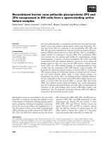
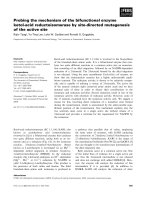
Fig. 1. Structure of the sTF–FVIIa complex [7]. sTF is displayed in
space filling and FVIIa in C-alpha ribbon. The TF1 domain is in dark
grey and the TF2 domain in light grey. The Gla domain of FVIIa is
showningreen;EGF1inmagenta;EGF2inlightblue;PDindark
blue. The sTF residues highlighted in red are located within the area of
interaction with FXa in the sTF–FVIIa–FXa–TFPI(1–161) complex
according to the fluorescence probing, whereas the two sTF residues in
yellow gave no significant fluorescence response upon FXa–TFPI(1–
161) binding.
Ó FEBS 2003 The sTF–FXa interface in sTF–FVIIa–FXa–TFPI (Eur. J. Biochem. 270) 2577
vesicles, total phospholipid concentration 5 l
M
) was studied
by mixing 0.1 n
M
FVIIa, 200 n
M
FX, and 200 n
M
sTF
variant in the buffer above. After a 10-min incubation, FXa
generation was stopped by excess EDTA and quantified
by the addition of 0.5 m
M
S-2765 (Chromogenix, Mo
¨
lndal,
Sweden). Each measurement was duplicated.
Fluorescence measurements
Fluorescence emission spectra were recorded on a Hitachi
F-4500 spectrophotometer with a thermostated cell com-
partment at a constant temperature of 23 °C. All measure-
ments were carried out using a 0.5-cm quartz cell and the
slits were set to 5 nm for both excitation and emission.
Fluorescence emission spectra for the IAEDANS-labeled
protein were recorded in the wavelength region 400–600 nm
after excitation at 350 nm. A spectrum was collected for
each labeled sTF-variant at a concentration of 0.3 l
M
.
Another spectrum was recorded for a sample containing
FVIIa in a 1.5-fold molar excess over labeled sTF.
TFPI(1)161) and FXa were incubated for 30 min in the
presence of 5 m
M
CaCl
2
, followed by the addition of sTF
and FVIIa. A spectrum was recorded for this mixture with
the final concentrations of 0.3 l
M
sTF, 0.45 l
M
FVIIa,
0.45 l
M
FXa, and 2.25 l
M
TFPI(1–161).
All reaction mixtures contained 50 l
M
PtdCho/PtdSer
in 50 m
M
Hepes, 0.15
M
NaCl, 5 m
M
CaCl
2
,pH7.5,and
the samples were incubated for 30 min. Each scan was
reproduced at least three times and the first derivative was
used to find the wavelength of the fluorescence emission
maximum.
Results
We created eight sTF variants, each with one position
mutated to a cysteine. We labeled the variants with the
fluorescent probe IAEDANS so that we could monitor the
binding of FXa by studying changes in the fluorescence
emission spectra upon AEDANS-sTF–FVIIa–FXa–
TFPI(1–161) complex formation. In this study, we focused
on the Stokes’ shifts caused by alterations in the environ-
ment around the probe when it came in contact with the
surface of FXa. Detection of such changes provides more
reliable results compared to detection of small changes in
fluorescence intensity, because the wavelength shifts of the
emission maximum do not depend on changes in concen-
trations. The effects on sTF-dependent FVIIa activity and
activation of FX as a result of mutation and labeling of sTF
were also investigated.
The ability of IAEDANS-labeled sTF variants
to support FVIIa activity
We performed a set of experiments to examine the impact
of mutation and labeling of sTF on FVIIa binding
and enhancement of FVIIa activity. The ability of the
IAEDANS-labeled sTF variants to stimulate FVIIa
was assessed and the K
d
values for FVIIa binding to
AEDANS-sTF were calculated (Table 1). These data show
that the AEDANS-sTF mutants maintained virtually
normal binding to FVIIa and normal cofactor activity
(Table 1).
Effects on fluorescence spectra upon FVIIa
and TFPI(1–161) binding
We conducted a series of measurements to ascertain
whether any fluorescence emission shifts accompanied
FVIIa binding to AEDANS-sTF, that is, we examined
possible changes in the environment surrounding the
fluorescent probe as a result of AEDANS-sTF–FVIIa
complex formation (Table 1). Three of the labeled sTF
variants (Y156C, S163C and K201C) showed a change in
emission spectra and in the rest of the mutants the probe
was not significantly affected by the sTF–FVIIa complex
formation.
As the purpose of our study was to map the
interaction between FXa and sTF in the quaternary
sTF–FVIIa–FXa–TFPI(1–161) complex, we also investi-
gated whether TFPI(1)161) contributed to the emission
changes sensed by the probe after addition of the FXa–
TFPI(1–161) complex to AEDANS-sTF–FVIIa. In this
control experiment, we used a concentration of TFPI(1–
161) that was 250 times higher than the IC
50
value that
Hamomoto et al. [6] reported when using TFPI(1–161) to
inhibit FVIIa bound to relipidated TF, which assured
stoichiometric binding of TFPI(1–161) to AEDANS-
sTF–FVIIa. Comparison of the emission spectra of
AEDANS-sTF–FVIIa with the corresponding spectra
after addition of TFPI(1–161) alone demonstrated that
the spectral shifts detected upon FXa–TFPI(1–161)
binding to AEDANS-sTF–FVIIa did not originate from
TFPI(1–161) (data not shown).
Effects on fluorescence spectra upon FXa binding
In the sTF–FVIIa complex, positions 156, 163 and 166 in
sTF are adjacent to the Gla domain of FVIIa, and positions
200 and 201 are situated near EGF1 and the so-called
hydrophobic stack, which is a linker between the Gla
domain and EGF1 of FVIIa (Fig. 1). All five of the
mentioned residues are located in the TF2 domain [7]. Three
of our mutants labeled in this region, sTF(S163C),
Table 1. Functional binding of IAEDANS-labeled sTF to FVIIa and
fluorescence emission shifts caused by formation of the AEDANS-sTF–
FVIIa complex. The K
d(app)
for wt-sTF is 7 n
M
[37]. The cofactor
activity is set to 100% for wt-sTF.
sTF
variant
K
d(app)
a
(n
M
)
Cofactor activity
a
(%)
Dk
Fmax
b
(nm)
E99C 10.6 ± 0.6 90 ± 3 ) 1.5 ± 0.0
L104C 3.8 ± 0.5 98 ± 6 + 0.3 ± 0.1
Y156C 4.0 ± 0.2 105 ± 2 ) 4.4 ± 0.2
S163C 3.8 ± 0.7 112 ± 6 ) 8.7 ± 0.1
K166C 8.3 ± 0.4 112 ± 5 + 0.8 ± 0.3
T197C 7.1 ± 1.0 93 ± 4 ) 0.4 ± 0.5
R200C 5.0 ± 0.6 100 ± 1 ) 0.3 ± 0.6
K201C 8.9 ± 0.9 90 ± 3 ) 15.0 ± 0.2
a
Calculated from amidolytic activity stimulation. The standard
error of the mean is given.
b
Dk
Fmax
¼ k
Fmax
(AEDANS-sTF–
FVIIa) – k
Fmax
(AEDANS-sTF). The calculated pooled standard
deviations are also given.
2578 K. Carlsson et al. (Eur. J. Biochem. 270) Ó FEBS 2003
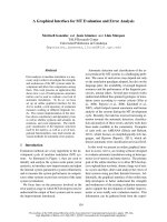
sTF(K166C), and sTF(R200C), displayed a blue shift of the
emission upon binding of FXa in the AEDANS-labeled
sTF–FVIIa–FXa–TFPI(1–161) complex (Fig. 2; Table 2).
On the other hand, FXa binding to AEDANS-sTF(K201C)
resulted in a red shift of the spectrum, probably due to
interaction with a charged group in FXa. The attached label
in position 156 did not sense any significant change in the
environment when FXa was associated with sTF in the
quaternary complex.
Residues E99 and L104 are located in the TF1 domain,
and T197 is situated in the N-terminal portion of the TF2
domain, i.e. close to the TF1 domain at the level of EGF1 in
FVIIa in the sTF–FVIIa complex (Fig. 1). Binding of FXa
caused blue shifts in the fluorescence emission spectra of
AEDANS-sTF(L104C) and AEDANS-sTF(T197C) but
did not affect the emission spectrum of AEDANS-
sTF(E99C) upon formation of the quaternary complex
(Fig. 2; Table 2).
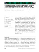
Fig. 2. Fluorescence emission spectra of the
IAEDANS-labeled sTF variants upon complex
formation. In complex with FVIIa (grey line)
and when part of the AEDANS-sTF–FVIIa–
FXa–TFPI(1–161) complex (black line).
Representative spectra are shown.
Table 2. Fluorescence emission shifts occurring upon formation of the
AEDANS-sTF–FVIIa–FXa–TFPI(1–161) complex and effects of
mutation and labeling of sTF on sTF–FVIIa-catalyzed activation of FX.
sTF variant Dk
Fmax
a
(nm)
FX activation rate (v
mutant
/v
wt
)
AEDANS-sTF
b
sTF
b
E99C ) 0.8 ± 0.2 0.93 ± 0.03 0.90 ± 0.01
L104C ) 4.8 ± 0.3 0.86 ± 0.04 1.00 ± 0.07
Y156C ) 1.0 ± 0.2 0.11 ± 0.02 0.37 ± 0.04
S163C ) 3.4 ± 0.2 0.01 ± 0.00 0.01 ± 0.00
K166C ) 5.0 ± 0.5 0.03 ± 0.02 0.01 ± 0.01
T197C ) 5.6 ± 0.5 0.57 ± 0.05 0.66 ± 0.02
R200C ) 6.3 ± 0.6 0.32 ± 0.03 0.23 ± 0.00
K201C + 4.6 ± 0.2 0.13 ± 0.01 0.78 ± 0.04
a
Dk
Fmax
¼ k
Fmax
[AEDANS-sTF–FVIIa–FXa–TFPI(1–161)] –
k
Fmax
(AEDANS-sTF–FVIIa). The calculated pooled standard
deviations are also given.
b
The standard error of the mean is given.
Ó FEBS 2003 The sTF–FXa interface in sTF–FVIIa–FXa–TFPI (Eur. J. Biochem. 270) 2579
The ability of the sTF variants to support FX activation
We used a two-stage amidolytic assay to assess the ability of
the labeled and unlabeled sTF variants to support FVIIa-
catalyzed activation of FX. In Table 2, the cofactor activity
of the individual sTF variants is expressed as a ratio between
the rates of FX activation with the mutated form of sTF and
wild-type sTF, respectively, bound to FVIIa. Replacement
of the wild-type residue by Cys decreased the FX activation
rate for all of the studied positions in sTF except sTF(E99C)
and sTF(L104C) which retained normal cofactor activity.
The effect on the FX activation rates as a result of
attachment of the fluorescent probe IAEDANS was also
monitored by comparing the data with the corresponding
values for the unlabeled sTF variant (Table 2). Linking
IAEDANStosTF(R200C)resultedinaslightrecoveryof
FX activation ability, whereas labeling of sTF(Y156C) and
sTF(K201C) led to a significantly decreased ability to
stimulate activation of FX. Labeling of the remaining
variants did not significantly affect the cofactor activity.
The rates of FX activation with IAEDANS-labeled
sTF(Y156C), sTF(S163C), sTF(K166C), sTF(R200C), and
sTF(K201C) were markedly decreased (to 1–30% of the
activation induced by wild-type sTF; Table 2). Similar
results have been obtained by Kirchhofer et al. [13] when
using mutants with primarily Ala substitutions to achieve
functional mapping of the sTF–FX interaction in the sTF–
FVIIa–FX ternary complex. Compared with wild-type sTF,
AEDANS-sTF(E99C) and AEDANS-sTF(L104C) were
nearly as effective in activating FX, and sTF(T197C) was
somewhat less efficient (i.e. it induced 57% of the activation
obtained with wild-type sTF; Table 2).
Discussion
Formation of the TF–FVIIa–FXa–TFPI complex is an
important event in the regulation of coagulation, because it
inhibits the function of TF–FVIIa. The quaternary complex
mediates translocation of the inhibited TF–FVIIa complex
into caveolae [4], and, in some types of cells, the presence of
TFPI influences the rate of internalization and degradation
of TF–FVIIa [5]. However, no crystallographic data have
been obtained on the large sTF–FVIIa–FXa–TFPI com-
plex. From functional studies only limited results have been
published regarding the binding area between sTF and FXa
in this complex.
Regarding the ternary sTF–FVIIa–FX complex other
investigators have performed functional analyses to investi-
gate the binding region between sTF and FX. Kirchhofer
and coworkers [13] reported mutagenesis of surface-exposed
residues of sTF and the effect of the modified sTF–FVIIa
complex on FX activation. In another study [17], these
authors also examined the interaction between FXa and
sTF–FVII in a similar way by observing the impact of
mutation in sTF on activation of FVII by FXa.
The aim of the present work is to map in more detail the
interaction area between sTF and FXa in the quaternary
complex than previously carried out. This will also allow a
comparison of the binding interface between sTF and FXa
in the quaternary and ternary complexes. We applied a site-
directed fluorescence labeling approach that can determine
whether the labeled positions are involved in the interaction
between the proteins in the complex. The main advantage of
this experimental strategy is that it permits direct mapping
of the contact area; by comparison, in methods such as
alanine scanning, structural effects caused by mutagenesis
can affect the function without the mutation necessarily
being located in the binding interface.
We selected eight amino acid residues in sTF for
individual cysteine replacement and used the cysteines as
handles to which we attached the fluorescent probe
IAEDANS. The residues were chosen on the basis of
X-ray data [7], which showed that they were not in direct
contact with FVIIa in the sTF–FVIIa complex. Accord-
ingly, the AEDANS-sTF mutants retained virtually normal
affinity for FVIIa (Table 1). For five of the mutants,
essentially no change in emission spectra could be detected
upon formation of the AEDANS-sTF–FVIIa complex
(Table 1). However, three of the mutants, AEDANS-
sTF(Y156C), AEDANS-sTF(S163C), and AEDANS-
sTF(K201C), showed significantly altered emission spectra
upon complex formation. This means that the introduced
fluorophore was affected by FVIIa association with sTF.
However, in this case it is unlikely that the probe is located
in the interaction area between FVIIa and sTF, as the
binding strength between these two proteins was not
decreased for the labeled variants (Table 1). In accordance
with this, we have previously observed rather large changes
in K
d
for sTF–FVIIa complexes when containing AEDANS
labels within the contact interface [27]. Thus it is likely that
the probes in positions 156, 163 and 201 are just pointing
towards the border between sTF and FVIIa.
Based on our fluorescence emission shift data, it can be
seen that part of the C-terminal region of sTF (i.e. positions
163, 166, 200 and 201) interacts with FXa in the sTF–
FVIIa–FXa–TFPI(1–161) complex (Figs 1 and 2; Table 2).
This agrees well with what Rao and Ruf [18] have suggested
for position 166 in TF when part of the quaternary complex
based on activity measurements. Interestingly, the observed
interaction pattern between sTF and FXa in the quaternary
complex seems to be similar in this region to the one
proposed for the ternary sTF–FVIIa–FX and sTF–FVII–
FXa complexes based on the results of functional studies
[11,13,17]. Combining the fluorescence shifts of the
AEDANS-sTF–FVIIa and AEDANS-sTF–FVIIa–FXa–
TFPI(1–161) complexes (Tables 1 and 2) gives further
information that indicates that FXa is in direct contact with
or is located very close to the border of the Gla domain of
FVIIa around position 163 and the hydrophobic stack
around position 201. This is implied because, according to
the AEDANS-sTF–FVIIa data, the reporter group from
these positions was pointing towards FVIIa, and association
of FXa gave rise to an additional spectral shift, thus both
FVIIa and FXa should have been in contact with the
fluorophore.
Based on the structural similarity between FVIIa and
FXa [28–30], as well as similar orientation of FVIIa and
FXa relative to the membrane [7,31–33], it is plausible that
the Gla domain of FXa is involved in binding to the
C-terminal domain of sTF. As we found that FXa is bound
next to the Gla domain of FVIIa, it is highly likely that the
Gla domains of FVIIa and FXa interact with each other in
the quaternary complex. Interestingly, previous reports
indicate that the Gla domain of FVIIa/FVII also plays a
2580 K. Carlsson et al. (Eur. J. Biochem. 270) Ó FEBS 2003
role in recognition of FX/FXa in the ternary complexes
[8–10,34].
On the other hand, the fluorescent label linked to position
156 in the C-terminal part of sTF between positions 163 and
201 (Fig. 1) was not affected by incorporation of FXa
into the AEDANS-sTF–FVIIa–FXa–TFPI(1)161) com-
plex. Hence, FXa appears to be positioned some distance
from the FVIIa Gla domain, probably over the side chain of
Tyr156 in sTF, as indicated by substrate activation meas-
urements in this study (Table 2). Similar measurements on
the ternary sTF–FVIIa–FX and sTF–FVII–FXa complexes
indicate that this residue indeed is involved in the FX/FXa
contact area [13,17].
Residues E99, L104, and T197 are located very close to
each other in an area of the C-terminal part of the TF1
domain and the N-terminal portion of the TF2 domain
(Fig. 1). Mutation and labeling of positions 99 and 104 had
little effect on the FX-activating ability of the AEDANS-
sTF–FVIIa complex. Mutation and labeling of position 197
resulted in a somewhat lower rate of FX activation
(Table 2). In addition, when using AEDANS-sTF(E99C),
we found no significant change in emission upon formation
of the AEDANS-sTF–FVIIa–FXa–TFPI(1)161) complex,
indicating that position 99 in sTF is not involved in the
binding of FXa. In contrast, for both AEDANS-
sTF(L104C) and AEDANS-sTF(T197C), formation of
the AEDANS-sTF–FVIIa–FXa–TFPI(1)161) complex
resulted in alterations in the environment of the probe,
detected as shifts in the emission spectra. These shifts imply
that positions 104 and 197 are involved in binding to FXa in
the quaternary complex, although the two residues, in
particular 104, are not essential for activation of the
substrate FX. FXa and FX seem to bind to sTF in a
similar way in the ternary sTF–FVII–FXa/FX complexes
[13,17,35]. Provided that in this region FX in the ternary
complex does not bind differently than FXa in the
quaternary sTF–FVIIa–FXa–TFPI(1–161) complex the
results of our activity experiments and our direct fluores-
cence binding studies for position 104 are contradictory.
This emphasizes the risk of arriving at definitive conclusions
concerning the binding interface between sTF and FXa
based solely on data provided by activity studies, especially
when the active site is located far from the site of mutation.
Thus, we have demonstrated that the region of inter-
action between sTF and FXa in sTF–FVIIa–FXa–
TFPI(1)161) involves not only the C-terminal part of
sTF, but also an area in the N-terminal part of TF2 (around
position 197) and a region in the C-terminal part of TF1
(around position 104) (Fig. 1). However, the entire
C-terminal part of TF1 is not involved in the FXa
interaction area, because position 99 does not participate
in the binding. This region is found at the level of EGF1 of
FVIIa in the sTF–FVIIa complex. However, in the previ-
ously mentioned studies by Kirchhofer et al. [13,17], these
positions were not probed, hence they might also be
involved in the interaction region in the ternary complexes
sTF–FVII–FXa and sTF–FVIIa–FX.
The structural analogy between FVIIa and FXa (dis-
cussed above) in combination with our fluorescence meas-
urement results suggests that, similar to the Gla domain,
EGF1 of FXa is involved in binding to sTF in the
quaternary sTF–FVIIa–FXa–TFPI(1–161) complex. In this
context, it should be noted that also for the ternary
TF–FVIIa–FX complex it has been demonstrated that FX
interactswithTFusing,inpart,theEGF1domain[36].
To conclude, we have described the interaction region
between sTF and FXa in the quaternary complex, at the
level of specific residues, and we have also shown major
similarities regarding this interaction area in the quaternary
and ternary complexes. Increased knowledge about the
interface between FX/FXa and TF in these complexes
would facilitate the design of small molecules with anti-
thrombotic effects. In any case, potential contacts between
sTF and FXa at the level of EGF2 and the PD of FVIIa in
the sTF–FVIIa–FXa–TFPI(1–161) complex remain to be
identified.
Acknowledgements
This work was supported by the Swedish Research Council (MS, UC)
and Magnus Bergvall Stiftelse (MS). We thank Helle Bak for technical
assistance.
References
1. Golino, P. (2002) The inhibitors of the tissue factor: factor VII
pathway. Thromb. Res. 106, V257–V265.
2. Broze, G.J. Jr, Girard, T.J. & Novotny, W.F. (1990) Regulation of
coagulation by a multivalent Kunitz-type inhibitor. Biochemistry
29, 7539–7546.
3. Girard, T.J., Warren, L.A., Novotny, W.F., Likert, K.M., Brown,
S.G., Miletich, J.P. & Broze, G.J. Jr (1989) Functional significance
of the Kunitz-type inhibitory domains of lipoprotein-associated
coagulation inhibitor. Nature 338, 518–520.
4. Sevinsky, J.R., Rao, L.V.M. & Ruf, W. (1996) Ligand-induced
protease receptor translocation into caveolae: a mechanism for
regulating cell surface proteolysis of the tissue factor-dependent
coagulation pathway. J. Cell. Biol. 133, 293–304.
5. Iakhiaev, A., Pendurthi, U.R., Voigt, J., Ezban, M. & Rao,
L.V.M. (1999) Catabolism of factor VIIa bound to tissue factor in
fibroblasts in the presence and absence of tissue factor pathway
inhibitor. J. Biol. Chem. 274, 36995–37003.
6. Hamamoto, T., Yamamoto, M., Nordfang, O., Petersen, J.G.L.,
Foster,D.C.&Kisiel,W.(1993)Inhibitorypropertiesoffull-
length and truncated recombinant tissue factor pathway inhibitor
(TFPI). Evidence that the third Kunitz-type domain of TFPI is
not essential for the inhibition of factor VIIa- tissue factor com-
plexes on cell surfaces. J. Biol. Chem. 268, 8704–8710.
7. Banner, D.W., D’Arcy, A., Che
`
ne, C., Winkler, F.K., Guha, A.,
Konigsberg, W.H., Nemerson, Y. & Kirchhofer, D. (1996) The
crystal structure of the complex of blood coagulation factor VIIa
with soluble tissue factor. Nature 380, 41–46.
8. Huang, Q., Neuenschwander, P.F., Rezaie, A.R. & Morrissey,
J.H. (1996) Substrate recognition by tissue factor-factor VIIa.
Evidence for interaction of residues Lys165 and Lys166 of tissue
factor with the 4-carboxyglutamate-rich domain of factor X.
J. Biol. Chem. 271, 21752–21757.
9. Ruf, W., Shobe, J., Rao, S.M., Dickinson, C.D., Olson, A. &
Edgington, T.S. (1999) Importance of factor VIIa Gla-domain
residue Arg-36 for recognition of the macromolecular substrate
factor X Gla-domain. Biochemistry 38, 1957–1966.
10. Martin, D.M.A., O’Brien, D.P., Tuddenham, E.G.D. & Byfield,
P.G.H. (1993) Synthesis and characterization of wild-type and
variant c-carboxyglutamic acid-containing domains of factor VII.
Biochemistry 32, 13949–13955.
11. Ruf, W., Miles, D.J., Rehemtulla, A. & Edgington, T.S. (1992)
Tissue factor residues 157–167 are required for efficient
Ó FEBS 2003 The sTF–FXa interface in sTF–FVIIa–FXa–TFPI (Eur. J. Biochem. 270) 2581
proteolytic activation of factor X and factor VII. J. Biol. Chem.
267, 22206–22210.
12. Rehemtulla, A., Ruf, W., Miles, D.J. & Edgington, T.S. (1992)
The third Trp-Lys-Ser (WKS) tripeptide motif in tissue factor is
associated with a function site. Biochem. J. 282, 737–740.
13. Kirchhofer, D., Lipari, M.T., Moran, P., Eigenbrot, C. & Kelley,
R.F. (2000) The tissue factor region that interacts with substrates
factor IX and factor X. Biochemistry 39, 7380–7387.
14. Roy, S., Hass, P.E., Bourell, J.H., Henzel, W.J. & Vehar, G.A.
(1991) Lysine residues 165 and 166 are essential for the cofactor
function of tissue factor. J. Biol. Chem. 266, 22062–22066.
15. Ruf, W., Miles, D.J., Rehemtulla, A. & Edgington, T.S. (1992)
Cofactor residues lysine 165 and 166 are critical for protein sub-
strate recognition by the tissue factor–factor VIIa protease com-
plex. J. Biol. Chem. 267, 6375–6381.
16. Dittmar, S., Ruf, W. & Edgington, T.S. (1997) Influence of
mutations in tissue factor on the fine specificity of macro-
molecular substrate activation. Biochem. J. 321, 787–793.
17. Kirchhofer, D., Eigenbrot, C., Lipari, M.T., Moran, P., Peek,
M. & Kelley, R.F. (2001) The tissue factor region that interacts
with factor Xa in the activation of factor VII. Biochemistry 40,
675–682.
18. Rao, L.V.M. & Ruf, W. (1995) Tissue factor residues Lys165 and
Lys166 are essential for rapid formation of the quaternary com-
plex of tissue factor-VIIa with Xa-tissue factor pathway inhibitor.
Biochemistry 34, 10867–10871.
19. Girard, T.J., MacPhail, L.A., Likert, K.M., Novotny, W.F.,
Miletich, J.P. & Broze, G.J. Jr (1990) Inhibition of factor VIIa-
tissue factor coagulation activity by a hybrid protein. Science 248,
1421–1424.
20. Warn-Cramer,B.J.,Rao,L.V.M.,Maki,S.L.&Rapaport,S.I.
(1988) Modifications of extrinsic pathway inhibitor (EPI) and
FXa that affect their ability to interact and to inhibit factor VIIa/
tissue factor: evidence for a two-step model of inhibition. Thromb.
Haemost. 60, 453–456.
21. Rao, L.V.M., Williams, T. & Rapaport, S.I. (1996) Studies of
the activation of factor VII bound to tissue factor. Blood 87,
3738–3748.
22. Owenius, R., O
¨
sterlund, M., Lindgren, M., Svensson, M., Olsen,
O.H., Persson, E., Freskga
˚
rd, P O. & Carlsson, U. (1999) Prop-
erties of spin and fluorescent labels at a receptor–ligand interface.
Biophys. J. 77, 2237–2250.
23. Gill, S.C. & von Hippel, P.H. (1989) Calculation of protein
extinction coefficients from amino acid sequence data. Anal.
Biochem. 182, 319–326.
24. Thim, L., Bjoern, S., Christensen, M., Nicolaisen, E.M., Lund-
Hansen, T., Pedersen, A.H. & Hedner, U. (1988) Amino acid
sequence and posttranslational modifications of human factor
VIIa from plasma and transfected baby hamster kidney cells.
Biochemistry 27, 7785–7793.
25. Petersen, J.G.L., Meyn, G., Rasmussen, J.S., Petersen, J., Bjørn,
S.E., Jonassen, I., Christiansen, L. & Nordfang, O. (1993) Char-
acterization of human tissue factor pathway inhibitor variants
expressed in Saccharomyces cerevisiae. J. Biol. Chem. 268, 13344–
13351.
26. Freskga
˚
rd, P O., Olsen, O.H. & Persson, E. (1996) Structural
changes in factor VIIa induced by Ca
2+
and tissue factor studied
using circular dichroism spectroscopy. Protein Sci. 5, 1531–1540.
27. Owenius, R., O
¨
sterlund, M., Svensson, M., Lindgren, M., Persson,
E., Freskga
˚
rd, P O. & Carlsson, U. (2001) Spin and fluorescent
probing of the binding interface between tissue factor and factor
VIIa at multiple sites. Biophys. J. 81, 2357–2369.
28. Pike, A.C.W., Brzozowski, A.M., Roberts, S.M., Olsen, O.H. &
Persson, E. (1999) Structure of human factor VIIa and its
implications for the triggering of blood coagulation. Proc. Natl
Acad. Sci. USA 96, 8925–8930.
29. Padmanabhan, K., Padmanabhan, K.P., Tulinsky, A., Park,
C.H., Bode, W., Huber, R., Blankenship, D.T., Cardin, A.D. &
Kisiel, W. (1993) Structure of human des (1–45) factor Xa at 2.2 A
˚
resolution. J. Mol. Biol. 232, 947–966.
30. Brandstetter, H., Ku
¨
hne, A., Bode, W., Huber, R., von der Saal,
W., Wirthensohn, K. & Engh, R.A. (1996) X-ray structure of
active site-inhibited clotting factor Xa. Implications for drug
design and substrate recognition. J. Biol. Chem. 271, 29988–29992.
31. Husten, E.J., Esmon, C.T. & Johnson, A.E. (1987) The active site
of blood coagulation factor Xa. Its distance from the phospholipid
surface and its conformational sensitivity to components of the
prothrombinase complex. J. Biol. Chem. 262, 12953–12961.
32. McCallum, C.D., Hapak, R.C., Neuenschwander, P.F., Morris-
sey, J.H. & Johnson, A.E. (1996) The location of the active site of
blood coagulation factor VIIa above the membrane surface and its
reorientation upon association with tissue factor. A fluorescence
energy transfer study. Biol. Chem. 271, 28168–28175.
33. McCallum,C.D.,Su,B.,Neuenschwander,P.F.,Morrissey,J.H.
& Johnson, A.E. (1997) Tissue factor positions and maintains the
factor VIIa active site far above the membrane surface even in the
absence of the factor VIIa Gla domain. A fluorescence resonance
energy transfer study. J. Biol. Chem. 272, 30160–30166.
34. Ruf, W., Kalnik, M.W., Lund-Hansen, T. & Edgington, T.S.
(1991) Characterization of factor VII association with tissue factor
in solution. High and low affinity calcium binding sites in factor
VII contribute to functionally distinct interactions. J. Biol. Chem.
266, 15719–15725.
35. Baugh, R.J., Dickinson, C.D., Ruf, W. & Krishnaswamy, S.
(2000) Exosite interactions determine the affinity of factor X for
the extrinsic Xase complex. J. Biol. Chem. 275, 28826–28833.
36. Zhong, D., Bajaj, M.S., Schmidt, A.E. & Bajaj, S.P. (2002) The
N-terminal epidermal growth factor-like domain in factor IX and
factor X represents an important recognition motif for binding to
tissue factor. J. Biol. Chem. 277, 3622–3631.
37. O
¨
sterlund, M., Owenius, R., Carlsson, K., Carlsson, U., Persson,
E., Lindgren, M., Freskga
˚
rd, P O. & Svensson, M. (2001) Prob-
ing inhibitor-induced conformational changes along the interface
between tissue factor and factor VIIa. Biochemistry 40, 9324–9328.
2582 K. Carlsson et al. (Eur. J. Biochem. 270) Ó FEBS 2003