Báo cáo khoa học: Procarboxypeptidase A from the insect pest Helicoverpa armigera and its derived enzyme potx
Bạn đang xem bản rút gọn của tài liệu. Xem và tải ngay bản đầy đủ của tài liệu tại đây (287.17 KB, 10 trang )
Procarboxypeptidase A from the insect pest
Helicoverpa armigera
and its derived enzyme
Two forms with new functional properties
Alex Baye
´
s
1
, Anka Sonnenschein
2
, Xavier Daura
3
, Josep Vendrell
1
and Francesc X. Aviles
1
1
Departament de Bioquı
´
mica i Biologia Molecular, Facultat de Cie
`
ncies and Institut de Biotecnologia i Biomedicina,
Universitat Auto
`
noma de Barcelona, Spain;
2
Klinik fu
¨
r Neurologie, Universita
¨
tsklinikum der Technischen Universita
¨
t,
Dresden, Germany;
3
Institucio
´
Catalana de Recerca i Estudis Avanc¸ ats (ICREA) and Institut de
Biotecnologia i Biomedicina, Universitat Auto
`
noma de Barcelona, Spain
Although there is a significant knowledge about mammalian
metallocarboxypeptidases, the data available on this family
of enzymes is very poor for invertebrate forms. Here we
present the biochemical characterization of a metallocarb-
oxypeptidase from the insect Helicoverpa armigera (Lepi-
doptera: Noctuidae), a devastating pest spread in subtropical
regions of Europe, Asia, Africa and Oceania. The zymogen
of this carboxypeptidase (PCPAHa) has been expressed at
high levels in a Pichia pastoris system and shown to display
the characteristics of the enzyme purified from the insect
midgut. The in vitro activation process of the proenzyme
differs significantly from the mammalian ones. The lysine-
specific endoprotease LysC activates PCPAHa four times
more efficiently than trypsin, the general activating enzyme
for all previously studied metalloprocarboxypeptidases.
LysC and trypsin independently use two different activation
targets and the presence of sugars in the vicinity of the LysC
activation point affects the activation process, indicating a
possible modulation of the activation mechanism. During
the activation with LysC the prodomain is degraded, while
the carboxypeptidase moiety remains intact except for a
C-terminal octapeptide that is rapidly released. Interestingly,
the sequence at the cleavage point for the release of the
octapeptide is also found at the boundary between the
activation peptide and the enzyme moieties. The active
enzyme (CPAHa) is shown to have a very broad substrate
specificity, as it appears to be the only known metallocarb-
oxypeptidase capable of efficiently hydrolysing basic and
aliphatic residues and, to a much lower extent, acidic resi-
dues. Two carboxypeptidase inhibitors, from potato and
leech, were tested against CPAHa. The former, of vegetal
origin, is the most efficient metallocarboxypeptidase
inhibitor described so far, with a K
i
in the p
M
range.
Keywords: metallocarboxypeptidase; zymogen; proteolytic
activation; substrate specificity; protein inhibitor.
The understanding of the digestive process in pest insects
is a key step in the design of many insecticides, including
insect-resistant transgenic plants [1]. Exopeptidases are
supposed to play a major role in protein digestion, as
peptides and proteins have to be converted into dipeptides
or single amino acids in order to be taken up efficiently by
the gut. These proteases are well described in mammals,
but little is known about the exoproteases of insect origin.
Helicoverpa armigera (Lepidoptera: Noctuidae), also
known as cotton worm or boll worm, has a widespread
distribution in tropical, subtropical and warm temperature
regions in Europe, Asia, Africa and Oceania. It is an
important pest of many crop plants, including cotton,
corn, maize, tomato, bean, sorghum, tobacco and certain
flower plants such as chrysanthemum or carnation. The
losses due to Helicoverpa zea and Helicoverpa virescens,
two butterflies that belong to the genre of Helicoverpa
armigera, were calculated to be one thousand million
dollars per year in the USA [2]. A midgut carboxypept-
idase from this lepidopter, first described by Bown et al.
[3], is the subject of the present work.
From a mechanistic point of view, two major types of
carboxypeptidases can be distinguished: serinecarboxy-
peptidases and metallocarboxypeptidases. In mammals the
metallocarboxypeptidase family is divided into subfamilies
Correspondence to J. Vendrell, Departament de Bioquı
´
mica i
Biologia Molecular, Facultat de Cie
`
ncies, Universitat Auto
`
noma de
Barcelona, E-08193 Bellaterra, Spain.
Fax: + 34 93 5811264, Tel.: + 34 93 5812375,
E-mail:
or F. X. Aviles, Institut de Biotecnologia i Biomedicina, Universitat
Auto
`
noma de Barcelona, E-08193, Bellaterra, Spain.
Fax: + 34 93 5812011, Tel.: + 34 93 5811315,
E-mail:
Abbreviations: PCPAHa, procarboxypeptidase from Helicoverpa
armigera;PCPAHaa, procarboxypeptidase a from Helicoverpa
armigera; CPAHa, carboxypeptidase from Helicoverpa armigera;
CPA1h, human carboxypeptidase A1; CPA2h, human carboxypepti-
dase A2; CPBh, human carboxypeptidase B; CPAb, bovine carb-
oxypeptidase A; LysC, lysyl endopeptidase; PCI, potato
carboxypeptidase inhibitor; LCI, leech carboxypeptidase inhibitor;
AOX1, alcohol oxidase gene; AAFP, N-(4-methoxyphenyl-
azoformyl)-
L
-phenylalanine; Cbz, carbobenzoxy; N-(3-(2-furyl),
acryloyl)-
L
-phenylalanyl-
L
-phenylalanine.
Enzymes: CPAHa SWP: O97434 (E.C. 3.4.17.1); CPA1 h SWP:
P15085 (E.C. 3.4.17.1); CPA2 h SWP: P48052 (E.C. 3.4.17.15); CPBh
SWP: P15086 (E.C. 3.4.17.2); CPAb SWP: P00730 (E.C. 3.4.17.1).
(Received 20 March 2003, revised 8 May 2003, accepted 21 May 2003)
Eur. J. Biochem. 270, 3026–3035 (2003) Ó FEBS 2003 doi:10.1046/j.1432-1033.2003.03681.x
A/B and N/E [4] which include, respectively, the formerly
called pancreatic-like and regulatory forms, the latter
referring to a number of enzymes involved in the processing
of bioactive peptides and hormones [4,5]. The carboxy-
peptidase of H. armigera belongs to the A/B subfamily and
contains a Zn
2+
atom directly involved in catalysis. From
its localization in the gut of the larvae, it is thought to
participate in the digestive process of the insect.
Two forms of pancreatic-like carboxypeptidases, CPA
and CPB, are involved in the degradation of dietary
proteins. The two isoforms of CPA, A1 and A2, differ in
specificity with the former having a preference for aliphatic
and aromatic C-terminal residues and the latter being more
restrictive for aromatic residues, particularly tryptophan
[5–7]. The B form is highly specific for basic residues.
Pancreatic-like carboxypeptidases are synthesized as pro-
enzymes. Upon tryptic activation, a 92–95 residue
N-terminal activation segment, that shields the entrance of
substrates to the active site, is released. This proregion,
besides acting as a potent inhibitor of the enzyme (in the n
M
range) [5,8], also behaves as an intramolecular chaperone
for the folding of the enzyme.
We have recently described [9] the three-dimensional
crystal structure of an A-type metalloprocarboxypeptidase
from H. armigera (PCPAHa), showing that its overall fold
and conformation is very much similar to other known zinc
procarboxypeptidases, indicating the conservation of these
features through evolution.
In the present study we report the production of this
zymogen at high yield in the methylotrophic yeast Pichia
pastoris, a fact that allowed the study of the biochemical
properties of both the proenzyme and enzyme forms.
Through the description of the proenzyme activation
process, the substrate specificity of the active enzyme and
the behaviour upon inhibition by two well known natural
inhibitors, a number of specific, distinctive features can be
deduced for this new member of the family of pancreatic-
like procarboxypeptidases.
Experimental procedures
Materials
Restriction endonucleases AvrII, SacIandXhoI, T4 DNA
ligase, Taq polymerase, deoxynucleotide stocks and N-gly-
cosidase F were purchased from Roche. Salts and media for
E. coli and P. pastoris growth were obtained from Sigma
and Hispanlab (Alcobendas, Spain), respectively. The
P. pastoris expression kit was purchased from Invitrogen.
Trypsin (treated with tosylphenylalanyl chloromethyl ke-
tone) was from Worthington (Lakewood, USA) and Lysyl
endopeptidase (from Achromobacter lyticus)fromWaco.
Chymotrypsin was from Merck. The peptides V14R, V15E
and V14W where synthesized by Diverdrugs (Barcelona,
Spain). Poly(vinylidene difluoride) (PVDF) membrane was
from Waters. Elastase, trifluoroacetic acid, cyanogen bro-
mide, synapinic acid, N-(3-(2-furyl) acryloyl)-
L
-phenylala-
nyl-
L
-phenylalanine (FAPP) and Cbz-Gly-Gly-Ser were
from Sigma. N-(4-Methoxyphenylazoformyl)-
L
-phenyl-
alanine (AAFP) and the rest of substrates used for the
kinetic measurements were from Bachem (Bubendorf,
Switzerland).
Plasmids constructs
DNA manipulations were carried out essentially as des-
cribed by Sambrook et al. [10] using E. coli strain MC1061
as host. Primers were synthesized to amplify the cDNA
containing the procarboxypeptidase by PCR. Sense primer
5¢-GATTCT
CTCGAGAAAAGAAAACATGAAATTT
ATGATGG-3¢; antisense primer 5¢-CTTCTTTGAGT
TATGACGAATT
GGATCCTAC-3¢. The original signal
peptide from this molecule is not included in the construct.
The underlined sequences indicate the restriction sites for
XhoIandAvrII introduced to be able to subclone the cDNA
into the P. pastoris expression vector pPIC9. The cDNA
was introduced between the 5¢ promoter and 3¢ terminator
of the alcohol oxidase gene (AOX1), resulting in a new
vector called pPIC9-PCPAHa. This vector provides the
a-mating factor signal for secretion of the recombinant
protein.
Transformation and selection of the productive clones
Prior to the transformation the vector was linearized with
SacI. The KM 71 strain of P. pastoris, which produces only
the slow growing phenotype, was transformed using the
spheroplasts method with the linearized vector. The cells
where then plated on minimal dextrose medium (MD) agar
(1.34% yeast nitrogen base, 0.00004% biotin, and 1%
dextrose) a medium devoid of histidine where only the
transformed cells can grow. To find a highly producing
clone, over 60 colonies were grown in 10 mL buffered
glycerol-complex (BMGY) medium (1% yeast extract, 2%
peptone, 90 m
M
potassium phosphate, pH 6.0, 0.00004%
biotin and 1% glycerol) at 30 °C for 3 days. Cells were
collected by centrifugation and resuspended in 2 mL
buffered methanol-complex medium (BMMY) medium
(same as BMGY but containing 1% methanol instead of
1% glycerol) and grown for 3 days more to induce the
production of recombinant protein. The supernatant of all
the clones was analysed by SDS/PAGE, followed by
densitometry to identify the most productive ones. The
functionality of the recombinant protein was tested with the
specific substrate FAPP(N-(3-(2-furyl) acryloyl)-
L
-phenyl-
alanyl-
L
-phenylalanine) [11] after activation of the pro-
enzyme with trypsin.
Expression and purification of the recombinant enzyme
Expression and purification procedures were carried out
essentially as described in [9]. In short, 1 L of BMGY
medium was grown at 30 °C and at 300 r.p.m. constant
shaking for 2 days until D
600
reached 20 units. The cells
were then collected by centrifugation at 1500 g and gently
resuspended in 200 mL of BMMY medium. In a first step,
the protein secreted to the supernatant was purified by
hydrophobic interaction chromatography in a butyl-
Toyopearl 650M column. The sample was loaded onto
the column after equilibration of its ionic strength to 30%
saturation with ammonium sulphate, and the protein was
eluted with a decreasing gradient of the same salt. After
overnight dialysis of the selected fractions, the protein was
finally purified on an FPLC system using a preparative
anion-exchange column (TSK-DEAE 5PW; TOSOH,
Ó FEBS 2003 A metalloprocarboxypeptidase from Helicoverpa armigera (Eur. J. Biochem. 270) 3027
Tokyo, Japan) and applying a 65 min gradient from 100%
buffer A (20 m
M
Tris, pH 7.0) to 15% buffer B (buffer A
plus 0.8
M
ammonium acetate).
Activity assays
Two different synthetic substrates were used to analyse
carboxypeptidase A activity. N-(3-(2-furyl) acryloyl)-
L
-
phenylalanyl-
L
-phenylalanine (FAPP) was used routinely
to measure carboxypeptidase activity and AAFP was
used to calculate the inhibition constants [12]. FAPP was
prepared at a 0.2 m
M
concentration in 50 m
M
Tris,
0.45
M
NaCl, pH 7.5, and the A
330
decrease at 25 °C. A
stock solution of 50 m
M
N-(4-methoxyphenylazoformyl)-
L
-phenylalanine (AAFP) in dimethylsulfoxide was diluted
immediately before use to 10 m
M
with 50 m
M
Tris, 0.1
M
NaCl, pH 8.0. From this solution, 10 lL were added to
1mL of 50m
M
Tris, 0.1
M
NaCl, pH 8.0. CPA activity
was measured by following the A
350
decrease at 25 °C.
Deglycosylation assay
Samples were deglycosylated with N-glycosidase F, an
enzyme that removes N-linked sugars by cleaving the bond
between the asparagines from the polypeptide chain and
the first N-acetylglucosamine. Glycosylated molecules were
concentrated at 1 mgÆmL
)1
in Tris 5 m
M
pH 8.0 and
appropriate volumes of N-glycosidase F at 1 unit lL
)1
were
added to achieve a final ratio of 100 : 1 v/v. The reaction
was left to proceed overnight at 37 °C.
Kinetic measurements
The rate of hydrolysis of the different substrates were
measured spectrophotometrically in 50 m
M
Tris, 0.5
M
NaCl, 1 l
M
ZnCl
2
, pH 8.0, at 25 °C. The wavelengths used
to monitor the various reactions were as follows: 226 nm
for Cbz-Gly-Gly-Ser, Cbz-Gly-Gly-Ala, Cbz-Gly-Gly-Leu,
Cbz-Gly-Gly-Val, Cbz-Gly-Gly-Phe and Cbz-Gly-Phe;
236 nm for Cbz-Gly-Gly-Tyr and Cbz-Gly-Tyr; and
302 nm for Cbz-Gly-Gly-Trp and Cbz-Gly-Trp. Initial
rates, determined from the first 5–10% of the time-trace of
each reaction, were obtained at substrate concentrations
close to the K
m
value whenever possible. The kinetic
parameters, k
cat
and K
m
, were obtained using 6–8 experi-
mental points by direct fit to a Michaelis–Menten curve
using the
ENZFITTER
program [13].
Activation studies of recombinant
H. armigera
PCPAHa
Recombinant enzyme at 1 mgÆmL
)1
in 5 m
M
Tris, 1 l
M
ZnCl
2
, pH 8.0, was treated with lysyl endopeptidase
(LysC) at a PCPAHa : LysC ratio of 40 : 1 (w/w) and at
37 °C. To avoid the action of active carboxypeptidase
upon the fragments generated, the potato carboxypep-
tidase inhibitor (PCI) was also added to the mixture at a
1 : 4 PCPHa/PCI molar ratio when enzymatic activity
was not going to be measured. During the activation
process, aliquots were taken for reverse-phase HPLC
analysis and activity measurements. Seventy microlitres
of the reaction mixture, with trifluoroacetic acid added to
a concentration of 0.05% (v/v) to stop the activation
reaction, were analysed in a Vydac C
4
column (250 ·
4.6 mm, 5 lmparticlesizeand0.3lm pore size). The
chromatographies were performed in the presence of
0.1% trifluoroacetic acid with an elution gradient
between water (solvent A) and 90% acetonitrile (solvent
B) according to the following steps: 10% solvent B from
0 to 10 min, 10–60% solvent B from 10 to 130 min.
Elution was followed by measuring the A
214
and the
isolated fractions were concentrated in an Speed-Vac
(Savant) and further analysed by MALDI-TOF spectr-
ometry, SDS/PAGE and N-terminal sequencing. Parallel
10 lL aliquots of the activation mixture were added to
190 lL of aprotinin (bovine pancreas trypsin inhibitor) at
0.1 mgÆmL
)1
in 20 m
M
Tris, 0.1
M
NaCl, 1 l
M
ZnCl
2
,
pH 8.0, and 10 lL of the resulting mixture were used to
measure enzyme activities using FAPP as a substrate.
To analyse the effect of sugars on the activation of
PCPAHa by LysC and bovine trypsin, glycosylated and
nonglycosylated PCPAHa were activated with increasing
PCPAHa/activating enzyme ratios at 37 °C for 2 h. One
microlitre of the reaction mixture was assayed against FAPP.
Triplicate measures were obtained for each data point.
Cyanogen bromide cleavage of PCPAHa
One hundred micrograms of PCPAHa and PCPAHa-a
were lyophilized separately in eppendorf tubes and resus-
pended with 50 lL of 70% formic acid, containing CNBr at
100 mgÆmL
)1
and tryptophan at 0.1 mgÆmL
)1
. The tube
was protected from light and the reaction left to proceed for
10 h at room temperature. The sample was subsequently
diluted 10 times with Milli-Q water (Millipore, France),
frozen and lyophilized. The resuspension-freezening-lyo-
philization cycle was repeated once. The sample was finally
dissolved in 5 lL of Milli-Q water and analysed by
MALDI-TOF.
Activity measurements using peptide substrates
The hydrolytic activity of CPAHa against the three different
peptide substrates V14R (VKKKARKAAGGAKR),
V14W [VKKKARKAAGC(Acm)AW] and V15E [VKK
KARKAAGC(Acm)AWE] was analysed by HPLC in a
Vydac C
18
column (250 · 4.6 mm, 5 lmparticlesizeand
0.3 lm pore size). Human carboxypeptidases A1 (CPA1 h),
A2 (CPA2 h), B (CPABh), a CPBh mutant (CPBh S251T,
D253K) [14] which hydrolyses acid C-terminal residues and
the H. armigera carboxypeptidase (CPAHa) were used in this
assay at an enzyme/substrate ratio of 1 : 1 (w/w) at 37 °C. At
desired times, the reactions were stopped by the addition of
trifluoroacetic acid to a final concentration of 0.05%. The
reaction products were analysed by HPLC using the
same column and solvents described for the activation
studies, but applying a linear gradient from 10–30% solvent
Bin60min.
Mass spectrometry and N-terminal sequence analysis
A MALDI-TOF spectrometer (Bruker; Bremen, Germany)
was used to analyse peptides and proteins. The matrix used
was synapinic acid and samples were mixed 1 : 1 (v/v). All
N-terminal sequences were obtained in a Beckman CF3000
3028 A. Baye
´
s et al.(Eur. J. Biochem. 270) Ó FEBS 2003
sequencer. Samples were analysed in solution or blotted
onto PVDF membranes and detected by Coomassie
staining.
Measurement of equilibrium dissociation constant (
K
i
)
To calculate the K
i
values, the method for reversible tight-
binding inhibitors described by Bieth [15] was used.
Carboxypeptidase concentration was left constant at
0.8 n
M
and increasing amounts of inhibitor were added.
At each point, the activity (v
i
) was measured against the
substrate AAFP. The activity of CPAHa in the absence
of inhibitor is defined as v
o
and the parameter a is defined as
v
i
/v
o
. By plotting [I]/1 ) a against 1/a, a line is obtained that
follows the equation: [I] ¼ [E](1 ) a) + K
iapp
(1 ) a)/a.
To correct for the effect of the substrate on the formation of
the complex EI, the following equation is applied:
K
i
¼ K
iapp
/(1 + [S]/K
m
), resulting in the final K
i
value.
Computational methods
The simulations were carried out using the
GROMOS
96
package of programs [16,17] and
GROMOS
96 45A3 force
field [16,18]. The ionisable groups were set to their
protonated or deprotonated state according to standard
pK
a
values of amino acids and a pH of 7. The SPC water
model [19] was used as solvent.
The CPAHa-PCI complex was modelled using the
coordinates of the CPAb-PCI complex (Protein Data Bank
entry 4CPA) as a template. The coordinates for the apo
form of CPAHa were obtained simply by removing the
prosegment in the Protein Data Bank entry 1JQG. CPAHa
was then superimposed onto the CPAb-PCI complex by
least-squares fitting of the two enzyme structures using
the C
a
atoms in conserved helices (residues 14–28, 74–88,
98–102, 112–121, 173–186, 215–231, 253–262, 285–306),
the catalytic triad, and the Zn
2+
atom.
A 500 ps molecular dynamics (MD) simulation at 298 K
and 1 atm under truncated–octahedron periodic boundary
conditions was carried out for each system (CPAb-PCI:
39372 atoms; CPAHa-PCI: 38703 atoms). Trajectory
coordinates and energies were stored at 0.5 ps intervals
from the time frame 100–500 ps and used for analysis.
Least-squares translational and rotational fitting of traject-
ory structures from the two complexes was based on the C
a
atoms found in conserved helical regions (residues 14–28,
74–88, 98–102, 112–121, 173–186, 215–231, 253–262,
285–306), the catalytic triad, and the Zn
2+
atom. The
atom-positional rmsd was calculated for the backbone
atoms (N-C
a
-C) of PCI.
Results
Overexpression, purification and initial characterization
of recombinant Pro-CPA from
H. armigera
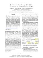
Analysis of more than 60 transformant colonies led to the
identification of a clone able to produce up to 40 mg of pure
protein from 1 L of initial culture. The product was highly
homogenous as assessed by SDS/PAGE and had the
expected molecular mass of 46.6 kDa (Fig. 1). In the first
purification step, the use of a hydrophobic interaction
chromatography partially eliminates components from the
culture supernatant and an additional anionic exchange
chromatography is sufficient to obtain a highly purified
enzyme that elutes at 6% of B buffer (0.8
M
ammonium
acetate). The N-terminal sequence determined for the
sample in peak B corresponded to the first 10 N-terminal
residues of the proenzyme, indicating that the a-mating
factor had been completely removed by KEX2, the
endoprotease from P. pastoris responsible for this action.
In initial activation tests, the purified recombinant
PCPAHa was activated with trypsin, the general activator
of mammalian pancreatic procarboxypeptidases. Peptidase
assays with the synthetic substrate FAPP showed that the
enzyme was completely activated at a 4 : 1 (w/w) PCPAHa/
trypsin ratio at 25 °C, and its specific activity was calculated
to be 150 lmol of substrate per minute and per mg of
protein. PCI, leech carboxypeptidase inhibitor (LCI), ben-
zylsuccinic acid and o-phenantroline completely inhibit the
active enzyme at concentrations of 5 l
M
,8l
M
,2m
M
and
5m
M
, respectively (results not shown), although no inhi-
bitory effect of EDTA could be detected. This is in
agreement with previous data obtained with the H. armigera
gut extracts for the first and the last of the tested inhibitors
[3]. Thus, the N-terminal sequence of the recombinant
enzyme, its ability to be activated by trypsin and its response
to different inhibitors together suggest that the protein is
properly folded and very similar to the native form.
Elucidation of the activating enzyme
Four serine proteases (elastase, chymotrypsin, LysC and
trypsin) were tested in the search for the type of proteolytic
activity that might be responsible for the physiological
activation of PCPAHa. A PCPAHa/activating enzyme ratio
of 8 : 1 (w/w) was used in all four cases and activation was
left to proceed at 23 °C for 60 min. As shown in Fig. 2A,
elastase and chymotrypsin were not able to activate the
enzyme, while LysC behaved as the best activator, as it only
needed half the time used by trypsin to generate a maximum
Fig. 1. Purification of PCPAHa. Electrophoretic analysis and anionic
exchange chromatography showing, respectively, the evolution of the
recombinant expression and the purification to homogeneity of PCPA-
Ha. Lanes 1–4 in the electrophoresis correspond to the analysis of the
protein expression culture supernatant at 16, 24, 36 and 46 h. Lane 5
corresponds to the eluate of the hydrophobic interaction chromato-
graphy and lanes 6, 7 and 8 correspond, respectively, to peaks A, B and
C from the anionic exchange chromatography shown.
Ó FEBS 2003 A metalloprocarboxypeptidase from Helicoverpa armigera (Eur. J. Biochem. 270) 3029
activity. The search for the mildest activating conditions for
LysC resulted in a PCPAHa/LysC ratio of 40 : 1 (w/w) at
37 °C (see below), while the mildest activation conditions
required for trypsin to reach the maximum CPA activity
required a fourfold higher ratio (10 : 1, w/w), also at 37 °C.
The activation of PCPAHa by LysC in those conditions is
shown in Fig. 2B. LysC and trypsin produced different
N-terminal sequences for the mature protein, as determined
by N-terminal sequencing. LysC activates the zymogen by
cleaving at position 99A after the motif (A)
5
-K, while
trypsin cleaves after R4, five residues downstream (Fig. 2C).
Activation studies; effect of glycosylation and
determination of species produced during activation
Some mammalian pancreatic procarboxypeptidases are not
able to release a full carboxypeptidase activity upon tryptic
activation even after complete cleavage of the limited
proteolysis target bond and full release of the mature
enzyme. This is due to the inhibitory capacity kept by the
activation segment fragment before it suffers extensive and
sufficient degradation. In these instances a biphasic curve is
obtained when representing the time-course of activity
generation [8]. In other cases the propiece is unable to
interact with the enzyme moiety in trans and a hyperbolic
curve is observed [20]. PCPAHa belongs to this second class
of zymogens as seen from the shape of the activation course
presented in Fig. 2B, in which the generation of activity
closely reflects the appearance of the mature enzyme as
followed by SDS/PAGE. Furthermore, no trace of activa-
tion domain of PCPAHa could be observed during the
course of LysC activation by SDS/PAGE analysis, and a
parallel HPLC follow-up confirmed that it is extensively
fragmented by cleavage at its seven internal Lys residues
(results not shown). In contrast to this, the enzyme moiety is
resistant to further proteolysis beyond the activating event.
The presence of a unique consensus glycosylation site in
the PCPAHa sequence at the border of the activation
targets for both LysC and trypsin (Fig. 2C) suggested that
glycosylation might affect the activation rate of the zymo-
gen. In order to study this, PCPAHa was treated with
N-glycosidase F, and both deglycosylated and glycosylated
PCPAHa were activated with decreasing amounts of LysC.
Figure 3 shows that PCPAHa is indeed glycosylated and
that this modification affects activation, because treated and
nontreated samples reach different levels of CPA activity
depending on the quantity of activating protease used.
Deglycosylated PCPAHa is fully activated at a ratio of
200 : 1 (w/w) whilst the glycosylated enzyme needs five times
more LysC to reach the maximum activity, evidence that the
presence of the sugar chain makes the access of LysC more
difficult for activation. The shift in electrophoretic mobility
produced by the deglycosylation is also clearly observed in
Fig. 4B. A similar experiment performed using trypsin as the
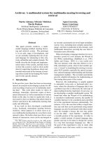
Fig. 2. Activation of PCPAHa by different serine proteases. (A) Activation was carried out at 23 °C at a PCPAHa:activating enzyme ratio of 8 : 1
(w/w) for 60 min. The amount of mature CPAHa produced at different times was detected with the substrate FAPP. The activating enzymes are:
(s)LysC(d) bovine trypsin (h) porcine elastase and (j) bovine chymotrypsin. (B) Generation of CPA activity from PCPAHa after activation
withLysCataPCPAHa:LysCratioof40:1(w/w)andat37°C. (C) Amino acid sequence of PCPA-Ha at the limit between the activation
segment and the mature enzyme, where cleavage is produced. The activation points for LysC (Lys99A) and trypsin (Arg4) are shown and the
consensus site for N-glycosylation is underlined.
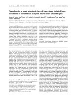
Fig. 3. Effect of glycosylation on the activation of PCPAHa with LysC.
Nonglycosylated and glycosylated PCPAHa were activated with
decreasing ratios of LysC for 2 h at 37 °C. Subsequently, 1 lLofthe
reaction mixture was assayed with the FAPP substrate to detect the
CPAHa activity generated, which is expressed as absorbance units per
min. Dark columns correspond to glycosylated PCPAHa, light col-
umns correspond to nonglycosylated. The data shown are the mean of
three measurements ± SD.
3030 A. Baye
´
s et al.(Eur. J. Biochem. 270) Ó FEBS 2003
activating enzyme showed that it was not affected by the
presence of the sugar chain (data not shown).
From the activation curve depicted in Fig. 2B it is
clear that active CPAHa is produced from the very
beginning of the process and that approximately 90 min
are needed to attain full activity and thus to generate a
maximum of mature enzyme. Analysis of the activation
process over time by HPLC shows that, besides the
PCPAHa precursor and the final CPAHa product, a
third protein species is also detected. This form, marked
as PCPAHaa in Fig. 4A, corresponds to a truncated
proenzyme which has no CPA activity. The generation of
PCPAHaa also starts immediately after activation, but it
reaches a maximum in only 5 min, thereafter gently
decreasing until complete disappearance in a process that
generates the fully mature CPAHa. N-terminal sequen-
cing and MALDI-TOF analysis (Fig. 4C) showed that
PCPAHaa shares the N-terminal of the original pro-
enzyme but has a molecular mass about 900 Da smaller.
An interesting feature of the insect proenzyme studied
here is, as commented above, the presence of an (A)
n
K
sequence at the LysC activating point, which is also
repeated at the end of the protein. A cleavage after this
second motif would result in a decrease of 897 Da of
mass and be responsible for the generation of PCPAHaa.
To assess this possibility, PCPAHA and PCPAHaa were
fragmented with cyanogen bromide and the peptides
produced analysed by MALDI-TOF spectrometry. This
fragmentation generates 11 peptides, Q356-A417 being
the one containing the C-terminal peptide in the
uncleaved proenzyme. The masses observed for the
corresponding fragments in PCPAHa and PCPAHa-a
were, respectively, 6.795 ± 22 and 5.922 ± 10, display-
ing a difference of 873 Da, close enough to 897 Da to
demonstrate that the variation is due to the removal of
the C-terminal octapeptide. The mass of the correspond-
ing fragment observed for the active, mature enzyme was
5.924 ± 14, confirming that the final product of the
activation is also lacking the C-terminal peptide.
In Fig. 4B the analysis of the species isolated from the
chromatograms in part A of the figure confirms that the
protein expressed in the Pichia pastoris system is glycosyl-
ated, and that the glycosylation takes place downstream of
the cleavage point for LysC, since the electrophoretic
mobility is affected in all three forms upon the addition of
N-glycosidase F.
Characterization of substrate specificities of CPA
from
H. armigera
A series of synthetic substrates with the same spectro-
photometric characteristics were used in the kinetic meas-
urements to calculate the values of K
m
, k
cat
and k
cat
/K
m
for
CPAHa and compare them to those of bovine CPA and
human CPA2, two A-type enzymes from mammals
(Table 1). These studies, as well as the inhibition kinetics
measurements (see below), were always performed with the
active enzyme generated by LysC, even though the enzyme
generated by trypsin showed similar enzymatic properties.
CPAHa is unable to hydrolyse synthetic substrates con-
taining C-terminal Trp residues, in contrast to CPA2. This,
together with its capability to cleave substrates containing
Phe or Tyr as C-terminal, allows to classify CPAHa as an
enzyme of the A1 subtype. In most instances, the insect
enzyme appears to be less efficient than the mammal
enzymes as judged by the k
cat
/K
m
values but, on the other
hand, displays a broader substrate specificity. Its ability to
hydrolyse Cbz-Gly-Gly-Ala is similar to rat CPA1 [6] the
only carboxypeptidase known able to hydrolyse this sub-
strate. It also displays activity against Cbz-Gly-Gly-Leu
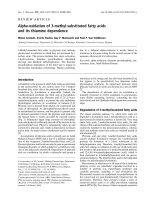
Fig. 4. Analysis of the species generated during the activation process. The activation of PCPHa with LysC was performed in the conditions of
Fig. 2B with the addition of the carboxypeptidase inhibitor from potato (PCI) at a 1–4 molar ratio. (A) At given times, samples from the reaction
mixture were made 0.05% in trifluoroacetic acid to stop the reaction and subsequently analysed by HPLC on a Vydac C
4
column. (B) SDS/PAGE
electrophoresis of the 3 species isolated from the chromatograms shown in part A of the figure; lanes 1 and 2, PCPAHa; lanes 2 and 4, PCPAHa-a;
lanes 5 and 6, CPAHa. Samples from lanes 2, 4 and 6 were treated with N-glycosidase F as described in the experimental procedures. Some bands
are numbered: 1, N-glycosidase F; 2, glycosylated CPAHa; 3, deglycosilated CPAHa. (C) Table containing the results of the N-terminal and mass
spectrometry analysis of all molecules and also the mass of the C-terminal fragment of the enzyme produced with cyanogen bromide fragmentation,
as determined by MALDI-TOF spectrometry.
Ó FEBS 2003 A metalloprocarboxypeptidase from Helicoverpa armigera (Eur. J. Biochem. 270) 3031
and a measurable k
cat
/K
m
for the substrate with a
C-terminal valine.
A remarkable difference between the insect enzyme and
the mammalian ones is the ability of the former to hydrolyse
short substrates. In contrast to the mammalian enzymes,
CPAHa hydrolytic efficiency for Cbz-Gly-X is very similar
to that displayed against Cbz-Gly-Gly-X, suggesting that
the importance of the secondary substrate binding subsites
is reduced in the insect enzyme.
Three different peptides were used as substrate models to
analyse the ability of CPAHa for cleaving acid, basic and
tryptophan C-terminal residues. In each assay, CPAHa was
compared with a similar carboxypeptidase, human CPA1,
and a second one chosen according to its specificity for the
residue being analysed (Fig. 5). Relative cleavage rates
were calculated on the basis of the time needed by each
carboxypeptidase to fully degrade the same amount of
initial substrate.
The ability of CPAHa to cleave all three peptides was
always better than that of CPA1 h, an observation specially
clear in the case of the peptide with a C-terminal arginine
(V14R), which is cleaved 120 times faster by CPAHa.
Compared with human CPB, a prototype enzyme for basic
residue specificity, CPAHa showed a relatively high affinity
for C-terminal arginine in peptide V14R since its relative
cleavage rate is only 2.5 times smaller, whilst human CPA1
can hardly hydrolyse this substrate at all. A similar result is
observed for V14W, where the relative cleavage rate for
human CPA2, a very specific enzyme for peptides with
tryptophan at the C-terminus, is three times larger than that
for CPAHa but 120 times larger than that of human CPA1.
Finally the cleavage of V15E by the mutant human CPB
wassixtimesfasterthanthatofCPAHa.
Measurement of equilibrium dissociation constant (
K
i
)
for protein inhibitors
The K
i
value was calculated for the recombinant forms
of two different carboxypeptidase inhibitors, PCI [21]
and LCI [22] (Table 2). The inhibition constant of LCI,
Table 1. Kinetic constants for peptide substrate hydroysis by H. armigera CPA (CPAHa), bovine CPA (CPAb) and human CPA2 (CPA2 h). NM: not
measurable.
Substrate
CPAHa CPAb CPA2 h
k
cat
(s
)1
)
K
m
(l
M
)
(
M
)1
Æs
)1
)
(k
cat
/K
m
)
·10
)5
k
cat
(s
)1
)
K
m
(l
M
)
(
M
)1
Æs
)1
)
(k
cat
/K
m
)
·10
)5
k
cat
(s
)1
)
K
m
(l
M
)
(
M
)1
Æs
)1
)
(k
cat
/K
m
)
·10
)5
Cbz-Gly-Gly-Phe 35.6 ± 1.8 506 ± 31 0.704 131.5 ± 3.1
a
172 ± 12 7.62 58.3 ± 2.4 372 ± 30 1.57
Cbz-Gly-Gly-Tyr 57.0 ± 3.8 238.71 ± 40 2.38 56.3 ± 2.0
a
102 ± 2 5.51 70.0 ± 5.3 125 ± 15 5.6
Cbz-Gly-Gly-Trp NM NM NM NM
b
NM NM 90.3 ± 7.0 146 ± 9 6.18
Cbz-Gly-Gly-Leu 49.4 ± 2.8 746 ± 150 0.662 63.4 ± 2.5
b
1180 ± 93 0.54 11.8 ± 1.1 5300 ± 1400 0.03
Cbz-Gly-Gly-Val 0.3 ± 0.013 1748 ± 321 1.72E10
)3
19.5 ± 2.0
c
3720 ± 390 0.052 NM NM NM
Cbz-Gly-Gly-Ala 6.25 ± 0.2 2618 ± 580 0.024 NM
c
NM NM NM NM NM
Cbz-Gly-Gly-Ser NM NM NM NM
c
NM NM NM NM NM
Cbz-Gly-Phe 35.6 ± 1.8 328 ± 40 1.095 41.7 ± 2.8
a
1093 ± 154 0.38 16.1 ± 1.3 2270 ± 200 0.07
Cbz-Gly-Tyr 58.2 ± 4.2 289 ± 68 2.01 16.0 ± 0.6
a
394 ± 29 0.41 9.7 ± 1.3 175 ± 10 0.56
Cbz-Gly-Trp NM NM NM 50.0 ± 4.3
a
3310 ± 430 0.15 33.8 ± 1.1 261 ± 12 1.29
Taken from
a
[6],
b
[23] and
c
[7].
Fig. 5. Analysis of substrate specificity of PCPAHa with peptides. Comparative analysis by reverse-phase HPLC of the degradation of
three synthetic substrates by CPAHa. Degradation of V15E (VKKKARKAAGC(Acm)AWE)
1
by CPA1 h, CPBh mutant 2 (cleaves acidic
C-ter residues) and CPAHa. Degradation of V14R (VKKKARKAAGGAKR) by CPA1 h, CPBh and CPAHa. Degradation of V14W
(VKKKARKAAGC(Acm)AW) by CPA1 h, CPA2 h and CPAHa. The numbers beside the chromatograms indicate the reaction times for each
enzyme-substrate combination. The chromatographic conditions are explained in Materials and methods.
3032 A. Baye
´
s et al.(Eur. J. Biochem. 270) Ó FEBS 2003
260 ± 32 p
M
, is similar to that of LCI for bovine CPA,
250–480 p
M
. However, the K
i
of PCI for CPAHa,
65 ± 7 p
M
, is 23 times lower than the K
i
of PCI for the
bovine homologue, which is 1.5 ± 0.6 n
M
.
Molecular modelling and dynamics simulation
To further investigate the nature of the important K
i
difference between the CPAHa–PCI and CPAb–PCI com-
plexes, a model structure of the former was generated. The
CPAHa–PCI complex was modelled using the known
crystal structure of the CPAb–PCI complex as template
(Protein Data Bank entry 4CPA). To avoid the presence of
unrealistic interactions in the model, the structure of the
CPAHa–PCI complex was relaxed under the conditions of a
molecular force field by means of a 500 ps molecular
dynamics simulation in aqueous solution. A reference
simulation of the CPAb–PCI complex was also carried out.
A cartoon representation of the superimposed complexes,
at simulation time t ¼ 0, is shown in Fig. 6. The atom-
position rmsd of the PCI backbone from its initial structure
in each of the complexes (t ¼ 0) is also given in Fig. 6 as a
function of time. The calculated rmsd values of the
inhibitor’s structure, which contain information about both
internal motions and motions relative to the CPA moiety,
are similar in the two systems. Although the amplitudes of
the rmsd fluctuations are smaller for the CPAHa–PCI
complex, they should not be considered statistically signi-
ficant because the timescale of the simulations is not
sufficient to draw conclusions about relative stabilities. The
purpose of the simulations was to relax the experimentally
determined structure of CPAb–PCI and the model structure
of CPAHa–PCI under the same molecular force field and
conditions, in order to facilitate the comparison of the
corresponding molecular interfaces.
In spite of the similar binding geometries (imposed by
the modelling strategy), the two complexes appear fairly
different in terms of specific interactions between enzyme
and inhibitor (results not shown). The difference in
average interaction energy between enzyme and inhibitor
in the simulation ()1074 kJÆmol
)1
CPAHa–PCI vs.
)1257 kJ mol
)1
for CPAb-PCI) is not sufficient to
explain the remarkably lower K
i
of the CPAHa–PCI
complex. However, we note that the free energy of
binding is equal to the work required to bring the two
molecules from free solution to the solvated complex,
and the above-mentioned interaction energy is only one
of the components of this free energy.
Discussion
The high expression yield of the procarboxypeptidase from
the insect pest Helicoverpa armigera attained in the meth-
ylotrophic yeast Pichia pastoris indicates both the suitability
of this organism to host the heterologous expression of this
class of enzymes [23,24] and the correct folding of the
proenzyme. The latter is further confirmed by its activation
by trypsin, its capability to degrade synthetic CP substrates
and its susceptibility to protein inhibitors, proved to be
effective on related metallocarboxypeptidases. Overall, the
Table 2. K
i
values of PCI and LCI against CPAHa compared to
previous data obtained for CPAb [21,22].
Carboxypeptidase
K
i
(p
M
)
PCI LCI
CPAHa 65 ± 7.3 260 ± 32.5
CPAb 1500 ± 600 250–480
Fig. 6. Molecular modelling and dynamics
simulation. Cartoon representation of a the
least-squares fitted complexes CPAb–PCI and
CPAHa–PCI, at simulation time t ¼ 0; CPAb
in cyan, CPAHa in red, PCI in yellow. Atom-
position rmsd of the PCI backbone from its
initial structure in each of the complexes
(t ¼ 0) as a function of time; CPAbPCI in
cyan, CPAHa–PCI in red.
Ó FEBS 2003 A metalloprocarboxypeptidase from Helicoverpa armigera (Eur. J. Biochem. 270) 3033
recombinant protein is thus indistinguishable from the
natural one and constitutes a good model to study it.
Although trypsin can activate PCPAHa, as with many
other procarboxypeptidases a lysine specific endopeptidase
(LysC) can activate it four times more efficiently. However,
activation with either protease releases an enzyme with
identical activity against the synthetic substrates FAPP and
AAFP. This is the first member of this family of proenzymes
that can be activated more efficiently by a protease other
than trypsin. The fact that several trypsin-like proteases
from H. armigera have been cloned and sequenced [25] and
that some of them show a higher degree of identity with
LysC than with bovine trypsin suggests that this insect
might possess a specific enzyme able to activate PCPAHa,
as LysC does in vitro.
The activation point for trypsin in vitro is R4, an
accessible residue located in an unstructured loop at the end
of the connecting region between the activation domain and
the enzyme moieties, a position very similar to that of most
mammal procarboxypeptidases [9]. LysC, a serine protease
that only recognizes lysine at the P1 position, is unable to
activate any human pancreatic procarboxypeptidases, as
previously observed in our laboratory. However, when
acting on PCPAHa, it generates the active enzyme by
cleavage at the carbonyl end of a lysine located four residues
upstream of R4 and after five consecutive alanines
(Fig. 2C), a sequence that could be a recognition motif for
a highly specific activating protease. This motif, not found
in any other protein, is repeated a second time near the
C-terminal end of this molecule, and also in this case LysC
has also been shown to be able to release the C-terminal
peptide after specific cleavage. Whether the dual presence of
the specific sequence motif is related to some hypothetical
mechanism of control of the activity will require further
investigation.
Between the two activation points described there is a
consensus target for glycosylation (Asn-Ser-Thr) which does
become glycosylated in the P. pastoris system, adding a mass
of around 1900 Da. The presence of sugars seems to affect
LysC activation, as demonstrated by the easier activation of
deglycosylated PCPAHa. This adds a further possible
regulatory mechanism which has never been observed
before in enzymes of this family.
To achieve a complete in vitro activation of PCPAHa in a
period of time similar to other activation studies performed
with mammalian procarboxypeptidases it was found that
the PCPAHa/LysC ratio needed was 40 : 1 (w/w) at 37 °C,
and the activation process was studied in detail in these
conditions. The timecourse of activity generation is hyper-
bolic and coincides with those described for procarboxy-
peptidases with a proregion that does not inhibit the enzyme
after cleavage [20,23]. This is consistent with the observation
that the prodomain is completely degraded during activa-
tion because LysC cleaves after all of the seven internal
lysines. Besides the removal and degradation of the
prodomain, LysC also causes the removal of a C-terminal
octapeptide, which is placed after an (A)
6
K motif, almost
identical to the sequence recognized by LysC at the border
between the activation peptide and the enzyme moiety. The
cleavage of the C-terminal peptide is much faster than the
elimination of the proregion because disappearance of full-
length PCPAHa occurs only 5 min after activation, while a
complete CPAHa activity is only reached after 90 min
(Figs 2B and 4). The parallel release of the active enzyme
and the C-terminal peptide due to the highly specific action
of an enzyme able to cleave after (A)
n
K might have some
physiological relevance.
From the analysis with a series of carbobenzoxy (Cbz)
substrates, and in a first instance, the mature enzyme
derived from PCPAHa should be classified as A1, as it
cleaves aliphatic and aromatic C-terminal residues but not
tryptophan. Surprisingly, further analysis shows that the
enzyme is also able to cleave C-terminal E, W and R
residues, with a particularly good efficiency for the latter.
In all cases, the insect enzyme was much more efficient
than human pancreatic CPA1. This is the first reported
case of a metallocarboxypeptidase showing such a wide
specificity spectrum. S255, located in the S1¢ pocket,
which replaces a conserved isoleucine in the A-type
carboxypeptidases and an equally conserved aspartate
residue in the B forms might be responsible for this
change in specificity [9].
The plant carboxypeptidase inhibitor PCI shows K
i
values in the p
M
range with CPAHa, in contrast to the n
M
values displayed against mammalian carboxypeptidases.
This supports the theory that PCI, which is expressed in
potato leaves in response to wounding [26], may inhibit the
digestive carboxypeptidases of potential insect pests. The
impact of H. armigera in many different crops makes this
efficient protein inhibitor very interesting in the design of
new insecticide strategies. To investigate the structural bases
of the strong binding of PCI to CPAHa, a molecular model
of the CPAHa–PCI complex was generated based on the
known structures of PCPAHa and the CPAb–PCI complex,
and it was submitted to relaxation and structural analysis by
a molecular dynamics approach. From these studies, the
differences in the K
i
values observed for CPAHa–PCI and
CPAb–PCI cannot apparently be readily explained in terms
of specific interactions in the model. This suggests that there
may be local conformational differences between the
structure of CPAHa in the proenzyme and in the complex
which are not reproduced by the model, that the geometry
of binding of PCI to CPAHa may differ from that assumed
with the model and that the difference in the binding free
energies of the two complexes may be dominated by other
than the intermolecular interaction energy (e.g. enthalpy
and/or entropy associated with desolvation and conform-
ational changes upon complex formation). To evaluate
these three possibilities, further computational studies are in
progress.
Overall, the procarboxypeptidase A from Helicoverpa
armigera and its derived enzyme, although apparently very
similar both functionally and structurally to their mamma-
lian counterparts, have some unique properties in terms of
activation, specificity and regulation, which make them an
interesting system that settles new questions on this family
of enzymes, both in the basic and applied fields.
Acknowledgements
This work was supported by grant BIO2001-2046 (MCYT, Ministerio
de Ciencia y Tecnologı
´
a, Spain) and by the Centre de Refere
`
ncia en
Biotecnologia (Generalitat de Catalunya, Spain). X. D. is grateful to
W. F. van Gunsteren for granting access to computational resources at
3034 A. Baye
´
s et al.(Eur. J. Biochem. 270) Ó FEBS 2003
the ETH Zurich. We wish to thank Drs John A. Gatehouse and David
P. Bown, from the University of Durham, UK, for kindly providing us
with the cDNA of PCPAHa and to Dr Sonia Segura for providing us
with purified CPBh mutant2. We also thank Dr Salvador Bartolome
´
(LAFEAL-UAB) and Dr Francesc Canals (IBB-UAB) for technical
assistance.
References
1. Terra, W.R. & Ferreira, C. (1994) Insect digestive enzymes:
properties, compartimentalization and function. Comp. Biochem.
Physiol. 109B, 1–62.
2. Johnson, S.J., King, E.G. & Bradley, J.R., eds. (1986) Theory and
tactics of Heliothis population management. I. Cultural and bio-
logical control. South. Coop. Series Bull. 316, 161.
3. Bown, D.P., Wilkinson, S. & Gatehouse, J.A. (1998) Midgut
carboxypeptidase from Helicoverpa armigera (Lepidoptera:
Noctuidae) larvae: enzyme characterization, cDNA cloning and
expression. Insect Biochem. Mol. Biol. 28, 739–749.
4. Wei,S.,Segura,S.,Vendrell,J.,Aviles,F.X.,Lanoue,E.,Day,R.,
Feng, Y. & Fricker, L.D. (2002) Identification and characteriza-
tion of three members of the human metallocarboxypeptidase
gene family. J. Biol. Chem. 277, 14954–14964.
5. Vendrell, J. & Aviles, F.X. (2000) Metallocarboxypeptidases and
their protein inhibitors. Biochem. Biophys. Acta. 1477, 284–298.
6. Gardell, S.J., Craik, C.S., Clauser, E., Goldsmith, E.J., Steward,
C.B., Graf, M. & Rutter, W.J. (1988) A novel rat carboxy-
peptidase, CPA2: characterization, molecular cloning and
evolutionary implications on substrate specificity in the carboxy-
peptidase gene family. J. Biol. Chem. 263, 17828–17836.
7. Reverter, D., Garcı
´
a-Saez, I., Catasu´ s, L., Vendrell, J., Coll, M. &
Avile
´
s, F.X. (1997) Characterisation and preliminary X-ray dif-
fraction analysis of human pancreatic procarboxypeptidase A2.
FEBS Lett. 420, 7–10.
8. Vendrell, J., Cuchillo, C. & Avile
´
s, F.X. (1991) The trypsin acti-
vation pathway of monomeric procarboxypeptidase A. J. Biol.
Chem. 265, 6949–6953.
9. Este
´
banez-Perpin
˜
a
´
,E.,Baye
´
s,A.,Vendrell,J.,Jongsma,M.A.,
Bown,D.,Gatehouse,J.A.,Huber,R.,Bode,W.,Aviles,F.X.&
Reverter, D. (2001) Crystal structure of a novel mid-gut pro-
carboxypeptidase from the cotton pest Helicoverpa armigera.
J. Mol. Biol. 313, 629–638.
10. Sambrook, J., Fritsch, E.F. & Maniatis, T. (2001) Molecular
Cloning: A Laboratory Manual, 3rd edn. Cold Spring Harbor
Laboratory, Cold Spring Harbor, NY.
11. Peterson, L.M., Holmquist, B. & Bethune, J.L. (1982) Unique
activity assay for carboxypeptidase A in human serum. Anal.
Biochem. 125, 420–426.
12. Mock, W.L. (1996) Arazoformyl peptide surrogates as spectro-
photometric kinetic assay substrates for carboxypeptidase A.
Anal. Biochem. 239, 218–222.
13. Leatherbarrow, R.J. (1987) Enzfitter: a non-linear regression data
analysis program for the IBM-PC. Elsvier Biosoft, Cambridge,
UK.
14. Edge,M.,Forder,C.,Hennam,J.,Lee,I.,Tonge,D.,Harden,I.,
Fitton, J., Eckersley, K., East, S., Shuffebotham, A., Blakey, D. &
Slater, A. (1998) Engineered human carboxypeptidase B enzymes
that hydrolyse hippuryl-
L
-glutamic acid: reversed-polarity
mutants. Protein Eng. 11, 1229–1234.
15. Bieth, J.G. (1995) Theoretical and practical aspects of proteinase
inhibition kinetics. Methods Enzymol. 248, 59–84.
16. van Gunsteren, W.F., Billeter, S.R., Eising, A.A., Hu
¨
nenberger,
P.H., Kru
¨
ger, P., Mark, A.E., Scott, W.R.P. & Tironi, I.G. (1996)
Biomolecular simulation: The GROMOS96 manual and user guide.
vdf Hochschulverlag AG an der ETH Zu
¨
rich and BIOMOS b.v.,
Zu
¨
rich, Groningen.
17. Scott, W.R.P., Hu
¨
nenberger, P.H., Tironi, I.G., Mark, A.E.,
Billeter, S.R., Fennen, J., Torda, A.E., Huber, T., Kru
¨
ger, P. &
van Gunsteren, W.F. (1999) The GROMOS biomolecular simu-
lation program package. J. Phys. Chem. A 103, 3596–3607.
18. Schuler, L.D., Daura, X. & van Gunsteren, W.F. (2001) An
improved GROMOS96 force field for aliphatic hydrocarbons in
the condensed phase. J. Comp. Chem. 22, 1205–1218.
19. Berendsen, H.J.C., Postma, J.P.M., van Gunsteren, W.F. &
Hermans, J. (1981) Interaction models for water in relation to
protein hydration. In Intermolecular Forces. (Pullman, B., ed.), pp.
331–342. D. Reidel Publishing Co., Dordrecht.
20. Burgos, F.J., Salva
`
, M., Villegas, V., Soriano, F., Me
´
ndez,E.&
Aviles, F.X. (1991) Biochemistry 30, 4082–4089.
21. Molina, M.A., Marino, C., Oliva, B., Avile
´
s, F.X. & Querol, E.
(1994) C-tail valine Is a key residue for stabilitzation of complex
between potato inhibitor and carboxypeptidase A. J. Biol. Chem.
269, 21467–21472.
22. Reverter, D., Vendrell, J., Canals, F., Horstmann, J., Aviles, F.X.,
Fritz, H. & Sommerhoff, C.P. (1998a) A carboxypeptidase
inhibitor from the medical leech Hirudo medicinalis.Isolation,
sequence analysis, cDNA cloning, recombinant expression and
characteritzation. J. Biol. Chem. 273, 32927–32933.
23. Reverter, D., Ventura, S., Villegas, V., Vendrell, J. & Aviles, F.X.
(1998b) Overexpression of human procarboxypeptidase A2 in
Pichia pastoris and detailed characteritzation of its activation
pathway. J. Biol. Chem. 273, 3535–3541.
24. Ventura, S., Villegas, V., Sterner, J., Larson, J., Vendrell, J.,
Hershberger, C.L. & Aviles, F.X. (1999) Mapping the pro-region
of carboxypeptidase B by protein engineering. Cloning over-
expression and mutagenesis of the porcine proenzyme. J. Biol.
Chem. 274, 19925–19933.
25. Bown, D.P., Wilkinson, H.S. & Gatehouse, J.A. (1997) Differen-
tially regulated inhibitor-sensitive and insensitive protease genes
from the phytophagus insect pest, Helicoverpa armigera, are
members of complex multigene families. Insect Biochem. Molec.
Biol. 27, 625–638.
26. Villanueva, J., Canals, F., Prat, S., Ludevid, D., Querol, E. &
Aviles, F.X. (1998) Characterization of the wound-induced
metallocarboxypeptidase inhibitor from potato. FEBS Lett.
440, 175–182.
Supplementary material
The following material is available from http://www.
blackwellpublishing.com/products/journals/suppmat/EJB/
EJB3681/EJB3681sm.htm
Appendix S1. Procarboxypeptidase A from the insect pest
Helicoverpa armigera and its derived enzyme. Two forms
with new functional properties.
Ó FEBS 2003 A metalloprocarboxypeptidase from Helicoverpa armigera (Eur. J. Biochem. 270) 3035