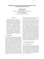
Báo cáo khoa học: Bovine tryptases cDNA cloning, tissue specific expression and characterization of the lung isoform ppt
Bạn đang xem bản rút gọn của tài liệu. Xem và tải ngay bản đầy đủ của tài liệu tại đây (1.13 MB, 11 trang )
Bovine tryptases
cDNA cloning, tissue specific expression and characterization of the lung isoform
Alessandra Gambacurta
1
*, Laura Fiorucci
1
*, Paolo Basili
1
, Fulvio Erba
1
, Angela Amoresano
2
and Franca Ascoli
1
1
Department of Experimental Medicine and Biochemical Sciences, University of Rome ‘Tor Vergata’, Rome;
2
Department of Organic Chemistry and Biochemistry, University of Naples ‘Federico II’, Naples, Italy
A complementary DNA encoding a new bovine tryptase
isoform (here named BLT) was cloned and sequenced
from lung tissue. Analysis of sequence indicates the pres-
ence of a 26-amino acid prepro-sequence and a 245 amino
acid catalytic domain. It contains six different residues
when compared with the previously characterized tryptase
from bovine liver capsule (BLCT), with the most signifi-
cant difference residing at the primary specificity S1
pocket. In BLT, the canonical residues Asp-Ser are pres-
ent at positions 188–189, while in BLCT these positions
are occupied by residues Asn-Phe. This finding was con-
firmed by mass fingerprinting of the peptide mixture
obtained upon in-gel tryptic digestion of BLT. Analysis by
gel filtration of the purified protein shows that BLT is
probably tetrameric, similar to the previously identified
tryptases from other species, with monomer migrating as
35–40 kDa multiple bands in SDS/PAGE. As expected,
the catalytic abilities of the two bovine tryptases are dif-
ferent. The specificity constant values (k
cat
/K
m
) assayed
with model substrates are 10- to 60-fold higher in the case
of BLT. The tissue-specific expression of the two tryptases
was evaluated at the RNA level by analysis of their dif-
ferent restriction patterns. In lung, only BLT was found to
be expressed, while in liver capsule only BLCT is present.
Both isoforms are distributed in similar amounts in heart
and spleen. Analysis of the two gene sequences reveals the
presence of several recognition sequences in the promoter
regions and suggest a role for hormones in governing the
mechanism of tissue expression of bovine tryptases.
Keywords: bovine tryptases; aprotinin; tissue expression;
promoter sequences; mass spectrometry.
Tryptases are trypsin-like proteinases stored in the secretory
granules of human [1–3], dog [4,5], rat [6–8], mouse [9,10],
bovine [11], gerbil [12] and sheep [13] mast cells. These
enzymes are released along with other mediators into the
extracellular medium upon mast cell activation/degranula-
tion. Although their patho-physiological role is not yet
understood, tryptases seem to be involved in several mast
cell-mediated allergic and inflammatory diseases. However,
the underlying molecular mechanism, as well as the
proenzyme/polypeptide target(s) of these enzymes have
not been identified yet, in spite of their involvement in a
variety of biochemical reactions in vitro [14–18]. Recently it
was shown that human tryptase activates by proteolytic
cleavage the proteinase-activated receptor 2, inducing
widespread inflammation by an unknown mechanism and
possibly contributing to the proinflammatory effects of mast
cells in human diseases [19].
Almost all tryptases are made of glycosylated 245 residue
identical subunits, which share many characteristics with the
prototype enzyme trypsin (225 residues), in terms of
sequence (identity around 45%) and overall folding. How-
ever, two main features are peculiar to tryptases. One
feature is the tetrameric structure of most tryptases studied
so far, which is necessary for biological activity and is
maintained in vivo through association with heparin; in
many cases this glycosaminoglycan is required for stabi-
lization of the enzyme after its release from mast cells [20,21].
In the 3 A
˚
crystal structure of the tetrameric bII human
enzyme (molecular mass 120–140 kDa), the active site of
each monomer faces a central oval pore, whose dimension
limits the accessibility for macromolecular substrates/inhi-
bitors [22]. A second common feature of tryptases seems to
be their occurrence as a multigene family: in humans, at
least four homologous tryptase cDNAs (tryptases a and
bI–III) have been isolated [23–25] and a gene cluster was
Correspondence to Franca Ascoli, Department of Experimental
Medicine and Biochemical Sciences, University of Rome
ÔTor VergataÕ, Via Montpellier 1, 00133 Rome, Italy.
Fax: + 39 06 72596477; Tel.: + 39 06 72596474;
E-mail:
Abbreviations: BLCT, bovine liver capsule tryptase; BLT, bovine lung
tryptase; Boc, t-butyloxycarbonyl; BPTI, bovine pancreatic trypsin
inhibitor; DFP, diisopropylfluoro-phosphate; MCA, methyl-
coumarin; MUGB, 4-methylumbelliferyl p-guanidinobenzoate;
STI, soybean trypsin inhibitor; Z, benzyloxycarbonyl.
Dedication: This paper is dedicated to the memory of Eraldo Antonini,
eminent biochemist, prematurely deceased twenty years ago,
on March 19th 1983.
Note: nucleotide sequence data are available in the GenBank database
with the accession numbers AF515641 (full-length bovine lung
tryptase cDNA), X94982 (full-length bovine liver capsule tryptase
cDNA), AF515642 (bovine lung tryptase promoter) and AF516175
(bovine liver capsule tryptase promoter).
*These authors contributed equally to this work.
(Received 8 October 2002, revised 20 November 2002,
accepted 29 November 2002)
Eur. J. Biochem. 270, 507–517 (2003) Ó FEBS 2003 doi:10.1046/j.1432-1033.2003.03406.x
identified for multiple human tryptases [26]; two tryptases
(mMCP-6 and mMCP-7) have been identified in mouse
[9,10], and their genes isolated [27,28].
In a previous paper [11], we reported isolation of a tryptase
isoform (BLCT) from bovine liver connective capsule
(Glisson capsule). This enzyme is made of 245 amino acid
(aa) subunits; its sequence was determined either biochemi-
cally on the purified protein or by isolating and sequencing its
cDNA [29]. The most peculiar and important difference
between BLCT and other tryptases analyzed so far occurs at
positions 188–189 of the primary specificity pocket S1, where
the basic side chain of the substrate P1 residue, Arg or Lys
(whose carbonyl group belongs to the scissile peptide bond of
the substrate), is accommodated. In BLCT, residues Asn188
and Phe189 replace the canonical residues Asp and Ser,
respectively, present in all other tryptases and in all trypsin-
like enzymes. However, these substitutions do not affect
significantly the substrate specificity of the bovine enzyme.
In this paper, we report cloning of a new cDNA from
bovine lung encoding a tryptase isoform (BLT) with the
usual doublet Asp-Ser in the S1 specificity pocket and
isolation of the corresponding protein. Sequence analysis by
mass spectrometry and partial characterization of BLT
revealed more similarities between this enzyme and b-type
tryptases from other species with respect to BLCT. Some
evidence on tissue-specific expression of the two isoforms in
different bovine tissues is also reported and in this light the
different sequence of the two tryptase gene promoter
regions are discussed.
Experimental procedures
Oligonucleotide primers and restriction enzymes
PCR primers were obtained from MWG Biotech (Italy),
Genset (France) or Pharmacia (Italy). Their numbering
refers to the first nucleotide (+1) of cDNA start codon.
Restriction enzymes were obtained from New England
Biolabs (USA).
Amplification reaction (PCR), cloning and sequence
analysis
Unless otherwise indicated, PCRs were conducted using
5U of Taq polymerase (Perkin Elmer, USA), 200 m
M
dNTPs, 1.5 m
M
MgCl
2
,10m
M
Tris/HCl, pH 8.3, 50 m
M
KCl (50 lL final volume). All PCR products were size-
fractionated by agarose gel electrophoresis and the bands
eluted, purified and subcloned in the PCRII
TM
TOPO
vector containing the lac promoter and the b-galactosidase
gene, using the TA Cloning Kit (Invitrogen, USA).
Transformation was performed in the TOP 10 cells, the
positive clones were isolated and their nucleotide sequence
determined. Sequence analysis was performed on both
strands by the dideoxy-chain termination method, either
using the Sequenase 2.0 Kit (Amersham Pharmacia Biotech
Italia) or automatically.
cDNA synthesis
mRNA was prepared from various bovine tissues using the
Fast Track kit (Invitrogen, USA). The first strand cDNA
was synthesized at 42 °C using 0.1–1 lgofmRNAwiththe
cDNA cycle kit (Invitrogen). To obtain partial cDNAs
encoding tryptases (see Results) PCRs were performed as
already described [29], using 2 lL of the RT reaction
products and the primer pair N9 (nt 127–153, 5¢-AGC
CTGAGAGTCAGCCGTCGGTACTGG-3¢)andN10
(nt 790–816 antisense, 5¢-TCAGGGCCCCTGGGGGAC
GTACTGGTG-3¢). Entire tryptase cDNAs were obtained
under the same conditions, at the annealing temperature of
58 °C, using the primer pair Met (nt 1–20, 5¢-ATG
CTCCATCTGCTGGCGCT-3¢, designed on the basis of
the 5¢ RACE experiments reported below) and Coda
[5¢-CGCGCGCG(T)
16
)3¢] [29] and sequenced.
5¢ Rapid amplification of cDNA ends (RACE)
5¢ RACE was carried out to determine the 5¢ nucleotide
sequence of the tryptase full-length transcripts, using the
RACE System from Gibco (Paisley, USA). One hundred
nanograms of bovine lung and hepatic capsule mRNAs
were reverse transcribed using oligo-dT as primer. After
purification of the first strand cDNA, a dC tail was added to
the 3¢ end using dCTP and terminal transferase. PCRs
were conducted on 5 lLoftheÔtailing reactionÕ,usingthe
5¢ RACE abridged universal amplification primer
AUAP with a 3¢-G tail (5¢-GGCCACGCGTCGACTAG
TACGGGGGGGGGGGGGG-3¢)as5¢ primer and C1
(nt 537–563 antisense, 5¢-TACTTCCTGTCACAGACAC
TGTTCTCC-3¢)as3¢ primer. Nested PCRs were then
performed using the same 5¢ primer and C2 (nt 372–
396 antisense, 5¢-GTGCCAGGAGATATTCACAAGCT
TG-3¢)as3¢ primer. Amplification reactions were con-
ducted using 40 pmol of each primer, under the following
conditions: 2 min at 94 °C (1 cycle), 1 min at 94 °C, 1 min
at 58 °C, 1 min at 72 °C (30 cycles) and 10 min at 72 °C
(1 cycle).
Evaluation of tissue distribution of bovine tryptases
In order to ascertain the expression of one or both tryptase
isoforms (see Results) in different bovine tissues, tryptase
cDNAs were prepared as described above from mRNAs
isolated from bovine liver capsule, lung, heart and spleen.
The amplification profile was optimized as follows: 1 min at
94 °C (1 cycle), 1 min at 94 °C, 1 min at 58 °C, 2 min at
72 °C (30–40 cycles) and 10 min at 72 °C(1cycle).The
RT-PCR products were separated by electrophoresis
through a 1.5% (w/v) agarose gel, eluted, cloned in a TA
vector and transformed in the TOP 10 competent cells. The
positive clones were identified by restriction analysis with
NspI (overnight at 37 °C) and sequenced.
Identification of 5¢ flanking sequences and UTRs
of bovine tryptase genes
A strategy similar to that described in the protocol of the
Universal Genome Walker Kit (CLONTECH, USA) was
employed to identify 5¢ flanking sequences and UTRs of
the tryptase genes.
Genomic DNA was obtained from bovine liver using the
DNA TURBOGEN Kit (Invitrogen, USA) at a final
concentration of 100 ngÆlL
)1
and the molecular weight was
508 A. Gambacurta et al. (Eur. J. Biochem. 270) Ó FEBS 2003
checked by 0.8% (w/v) agarose gel electrophoresis. Genomic
DNA(500ng)wasthendigestedwith10Uofthe
restriction enzymes HincII, EcoRV, MscI, SspI, in four
separate reactions. Each digested sample was ligated with
the annealed adaptor oligonucleotides A1 (5¢-GTAATAC
GACTCACTATAGGGCACGCGTGGTCGAC-3¢)and
A2 (5¢-GTCGACCACGCGTGC-3¢, complementary to
15 nt of the A1 3¢ region).
Amplification reactions were then conducted for each
digested and ligated genomic DNA sample (10 lL), using
20 pmoles of each primer (see below) and 5 U of the
ÔElongase enzyme mixÕ (Gibco) in 60 m
M
Tris sulfate pH 9.1,
18 m
M
ammonium sulfate, 1 m
M
magnesium sulfate and
1.5 m
M
magnesium chloride, in a final reaction volume of
50 lL. The conditions used were: 1 min at 94 °C(1cycle),
1minat94°C, 1 min at 55 °C(5¢ region) or at 52 °C(3¢
region), 4 min at 68 °C (32 cycles) and 5 min at 68 °C(1
cycle). Two microliters of each PCR was then used as a
template in a nested PCR under the same conditions. The
following oligonucleotides were used as primers: AP1 (5¢-
GTAATACGACTCACTATAGGGC-3¢, identical to 22 nt
of the A1 5¢ region); AP2 (5¢-ACTATAGG GCACGCGTG
GT-3¢, identical to 12 internal nt of A1); C3 (nt 41–61
antisense, 5¢-CCTGGCCAGGGGCTGCG GAGA-3¢); C4
(nt 34–54 antisense, 5¢-AGGGGCTGCGGAGACCAGG
CT-3¢). The primer pairs AP1/C3 and AP2/C4 were used in
the first and in the nested PCR, respectively.
In order to assign the two 5¢ sequences obtained (from the
genomic DNA sample digested with HincII, see Results) to
the two bovine tryptase genes, two different PCRs were
conducted, using as a template genomic DNA and the
primer pairs U1a/N10 and U1b/N10, respectively. Primers
U1a (5¢-AGATGAAGGAATTAGTAGTTTAATGG-3¢,
nt ) 374 to )399) and U1b (5¢-ATTAATTTCAGTTTA
AAAGAGCTACT-3¢,nt) 374 to ) 399) were designed on
thebasisofthe5¢ sequences obtained (a and b). N10
sequence is reported above. Amplification was conducted
using 20 pmol of each primer and 100 ng digenomic DNA,
with the following parameters: 1 min at 94 °C(1cycle);
1mina94°C; 1 min at 64 °C; 4 min at 72 °C (32 cycles);
5 min at 72 °C (1 cycle). The PCR products were size-
fractionated by electrophoresis through a 1% (w/v) agarose
gel. After cloning, the PCR II
TM
TOPO vectors, containing
the inserts, were digested with the restriction enzyme NspIto
distinguish between the sequences encoding the two differ-
ent bovine tryptases (see Results).
Organization of bovine tryptase genes and location
of intron II–V
Intron II–V length of the two genes encoding bovine
tryptases was evaluated by amplification of bovine genomic
DNA, using the following primer pairs: Met/C7 for intron
II amplification; N9/C6 for intron III amplification;
C8/C1 for intron IV amplification; C5/N10 for intron V
amplification. Sequences of primers Met, C1, N9 and
N10 are reported above. Other primers used are: C5,
5¢-CCGTCGTGGAGAACAGTGTC-3¢ (nt 530–549); C6,
5¢-TGTCCGCCCCGTTCTTAACGCTGTA-3¢ (nt 328–
352, antisense); C7, 5¢-ACGATGCCCGCGCGCTG-3¢
(nt 67–83, antisense); C8, 5¢-ACGGGCTGGGGCAA
CGTGG-3¢ (nt 460–478). The primer pair sequences
correspond to cDNA sequences at the intron/exon
junctions, deduced from the homologous sequences of
human and murine tryptase genes. PCRs were conducted
using 100 ng of genomic DNA as a template, 20 pmol of
each primer, and the following conditions for amplification:
3 min at 94 °C (1 cycle), 1 min at 94 °C; 1 min at the
annealing temperature; 30 s at 72 °C (30 cycles); 5 min at
72 °C (1 cycle). Annealing temperatures were: 58 °Cfor
amplification of introns II and III, 60 °C for intron IV and
62 °C for intron V. The PCR products were size-fraction-
ated by electrophoresis through a 1% (w/v) agarose gel,
eluted, cloned in the PCR II
TM
TOPO vectors and
sequenced.
Purification of bovine tryptases
BLCT and BLT were purified as previously described for
bovine liver capsule tryptase [11], except that, in the case of
the lung enzyme, the three step procedure (high-salt
extraction followed by hydrophobic chromatography on
octyl sepharose and then an heparin affinity column) was
carried out using pH 5.5 buffers. Tryptase enzymatic
activity was routinely assayed at 30 °C monitoring the
fluorescence of 7-amino-4-methyl-coumarin released from
Boc-Phe-Ser-Arg-MCA substrate (Sigma Chemical Co.,
USA), as reported previously [11]. The tryptase-containing
fractions eluted from the heparin column were concentrated
with an Amicon stirred-cell concentrator equipped with a
30 kDa cut-off membrane and stored at )20 °Cinthe
heparin column elution buffer containing 20% (v/v)
glycerol. Lung tryptase was purified further by gel filtration
chromatography. The enzyme sample was diluted with four
volumes of 10 m
M
Mes pH 5.5 and injected (100 lL) at a
50 lLÆmL
)1
flow rate onto a Superose 12PC column
(Pharmacia, Italy) pre-equilibrated with the gel filtration
buffer (10 m
M
Mes, 0.4
M
NaCl, pH 5.5). Protein was
detected spectrophotometrically at 280 nm and 100 lL
fractions were collected. Tryptase activity in each fraction
was measured as described previously. The fractions
containing tryptase activity were pooled and used for
characterization of the enzyme. For determination of BLT
molecular weight, the three most active fractions were
pooled, preincubated with heparin (10 lgÆmL
)1
,10minat
room temperature), and reloaded (20 lL) on the gel
filtration column as above.
Tryptase concentrations were determined by active site
titration with 4-methylumbelliferyl p-guanidinobenzoate
(MUGB) (Sigma Chemical Co., USA) for the lung enzyme
as reported in [30], and with radioactive diisopropylfluoro-
phosphate ([
3
H]DFP) (New England Nuclear, UK) for the
liver capsule enzyme, as already described [11]. Western
blotting was performed as already reported using an anti-
(178/191-tryptase-peptide) Ig [31].
Mass spectrometry analysis
Mass spectrometric analysis was performed on the Coo-
massie blue-stained BLT protein excised from a preparative
SDS electrophoresis on a 14% (w/v) polyacrylamide gel.
The excised band was washed first with acetonitrile and then
with 0.1
M
ammonium bicarbonate. Protein samples were
reduced by incubation in 10 m
M
dithiothreitol for 45 min at
Ó FEBS 2003 Tissue-specific expression of bovine tryptases (Eur. J. Biochem. 270) 509
56 °C. The gel particles were then washed with ammonium
bicarbonate and acetonitrile. Enzymatic digestion was
carried out with trypsin (Sigma Chemical Co., USA) at a
final concentration of 15 ngÆmL
)1
in 50 m
M
ammonium
bicarbonate pH 8.5, at 4 °C for 4 h. The buffer solution was
then removed and a new aliquot of the enzyme/buffer
solution was added for 18 h at 37 °C. A minimum reaction
volume, sufficient for complete rehydration of the gel was
used. Peptides were then extracted washing the gel particles
with 20 m
M
ammonium bicarbonate and 0.1% (v/v)
trifluoroacetic acid in 50% (v/v) acetonitrile at room
temperature and then lyophilized.
MALDI mass spectra were recorded using a Applied
Biosystem Voyager DE-Pro reflector instrument. A mixture
of analyte solution and a-cyanohydroxycinnamic acid
[10 mgÆmL
)1
in acetonitrile/ethyl alcohol/0.1% trifluoro-
acetic acid (1 : 1 : 1 v/v/v)] was applied to the metallic
sample plate and dried under vacuum. Mass calibration was
performed using external standards. Raw data were
analyzed using computer software provided by the manu-
facturer and reported as monoisotopic masses.
Enzymatic assays
Rate assays for the determination of kinetic constants with
7-amino-4-methyl-coumarin (MCA) peptide substrates
(Sigma Chemical Co., USA) were started by addition of
the enzyme (BLT or BLCT) to 0.1
M
Tris/HCl, pH 8.0,
containing the various substrates in a total reaction volume
of 2.0 mL maintained at 25 °C during measurements.
Hydrolysis of MCA substrates was monitored using an
excitation wavelength of 370 nm and an emission wave-
length of 460 nm in a Kontron spectrofluorimeter. k
cat
/K
m
values were determined under pseudo first-order conditions.
For all substrates [S
°
]was K
m
. Progress curves were fitted
using an exponential function to obtain k
obs
; k
obs
/[E] was
usedtoobtaink
cat
/K
m
, where [E] represents the enzyme
concentration.
To test for susceptibility of BLT to inhibition, the enzyme
(5 n
M
active sites) and various inhibitors were mixed in
2 mL of the assay buffer and maintained at 30 °Cfor
30 min. Then 20 lLof1.5m
M
Boc-Phe-Ser-Arg-MCA
were added and residual activity was determined as
described above by comparison with that of an identical
enzyme incubation mixture containing no inhibitor.
Results
Cloning and sequence analysis of full-length tryptase
cDNAs
A partial cDNA (690 bp) encoding a new bovine tryptase
isoform (BLT) was obtained from lung mRNA by
RT-PCR, using primers N9 and N10, and by subsequent
cloning and sequencing. Based on this partial sequence, 5¢
RACE experiments and RT-PCR (using the primer pair
Met and Coda) were performed as described in the
Experimental Procedures. The full-length BLT cDNA
consists of 1078 bp, including the 5¢ untranslated 20 nt.
Its sequence is reported in Fig. 1A, with the deduced protein
sequence. An ATG codon is present 20 nt downstream of
the 5¢-end, the stop codon following after 813 nt. Thus, a
271 residue protein precursor chain is encoded by a single
open reading frame. The 242 bp 3¢-UTR, with a polyade-
nylation signal at nt 1039–1043, is identical in the initial
100bptothe3¢-UTR of BLCT cDNA [29], with an overall
difference in 71 positions.
Full-length BLCT cDNA sequence of 1031 nt (Fig. 1B)
was similarly obtained from liver capsule mRNA, by 5¢
RACE experiments and RT-PCR. The BLCT sequence
previously reported [29] is now confirmed by the sequence of
the full-length BLCT cDNA, except for residue 11 of the
mature protein, in that it possesses Arg rather than Gln in
this position (see Fig. 2).
When the deduced amino acid sequence of BLT is
compared with that of BLCT and other tryptases (Fig. 2), it
is evident that the first 26 aa residues of both bovine
isoforms represent the prepro-sequence, the mature protein
starting with residues IVGG, the canonical N-terminal
sequence of tryptases. The serine protease catalytic triad
residues (His44, Asp91 and Ser194) and eight cysteine
residues building the predicted intrachain disulfide bonds
are well conserved, as are many other sequence regions.
Three putative N-linked glycosylation sites at positions 102
(NIS), 171 (NVS) and 203 (NGT) are present in BLT,
whereas only two glycosylation sites were found in BLCT
[29], gerbil tryptase [12] and sheep tryptases 1 and 2 [13]. The
sequence identity of BLT is about 98% with BLCT
(corresponding to six different residues), 70–74% with
tryptases from other species, except in the case of sheep
tryptases 1 and 2 [13], where the identity reaches 82–83%.
The major and more significant difference between BLT
and BLCT resides at positions 188–189 of the S1 specificity
pocket. In BLCT they are occupied by residues Asn-Phe
(from full-length cDNA sequencing, in agreement with
previously reported partial cDNA and protein sequencing
[29]), while in BLT the canonical residues Asp-Ser are
present, as in all tryptases from other species (see also below
for the biochemical analysis of the purified protein).
Tissue-distribution and expression pattern
of bovine tryptases
Another interesting difference between the two bovine
tryptase isoforms occurs at residue 179, which is Met in
BLCT, as in many other tryptases, and is Asn in BLT (see
Fig. 2), while residues 178 and 180 are identical in the two
enzymes. This results, only in BLCT cDNA, in a restriction
site (ACATGT) for NspI endonuclease. Thus, when treated
with this enzyme, BLT and BLCT cDNAs, cloned into the
TA vector, show a different restriction pattern. BLT insert
results in an undigested band, while in the BLCT insert the
presence of the restriction site gives rise to two bands. We
took advantage of this different restriction pattern with
NspI to evaluate the distribution of bovine tryptases in
different tissues (lung, heart, spleen and liver capsule). The
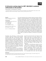
results, reported in Fig. 3, show that in lung only BLT is
expressed, while in liver capsule only BLCT cDNA is
present, in agreement with our previous results [29]. On the
contrary, in heart and spleen both isoforms are expressed.
We were unable to detect BLCT mRNA in lung and BLT
mRNA in the liver capsule, even when 40 cycles of PCR
were performed to allow identification of low abundant
transcripts.
510 A. Gambacurta et al. (Eur. J. Biochem. 270) Ó FEBS 2003
A
-20
AGCAGCCTGGACCTGCCAAG -1
ATGCTCCATCTGCTGGCGCTCGCCCTCCTGCTGAGCCTGGTCTCCGCAGCCCCTGGCCAGGCCCTGCAGCGC 72
M L H L L A L A L L L S L V S A A P G Q A L Q R (-3)
GCGGGCATCGTCGGGGGGCAGGAGGCCCCTGGGAGCAGATGGCCCTGGCAGGTGAGCCTGAGAGTCAGCCGT 144
A G I V G G Q E A P G S R W P W Q V S L R V S R (22)
CGGTACTGGAGGCACCACTGCGGGGGCTCCCTGATCCACCCCCAGTGGGTGCTGACCGCAGCCCACTGCGTC 216
R Y W R H H C G G S L I H P Q W V L T A A H C V (46)
•
GGACCGGAAGTCCATGGCCCCTCATACTTCAGGGTGCAGCTGCGTGAGCAGCACCTGTATTACCAGGACCAG
288
G P E V H G P S Y F R V Q L R E Q H L Y Y Q D Q (70)
CTGCTGCCCATCAGCAGGATCATCCCCCACCCCAACTACTACAGCGTTAAGAACGGTGCGGACATCGCCCTG 360
L L P I S R I I P H P N Y Y S V K N G A D I A L (94)
•
CTGGAGCTGGACAAGCTTGTGAATATCTCCTGGCACGTCCAGCTGGTCACCCTGCCCCCTGAGTCGGAGACC
432
L E L D K L V N I S W H V Q L V T L P P E S E T (118)
*
TTTCCCCCGGGGACGCAGTGCTGGGTGACGGGCTGGGGCAACGTGGACAATGGAAGGCGCCTGCCGCCCCCA
504
F P P G T Q C W V T G W G N V D N G R R L P P P (142)
TTCCCCCTGAAGCAGGTGAAGGTGCCCGTCGTGGAGAACAGTGTCTGTGACAGGAAGTACCACTCTGGCCTG 576
F P L K Q V K V P V V E N S V C D R K Y H S G L (166)
TCCACAGGGGACAACGTATCCATAGTGCAGGAGGATAACTTGTGTGCTGGGGACAGCGGGAGGGACTCCTGC 648
S T G D N V S I V Q E D N L C A G D S G R D S C (190)
*
CAGGGCGACTCTGGAGGGCCCCTGGTCTGCAAGGTGAATGGCACCTGGCTGCAGGCGGGGGTGGTCAGCTGG
720
Q G D S G G P L V C K V N G T W L Q A G V V S W (214)
• *
GGCGATGGTTGCGCGAAGCCCAACCGGCCCGGCATCTACACCCGCGTCACCTCCTACCTGGACTGGATCCAC
792
G D G C A K P N R P G I Y T R V T S Y L D W I H (238)
CAGTACGTCCCCCAGGGGCCCtgagcctggtccccaggccgccccctggtcagcggaggagctggccccctc 864
Q Y V P Q G P ♦ (245)
tgtcccctcagcgctgcttccggcccgaggaggagaccttcccccaccttccctggccccctgcccaatgcc 936
cacccctggctgacccctctctgctgacccctccctgccctgaacccctgccccagccccctccccactagc 1008
tcagggcgctggcaggggctgctgacactcataaaaagcatggagagcag 1058
B
-20
AGCAGCCTGGACCTGCCAAG -1
ATGCTCCATCTGCTGGCGCTCGCCCTCCTGCTGAGCCTGGTCTCCGCAGCCCCTGGCCAGGCCCTGCAGCGC 72
GCGGGCATCGTCGGGGGGCAGGAGGCCCCTGGGAGCAGATGGCCCTGGCAGGTGAGCCTGAGAGTCAGCCGT 144
CGGTACTGGAGGCACCACTGCGGGGGCTCCCTGATCCACCCCCAGTGGGTGCTGACCGCAGCCCACTGCGTC 216
GGACCGGAAGTCCATGGCCCCTCATACTTCAGGGTGCAGCTGCGGGAGCAGCACCTGTATTACCAGGACCAG 288
CTGCTGCCCATCAGCAGGATCATCCCCCACCCCAACTGCTACAGCGTTAAGAACGGGGCGGACATCGCCCTG 360
CTGGAGCTGGACAAGCTTGTGAATATCTCCTGGCACGTCCAGCCGGTCACCCTGCCCCCTGAGTCGGAGACC 432
TTCCCCCCGGGGACGCAGTGCTGGGTGACGGGCTGGGGCAACGTGGACAATGGAAGGCGCCTGCCGCCCCCA 504
TTCCCCCTGAAGCAGGTGAAGGTGCCCGTCGTGGAGAACAGTGTCTGTGACAGGAAGTACCACTCTGGCCTG 576
TCCACAGGGGACAACGTCCCCATCGTGCGGGAGGACATGCTGTGTGCTGGGGACAGCGGGAGGAACTTCTGC 648
CAGGGCGACTCTGGAGGGCCCCTGGTCTGCAAGGTGAATGGCACCTGGCTGCAGGCGGGGGTGGTCAGCTGG 720
GGCGATGGTTGCGCGAAGCCCAACCGGCCCGGCATCTACACCCGCGTCACCTCCTACCTGGACTGGATCCAC 792
CAGTACGTCCCCCAGGGGCCCtgagcctggtccccaggccgccccctgggtcagcggaggagctggccccca 864
♦
cagtcccctcaacactgcttccggccgaggaggagaccttcccccaccttccccggccccctgtcccagtgc 936
ccacacctgatgaccccactcctggctgtacccctctcccgctcagctcacccccccgcaggggctgctgac 1008
actcattaaagagcatggagagg 1031
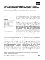
Fig. 1. Full-length bovine tryptase cDNAs. Nucleotide numbering begins at the first nucleotide of the preprosequence. Stop codon (r)and
polyadenylation signal (underlined) are indicated. (A) BLT cDNA and deduced amino acid sequence. Potential N-linked glycosylation sites (w),
residues of the serine protease catalytic triad (d) and residues identified by mass spectrometry (underlined) are indicated. Amino acid numbering
startsatthefirstresidueofthematureprotein.(B)BLCTcDNA(seealso[29]).
Ó FEBS 2003 Tissue-specific expression of bovine tryptases (Eur. J. Biochem. 270) 511
Promoter analysis and organization of tryptase genes
In order to obtain information on the promoter regions of
the two tryptase genes, the amplification products obtained
from bovine genomic DNA, digested and modified as
described above, were analyzed on agarose gel. Two distinct,
prominent bands were obtained in the case of genomic
DNA digested with HincII. After cloning and sequencing, it
was possible to assign each 5¢ region to one of the two
tryptase (BLT and BLCT) genes, which were amplified with
the proper primer pairs and subjected to restriction analysis
with NspI (see Materials and methods). The two promoter
sequences and 5¢-UTRs are reported in Fig. 4, where the
regulatory sequences found using the
TRANSFAC
4.0
program are highlighted. The same figure shows that in
both sequences intron I is present in phase 0 just upstream
the initiation codon, as found in most tryptases. Location
and phase of introns II–V were evaluated as reported under
Material and methods and were found identical (data not
shown) to those of human tryptases [26]. Moreover, in
searching for location and phase of intron V, we sequenced
exon regions of BLT and BLCT genes corresponding to
amino acid residues 158–245. These regions include the five
residues (out of six) which are different in the two tryptases
and the results confirm once again the presence of Asn188-
Phe189 in BLCT, unlike BLT and tryptases from other
species which contain Asp-Ser in those positions.
Isolation and characterization of bovine lung tryptase
We routinely purified tryptase from bovine liver capsule
using an high salt extraction followed by a two-column
purification. The whole procedure was carried out at
pH 6.1. Based on active site titration with [
3
H]DFP, a
typical heparin pooled fraction contains 0.3 nmol of active
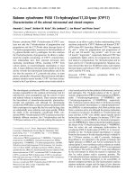
Fig. 2. Comparison of amino acid sequences of
BLT, BLCT and tryptases from other species.
The compared sequences are: BLT (this
work), BLCT [29], human tryptase bII [25],
human tryptase a [23,42] and sheep tryptase 1
[13]. Residues identical in BLT and other
tryptases are indicated by a point (Æ). Num-
bering begins at the first residue of the mature
proteins.
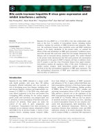
Fig. 3. Differential NspI restriction of bovine tryptase cDNAs in dif-
ferent tissues. Agarose 1.7% (w/v) gel electrophoresis of the TA vector-
cloned-bovine tryptase cDNAs obtained from bovine lung, heart,
spleen and liver capsule, was performed after treatment with NspI
endonuclease. Arrows indicate the bands corresponding to the NspI-
undigested BLT insert (lung; heart, lane b; spleen, lane b) and to the
NspI-digestion products of the BLCT insert (heart, lane a; spleen, lane
a; liver capsule). The two upper bands in all lanes represent the NspI-
digested TA vectors. Lane MW, DNA marker (1 kb ladder).
512 A. Gambacurta et al. (Eur. J. Biochem. 270) Ó FEBS 2003
sitesÆg
)1
of wet tissue. The specific activity of BLCT in the
standard assay containing 75 l
M
Boc-Phe-Ser-Arg-MCA
is 190 pmol MCA min
-1
(pmol of tryptase subunit)
)1
.
BLCT is stable and maintains its full activity for about
two to three weeks when kept at 4 °Cinhighsaltat
pH 6.1.
Inthecaseofbovinelung,wewereabletoisolatetryptase
only after lowering the buffer pH to 5.5. The lower pH
resulted in increased adsorption to the resins and increased
stability of tryptase activity. Fractions with tryptic activity,
eluted from the heparin column, were pooled, concentrated
and analyzed by SDS/PAGE. To further purify the still
heterogeneous sample, the concentrated pooled fraction
was loaded on a gel filtration column and the elution
profile at 280 nm showed several peaks. The fractions
containing tryptic activity were then pooled and reacted
with radiolabeled DFP. SDS/PAGE followed by fluoro-
graphy yielded bands only in the 35–40 kDa range (data not
shown). The same multiple bands were detected with the
anti-(178/191 tryptase-peptide) Ig, using BLCT as control
[31] (inset of Fig. 5). The multiple banding pattern is
probably due to variable glycosylation of two/three different
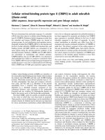
sites. Reloading of the BLT sample, preincubated with
heparin, on the gel filtration column yielded a symmetrical
peak displaying enzymatic activity and migrating with an
apparent molecular weight of 200 kDa (Fig. 5). This size
is in reasonable agreement with a tetramer bound to
heparin. A minor peak with no activity and an elution
volume equivalent to a molecular mass of 35 kDa was
present. To test for catalytic activity, BLT was titrated with
the burst titrant MUGB. Approximately 6 lgofactivelung
tryptase was obtained with this procedure (4 pmol active
sitesÆg
)1
of wet tissue, assuming M
r
¼ 35 000). The specific
activity of BLT in the standard assay containing 75 l
M
Boc-Phe-Ser-Arg-MCA is 870 pmol MCA min
)1
(pmol of
tryptase subunit)
)1
. Based on a protein assay and active site
titration with MUGB, our tryptase preparation was 52%
active.
MALDI mass spectrometry analysis was performed on
about 50 pmol of Coomassie blue-stained BLT, from the
SDS/PAGE, which were subjected to in-gel tryptic diges-
tion. We took precautions during gel electrophoresis to
avoid formation of acrylamide adducts and used only the
best and purest chemicals and solvents available throughout
the entire purification process. The peptides were extracted
from the gel as described above and the mixture was directly
analyzed by MALDI mass spectrometry. This gave
predominantly singly charged fragments, allowing easier
interpretation of masses observed for peptide mixtures than
is the case for spectra generated by electrospray mass
spectrometry. From the MALDI mass spectra (Fig. 6), it
was possible to recover and identify peptides from the entire
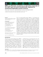
sequence of BLT. As shown in Table 1, we were able to
match all peptide masses with the amino acid sequence as
deduced from the cDNA clone. More than 80% of BLT
sequence (see also underlined residues in Fig. 1) was covered
with an adequate mass accuracy (better than 0.1%,
Table 1). In particular, the products with ion signals at
m/-z 1490.7 and 1366.2 represent the peptides that
characterize BLT isoform with respect to BLCT isoform
(corresponding to peptides 167–180 and 188–201, with
theoretical M
r
s of 1490.5 and 1365.7, respectively).
BLT was highly reactive toward tripeptide coumarin-
containing substrates, especially those with basic amino acid
in P1 and P2 positions exhibiting k
cat
/K
m
values of about
10
6
M
)1
Æs
)1
. In Table 2 the catalytic efficiency vs. tripeptide
and single residue substrates is reported in comparison with
the activity of BLCT. k
cat
/K
m
values for both enzymes were
determined at the same pH and temperature and under
pseudo first-order conditions. For all substrates BLT was a
more efficient catalyst then BLCT (10- to 60-fold) and both
enzymes exhibited a dramatic drop in catalytic ability in
Fig. 4. Comparison of promoter sequences and
5¢-UTRs in BLT and BLCT genes. Identical
nucleotides are indicated (w). Putative TATA,
CAAT and GC box sequences are underlined.
Binding sites for specific transcription factors
(indicated) are boxed. Intron I sequence is
reported in lower case. Numbering begins at
the transcription initiation site (in bold).
Ó FEBS 2003 Tissue-specific expression of bovine tryptases (Eur. J. Biochem. 270) 513
going from tripeptide to single-residue substrates (about 10
4
and 10
2
M
)1
Æs
)1
, respectively).
As shown in Table 3, BLT was inactivated by low
molecular weight inhibitors of tryptic proteases. Like other
tryptases it is essentially unaffected by large serine protease
inhibitors as STI and a-1-antitrypsin. However, BPTI (or
aprotinin, the trypsin inhibitor present in bovine mast cells),
causes a significant reduction in BLT activity, similarly to
what previously found for BLCT [11,32].
Discussion
In previous studies, we reported isolation of tryptase (then
named BLCT) from bovine liver capsule and its characteri-
zation [11,29]. BLCT was the only tryptase found in that
Fig. 5. Gel filtration analysis of purified BLT. BLT, preincubated with
heparin (10 lgÆmL
)1
), was chromatographed on a Superose 12PC
column preequilibrated with 10 m
M
Mes, 0.4
M
NaCl,pH 5.5.Protein
was detected spectrophotometrically at 280 nm and 100 lLfractions
were collected. Tryptase activity in each fraction was measured as
described in the text and reported as percent of the most active
fraction. Elution positions of blue dextran (void volume), catalase
(220 kDa), ovalbumin (43 kDa) and ribonuclease (13.7 kDa) are
indicated by arrows. In the inset the immunodetection of purified
BLCT (a) and of BLT (b) is shown.
Fig. 6. MALDI mass spectrometry analysis of peptides obtained from
the in-gel tryptic digestion of BLT. The peptides correspond to (MH)
+
masses. Ion masses £ 1150 and ‡ 1600 Da are not shown. The marked
products represent the peptides that characterize the BLT isoform with
respect to BLCT isoform (peptides 167–180 and 188–201).
Table 1. MALDI MS analysis of the peptide mixture extracted from
BLT gel spot.
MH
+
experimental
MH
+
theoretical
Peptide
2693.1 2693.7 162–187
2324.7 2324.1 41–61
1947.3 1947.9 202–220
1903.1 1903.5 62–76 230–245
1576.0 1576.3 113–126
1490.7 1490.5 167–180
1442.5 1442.1 88–100
1366.2 1365.7 188–201
1344.5 1344.4 150–161
1330.8 1330.4 77–87
1272.4 1271.9 88–99
1263.7 1263.1 139–149
1217.5 1217.0 150–160
1174.1 1174.5 127–137
1072.2 1072.0 12–19
1071.2 1071.0 1–11
1064.3 1064.5 138–146
909.1 909.2 139–146
680.4 680.9 23–26
Table 2. Specificity constants for the hydrolysis of model substrates by
BLT and BLCT. Assay conditions were 0.1
M
Tris/HCl, pH 8.0, and
25 °C. Enzyme concentration was 3 n
M
. Values were determined un-
der pseudo first-order conditions and are the averages of four different
experiments. SDs were £ 8% of the averages.
Substrate
10
5
· k
cat
/K
m
(
M
)1
Æs
)1
)
BLT BLCT
Boc-Gly-Lys-Arg-MCA 30 2.3
Boc-Gly-Gly-Arg-MCA 20 0.7
Boc-Phe-Ser-Arg-MCA 17 1.0
Boc-Val-Pro-Arg-MCA 18 0.5
Z-Arg-MCA 0.13 0.002
Table 3. Effect of serine protease inhibitors on BLT activity. Assay
conditions were 0.1
M
Tris/HCl, pH 8.0 and 30 °C. Residual activity
was determined using Boc-Phe-Ser-Arg-MCA as substrate. BLT
concentration was 5 n
M
active sites. Values are the averages of three
determinations.
Inhibitor Concentration % BLT activity
None 0 100
DFP 2 m
M
0
Benzamidine 2 m
M
0
TLCK 2 m
M
0
a-1-Antitrypsin 0.1 mgÆmL
)1
96
STI 0.1 mgÆmL
)1
100
BPTI (Aprotinin) 0.1 mgÆmL
)1
35
514 A. Gambacurta et al. (Eur. J. Biochem. 270) Ó FEBS 2003
tissue, at the protein level and at the transcription level, as
confirmed here. Here we describe isolation of a bovine
tryptase gene and cDNA encoding a new tryptase isoform
(BLT), which is in turn the only one isoform expressed in
bovine lung. Furthermore, analysis of BLCT and BLT
expression in bovine heart and spleen has shown that both
enzymes are present in these tissues at the mRNA level. The
simultaneous expression of the two isoforms could be due to
similar regulatory mechanisms in these specific tissues.
The coexistence in the same organism of multiple tryptase
genes, as found here, is in linewith findings reported by others
for human [23–26], and mouse [27,28] tryptases. What is
peculiar here is the presence in the same organism of
isoforms, BLCT and BLT, whose primary structure predicts
a different functional efficiency, despite their 98% sequence
identity, as confirmed by the catalytic activity of the isolated
proteins (see also later). BLT differs from BLCT at only six of
the 245 residues forming the catalytic domain, two of them
(residues 188–189) being in sites thought to be critical
determinants of function. BLT is structurally more similar
than BLCT to most tryptases, in particular for the presence of
the canonical residues Asp188 and Ser189 in the S1 specificity
pocket, whereas residues Gly215 and Gly225, found in all
b-type tryptases, are present in both the bovine enzymes.
These results, indicating a possible tissue specific function
of the two isoforms, prompted us to analyze the organiza-
tion and the promoter sequences (Fig. 4) of the two tryptase
genes. The length of both genes, as evaluated from the size
of the PCR products, obtained from genomic DNA with
proper primers, is around 1800 bp, similar to that of human
bI tryptase gene [25]. The two genes share with human, dog
and mouse MCP-6 tryptase genes the same organization
with six exons separated by five introns, the same and
unique position of intron I (189 bp), immediately upstream
the initiation codon, and the location/phase of introns II–V.
It is interesting to note that five codons (out of six) encoding
different residues in BLT, with respect to BLCT, are all
located in exon V, which encodes residues 137–191 of the
mature proteins; this is the same region where the greatest
disparity among human tryptases was found [26]. The
prepro-sequences of BLT and BLCT (26 residues) are
identical. Although four residue shorter, these sequences are
very similar to the corresponding sequences of human a and
bI-III tryptases [26]. Their C-terminal portions (10 residues)
are identical to those of b tryptases, in agreement with their
role as activation peptides. The presence of Arg in )3
position (relative to the mature proteins) is a key feature of
b-like tryptases [26].
The 5¢ flanking regions (about 190 bp) of the BLT and
BLCT genes (Fig. 4) are 70% identical; their last 100 bp
are similar (about 60% identity) to the same regions of
human bIandbII tryptase genes [25,26] and contain the
same putative TATA box (ATAAA) in a similar position
() 33/)32). BLCT also contains a canonical TATA box
in an unusual position ()91) and a CAAT box at position
)161. Both promoters contain a GC box (positions
)68/)67) and other regulatory sequences (boxed in
Fig. 4). In BLCT, binding sequences are present for
positive transcription factors, such as APF and androgen
receptors, AR [33]. APF is homologous to HNF-1
(hepatocyte nuclear factor I) which is responsible for the
tissue specific activation of human a1-antitrypsin [34].
Likewise, BLT promoter contains several recognition
sequences for positive transcription factors such as AR,
NFIII (nuclear factor III) functionally identical to tran-
scription factor OTF-1 [35], and AP-1, which is known to
bind specific sequences present in promoters or enhancers
[36]. The presence in both genes of AR sequences could
suggest an hormone-regulated expression. Interestingly,
the BLT promoter contains a recognition sequence for a
negative transcription factor, COUP-TF (chick ovalbumin
upstream promoter-transcription factor), which has been
identified in many different species [37]. COUP-TFs
belong to the steroid/thyroid hormone receptor (TR)
superfamily and have been shown to down regulate the
hormonal induction of TR-dependent activation of speci-
fic genes, acting as inhibitors of transcriptional activity
[37]. Thus, the interplay of positive or negative transcrip-
tion factors may regulate, in a tissue-specific fashion, the
expression of BLT and BLCT proteins.
For the isolation of tryptase from lung we used a more
acidic pH than that used in the liver capsule tryptase
purification procedure, with the aim of increasing adsorp-
tion of the enzyme to the resin and its stability. The
heterogeneous sample needed to be purified by a further
chromatographic step. However, some contaminating
proteins were still present after this step, but the only serine
protease detected by fluorography after labeling with
radioactive DFP showed to be immunoreactive with specific
anti-tryptase Igs. Our results show that in a gel chromato-
graphy analysis of native BLT preincubated with heparin,
theenzymeelutedasan 200 kDa protein. This size is in
reasonable agreement with a tetramer bound to heparin [38]
considering that the BLT monomer has a size of 35 kDa
and that the elution position may be anticipated by the
presence of the anionic heparin glycosamminoglycan. BLT
subunit concentration was measured by burst tritation with
MUGB. The procedure, whose success depends on the
rapid acylation of the enzyme with release of a fluorescent
leaving group followed by a very slow deacylation, was
less satisfactory with BLCT. The instability of the
guanidinobenzoyl-enzyme intermediate was probably due
to the replacement of Asp188 with Asn in the S1 pocket of
the protease. However, BLCT could be labeled with
radioactive DFP, indicating that the catalytic machinery
of the protease was functional [11,29]. In this regard, it is
worthwhile to underscore the difference in specific activity
between BLT and BLCT for the hydrolysis of Boc-Phe-Ser-
Arg-MCA [870 and 190 pmol MCA min
)1
Æ (pmol of
tryptase subunit)
)1
, respectively].
To investigate the structural features responsible for the
functional differences between BLT and BLCT, we decided
to support the sequence information obtained from cDNA
analysis by protein sequence analysis. After column puri-
fication, lung tryptase identified by SDS/PAGE was
subjected to in-gel tryptic fragmentation followed by
analysis of the peptide mixture by MALDI mass spectros-
copy. Mass fingerprinting of BLT tryptic fragments allowed
us to screen the entire protein sequence for the presence of
peptides that characterize lung tryptase in comparison with
the isoform isolated from liver capsule.
Thepreferenceoftryptaseforcleavingsmallsynthetic
substrates with two basic residues was previously suggested
for human pituitary tryptase [2] and for BLCT [11,31]. In
Ó FEBS 2003 Tissue-specific expression of bovine tryptases (Eur. J. Biochem. 270) 515
particular, the latter was shown to cleave peptide substrates
that reproduce precursor sequences around putative clea-
vage loci [31]. However, no conclusions can be drawn at this
stage on the BLT preference for substrates with two
terminal basic residues, in spite of the similar trend found
in the catalytic efficiency of BLT and BLCT toward some
synthetic substrates. Moreover, for all substrates examined,
BLT exhibited k
cat
/K
m
values that were 10- to 60-fold
greater than those of BLCT. The difference in catalytic
properties between the two enzymes may be related to the
sequence of the region forming the primary specificity S1
pocket. An Asp residue is located at position 188 in BLT,
human, sheep and other tryptases and confers specificity for
binding basic P1 amino acid residues. In BLCT, the
presence of the Asn residue in that position results in a
decrease negative charge at the bottom of the pocket and a
consequent weaker interaction of substrates when compared
with BLT and the other tryptases. The usual substrate
specificity of BLCT was explained by assuming some
conformational change of the active sites [29] and/or
involving the role of additional interactions occurring
between the active sites and substrates. In this regard,
modeling studies showed that the carbonyl oxygen atom of
the properly oriented Phe190 may form a hydrogen bond
with the c-guanidino group of the P1 Arg residue in the
inhibitor and/or substrate molecule [39]. Additional inter-
actions in the interior of the extended substrate binding-site
may also explain the consistently greater catalytic efficiency
of BLT and BLCT on tripeptide substrates when compared
with a single residue substrate. As to the inhibition by
standard serine protease inactivators, it is worth mentioning
that, similarly to BLCT [11,32], BLT is sensitive to
aprotinin, the trypsin inhibitor of bovine origin.
On the whole, the results reported in this study suggest a
tissue-specific expression and a different competence for
catalysis of BLT and BLCT. Thus, cattle could be a useful
model for investigating heterogeneity of tryptases. Such
heterogeneity is probably linked to different patterns of
tryptase action following release from bovine mast cells in
different tissues. The physiological meaning and the mech-
anism underlying the differential expression of granule
proteinases are not yet fully understood for humans,
rodents, dog and sheep mast cells [40]. It is interesting to
recall that in rat lung, chymase expression is modified by
nematode infection [41]. It may be argued that the role of
tissue microenvironment on mast cell phenotype must be
linked to proteinase function in the various tissues. As yet,
there are no obvious clues as to why such mechanism may
be correlated to different in vivo functions. Further studies
are required to explore the role of bovine tryptases and to
identify their target substrates.
Acknowledgements
This investigation was supported by MURST and MURST-CNR
Biotechnology Program L.95/95, Italy.
References
1. Smith, T.J., Houghl, M.W. & Johnson, D.A. (1984) Human lung
tryptase: purification and characterization. J. Biol. Chem. 259,
11046–11051.
2. Cromlish, J.A., Seidah, N.G., Marcinkiewicz, M., Hamelin, J.,
Johnson, D.A. & Chretien, M. (1987) Human pituitary tryptase:
molecular forms, NH
2
-terminal sequence, immunocytochemical
localization and specificity with prohormone and fluorogenic
substrates. J. Biol. Chem. 262, 1363–1373.
3. Harvima, I.T., Schechter, N.M., Harvima, R.J. & Fraki, J.E.
(1988) Human skin tryptase: purification, partial characterization
andcomparisonwithhumanlungtryptase.Biochim. Biophys.
Acta 957, 71–80.
4. Caughey, G.H., Viro, N.F., Ramachandran, J., Lazarus, S.C.,
Borson, D.B. & Nadel, J.A. (1987) Dog mastocytoma tryptase:
affinity purification, characterization and amino-termoinal
sequence. Arch. Biochem. Biophys. 258, 555–563.
5. Vanderslice, P., Craik, C.S., Nadel, J.A. & Caughey, G.H. (1989)
Molecular cloning of dog mast cell tryptases and a related pro-
tease:structuralevidenceofauniquemodeofserineprotease
activation. Biochemistry 28, 4148–4155.
6. Ide, H., Itoh, H., Tomita, M., Murakumo, Y., Kobayashi, T.,
Maruyama, H., Osada, Y. & Nawa, Y. (1995) Molecular cloning
and expression of rat mast cell tryptase. J. Biochem. (Tokyo) 118,
210–215.
7. Kido, H., Fukusen, N. & Katunuma, N. (1985) Chymotrypsin
and trypsin-type serine proteases in rat mast cells: properties and
functions. Arch. Biochem. Biophys. 239, 436–443.
8. Braganza, V.J. & Simmons, W.H. (1991) Tryptase from rat skin:
purification and properties. Biochemistry 30, 4997–5007.
9. Reynolds, D.S., Gurley, D.S., Austen, K.F. & Serafin, W.E.
(1991) Cloning of cDNA and gene of mouse mast cell protease-6.
J. Biol. Chem. 266, 3847–3853.
10. Mc. Neil, H.P., Reynolds, D.S., Schiller, V., Ghildyal, N., Gurley,
D.S., Austen, K.F. & Stevens, R.L. (1992) Isolation, characteri-
zation and transcription of the gene encoding mouse mast cell
protease 7. Proc. Natl Acad. Sci. USA 89, 11174–11178.
11. Fiorucci, L., Erba, F. & Ascoli, F. (1992) Bovine tryptase: puri-
fication and characterization. Biol. Chem. Hoppe-Seyler 373,
483–490.
12. Murakumo,Y.,Ide,H.,Itoh,H.,Tomita,M.,Kobayashi,T.,
Maruyama, H., Horii, Y. & Nawa, Y. (1995) Cloning of the
cDNA encoding mast cell tryptase of Mongolian gerbil, Meriones
unguiculatus, and its preferential expression in the intestinal
mucosa. Biochem. J. 309, 921–926.
13. Pemberton, A.D., McAleese, S.M., Huntley, J.F., Collie, D.D.S.,
Scudamore, C.L., McEuen, A.R., Walls, A.F. & Miller, H.R.P.
(2000) cDNA sequence of two sheep mast cell tryptases and the
differential expression of tryptase and sheep mast cell proteinase-1
in lung, dermis and gastrointestinal tract. Clin. Exper. Allergy 30,
818–832.
14. Schwartz, L.B., Bradford, T.R., Litman, B.L. & Wintroub, B.U.
(1985) The fibrinogenolytic activity of purified tryptase from
human lung mast cells. J. Immunol. 135, 2762–2767.
15. Caughey, G.H., Leidig, F., Viro, N.F., Gold, W.M. & Nadel, J.A.
(1988) Substance P and vasointestinal peptide degradation by
mast cell tryptase and chymase. J. Pharmacol. Exp. Ther. 244,
133–137.
16. Ruoss, S.J., Hartmann, T. & Caughey, G.H. (1991) Mast cell
tryptase is a mitogen for cultured fibroblasts. J. Clin. Invest. 88,
493–499.
17. Stack, M.S. & Johnson, D.A. (1994) Human mast cell tryptase
activates single chain urinary-type plasminogen activator
(pro-urokinase). J. Biol. Chem. 269, 9416–9419.
18. Blair, R.J., Meng, H., Marchese, M.J., Ren, S., Schwartz, L.B.,
Tonnesen, M.G. & Gruber, B.L. (1997) Human mast cells
stimulate vascular tube formation: tryptase is a novel, potent
angiogenic factor. J. Clin. Invest. 99, 2691–2700.
19. Steinhoff, M., Vergnolle, N., Young, S.H., Tognetto, M.,
Amadesi, S., Ennes, H.S., Trevisani, M., Hollenberg, M.D.,
516 A. Gambacurta et al. (Eur. J. Biochem. 270) Ó FEBS 2003
Wallace, J.L., Caughey, G.H., Mitchell, S.E., Williams, L.M.,
Geppetti, P., Mayer, E.A. & Bunnett, N.W. (2000) Agonists of
proteinase-activated receptor 2 induce inflammation by a neuro-
genic mechanism. Nat. Med. 6, 151–158.
20. Schwartz, L.B. & Bradford, T.R. (1986) Regulation of tryptase
from human lung mast cells by heparin: stabilization of the active
tetramer. J. Biol. Chem. 251, 7372–7379.
21. Schechter, N.M., Eng, G.Y., Selwood, T. & McCaslin, D.R.
(1995) Structural changes associated with the spontaneous
inactivation of the serine proteinase human tryptase. Biochemistry
34, 10628–10638.
22. Pereira, P.J.B., Bergner, A., Macedo-Ribeiro, S., Huber, R.,
Matschiner, G., Fritz, H., Sommerhoff, C.P. & Bode, W. (1998)
Human beta-tryptase is a ring-like tetramer with active sites facing
acentralpore.Nature 392, 306–311.
23. Miller, J.S., Westin, E.H. & Schwartz, L.B. (1989) Cloning and
characterization of complementary DNA for human tryptase.
J. Clin. Invest. 84, 1188–1195.
24. Miller, J.S., Moxley, G. & Schwartz, L.B. (1990) Cloning and
characterization of a second complementary DNA for human
tryptase. J. Clin. Invest. 86, 864–870.
25. Vanderslice, P., Ballinger, S.M., Tam, E.K., Goldstein, S.M.,
Craik, C.S. & Caughey, G.H. (1990) Human mast cell tryptase:
multiple cDNAs and genes reveal a multigene serine protease
family. Proc. Natl Acad. Sci. USA 87, 3811–3815.
26. Pallaoro, M., Fejzo, M.S., Shayesteh, L., Blount, J.L. & Caughey,
G.H. (1999) Characterization of genes encoding known and novel
human mast cell tryptases on chromosome 16p13.3. J. Biol. Chem.
274, 3355–3362.
27. Gurish, M.F., Nadeau, J.H., Johnson, K.R., McNeil, H.P.,
Grattan, K.M., Austen, K.F. & Stevens, R.L. (1993) A closely
linked complex of mouse mast cell-specific chymase genes on
chromosome 14. J. Biol. Chem. 268, 11372–11379.
28. Gurish, M.F., Johnson, K.R., Webster, M.J., Stevens, R.L. &
Nadeau, J.H. (1994) Location of the mouse mast cell protease 7
gene (MCP7) to chromosome 17. Mammal Genome 5, 656–657.
29. Pallaoro, M., Gambacurta, A., Fiorucci, L., Mignogna, G., Barra,
D. & Ascoli, F. (1996) cDNA cloning and primary structure of
tryptase from bovine mast cells, and evidence for the expression of
bovine pancreatic trypsin inhibitor mRNA in the same cells. Eur.
J. Biochem. 237, 100–105.
30.Jameson,G.W.,Roberts,D.V.,Adams,R.W.,Kyle,W.S.&
Elmore, D.T. (1973) Determination of the operational molarity of
solutions of bovine alpha-chymotrypsin, trypsin, thrombin and
factor Xa by spectrofluorimetric titration. Biochem. J. 131,107–
117.
31. Fiorucci, L., Pallaoro, M., Erba, F., Colombo, A.P., Rholam, M.,
Cohen, P. & Ascoli, F. (1998) Structural and functional properties
of Bos taurus tryptase: a search for a possible propeptide
processing role. Comp. Biochem. Physiol. B Biochem. Mol. Biol.
120, 239–245.
32. Fiorucci, L., Erba, F., Coletta, M. & Ascoli, F. (1995) Evidence
for multiple interacting binding sites in bovine tryptase. FEBS
Lett. 363, 81–84.
33. Faber, P.W., van Rooij, H.C., van der Korput, H.A., Baarends,
W.M., Brinkmann, A.O., Grootegoed, J.A. & Trapman, J. (1991)
Characterization of the human androgen receptor transcription
unit. J. Biol. Chem. 266, 10743–10749.
34. Monaci, P., Nicosia, A. & Cortese, R. (1988) Two different liver-
specific factors stimulate in vitro transcription from the human
a1-antitrypsin promoter. EMBO J. 7, 2075–2087.
35. O’Neill, E.A., Fletcher, C., Burrow, C.R., Heintz, N., Roeder,
R.G. & Kelly, T.J. (1988) Transcription factor OTF-1 is func-
tionally identical to the DNA replication factor NF-III. Science
241, 1210–1213.
36. Bohmann, D., Bos, T.J., Admon, A., Nishimura, T., Vogt, P.K. &
Tjian, R. (1987) Human proto-oncogene c-jun encodes a DNA
binding protein with structural and functional properties of tran-
scription factor AP-1. Science 238, 1386–1392.
37. Tsai, S.Y. & Tsai, M.J. (1997) Chick ovalbumin upstream pro-
moter-transcription factors (COUP-TFs): coming of age. Endocr.
Rev. 18, 229–240.
38. Hallgren, J., Estrada, S., Karlson, U., Alving, K. & Pejler, G.
(2001) Heparin antagonists are potent inhibitors of mast cell
tryptase. Biochemistry 40, 7342–7349.
39. Erba,F.,Fiorucci,L.,Pascarella,S.,Menegatti,E.,Ascenzi,P.&
Ascoli, F. (2001) Selective inhibition of human mast cell tryptase
by gabexate mesylate, an antiproteinase drug. Biochem. Pharma-
col. 61, 271–276.
40. Miller, H.R. & Pemberton, A.D. (2002) Tissue-specific
expression of mast cell granule serine proteinases and their
role in inflammation in the lung and gut. Immunology 105,
375–390.
41. Tomita, M., Itoh, H., Kobayashi, T., Onitsuka, T. & Nawa, Y.
(1999) Expression of mast cell proteases in rat lung during hel-
minth infection: mast cells express both rat mast cell protease II
and tryptase in helminth infected lung. Int. Arch. Allergy Immunol.
120, 303–309.
42. Huang,R.,Abrink,M.,Gobl,A.E.,Nilsson,G.,Aveskogh,M.,
Larsson, L.G., Nilsson, K. & Hellman, L. (1993) Expression of a
mast cell tryptase in the human monocytic cell lines U-937 and
Mono Mac 6. Scand. J. Immunol. 38, 359–367.
Ó FEBS 2003 Tissue-specific expression of bovine tryptases (Eur. J. Biochem. 270) 517