Báo cáo khoa học: Novel aspects of calmodulin target recognition and activation pptx
Bạn đang xem bản rút gọn của tài liệu. Xem và tải ngay bản đầy đủ của tài liệu tại đây (333.37 KB, 11 trang )
REVIEW ARTICLE
Novel aspects of calmodulin target recognition and activation
Stefan W. Vetter
1
and Estelle Leclerc
2
1
Department of Molecular Biology and
2
Department of Immunology, The Scripps Research Institute, La Jolla, CA, USA
Several crystal and NMR structures of calmodulin (CaM) in
complex with fragments derived from CaM-regulated pro-
teins have been reported recently and reveal novel ways for
CaM to interact with its targets. This review will discuss and
compare features of the interaction between CaM and its
target domains derived from the plasma membrane
Ca
2+
-pump, the Ca
2+
-activated K
+
-channel, the Ca
2+
/
CaM-dependent kinase kinase and the anthrax exotoxin.
Unexpected aspects of CaM/target interaction observed in
these complexes include: (a) binding of the Ca
2+
-pump
domain to only the C-terminal part of CaM (b) dimer for-
mation with fragments of the K
+
-channel (c) insertion of
CaM between two domains of the anthrax exotoxin (d)
binding of Ca
2+
ions to only one EF-hand pair and (e)
binding of CaM in an extended conformation to some of its
targets. The mode of interaction between CaM and these
targets differs from binding conformations previously
observed between CaM and peptides derived from myosin
light chain kinase (MLCK) and CaM-dependent kinase IIa
(CaMKIIa). In the latter complexes, CaM engulfs the CaM-
binding domain peptide with its two Ca
2+
-binding lobes and
forms a compact, ellipsoid-like complex. In the early 1990s, a
model for the activation of CaM-regulated proteins was
developed based on this observation and postulated activa-
tion through the displacement of an autoinhibitory or
regulatory domain from the target protein upon binding of
CaM. The novel structures of CaM-target complexes dis-
cussed here demonstrate that this mechanism of activation
may be less general than previously believed and seems to be
not valid for the anthrax exotoxin, the CaM-regulated K
+
-
channel and possibly also not for the Ca
2+
-pump.
Keywords: calmodulin; CaM-binding domains; Ca
2+
-
signaling; cellular signaling; CaM-target activation.
Ca
2+
signaling and calmodulin
The calcium metal ion (Ca
2+
) plays an uniquely important
role in the physiology of higher organisms and is involved in
the regulation of many cellular processes ranging from gene
transcription and neurotransmitter release to muscle con-
traction and cell survival [1–4]. The intracellular concentra-
tion of free Ca
2+
is tightly controlled and usually very low
inside the cytosol (0.1 l
M
) whereas the extracellular con-
centration of Ca
2+
is roughly 10 000-fold higher (1 m
M
).
Various stimuli, such as changes in membrane polarization
or small receptor ligands can trigger the opening of calcium
channels residing in the plasma membrane, resulting in the
influx of Ca
2+
ions into the cytosol. In addition, several
intracellular organelles function as Ca
2+
stores, which can
release Ca
2+
upon stimulation by, for instance, inositol-
1,4,5-trisphosphate (InsP
3
) or cyclic ADP-ribose [5–7]. The
sarcoplasmic reticulum (SR) and endoplasmic reticulum
(ER) are major Ca
2+
stores, but mitochondria and the
nucleus also participate actively in the release of Ca
2+
through the InsP
3
-receptor or ryanodine receptor [3,4,8–11].
The export of Ca
2+
ions from the cytosol into the
extracellular space or into intracellular organelles is
achieved by ATP-driven Ca
2+
-pumps and exchangers.
The key proteins involved in the transport of Ca
2+
ions,
(e.g. voltage gated Ca
2+
channels, the InsP
3
-receptor and
ryanodine receptors, sarcoplasmic Ca
2+
-pump (SERCA),
endoplasmic Ca
2+
-pump, plasma membrane Ca
2+
-pump
and others) are regulated by many different signals inclu-
ding secondary messenger molecules (e.g. InsP
3
,cADPR,
etc.), protein phosphorylation, neurotransmitters, mem-
brane potential and of course by the concentration of Ca
2+
ions itself [12–15]. In addition, Ca
2+
ions are involved in the
regulation of phospholipase C, the enzyme generating InsP
3
from PtdIns3,4P
2
, PtdIns-3-kinase, the enzyme metaboli-
zing InsP
3
to InsP
4
and in the regulation of protein kinase C
(PKC), which can phosphorylate the InsP
3
receptor [16,17].
These interdependencies generate an intricate network of
feedback loops which can result in oscillations of Ca
2+
concentration of defined amplitude, frequency and location
[18–25]. The mechanism of how the information contained
in Ca
2+
oscillation is decoded is not clear, but it has been
shown, for example, that the kinase activity of CaM-
dependent kinase II is sensitive to Ca
2+
oscillations [26–29].
The approximately 100-fold increase in free Ca
2+
con-
centration upon stimulation of a cell allows Ca
2+
-binding
proteins to bind Ca
2+
ions. Several hundred Ca
2+
-binding
proteins have been identified and most of them share a
common Ca
2+
binding motif [30–33]. This motif comprises
Correspondence to S. W. Vetter, Department of Molecular Biology,
10550 North Torrey Pines Road, La Jolla, CA 92037, USA.
Fax: + 1 858 784 2857, Tel.: + 1 858 784 2425,
E-mail:
Abbreviations: CaM, calmodulin; MLCK, myosin light chain kinase;
CaMKIIa, CaM-dependent kinase IIa; ER, endoplasmic reticulum;
SR, sarcoplasmic reticulum; SERCA, sarcoplasmic Ca
2+
pump;
PKC, protein kinase C; PtdIns, phosphatidylinositol; CaMKK,
CaM-dependent kinase kinase; InsP
3
, inositol-1,4,5-trisphosphate.
(Received 10 September 2002, revised 2 December 2002,
accepted 5 December 2002)
Eur. J. Biochem. 270, 404–414 (2003) Ó FEBS 2003 doi:10.1046/j.1432-1033.2003.03414.x
about 30 amino acids and consists of a helix-loop-helix
where the two helices are arranged similar to the extended
thumb and index finger of a hand: it is commonly called the
EF-hand motif. In almost all Ca
2+
-binding proteins two
EF-hand motifs are in close proximity forming an EF-hand
pair. This consists of four helices arranged in the form of a
twisted four helix bundle [34,35]. Structurally different
Ca
2+
-binding motifs are found in pentraxins, annexins and
in C
2
domains [36–39]. The C
2
domains consist of about 130
amino acids forming a compact b-sandwich composed of
two four-stranded antiparallel b-sheets and bind up to three
Ca
2+
ions in the loops connecting the strands [40,41]. Over
100 intracellular proteins have been found to contain C
2
domains and several proteins important for signal transduc-
tion are regulated by Ca
2+
binding to C
2
domains. Ca
2+
binding enables phospholipid binding to the C2 domain and
subsequent recruitment of the enzymes to phospholipid
membranes. Examples of such proteins are cytoplasmic
phospholipase A, phosphoinositide-specific phospholipase
C, PtdIns-3-kinase, protein kinase C, RAS-GTPase activa-
ting protein and many others [42–48]. In synaptic vesicles a
group of transmembrane proteins called synaptotagmins
are involved in the Ca
2+
-dependent neurotransmitter
release and C2 domains function as Ca
2+
effector domains
[49–52].
Calmodulin is a highly conserved, soluble, intracellular,
15 kDa Ca
2+
-binding protein ubiquitously found in ani-
mals, plants, fungi and protozoa, and is regarded as a major
transducer of Ca
2+
signals in mammalian cells [53,54].
Many proteins involved in Ca
2+
signal transduction alter
their activity in response to changes in free Ca
2+
levels, but
are themselves not able to bind Ca
2+
ions. Some of these
proteins utilize CaM as a sensor and mediator of the initial
Ca
2+
signal. CaM relays the Ca
2+
signal by binding free
Ca
2+
ions to its C- and N-terminal EF-hand pairs, which
causes a conformational change and enables Ca
2+
/CaM to
bind to specific CaM-binding domains. The binding of
Ca
2+
/CaM to its target proteins alters their activity in a
calcium dependent manner.
The details of the actual mechanism of activation are only
partially understood for most CaM-regulated proteins. A
major obstacle in obtaining detailed insights in the struc-
tural basis for CaM/target recognition and activation is the
fact that many CaM targets are large multimeric or
transmembrane proteins which makes it difficult to obtain
NMR or crystal structures of CaM in complex with
functional target proteins.
Structure of calmodulin and structural
changes upon Ca
2+
binding
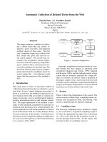
The overall structure of Ca
2+
/CaM as determined by X-ray
crystallography is shown in Fig. 1 [55,56]. The protein is
dominated by two EF-hand pairs forming the C- and
N-terminal lobes and a long a-helix connecting the two
lobes. The two EF-hand pairs share 48% sequence identity
and 75% sequence homology and the peptide backbone of
the two lobes can be superimposed with a mean square
derivation of 0.7 A
˚
[57]. Yet, reflecting the differences
in sequence, the Ca
2+
-binding affinities are different for the
N- and C-terminal lobe, with the C-terminal lobe having a
10-fold higher Ca
2+
-binding affinity (K
d
0.2 l
M
) than
the N-terminal lobe (K
d
2 l
M
) [58,59]. Mg
2+
and K
+
ions also bind to CaM, but with 10
3
)10
4
lower affinity than
Ca
2+
enabling CaM to respond specifically to increases in
Ca
2+
concentration [60].
The dissociation constant (K
d
) of CaM for Ca
2+
decreases significantly in ternary complexes when Ca
2+
/
CaM is bound to target peptides, mainly as the result of a
decrease in the dissociation rate k
off
of the Ca
2+
ion from
the EF-hands [61–63]. CaM–peptide complex dissociation
has been suggested to be initiated by the loss of Ca
2+
ions
from the N-terminal CaM lobe, likely followed by loss of
Ca
2+
from the C-terminal lobe and subsequently followed
by rapid loss of the peptide [62].
Due to the presence of four EF-hand (helix-loop-helix)
motifs, each of which is about 30 residues in length, only a
small portion of the 148 residue long CaM molecule does
not participate in Ca
2+
binding. This includes 8–10 residues
in the central region of the protein sequence (residues 76–
84). In the crystal structure, this central linker region is
folded into a long a-helix, connecting the fourth helix of the
N-terminal lobe with the first helix of the C-terminal lobe.
The length and expected rigidity of the central helix, as
suggested from the crystal structure, does not agree well
with data from small angle X-ray scattering and fluores-
cence energy transfer measurements in solution, which
generally indicate a shorter distance between the C- and
N-terminal lobes of CaM [64–69]. The conformation and
functional significance of this linker region has been subject
to intensive debate. Mutations within the central linker with
the aim to disrupt or to stabilize the helical conformation, or
to change the length of the helix, did not abolish the ability
of CaM to recognize and to activate its targets [70–74]. It
was ultimately shown beyond doubt by NMR as well as by
analyzing X-ray diffraction data that the observed a-helical
conformation of the central linker is a consequence of
crystal packing [75,76]. In solution, the central linker region
is very flexible and unstructured. Consequently, the N- and
C-terminal lobes do not adopt a defined orientation relative
to each other in solution, but display a tumbling motion,
being held together by the central linker.
Comparison of the structures of apo-CaM and Ca
2+
/
CaM determined by NMR and X-ray crystallography
reveals that the overall structure and distribution of
secondary structure elements is very similar in both
molecules (Fig. 1) [77–79]. The major conformational
change induced by the binding of Ca
2+
ions into the
EF-hand pairs is a significant alteration of the relative
orientation of the helices in each lobe [31,58,80]. As the
result of a twist-like motion, the relative angles between the
helices change from 121°)144° in apo-CaM to 86°)116° in
Ca
2+
/CaM [79]. This rearrangement of the helices leads to
the exposure of several hydrophobic residues to the solvent,
which form a large hydrophobic, concave patch or channel
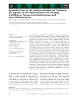
on the surface of each lobe (Fig. 2).
Structure of CaM bound to short peptides
Most physiological relevant CaM targets are proteins, but
CaM also binds to a number of synthetic peptides
corresponding to CaM binding domains, to peptide
hormones and toxins, as well as to small drug like molecules
[81–84]. Analysis of CaM binding peptides revealed that
Ó FEBS 2003 CaM target recognition and activation (Eur. J. Biochem. 270) 405
they share no sequence homology, but fulfill minimal
structural characteristics: they all have the potential to fold
into a basic, amphiphilic a-helix [85–89]. They display large
hydrophobic residues in conserved positions either 1-5-10 or
1-8-14, which point to one face of in a presumed a-helical
conformation. These structural characteristics have been
used to predict several dozen potential CaM-binding sites
from DNA and protein databases, as well as to engineer
synthetic CaM-binding peptides [85,86,90].
Biophysical characterization of complexes between CaM
and synthetic peptides indicated that the ternary complexes
have a more compact shape than Ca
2+
/CaM by itself [64–
66,91]. A collapsed structure of ternary Ca
2+
/CaM/peptide
complexes was also concluded from photoaffinity and cross-
linking experiments [92,93]. In order to reduce the distance
between the C- and N-terminal lobes it is necessary to allow
a kink or bend in the central helix. An early model,
proposed before the first structure of a CaM/peptide
complex had been solved, suggested a 45° bend within the
central helix at residue Ser81 [94,95].
Some speculation about the true binding mode of
peptides to CaM was finally resolved when the atomic
resolution structures of three CaM-peptide complexes from
smooth muscle myosin light chain kinase (smMLCK),
skeletal myosin light chain kinase (skMLCK) and
CaM-dependent kinase IIa (CaMKIIa) were solved by
Fig. 1. Ribbon presentations of CaM and CaM in complex with target peptides. CaM is colored blue, the CaM targets are red, Ca
2+
ions are yellow.
The N-terminal lobe of CaM is orientated to the top, the C-terminal lobe to the bottom of the figures. Structural data were taken from the Protein
Data Bank, accession codes: apo-CaM (1CFD) Ca
2+
/CaM (1CLL); CaM/CaMKIIa (1CM1); CaM/CaMKK (1IQ5); CaM/Ca
2+
-pump (1CFF);
CaM/K
+
-channel (1G4Y), CaM/anthrax exotoxin (1K93).
Table 1. Key features of CaM/peptide complexes.
CaM target
Target size
(amino acids)
CaM
conformation
Target
conformation
Anchor
residues
Ca
2+
ions
bound Method Year
smMLCK 20 Collapsed Helix 1-5-8-14 4 NMR 1992
skMLCK 26 Collapsed Helix 1-8-14 4 X-ray 1992
CaMKIIa 25 Collapsed Helix 1-5-10 4 X-ray 1992
CaMKK 27 Collapsed Helix + b-hairpin loop 1-5-16 4 NMR 1999
Ca
2+
-pump 20 Extended Helix, binds to C-terminal lobe only 1-5-8 4 NMR 1999
K
+
-channel 95 Extended 3 Helices, forms dimer Nonclassical 2 (N-terminal lobe) X-ray 2001
Anthrax exotoxin 510 Extended Complex Nonclassical 2 (C-terminal lobe) X-ray 2002
406 S. W. Vetter and E. Leclerc (Eur. J. Biochem. 270) Ó FEBS 2003
multidimensional NMR and X-ray crystallography in 1992
(Table 1) [96–98]. The structures of all three complexes are
quite similar and only the complex between CaM and the
peptide derived from CaMKIIa will be discussed here to
highlight the main features.
The CaM/CaMKIIa complex is of ellipsoidal shape and
much more compact (50 · 30 · 30 A
˚
3
) than Ca
2+
/CaM
without the peptide (maximal length of 65 A
˚
) (Fig. 1). The
central linker region is unwound and allows the C- and
N-terminallobestobendby100° and to rotate by 120°
relative to their orientation seen in the Ca
2+
/CaM crystal
structure. The peptide is bound in an a-helical conformation
and is engulfed by Ca
2+
/CaM into a hydrophobic channel
formed by bringing the C- and N-terminal lobes close
together (Fig. 2).
The binding of CaM-binding peptides is largely driven by
hydrophobic interactions between hydrophobic anchor
residues of the peptide with the hydrophobic surface cavities
of CaM. Methionine residues, unusually abundant in CaM,
play a particularly important role in the binding of target
peptides. The methionine side chains are very flexible and
the sulfur atom has a larger polarizability than carbon,
resulting in stronger van der Waals interactions.
The hydrophobic patches of each lobe are surrounded by
several charged residues, creating charged binding channel
outlets. The C-terminal end of the peptide-binding channel
has a negatively charged rim, whereas the N-terminal
hydrophobic patch has clusters of negatively and positively
charged residues. This charge distribution on the molecular
surface contributes to peptide binding via electrostatic inter-
actions and determines the relative binding orientation of
CaM-binding domain peptides. Basic residues at the N-ter-
minus of the peptide form salt bridges with acidic residues
surrounding the peptide-binding channel of the C-terminal
lobe of CaM (Fig. 2). The peptide-binding orientation is also
discussed for the complex of CaM with the peptide derived
from CaM-dependent kinase kinase (CaMKK).
The binding mode of CaM to target peptides observed in
these early structures was in agreement with a proposed
model for the regulation of CaM-targets by auto-inhibitory
domains. Limited proteolysis or mutation of the plasma
membrane Ca
2+
-pump, the cAMP phosphodiesterase,
myosin light chain kinase, calcineurin or neuronal nitric
oxide synthase led to constitutively activated enzymes [99–
103]. In the early 1990s, a model was developed in which an
auto-inhibitory domain becomes displaced from the active
site upon binding of CaM, leading to the activation of the
enzymes.
Plasma membrane Ca
2+
-pump
However, it has always been clear that the binding mode,
where CaM engulfs the target domain in a collapsed
conformation, could not generally apply to all CaM-
regulated enzymes. The CaM-binding domain of the
Ca
2+
-pump has hydrophobic anchor residues in positions
Fig. 2. The CaM/CaMKIIa complex. The N-terminal lobe of CaM is rendered as a ribbon, the C-terminal lobe is represented by its electrostatic
surface, the CaMKIIa peptide is shown as a blue ribbon. Negative electrostatic potential is indicated in red. The concave shape and hydrophobic
lining of the peptide-binding surface is visible.
Ó FEBS 2003 CaM target recognition and activation (Eur. J. Biochem. 270) 407
1-8-14. However, a splice site within the CaM binding
domain gives rise to splice isoforms, which can lack the last
anchor residue in position 14 [104]. Interestingly, the Ca
2+
-
pump of the plasma membrane can be activated not only by
full length Ca
2+
/CaM, but also with just the C-terminal
lobe of CaM alone [105]. To address this observation the
NMR structure of CaM in complex with the peptide C20W
has been solved recently [106]. The peptide C20W corres-
ponds to the N-terminal part of the CaM-binding domain
whichiscommontoallCa
2+
-pump isoforms, but lacks the
third hydrophobic anchor residue in position 14 (Table 1).
The structure of the C20W/CaM is shown in Fig. 1 and
reveals some unexpected features. Most significantly, the
complex does not collapse into an ellipsoid shaped complex,
but remains in an extended conformation. The central linker
region appears flexible between Arg74 and Glu84 and
medium range NOEs, which would indicate an a-helical
conformation, are not observed in the NMR experiment.
The two lobes do not contact each other and show large
variations in their relative orientation.
Secondly, the peptide C20W is bound only to the
C-terminal lobe. It is bound in an a-helical conformation
and is anchored to the peptide binding channel of the
C-terminal lobe through hydrophobic interactions involving
three hydrophobic residues (Trp1, Leu5 and Ile8). All four
methionine residues (Met109, Met124, Met144 and
Met145) of the C-terminal lobe of CaM interact with the
peptide, underlining the significance of methionine residues
for target binding. The hydrophilic face of the amphiphilic
peptide helix is exposed to the solvent. Overall, the binding
of the hydrophobic face of the C20W peptide to the
C-terminal lobe of CaM is equivalent to the binding mode
seen in complexes with other peptides (MLCK or CaMKII),
except that the C20W peptide does not induce the transition
of CaM into a collapsed conformation.
It can be speculated that the missing hydrophobic
anchor residue in position 14 is responsible for the
observed open conformation of the CaM/C20W complex.
This is supported by small-angle X-ray scattering experi-
ments on the complexes of CaM with the peptides C20W
and C24W. The peptide C24W is slightly longer than
C20W and contains the third hydrophobic anchor residues
of the full length CaM-binding domains. The scattering
data indicate an extended conformation of the CaM/C20W
complex and a collapsed structure of the CaM/C24W
complex [67].
The binding of the C20W peptide of the Ca
2+
-pump
CaM-binding domain to only the C-terminal lobe of CaM
demonstrates that Ôwrapping aroundÕ a CaM binding
domain, as seen in the complexes with MLCK and
CaMKIIa, is not necessary to activate certain CaM-
regulated proteins.
CaM in complex with CaMKK
Synthetic CaM-binding peptides have been successfully
designed based on the motif of a Ôbasic amphiphilic a-helixÕ
and the peptides derived from MLCK, CaMKIIa and the
Ca
2+
-pump do bind to CaM in a helical conformation. It
was therefore surprising to see a variation of this binding
mode for the CaM binding domain peptide derived from
Ca
2+
/CaM-dependent kinase kinase [107]. CaMKK is a
Ca
2+
/CaM dependent serine/threonine kinase which phos-
phorylates CaM-dependent kinases I and IV, modulating a
signal transduction cascade leading to Ca
2+
-regulated gene
transcription [108].
The CaM-binding domain of CaMKK shows some
differences compared to other CaM binding peptides. The
hydrophobic anchor residues are further apart, separated by
14 residues, rather then the usual 8 or 12 spacing residues
(Table 1). A cluster of basic residues is located near the
C-terminal anchor residue, whereas other CaM-binding
domains (MLCK, CaMKIIa and the Ca
2+
-pump) possess
basic residues near their N-terminal anchor residue. The
structure of the complex has been determined by NMR and
is shown in Fig. 1 [107]. The complex adopts a collapsed
conformation similar to the CaM/CaMKIIa complex and
the peptide is engulfed between the N- and C-terminal lobes
of CaM. Two features of the complex are distinct from
other complexes: the peptide is bound in an ÔinvertedÕ
orientation, i.e. binding with its C-terminus to the
C-terminal lobe of CaM. In contrast, the MLCK,
CaMKKIIa and Ca
2+
-pumppeptidesbindwiththeir
N-termini to the C-terminal lobe of CaM. This suggests that
the position of basic residues at either the C- or N-terminal
end of the binding domain peptides and charge comple-
mentarity of binding surfaces determine the binding orien-
tation of the peptide in complex with CaM.
The second interesting feature is the conformation of the
CaMKK binding domain peptide in complex with CaM.
The CaMKIIa and MLCK peptides are bound in a largely
a-helical conformation, and some of the terminal residues of
the peptides are not unexpectedly flexible and structurally
not defined. The CaMKK binding domain peptide in
contrast is only partially a-helical and contains an addi-
tional well-defined b-hairpin like loop. The helical part of
the peptide contains a hydrophobic anchor residue Trp444
which interacts with the hydrophobic patch of the
N-terminal lobe. The b-hairpin loop contains a hydropho-
bic anchor residue Phe459 which interacts with hydrophobic
residues of the C-terminal lobe of CaM (Table 1). The
spacing between the two anchor residues is 14 residues,
which would possibly prevent binding of the anchor
residues to the hydrophobic surface cavities of CaM if the
peptide would fold into a straight helix. However, the loop
allows the peptide to fold backwards and to position the
second hydrophobic residues in such a way that it can bind
into the C-terminal hydrophobic recognition pocket of
CaM.
The binding mode of the CaMKK peptide is a
significant variation from the ÔclassicalÕ binding mode of
a-helical peptides to CaM and has to be added to the
growing collection of the structural motifs recognizable by
CaM.
Ca
2+
activated K
+
-channel
A radically novel target binding mode and possibly a novel
mode of target protein activation through dimerization has
been revealed with the crystal structure of CaM in complex
with a fragment of the Ca
2+
-activated, small conductance
K
+
-channel [109]. The ion-guiding pore of the K
+
-channel
is formed by four a-subunits, each spanning the membrane
several times and is gated by the intracellular Ca
2+
408 S. W. Vetter and E. Leclerc (Eur. J. Biochem. 270) Ó FEBS 2003
concentration [110,111]. The C-terminal cytosolic portion of
each a-subunit has one CaM molecule constitutively bound
via a domain that shares no similarity to other CaM binding
domains and is not Ca
2+
dependent.
The structure of the complex between CaM and a 96
residue fragment corresponding to the C-terminal cytosolic
portion of the K
+
-channel provides interesting insights into
the mechanism of Ca
2+
activated gating of the channel and
presents a new way of CaM binding domain recognition.
The structure of the complex is shown in Fig. 1. Most
strikingly, CaM remains in an extended conformation and
forms a dimeric complex: two K
+
-channel fragments
are clamped together by two molecules of CaM. The
K
+
-channel fragment consists of a long a-helix and a
shorter a-helix folded back antiparallel against the longer
helix. In the complex, the K
+
-channel fragment is oriented
almost perpendicularly to the long axis of the extended
CaM molecule. The portion of the K
+
-channel fragment
containing the two helices is bound to the C-terminal lobe of
CaM. The EF-hand pair of the C-terminal lobe remains
Ca
2+
free and the binding of this portion of the K
+
-channel
peptide is Ca
2+
-independent. The C-terminal end of the
fragment K
+
-channel peptide protrudes sideways and binds
to the N-terminal lobe of a second CaM molecule. The
N-terminal lobe of CaM binds two Ca
2+
ions and exposes
subsequently the hydrophobic patch typical for Ca
2+
loaded CaM. The hydrophobic patch of CaM interacts
with the C-terminus of the long helix of the second
K
+
-channel fragment. This results effectively in the binding
of three helices from two different peptide chains by each
CaM molecule, compared to the binding of just one helix as
seen in all other CaM structures. To accommodate the three
helices CaM has to stretch, which is rendered possible by
partial unfolding of the central linker.
The dimeric CaM/K
+
-channel peptide complex shows
the two long C-terminal helices of the K
+
-channel
fragments in an anti-parallel, side-by-side orientation. The
N-termini of the peptides are facing each other and pointing
away from the plane formed by the two C-terminal helices.
When viewed from the side, the two K
+
-channel peptides
resemble an isosceles triangle. The N-termini of the
K
+
-channel peptides connect to a transmembrane helix,
which was not present in the structure of the CaM/
K
+
-channel peptide complex, but could be perpendicular to
the C-terminal helices. The geometry of two perpendicular
helices (C-terminal helix and transmembrane helix) con-
nected by the short N-terminal helix resembles a molecular
wrench. Movement of the C-terminal helix as the result of
Ca
2+
binding to CaM and subsequent dimerization of the
a-subunits of the K
+
-channel could translate into a rotation
of the transmembrane helices and thus could allow K
+
ions
to flow through the pore.
Anthrax adenylate cyclase exotoxin
The crystal structures of the C-terminal fragment (residues
291–800) of the exotoxin from Bacillus anthracis,withand
without its activator calmodulin, is the first structure of a
CaM-complex with a catalytically functional CaM-target
[112–114]. Anthrax exotoxin (edema factor) is a calmo-
dulin-dependent adenylate cyclase, but shares no overall
structural homology with mammalian adenylate cyclases
and uses a slightly different catalytic mechanism. The
exotoxin enters host cells through a transporter (protective
antigen) produced by the pathogen. Inside the cell, it
acquires CaM, becomes activated and then converts large
amounts of ATP to cAMP [115]. The structure of this large
CaM complex is particularly interesting because it allows
to compare the details of target activation by CaM at
atomic resolution (about 3 A
˚
) by comparison of three
structures: (a) exotoxin without CaM (b) exotoxin with
CaM and (c) exotoxin with bound CaM and the pseudo-
substrate 3¢-dATP.
The overall structure of CaM/exotoxin complex is shown
in Fig. 1. Striking features of the complex are that CaM is
inserted deeply between two domains of exotoxin and not
merely bound onto exposed helix or surface loop. CaM is
bound in an extended conformation, the central section
(residues 79–81) is partially unwound and only the
C-terminal EF-hand pair is loaded with Ca
2+
.
CaM is tightly squeezed between two domains of
exotoxin and this results in a large number of interacting
residues: 53 CaM residues interact with 63 exotoxin resi-
dues, leading to a very large buried surface area of 5.900 A
˚
2
.
The insertion of CaM between the two domains of exotoxin
causes large conformational changes in the relative domain
orientation (Figs 1 and 3). The 15 kDa large C-terminal
portion of the protein is displaced by 15 A
˚
and rotated by
30° upon binding of CaM. In addition, several short sheet
structures, helices and loops of exotoxin rearrange and
change their conformation.
The C-terminal lobe of CaM contains two Ca
2+
ions
and its conformation is very similar to other structures of
Ca
2+
-loaded CaM. The hydrophobic patch typical for
Ca
2+
/CaM is exposed and forms a peptide-binding channel,
which interacts with an a-helix of exotoxin. This a-helix
shows many properties of a potential CaM-binding site: it
forms a positively charged amphiphilic a-helix and contains
hydrophobic anchor residues in positions 1-5-10. Fragments
of exotoxin and synthetic peptides corresponding to this
helical region have been demonstrated to bind CaM in vitro
in a Ca
2+
-dependent manner. NMR experiments indicate
further that the peptide is bound in an a-helical conforma-
tion and fluorescence energy transfer experiments showed a
reduced interdomain distance in the CaM/peptide complex,
pointing towards a collapsed conformation of the CaM/
peptide complex [112,116]. These experimental observations
are very similar to results obtained for synthetic peptides
derived from other CaM-binding domains, such as MCLK
and CaMKIIa. While there is no NMR or crystal structure
of the CaM/exotoxin peptide complex available, it is
reasonable to assume that the complex adopts a conforma-
tion similar to the complexes with the MLCK or CaMKIIa
peptides discussed above. However, the binding of CaM to
the functional exotoxin fragment shows that CaM does not
collapse around this helix, but instead binds to exotoxin in
an extended conformation.
The EF-hand pair of the N-terminal lobe of CaM is
Ca
2+
-free in the CaM/exotoxin complex and shows a
conformation typical for apo-CaM. The EF-hand helices
are packed against several helices of the C-terminal
domain of exotoxin (Fig. 3). The absence of Ca
2+
from
the N-terminal lobe is remarkable, because the complex
was crystallized in the presence of Ca
2+
,makingit
Ó FEBS 2003 CaM target recognition and activation (Eur. J. Biochem. 270) 409
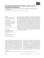
Fig. 3. Structural changes occurring in the anthrax exotoxin upon binding of CaM. The N-terminal part of the protein (residues 280–450) was used to
superimpose the structures of the exotoxin without CaM (blue) and exotoxin in complex with CaM (yellow) and the pseudosubstrate 3¢dATP
(electrostatic surface potential shown) bound into the active site. Calmodulin has been omitted in the upper panel for clarity. The lower panel shows
calmodulin (electrostatic surface rendering) inserted between the domains of the exotoxin and the relative distance from the substrate bound into the
active site.
410 S. W. Vetter and E. Leclerc (Eur. J. Biochem. 270) Ó FEBS 2003
necessary for CaM to ÔloseÕ two Ca
2+
ions during its
binding to exotoxin.
The binding of CaM to exotoxin increases the catalytic
activity of the enzyme approximately 1000-fold. Surpris-
ingly, the activation of the enzyme is not achieved by
dramatically changing the architecture of the active site, nor
by removing an auto-inhibitory domain as in other CaM
activated enzymes. Indeed, the binding of CaM does not
even occur close to the active site. The N-terminal part of
the exotoxin alone and in the presence of CaM can be
superimposed and show no dramatic conformational
changes as the result of CaM binding (Fig. 3). Instead,
several loops close to the active site become stabilized. This
relatively minor change around the active site enhances the
catalytic activity through better substrate binding and
positioning in the active site.
Conclusion
Target recognition and activation by CaM has been studied
intensively over the last two decades and today we are
further away from a general model than ever before. The
CaM-target complexes discussed here (CaMKIIa,CaM-
KK, Ca
2+
-pump, K
+
-channel, anthrax exotoxin) represent
several structurally different CaM-complex architectures.
With short, amphiphilic, a-helical peptides (CaMKIIa)a
collapsed complex conformation is observed, but also
b-hairpin-like loops can be accommodated in such a
collapsed complex (CaMKK). Clustering of basic residues
at the C- or N-terminus of CaM-binding peptides defines
their relative orientation (parallel or antiparallel) in the
peptide-binding channel of CaM. Binding of a target
peptide to only the C-terminal lobe is observed for a
truncated CaM-binding domain (Ca
2+
-pump) and can be
attributed to a missing hydrophobic anchor residue.
Binding of CaM to its targets in an extended, open
conformation was not expected based on the structures of
CaM complexed to peptides derived from smMLCK,
skMLCK and CaMKIIa, but was found in three out of
the four recent structures.
The structure of the CaM/K
+
-channel complex shows
three helices bound to CaM, which prevents CaM from
adopting a collapsed conformation. In addition, two of
these three helices interact with the Ca
2+
free N-terminal
EF-hand pair, allowing CaM to be constitutively bound to
the K
+
-channel and utilizing only the C-terminal lobe for
detecting changes in Ca
2+
concentration. Also, the structure
of CaM in complex with the K
+
-channel fragment suggests
that some CaM targets can be activated through dimeriza-
tion.
For several Ca
2+
/CaM-regulated proteins a mechanism
of activation through replacement of auto-inhibitory
domains upon CaM binding has been demonstrated
through biochemical studies. A model for the binding of
CaM to its targets was based on the X-ray and NMR
structures which showed the CaMKIIa and MLCK pep-
tides engulfed by CaM. However, this binding seems less
general than previously assumed as demonstrated by the
structures discussed here.
Particularly informative is the structure of CaM in
complex with the anthrax exotoxin. Studies using short
peptides derived from exotoxin had indicated a collapsed
CaM/peptide complex. However, CaM binds in an exten-
ded conformation to anthrax exotoxin and activates the
enzyme through subtle changes in the surrounding of the
active site.
There is no doubt that structures of CaM in complex with
large target fragments yield much more information about
the mechanisms of CaM target recognition and activation
than structures of complexes with short peptides. With this
in mind we can look forward to up-coming structures of
CaM in complex with full length targets and we are certain
that CaM has many more ways to activate its targets than
we can imagine today.
References
1. Evenas, J., Malmendal, A. & Forsen, S. (1998) Calcium. Curr.
Opin. Chem. Biol. 2, 293–302.
2. Carafoli, E. & Klee, C. (1999) Calcium as a Cellular Regulator.
Oxford University Press, Oxford, UK.
3. Brini, M. & Carafoli, E. (2000) Calcium signaling: a historical
account, recent developments and future perspectives. Cell. Mol.
Life Sci. 57, 354–370.
4. Carafoli, E. (2002) Calcium signaling: a tale for all seasons. Proc.
NatlAcad.Sci.USA99, 1115–1122.
5. Endo, M., Tanaka, M. & Ogawa, Y. (1970) Calcium induced
calcium release of calcium from the carcoplasmatic reticulum of
skinned skeletal fibres. Nature 228, 34–36.
6. Berridge, M.J. (1993) Inositol and calcium signalling. Nature 361,
315–325.
7. Galione, A. (1994) Cyclic ADP-ribose, the ADP-ribosyl cyclase
pathway and calcium signalling. Mol. Cell. Endocrinol. 98, 125–
131.
8. Pozzan, T., Rizzuto, R., Volpe, P. & Meldolesi, J. (1994) Mole-
cular and cellular physiology of intracellular calcium stores.
Physiol. Rev. 74, 595–636.
9. Babcock, D.F., Herrington, J., Goodwin, P.C., Park, Y.B. &
Hille, B. (1997) Mitochondrial participation in the intracellular
Ca
2+
network. J. Cell. Biol. 136, 833–844.
10. Horne, J.H. & Meyer, T. (1997) Elementary calcium-release units
induced by inositol trisphosphate. Science 276, 1690–1693.
11. Carafoli, E., Santella, L., Branca, D. & Brini, M. (2001) Gen-
eration, control, and processing of cellular calcium signals. Crit.
Rev. Biochem. Mol. Biol. 36, 107–260.
12. Meyer, T., Wensel, T. & Stryer, L. (1990) Kinetics of calcium
channel opening by inositol 1,4,5-trisphosphate. Biochemistry 29,
32–37.
13. Finch, E.A., Turner, T.J. & Goldin, S.M. (1991) Calcium as a
coagonist of inositol 1,4,5-trisphosphate-induced calcium release.
Science 252, 443–446.
14. Bezprozvanny, I., Watras, J. & Ehrlich, B.E. (1991) Bell-shaped
calcium-response curves for Ins(1,4,5)P
3
- and calcium-gated
channels from endoplasmatic reticulum of cerebellum. Nature
351, 751–754.
15. Marchant, J.S. & Taylor, C.W. (1997) Cooperative activation of
IP
3
receptors by sequential binding of IP
3
and Ca
2+
safeguards
against spontanous activity. Curr. Biol. 7, 510–518.
16. Bezprozvanny, I. & Ehrlich, B.E. (1995) The inositol-1,4,5-tris-
phosphate (InsP
3
) receptor. J. Membr. Biol. 145, 205–216.
17. Rebecchi,M.J.&Pentyala,S.N.(2000)Structure,function,and
control of phosphoinositide-specific phospholipase C. Physiol.
Rev. 80, 1291–1335.
18. Cuthbertson, K.S.R. & Cobbold, P.H. (1985) Phorbol ester and
sperm activated mouse oocytes by inducing sustained oscillations
in cell Ca
2+
. Nature 316, 541–542.
Ó FEBS 2003 CaM target recognition and activation (Eur. J. Biochem. 270) 411
19. Woods, N.M., Cuthbertson, K.S.R. & Cobbold, P.H. (1986)
Repetitive transient rises in cytoplasmatic free calcium in hor-
mone-stimulated hepatocytes. Nature 319, 600–602.
20. Berridge, M.J. (1990) Calcium oscillations. J. Biol. Chem. 265,
9583–9586.
21. Meyer, T. & Stryer, L. (1991) Calcium spiking. Ann. Rev. Bio-
phys. Chem. 20, 153–174.
22. Dupont, G. & Erneux, C. (1997) Simulations of the effects of
inositol 1,4,5-trisphosphate 3-kinase and 5-phosphate activities
on Ca
2+
oscillations. Cell Calcium 22, 321–331.
23. Berridge, M., Bootman, M.D. & Lipp, P. (1998) Calcium – a life
and death signal. Nature 395, 645–648.
24. Schuster, S., Marhl, M. & Hofer, T. (2002) Modelling of simple
and complex calcium oscillations. Eur. J. Biochem. 269, 1333–
1355.
25. Barbara, J.G. (2002) IP
3
-dependent calcium-induced calcium
release mediates bidirectional calcium waves in neurons: func-
tional implications for synaptic plasticity. Biochim. Biophys. Acta
1600, 12–18.
26. Hanson, P.I. & Schulman, H. (1992) Neuronal Ca
2+
/calmodulin-
dependent protein kinases. Annu. Rev. Biochem. 61, 559–601.
27. De Koninck, P. & Schulman, H. (1998) Sensitivity of CaM kinase
II to the frequency of Ca
2+
oscillations. Science 279, 227–230.
28. Schulman, H. & Braun, A. (1999) Calcium/calmodulin depen-
dent kinases. In Calcium as a Cellular Regulator (Carafoli, E. &
Klee, C., eds), pp. 311–343. Oxford Univerisity Press, Oxford,
UK.
29. Bayer, K.U., De Koninck, P. & Schulman, H. (2002) Alternative
splicing modulates the frequency-dependent response of CaMKII
to Ca
2+
oscillations. EMBO J. 21, 3590–3597.
30. Nakayama, S. & Kretsinger, R.H. (1994) Evolution of the EF-
hand family of proteins. Annu. Rev. Biophys. Biolmol. Struct. 23,
4673–4507.
31. Ikura, M. (1996) Calcium binding and conformational response
in EF-hand proteins. Trends Biochem. Sci. 21, 14–17.
32. Nelson, M.R. & Chazin, W.J. (1998) Structures of EF-hand
Ca
2+
-binding proteins: diversity in the organization, packing and
resonse to Ca
2+
-binding. Biometals 11, 297–318.
33. Lewit-Bentley, A. & Rety, S. (2000) EF-hand calcium-binding
proteins. Curr. Opin. Struct. Biol. 10, 637–643.
34. Kretsinger, R.H. (1996) EF-hands reach out. Nat. Struct. Biol. 3,
12–15.
35. Chazin, W.J. (1995) Releasing the calcium trigger. Nat. Struct.
Biol. 2, 707–710.
36. Dibois, T., Oudinet, J.P., Mira, J.P. & Russo-Marie, F. (1996)
Annexins and protein kinases C. Biochim. Biophys. Acta 1313,
290–294.
37. Nelsestuen, G.L. & Gaul Ostrowski, B. (1999) Membrane
association with multiple calcium ions: vitamin-K-dependent
proteins, annexins and pentaxins. Curr. Opin. Struct. Biol. 9, 433–
437.
38. Rizo, J. & Sudhof, T.C. (1998) C2-domains, structure and
function of a universal Ca
2+
-binding domain. J. Biol. Chem. 273,
15879–15882.
39. Nalepski, E.A. & Falke, J.J. (1996) The C2 domain calcium-
binding motif: structural and functional diversity. Protein Sci. 5,
2375–2390.
40. Essen, L.O., Perisic, O., Cheung, R., Katan, M. & Williams, R.L.
(1996) Crystal structure of a mammalian phosphoinositide-spe-
cific phospholipase Cd. Nature 380, 595–602.
41. Shao, X., Davletov, B.A., Sutton, R.B., Sudhof, T.C. & Rizo, J.
(1996) Bipartite Ca
2+
-binding motif in C2 domains of synapto-
tagmin and protein kinase C. Science 273, 248–251.
42. Trahey, M., Wong, G., Halenbeck, R., Rubinfeld, B., Martin,
G.A.,Ladner,M.,Long,C.M.,Crosier,W.J.,Watt,K.,Koths,
K. & McCormick, F. (1988) Molecular cloning of two types of
GAP complementary DNA from human placenta. Science 242,
1697–1700.
43. Nishizuka, Y. (1988) The molecular heterogeneity of protein
kinase C and its implication for cellular regulation. Nature 334,
661–665.
44. Clark, J.D., Lin, L.L., Kriz, R.W., Ramesha, C.S., Sultzman,
L.A., Lin, A.Y., Milona, N. & Knopf, J.L. (1991) A novel ara-
chidonic acid-sensitive cytosolic PLA2 contains a Ca
2+
-depend-
ent translocation domain with homology tp PKC and GAP. Cell
65, 1043–1051.
45. Rhee, S.G., Suh, P G., Ryu, S H. & Lee, S.Y. (1989) Studies
of inositol phospholipid-specific phospholipase C. Science 244,
546–550.
46. Hiles, I.D., Otsu, M., Volinia, S., Fry, M.J., Gout, I., Dhand, R.,
Panayotou, G., Ruiz-Larrea, F., Thompson, A., Totty, N.F.,
Hsuan, J.J., Courtneidge, S.A., Parker, P.J. & Waterfield, M.D.
(1992) Phosphatidylinositol 3-kinase: structure and expression of
the 110kD catalytic subunit. Cell 70, 419–429.
47. Coussens, L., Parker, P.J., Rhee, L., Yang-Feng, T.L., Chen, E.,
Waterfield, M.D., Francke, U. & Ulrich, A. (1986) Multiple,
distinct forms of bovine and human protein kinase C suggest
diversity in celular signaling pathways. Science 233, 859–866.
48. Knopf, J.L., Lee, M.H., Sultzman, L.A., Kriz, R.W., Loomis,
C.R., Hewick, R.M. & Bell, R.M. (1986) Cloning and expression
of multiple protein kinase C cDNAs. Cell 46, 491–502.
49. Perin, M.S., Fried, V.A., Mignery, G.A., Jahn, R. & Sudhof, T.C.
(1990) Phospholipid binding by a synaptic vesicle protein
homologous to the regulatory region of protein kinase C. Nature
345, 260–263.
50. Perin, M.S., Brose, N., Jahn, R. & Sudhof, T.C. (1991) Domain
structure of synaptotagmin (p65). J. Biol. Chem. 266, 623–629.
51. Brose, N., Petrenko, A.G., Sudhof, T.C. & Jahn, R. (1992)
Synaptotagmin: a calcium sensor on the synaptic vesicle surface.
Science 256, 1021–1025.
52. Geppert, M., Goda, Y., Hammer, R.E., Li, C., Rosahl, T.W.,
Stevens, C.F. & Sudhof, T.C. (1994) SynatotagminI: a major
Ca
2+
sensor for transmitter release at a central synase. Cell 79,
717–727.
53. Cohen, P. & Klee, C.B. (1988) Calmodulin. Elsevier, Amsterdam,
the Netherlands.
54. Eldik, L.V. & Watterson, D.M. (1998) Calmodulin and signal
transduction (Van Eldrick, L.J. & Watterson, D.M., eds).
Academic Press, New York, USA.
55. Babu, Y.S., Sack, J.S., Greenhough, T.J., Bugg, C.E., Means,
A.R. & Cook, W.J. (1985) Three-dimensional structure of cal-
modulin. Nature 315, 37–40.
56. Babu, Y.S., Bugg, C.E. & Cook, W.J. (1988) Structure of cal-
modulin refined at 2.2 A
˚
resolution. J. Mol. Biol. 204, 191–204.
57. Reference withdrawn.
58.Potter,J.D.,Strang-Brown,P.,Walker,P.L.&Lida,S.
(1983) Ca
2+
binding to Calmodulin. Methods Enzymol. 102,
135–143.
59. Ogawa, Y. & Tanokura, M. (1984) Calcium binding to calmo-
dulin: effects of ionic strength, Mg
2+
,pHandtemperature.
J. Biochem. 95, 19–28.
60. Haiech, J., Klee, C.B. & Demaille, J.G. (1981) Effects of cations
on affinity of calmodulin for calcium: ordered binding of calcium
ions allows the specific activation of calmodulin-stimulated
enzymes. Biochemistry 20, 3890–3897.
61. Peersen, O.B., Madsen, T.S. & Falke, J.J. (1997) Intermolecular
tuning of calmodulin by target peptides and proteins: differential
effects on Ca
2+
binding and implications for kinase activation.
Protein Sci. 6, 794–807.
62. Brown, S.E., Martin, S.R. & Bayley, P.M. (1997) Kinetic control
of the dissociation pathway of calmodulin-peptide complexes.
J. Biol. Chem. 272, 3389–3397.
412 S. W. Vetter and E. Leclerc (Eur. J. Biochem. 270) Ó FEBS 2003
63. Mirzoeva, S., Weigand, S., Lukas, T.J., Shuvalova, L., Anderson,
W.F. & Watterson, D.M. (1999) Analysis of the functional
coupling between calmodulin’s calcium binding and peptide
recognition properties. Biochemistry 38, 3936–3947.
64. Small, E.W. & Anderson, S.R. (1988) Fluorescence anisotropy
decay demonstrates calcium-dependent shape changes in photo-
cross-linked calmodulin. Biochemistry 27, 419–428.
65. Chapman, E.R., Alexander, K., Vorherr, T., Carafoli, E. &
Storm, D.R. (1992) Fluorescence energy transfer analysis of
calmodulin-peptide complexes. Biochemistry 31, 12819–12825.
66. Seaton, B.A., Head, J.F. & Richards, F.M. (1985) Calcium-
induced increase in the radius of gyration and maximum
dimension of calmodulin measured by small-angle X-ray scat-
tering. Biochemistry 24, 6740–6743.
67. Kataoka, M., Head, J.F., Vorherr, T., Krebs, J. & Carafoli, E.
(1991) Small-angle x-ray scattering study of calmodulin bound to
two peptides corresponding to parts of the calmodulin-binding
domain of the plasma membrane Ca-pump. Biochemistry 30,
6247–6251.
68. Meador, W.E., George, S.E., Means, A.R. & Quicho, F.A.
(1995) X-ray analysis reveals conformational adaptation of the
linker in functional calmodulin mutants. Nat. Struct. Biol. 2,
943–945.
69. Trewhella, J., Blumenthal, D.K., Rokop, S.E. & Seeger, P.A.
(1990) Small-angle scattering studies show distinct conformations
of calmodulin in its complexes with two peptides based on the
regulatory domain of the catalytic subunit of phosphorylase
kinase. Biochemistry 29, 9316–9324.
70. Putkey, J.A., Ono, T., VanBerkum, M.F.A. & Means, A.R.
(1988) Functional significance of the central helix in calmodulin.
J. Biol. Chem. 262, 11242–11249.
71. Persechini, A. & Kretsinger, R.H. (1988) The central helix of
calmodulin functions as a flexible tether. J. Biol. Chem. 263,
12175–12178.
72. Persechini, A., Blumenthal, D.K., Jarrett, H.W., Klee, C.B.,
Hardy, D.O. & Kretsinger, R.H. (1989) The effects of deletions in
the central helix of calmodulin on enzyme activation and peptide
bindng. J. Biol. Chem. 264, 8052–8058.
73. Persechini, A., Kretsinger, R.H. & Davis, T.N. (1991) Calmodulin
with deletions in the central helix functionally replace the native
protein in yeast cells. Proc.NatlAcad.Sci.USA88, 449–452.
74. Tabernero, L., Taylor, D.A., Chandross, R.J., VanBerkum,
M.F.,Means,A.R.,Quiocho,F.A.&Sacks,J.S.(1997)The
structure of a calmodulin mutant with a deletion in the central
helix: implications for molecular recognition and protein binding.
Structure 5, 613–622.
75. Barbato, G., Ikura, M., Kay, L.E., Pastor, R.W. & Bax, A.
(1992) Backbone dynamics of calmodulin studied by N
15
relaxation using inverse detected two-dimensional NMR spec-
troscopy: the central helix is flexible. Biochemistry 31, 5269–5278.
76. Wall, M.E., Clarage, J.B. & Phillips, G.N. (1997) Motions of
calmodulin characterized using both Bragg and diffuse X-ray
scattering. Structure 5, 1599–1612.
77. Kuboniwa, H., Tjandra, N., Grzesiek, S., Ren, H., Klee, C.B. &
Bax, A. (1995) Solution structure of calcium-free calmodulin.
Nat. Struct. Biol. 2, 768–776.
78. Finn,B.E.,Evenas,J.,Drakenberg,T.,Waltho,J.P.,Thulin,E.&
Forsen, S. (1995) Calcium-induced structural changes and
domain autonomy in calmodulin. Nat. Struct. Biol. 2, 777–783.
79. Zhang, M., Tanaka, T. & Ikura, M. (1995) Calcium-induced
conformational transition revealed by the solution structure of
apo calmodulin. Nat. Struct. Biol. 2, 758–767.
80. Chou, J.J., Li, S., Klee, C.B. & Bax, A. (2001) Solution structure
of Ca
2+
-calmodulin reveals flexible hand-like properties of its
domains. Nat. Struct. Biol. 8, 990–997.
81. Vandonselaar, M., Hickie, R.A., Quail, J.W. & Delbaere, L.T.J.
(1994) Trifluoperazine-induced conformational change in Ca
2+
-
calmodulin. Nat. Struct. Biol. 1, 795–801.
82. Malencik, D.A. & Anderson, S.R. (1993) Binding of hormones
and neuropeptides by calmodulin. Biochemistry 22, 1995–2001.
83. Malencik, D.A. & Anderson, S.R. (1982) Binding of simple
peptides, hormones and neurotransmitters by calmodulin. Bio-
chemistry 21, 3480–3486.
84. Osawa, M., Swindells, M.B., Tanikawa, J., Tanaka, T., Mase, T.,
Furuya, T. & Ikura, M. (1998) Solution structure of calmodulin-
W-7 complex: the basis of diversity in molecular recognition.
J. Mol. Biol. 276, 165–176.
85. DeGrado, W.F., Prendergast, F.G., Wolfe, H.R.J. & Cox, J.A.
(1985) The design, synthesis, and characterization of tight-bind-
ing inhibitors of calmodulin. J. Cell. Biochem. 29, 83–93.
86. Cox, J.A., Comte, M., Fitton, J.E. & DeGrado, W.F. (1985) The
interaction of calmodulin with amphiphilic peptides. J. Biol.
Chem. 260, 2527–2534.
87. Erickson-Viitanen, S. & DeGrado, W.F. (1987) Recognition and
characterization of calmodulin-binding sequences in peptides and
proteins. Methods Enzymol. 139, 455–478.
88. DeGrado, W.F. (1988) Design of peptides and proteins. Adv.
Protein Chem. 39, 51–125.
89. O’Neil, K.T. & DeGrado, W.F. (1990) How calmodulin binds its
targets: sequence independent recognition of amphiphilic alpha-
helices. Trends Biochem. Sci. 15, 59–64.
90. Rhoads, A.R. & Friedberg, F. (1997) Sequence motifs for cal-
modulin recognition. FASEB J. 11, 331–340.
91. Heidorn, D.B., Seeger, P.A., Rokop, S.E., Blumenthal, D.K.,
Means, A.R., Crespi, H. & Trewhella, J. (1989) Change in the
structure of calmodulin induced by a peptide based on the
calmodulin-binding domain of myosin light chain kinase.
Biochemistry 28, 6757–6764.
92. Vorherr, T., Quadroni, M., Krebs, J. & Carafoli, E. (1992)
Photoaffinity labeling study of the interaction of calmodulin with
theplasmamembraneCa
2+
pump. Biochemistry 35, 8245–8251.
93. Vorherr, T., James, P., Krebs, J., Enyedi, A., McCormick, D.J.,
Penniston, J.T. & Carafoli, E. (1990) Interaction of calmodulin
with the calmodulin binding domain of the plasma membrane
calcium pump. Biochemistry 29, 355–365.
94. O’Neil, K.T. & DeGrado, W.F. (1989) The interaction of
calmodulin with fluorescent and photoreactive model peptides:
evidence for a short interdomain separation. Proteins 6, 284–
293.
95. Vorherr,T.,Kessler,O.,Mark,A.&Carafoli,E.(1992)Con-
struction and molecular dynamics simulation of calmodulin in
the extended and in a bent conformation. Eur. J. Biochem. 204,
931–937.
96. Ikura, M., Clore, G.M., Gronenborn, A.M., Zhu, G., Klee, C.B.
& Bax, A. (1992) Solution structure of a calmodulin target pep-
tide complex by multidimensional NMR. Science 256, 632–638.
97. Meador, W.E., Means, A.R. & Quiocho, F.A. (1992) Target
enzyme recognition by calmodulin: 2.4A
˚
structure of calmodulin-
peptide complex. Science 257, 1251–1255.
98. Meador, W.E., Means, A.R. & Quiocho, F.A. (1993) Modula-
tion of calmodulin plasticity in molecular recognition on the basis
of X-ray structures. Science 262, 1718–1721.
99. Zurini, M., Krebs, J., Penniston, J.T. & Carafoli, E. (1984)
Controlled proteolysis of the purified Ca
2+
-ATPase of the
erythrocyte membrane. A correlation between the structure and
the function of the enzyme. J. Biol. Chem. 259, 618–664.
100. Edelman, A.M., Takio, A., Blumenthal, D.K., Hansen, R.S.,
Walsh, K.A., Titani, K. & Krebs, E.G. (1985) Characterization of
the calmodulin-binding and catalytic domains in skeletal muscle
myosin light chain kinase. J. Biol. Chem. 260, 11275–11285.
Ó FEBS 2003 CaM target recognition and activation (Eur. J. Biochem. 270) 413
101. Olsen, N.J., Pearson, R.B., Needleman, D.S., Hurwitz, M.Y.,
Kemp, B.E. & Means, A.R. (1990) Regulatory and structural
motifs of chicken myosin light chain kinase. Proc. Natl Acad. Sci.
USA 87, 2284–2288.
102. Montgomery, H.J., Romanov, V. & Guillemette, J.G. (2000)
Removal of a putative inhibitory element reduces the calcium-
dependent calmodulin activation of neuronal nitric-oxide syn-
thase. J. Biol. Chem 275, 5052–5058.
103. Hubbard, M.J. & Klee, C.B. (1989) Functional domain structure
of calcineurin A: mapping by limited proteolysis. Biochemistry 28,
1868–1874.
104. Strehler, E.E. (1991) Recent advances in the molecular charac-
terization of the plasma membrane Ca
2+
pump. J. Membr. Biol.
120, 1–15.
105. Guerini, D., Krebs, J. & Carafoli, E. (1984) Stimulation of the
purified erythrocyte Ca-ATPase by tryptic fragments of calmo-
dulin. J. Biol. Chem. 259, 15172–15177.
106. Elshorst, B., Henning, M., Forsterling, H., Diener, A., Maurer,
M., Schulte, P., Schwalbe, H., Griesinger, C., Krebs, J., Schmid,
H., Vorherr, T. & Carafoli, E. (1999) NMR solution structure of
a complex of calmodulin with a binding peptide of the Ca
2+
pump. Biochemistry 38, 12320–12332.
107. Osawa, M., Tokumits, H., Swindells, M.B., Kurihara, H., Orita,
M.,Shibanuma,T.,Furuya,T.&Ikura,M.(1999)Anoveltarget
recognition revealed by calmodulin in complex with Ca
2+
-cal-
modulin-dependent kinase kinase. Nat. Struct. Biol. 6, 819–824.
108. Soderling, T.R. (1999) The Ca-calmodulin-dependent protein
kinase cascade. Trends Biochem. Sci. 24, 232–236.
109. Schumacher, M.A.F.R.A., Bachinger, H.P. & Adelman, J.P.
(2001) Structure of the gating domain of a Ca
2+
-activated K
+
channel complexed with Ca
2+
/calmodulin. Nature 410, 1120–
1124.
110. Keen, J.E., Khawaled, R., Farrens, D.L., Neelands, T., Rivard,
A.,Bond,C.T.,Janowsky,A.,Fakler,B.,Adelman,J.P.&
Maylie, J. (1999) Domains responsible for constitutive and
Ca
2+
–dependent interactions between calmodulin and small
conductance Ca
2+
-activated potassium channels. J. Neurosci. 19,
8830–8838.
111. Xia, X M., Fakler, B., Rivard, A., Wayman, G., Johnson-Pais,
T.,Keen,J.E.,Ishii,T.,Hirschberg,B.,Bond,C.T.,Lutsenko,S.,
Maylie, J. & Adelman, J.P. (1998) Mechanism of calcium gating
in small-conductance calcium-ativated potassium channels.
Nature 395, 503–507.
112. Drum, C.L., Yan, S Z., Bard, J., Shen, Y Q., Lu, D., Soelaiman,
S., Grabarek, Z., Bohm, A. & Tang, W J. (2002) Structural basis
for the activation of anthrax adenylyl cyclase exotoxin by cal-
modulin. Nature 415, 396–402.
113. Drum,C.L.,Shen,Y.,Rice,P.A.,Bohm,A.&Tang,W.J.(2001)
Crystallization and preliminary X-ray study of the edema factor
exotoxin adenylyl cyclase domain from Bacillus anthracis in the
presence of its activator, calmodulin. Acta Crystallogr. D 57,
1881–1884.
114. Drum, C.L., Yan, S.Z., Sarac, R., Mabuchi, Y., Beckingham, K.,
Bohm, A., Grabarek, Z. & Tang, W.J. (2000) An extended
conformation of calmodulin induces interaction between the
structural domains of adenylyl cyclase from Bacillus anthracis to
promote catalysis. J. Biol. Chem. 275, 36334–36340.
115. Leppla, S.H. (1982) Anthrax toxin edema factor, a bacterial
adenylate cyclase that increases cyclic AMP concentrations of
eukaryotic cells. Proc. Natl Acad. Sci. USA 79, 3162–3166.
116. Munier, H., Blance, F.J., Precheur, B., Diesis, E., Nieto, J.L.,
Craescu, C.T. & Barzu, O. (1993) Characterization of a synthetic
calmodulin-binding peptide derived from Bacillus anthracis ade-
nylate cyclase. J. Biol. Chem. 268, 1695–1701.
414 S. W. Vetter and E. Leclerc (Eur. J. Biochem. 270) Ó FEBS 2003