Báo cáo khoa học: Interaction of caspase-3 with the cyclic GMP binding cyclic GMP specific phosphodiesterase (PDE5a1) potx
Bạn đang xem bản rút gọn của tài liệu. Xem và tải ngay bản đầy đủ của tài liệu tại đây (278.51 KB, 9 trang )
Interaction of caspase-3 with the cyclic GMP binding cyclic GMP
specific phosphodiesterase (PDE5a1)
Mhairi J. Frame
1
, Rothwelle Tate
1
, David R. Adams
2
, Keith M. Morgan
3
, M. D. Houslay
4
,
Peter Vandenabeele
5
and Nigel J. Pyne
1
1
Department of Physiology and Pharmacology, Strathclyde Institute for Biomedical Sciences, University of Strathclyde, Glasgow,
Scotland;
2
Department of Chemistry, Heriot-Watt University, Riccarton, Edinburgh, Scotland;
3
School of Textiles,
Heriot-Watt University, Scottish Borders Campus, Galashiels, Scotland;
4
Molecular Pharmacology Group,
Division of Biochemistry & Molecular Biology, Institute of Biological and Life Sciences, University of Glasgow, Scotland;
5
Department of Molecular Biology, Institute of Biotechnology, Flanders Interuniversity, University of Ghent, Belgium
Here, we show that recombinant bovine PDE5A1 is pro-
teolysed by recombinant caspase-3 in in vitro and transfected
Cos-7 cells. In addition, the treatment of PDE5A1-trans-
fected Cos-7 and PC12 cells with staurosporine, an apoptotic
agent that activates endogenous caspase-3, also induced
proteolysis and inactivation of PDE5A1. These findings
suggest that there is specificity in the interaction between
caspase-3 and PDE5A1 that requires application of an
apoptotic stimulus. The potential proteolysis of the
[778]DQGD[781] site in PDE5A1 by caspase-3 might affect
cGMP’s hydrolyzing activity as this is within the boundary
of the active site. We therefore created a truncated D781
mutant corresponding exactly to the potential cleavage
product. This mutant was expressed equally well compared
with the wild-type enzyme in transfected Cos-7 cells and was
inactive. Inactivity of the truncated mutant was not due
to potential misfolding of the enzyme as it eluted from
gel filtration chromatography in the same fraction as the
wild-type enzyme. Homology model comparison with the
catalytic domain of PDE4B2 was used to probe a func-
tional role for the region in PDE5A1 that might be cleaved
by caspase-3. From this, we can predict that a caspase-3-
mediated cleavage of the [778]DQGD[781] motif would
result in removal of the C-terminal tail containing Q807 and
F810, which are potentially important amino acids required
for substrate binding.
Keywords: apoptosis; caspases; cyclic GMP; phospho-
diesterase; proteases.
Members of the phosphodiesterase (PDE) family catalyze
the hydrolysis of cyclic nucleotides to inactive 5¢ nucleotides.
Therefore, they terminate the action of agents, such as
b-adrenergic agonists and nitric oxide, which use cAMP and
cGMP as Ôsecond-messengersÕ, respectively, to initiate
cellular responses.
There are at least 11 members of the PDE family (PDE1-
11) that are encoded by different genes. These isoforms have
different specificities for cAMP and cGMP, are regulated by
several different protein kinases, e.g. protein kinase A,
protein kinase B (Akt pro-oncogene), extracellular signal-
regulated kinase (ERK) and CAM kinase, and allosteric
molecules (e.g. cyclic nucleotides, Ca
2+
) and display distinct
tissue distribution [1–3]. PDE5A1 is a major cGMP-binding
protein expressed in lung [4] where it is believed to have a key
role in regulating nitric oxide signaling. There are at least two
isoforms (termed PDE5A1 and 2) [4]. The enzyme has a high-
affinity for cGMP at both noncatalytic (GAF domains) and
catalytic sites, is a dimeric protein with a subunit molecular
mass of 93–98 kDa [5]. The enzyme is phosphorylated at S92
and activated by both protein kinase A and protein kinase G
[6–7]. Here we explore the possibility that PDE5A1 may be
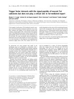
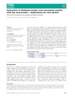
regulated by caspase-3 as sequence inspection shows that the
bovine enzyme contains five putative caspase consensus
sites: DHWD(26–29), DEGD(134–137), DEKD(289–292),
DCSD(365–368) and DQGD(778–781) (Fig. 1). Of these
sites, only two show strong consensus for caspase-3:
DHWD(26–29) and DQGD(778–781). Indeed, we have
shown that in the presence of the inhibitory protein (PDEc)
of the rod photoreceptor PDE6, PDE5A1 is a substrate for a
low activity preparation of purified caspase-3 [8]. Site-
directed mutagenesis studies have defined the position of the
GAF domains [9–11] and key amino acid residues involved in
the metal ion coordination and catalytic activity [12–15].
These are shown in Fig. 1 to define their relative position of
the putative caspase sites.
The caspase family is composed of 13 distinct gene
products, each with different substrate preferences and
inhibitor sensitivities [16]. The first caspase was identified as
ICE (caspase-1), which converts pro-interleukin-1b into
bioactive interleukin-1b [17]. Subsequently, several human
and murine caspases have been cloned. These enzymes show
sequence homology with CED-3 from the nematode,
Caenorrhabdidtis elegans [18]. The overexpression of
Correspondence to N. J. Pyne, Department of Physiology and
Pharmacology, Strathclyde Institute for Biomedical Sciences,
University of Strathclyde, 27 Taylor Street, Glasgow,
G4 ONR, Scotland, UK.
Fax: + 141 5522562, Tel.: + 141 5524400 ext 2659,
E-mail:
Abbreviations: DMEM, Dulbecco’s modified Eagle’s medium; PDE,
phosphodiesterase; PARP, poly (ADP-ribose) polymerase.
(Received 5 November 2002, revised 8 January 2003,
accepted 16 January 2003)
Eur. J. Biochem. 270, 962–970 (2003) Ó FEBS 2003 doi:10.1046/j.1432-1033.2003.03464.x
different caspases in cells induces apoptosis and/or inflam-
matory mediator production [19,20]. Caspases are synthes-
ized in the cell as inactive proenzymes. These are activated
by proteolysis at internal sites and are subdivided into
initiators and effector enzymes. Initiator caspases (e.g.
caspase-8 and -9) are activated by proximity induced
proteolysis by adaptor-dependent recruitment in the recep-
tosome or apoptosome complex. Once activated, these will
further propagate the cascade by activating the downstream
effector caspases. The effector (executioner) enzymes
include caspase-3, and proteolyse a number of substrates
resulting in structural changes, such as gelsolin, nuclear
changes such as ICAD and signal transduction such as
MEK kinase [21], Mst-1 [22], PAK-2 [23], PI3K/Akt [24],
PKCf [25] and FAK [26]. Caspase-3 and -7 cleave proteins
at a
4
DX
3
X
2
D
1
consensus site, where apolar amino acids at
position 2 are preferred. The cyclic nucleotides, cGMP and
cAMP have been shown to promote apoptosis of certain
mammalian cells. For instance, nitric oxide stimulates
apoptosis in cardiomyocytes and endothelial cells via a
cGMP-dependent pathway [27–29]. cGMP is also required
for nerve cell death caused by glutathione depletion, via
modulation of calcium channel activity [30]. In addition,
Huston and colleagues have shown that PDE4A5, which
specifically hydrolyses cAMP, is proteolysed by caspase-3.
This removes the N-terminal tail that contains specific
binding sites for the lyn kinase [31]. These findings provide a
rationale for investigating whether caspase-mediated path-
wayscaninteractwithPDE5inintactcells.
In this article, we show that caspase-3 either directly or
indirectly via caspase-3 activated proteases results in
cleavage and inactivation of PDE5A1. Homology model
comparison with the catalytic domain of PDE4B2 was
used to probe a functional role for the region in
PDE5A1 that might be cleaved by caspase-3. Residues
in PDE5 identified by Turko and colleagues [13,14]
H603, H607, H643, D644, E762, H675, T713, D754,
Q765 and Q779 were used for the modeling. Mutations
of T713 and H675 that are cognate residues of those that
orientate the magnesium ion via H-bonds to water
ligands in PDE4B produce comparatively little impact on
catalytic activity in PDE5. From the modeling, it is
possible that inactivation of PDE5A1 by caspase-3 might
occur via removal of key regions which constitute part of
the wall of the catalytic site of PDE5A1. We also suggest
a possible interaction between PDE5A1 and an uniden-
tified caspase-3-initiated protease(s) that may constitute a
novel signaling event.
Experimental procedures
Materials
All biochemicals were from Boehringer Mannheim (Mann-
heim, Germany), while general chemicals and snake venom
were from Sigma Chemical Co. (Poole, UK). [
3
H]cGMP
and [
35
S]methionine were from Amersham International
(Amersham, Buckinghamshire, UK). Cell culture supplies
were from Life Technologies (Paisley, UK). Ac-DEVD-
CHO and anti-PDE5 IgG was from Calbiochem (UK). The
pCAGGS-Casp-3 plasmid construct was kindly provided by
T. Miyazaki, The Burnham Institute, La Jolla, CA, USA.
Protein purification
Recombinant murine caspases were purified according to
[19]. The proteolytic activity of these enzymes on procaspase
substrates has been described previously [32].
Sub-cloning
Bovine PDE5A1 cDNA (GenBank accession number
L16545) in pBacPac9 (Clontech, CA, USA) was a gift from
J. Corbin (Vanderbilt University, USA). It was subcloned
into pcDNA3.1/Zeo(–) (Invitrogen, the Netherlands) by
amplifying the ORF using primers ApaI-Koz-PDE5A1-
FOR(AAGGGCCCGCCACCATGGAGAGGG
CCG GCC CCG GCT) and XbaI-PDE5A1REV (GCT
TCTAGACTCAGTTCCGCTTGGTCTGGCTGC
TTT CAC), digesting the product and the vector with ApaI
and XbaI (Promega, UK), ligating, and then transforming
into TOP10 Escherichia coli (Invitrogen). Positive clones
were selected and sequenced by BigDye terminator cycle
sequencing (PE Biosystems, UK) using a PE373A auto-
mated DNA sequencer.
Site-directed mutagenesis of D781 (DfiA) in the
caspase-3 consensus site was carried out using Stratagene’s
QuikChange Mutagenesis kit (Stratagene, UK). This was
achieved using pcDNA3.1-PDE5 Zeo(–) plasmid with
125 ng of the forward primer, PDE5MUTF (GAC CAA
GGA GCT AGA GAG AGG AAA GAA CTC) and the
reverse primer, PDE5MUTR (GAG TTC TTT CCT CTC
TCT AGC TCC TTG GTC) in a 50-lL PCR containing
50 ng of pcDNA3.1-PDE5 Zeo(–) plasmid, 10 m
M
KCl,
10 m
M
(NH
4
)
2
SO
4
,20m
M
Tris/HCl (pH 8.8), 2 m
M
MgSO
4
,0.1%TritonX-100,0.5lg bovine serum albumin,
0.4 m
M
dNTPs and 2.5 U PfuTurbo DNA polymerase. The
cycling conditions were 95 °C for 30 s then 12 cycles of
95 °C for 30 s, 55 °C for 1 min and 68 °C for 15 min. Ten
units of DpnI restriction enzyme was added to the reaction
following PCR. Two microliters of reaction mixture was
used in a transformation reaction with TOP10 competent
E. coli. Positive clones were selected and sequenced to
confirm the mutagenesis.
Truncation of PDE5A1 at D781 involved the use of
primers that carry a single point mutation to introduce a
premature stop codon at 782R. The forward primer has the
sequence GAC CAA GGA GAT TGA GAG AGG AAA
GAA CTC, while the reverse primer was GAG TTC TTT
CCT CTC TCA ATC TCC TTG GTC. Twelve cycles of
Fig. 1. Schematic showing the positions of
caspase motifs in PDE5A1.
Ó FEBS 2003 Interaction of caspase-3 with PDE5A1 (Eur. J. Biochem. 270) 963
95 °C for 30 s, 55 °C for 1 min and 68 °C for 16 min were
used for the PCR.
Transfection
Cos-7 cells or PC12 cells were grown to 50–70% confluence
in Dulbecco’s modified Eagle’s medium (DMEM) contain-
ing 10% (v/v) fetal bovine serum. Five micrograms of
pcDNA-3.1-PDE5 or 0.1–1 lgofpCAGGS-Casp-3was
added to DMEM and DEAE-dextran (10 mgÆmL
)1
), mixed
thoroughly and incubated at room temperature for 15 min.
Cells were incubated at 37 °C for 1 h with this medium,
before this was removed and 1 mL of 10% (v/v) dimethyl-
sulfoxide added for 30 s. The medium was then aspirated
and the cells washed twice with DMEM containing 10%
(v/v) fetal bovine serum.The cells werethenplacedinDMEM
containing 10% (v/v) fetal bovine serum, grown to conflu-
ence and harvested 48 h after transfection. Alternatively,
cells were transfected with the plasmid construct following
complex formation with LipofectAMINE
TM
2000, accord-
ing to the manufacturer’s instructions. The cDNA contain-
ing media was then removed following incubation for 24 h
at 37 °C, and the cells incubated for a further 24 h.
Cell lysates
Cos-7 and PC12 cell lysates were prepared by adding 0.25
M
sucrose, 1 m
M
EDTA, 10 m
M
Tris/HCl, pH 7.4, 2 m
M
benzamidine and 0.1 m
M
phenylmethanesulfonyl fluoride.
Cells were scraped into this buffer and homogenized by
passing through a 0.24-mm gauge syringe needle. The lysates
were either used for caspase activity assays or combined with
boiling electrophoresis sample buffer for SDS/PAGE.
Immunoblotting
Nitrocellulose membranes were blocked for 1 h at 4 °C in
10 m
M
phosphate-buffered saline (NaCl/P
i
) and 0.1% (v/v)
Tween-20 containing 5% (w/v) non fat dried milk and
0.001% (w/v) thimerisol. The nitrocellulose sheets were then
incubated overnight at 4 °C with antibodies in blocking
solution. The sheets were then washed with NaCl/P
i
and
0.1%(v/v) Tween-20 prior to incubation with horseradish
peroxidase-linked anti-rabbit IgGs in blocking solution for
2 h at room temperature. After washing the blots as above,
the immunoreactive bands were detected using an enhanced
chemiluminescence kit.
PDE assay
Unless otherwise stated, the assay of PDE activity was by the
two-step radiotracer method [33] using 0.5 l
M
[
3
H]cGMP.
PDE activity measurements were performed under condi-
tions of linear rate product formation and where less than
10% of the substrate was utilized during the assay.
[
35
S]Methionine-labeled PDE5A1 and poly (ADP-ribose)
polymerase (PARP)
One microgram of pcDNA-3.1-PDE5 or pGEM-PARP
was combined with an in vitro transcription/translation kit
reaction (Promega, UK) to produce [
35
S]methionine-labeled
proteins.
Purified caspases
[
35
S]Methionine-labeled PDE5A1 was combined with an
incubation mix (25 lL) containing 50 m
M
Hepes, pH 7.4,
1m
M
EDTA and 10 m
M
dithiothreitol with 60 ng per assay
purified recombinant murine caspases. Incubations were for
2 h at 30 °C and were terminated by addition of boiling
sample buffer for SDS/PAGE.
Caspase-3 activity assays
Caspase-3 activity in cell lysates was measured using
[
35
S]methionine-labeled PARP as a substrate. In each assay,
equal amounts of cell lysate protein ( 5 lg/assay)
were used and incubated with PARP for 2 h at 30 °C.
Incubations were terminated by addition of boiling electro-
phoresis sample buffer for SDS/PAGE. Inhibition of
caspase-3 activity was achieved using the reversible inhibitor
acetyl-Asp(OMe)-Glu(OMe)-Val-Asp(OMe)-aldehyde
(Ac-DEVD-CHO).
Molecular modeling
Modeling studies were performed on an SGI Octane
workstation using the automated homology-modeling
program,
MODELER
,within
INSIGHTII
(Accelerys Inc., San
Diego, CA, USA). This program generates an all-atom
model based on a specified sequence alignment and
reference protein structure. The crystal structure of the
PDE4B2B core catalytic domain published by Xu et al.
[34] was used as a reference model (Protein Data Bank
accession code 1FOJ, chain B). A truncated PDE5A1
sequence, corresponding to the region spanning helices
5–16 of the 1FOJ structure, was matched to the
PDE4B2 sequence. This region embodies the metal ion
and substrate-binding pocket together with flanking
helices and exhibits close homology to the PDE5A1
sequence (27% identity). Thirty models were generated
using the program’s highest optimization level of
molecular dynamics simulated annealing, and the model
with the lowest overall probability density function
violation was taken forward for further energy minimi-
zation. Key residues in the PDE5A1 model that
contribute to the metal binding environment (H603,
H607, H643, D644, E672, H675, T713, D754) overlaid
the corresponding residues in the PDE4B2 reference
structure (H234, H238, H274, D275, E304, H307, T345
and D392) with little deviation. The coordinates of these
residues and the a-carbon centers of Q765, Q779 and
Q807 were frozen during subsequent minimization,
which was carried out using the cvff forcefield imple-
mented in the
DISCOVER
module of
INSIGHTII
.The
dielectric constant was set to 4.00 and the model refined
through 3000 steps of steepest descent energy minimiza-
tion followed by conjugate gradient energy minimization
to convergence with a 0.001 kcalÆmol
)1
ÆA
˚
)1
root mean
square energy gradient difference between successive
minimization steps.
964 M. J. Frame et al. (Eur. J. Biochem. 270) Ó FEBS 2003
Results and discussion
Proteolysis of PDE5A1 by caspase-3
Recombinant PDE5A1 was produced from a pcDNA-1-
PDE5 plasmid construct (which has a T7 polymerase
initiation site) using an in vitro reticulocyte transcription/
translation kit. A major 98 kDa [
35
S]methionine-labeled
protein corresponding to PDE5A1 was produced from the
plasmid construct and resolved on SDS/PAGE (Fig. 2).
Several minor lower molecular mass [
35
S]methionine-labeled
proteins were also produced in the transcription and
translation reaction. These are probably derived by differ-
ential internal translation initiation or proteolysis of
PDE5A1 by reticulocyte proteases. Figure 2 shows that
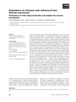
caspase-3 cleaved PDE5A1 to a major 82 kDa fragment.
This in vitro reaction showed specificity for caspase-3
because caspase-2, -12 and -14 did not significantly cleave
the enzyme. Consensus sites for caspase-2, -12 and -14 are
not present in PDE5A1. Assays were deliberately designed
such that the final concentration of caspase-3 in the
incubation was 40 n
M
, which is equivalent with its concen-
tration in mammalian cells [32]. These conditions were used
to best predict the extent and nature of the proteolysis that
might occur in intact cells. Higher concentrations of
caspase-3 or extended incubation times cause extensive
proteolysis of the 82 kDa fragment into smaller polypep-
tides and is therefore, less stringent.
Caspase-3 cleaved 50% of the PDE5A1 under the
assay conditions used. Proteolysis is dependent upon both
the specific activity of the caspase-3, which might be
limiting, and the affinity of interaction, which in vitro may
reflect reduced efficiency compared with in vivo.The
findings show that PDE5A1 is a substrate for caspase-3
in vitro, consistent with the presence of consensus caspase-3
sites in PDE5A1. They also support our previous results
showing that PDE5A1 is proteolysed by low activity
purified caspase-3 in the presence of PDEc [8].
PDE5A1 cleavage by caspase-3 and/or caspase-3
activated proteases in Cos-7 cell and PC12 cells
Cos-7 cells were transiently transfected with PDE5 and/or
caspase-3 plasmid constructs. The main objective here was
to establish whether the overexpression of recombinant
caspase-3 induces the proteolysis of PDE5A1 in an intact
cell system.
cGMP hydrolysing activity was increased 10- to 20-fold
in PDE5A1-transfected vs. mock-transfected cells (n > 20),
and was inhibited by > 90% by addition of the selective
PDE5 inhibitor, zaprinast (10 l
M
) to the assay. A major
98 kDa protein was detected on Western blots probed with
specific anti-PDE5 IgGs in lysates from PDE5A1-transfect-
ed but not mock-transfected cells and which comigrated
with recombinant PDE5A1 (see later). These results are
Fig. 2. The effect of recombinant caspases on PDE5A1. Autoradio-
graph showing the effect of purified caspase-2, 3, 12 and 14
(60 ngÆassay
)1
)on[
35
S]methionine PDE5A1. Control represents no
addition to PDE5A1. Radioactive-labeled molecular mass standards
are shown (M
r
¼ 200–33 kDa). This is a representative result of three
separate experiments.
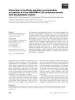
Fig. 3. Caspase-3 in transfected Cos-7 cells. Cells were transfected with
pCAGGS-Casp-3 cDNA (1 lg) and/or wild-type pcDNA-3.1-PDE-5
(5 lg). (A) Western blot probed with anti-caspase-3 antibodies
showing the expression of recombinant caspase-3 in pCAGGS-
Casp-3-transfected cells. (B) Autoradiograph showing the effect of
Ac-DEVD-CHO (100 l
M
) (added at the time of transfection) and
recombinant PDE5A1 on caspase-3 activity in Cos-7 cells. Caspase-3
activity was measured using [
35
S]methionine-labeled PARP as a sub-
strate. These are representative results of three experiments. C3 denotes
caspase-3.
Ó FEBS 2003 Interaction of caspase-3 with PDE5A1 (Eur. J. Biochem. 270) 965
consistent with previous reports showing expression of
functionally active recombinant PDE5 in Cos-7 cells [6].
Western blot analysis with anti-caspase-3 IgG confirmed
expression of recombinant caspase-3 in pCAGGS-Casp-3-
transfected cells. Figure 3A shows that the antibody
reacted with five polypeptides of molecular mass corres-
ponding to 35, 30, 17, 12 and 9 kDa in lysates from
caspase-3-transfected cells. These proteins were not detected
in lysates from mock-transfected cells. These polypeptide
fragments are formed from auto-processing of the protease.
Internal cleavage of native protein results in the formation
of p17 and p12, which are catalytically active toward
endogenous protein substrates. The formation of p9 might
be due to extensive cleavage of intermediate fragments, as a
result of particularly good overexpression of the enzyme in
Cos-7 cells. Cotransfection of PDE5A1 did not affect the
auto-activation of caspase-3. Caspase-3 activity in cell
lysates was also measured using [
35
S]methionine-labeled
PARP (M
r
¼ 115 kDa) as a substrate. Figure 3B shows
that there is substantial endogenous caspase-3 activity in
lysates from mock-transfected cell, possibility activated as a
consequence of stressing cells during the transfection
procedure. In the current study, endogenous caspase-3
activity converted 70% of the 115 kDa PARP to an 85-
kDa fragment (p85). The overexpression of recombinant
caspase-3 in Cos-7 cells resulted in more extensive proteo-
lysis of the exogenous 115 kDa PARP in the assay
(Fig. 3B). Caspase-3 activity was completely abolished by
treatment of the cells with the caspase-3/7 inhibitor,
Ac-DEVD-CHO (added at the time of transfection with
caspase-3 plasmid construct). Overexpression of PDE5A1
did not inhibit the auto-activation of caspase-3 (Fig. 3B).
This is in line with results showing that PDE5A1 did not
affect auto-proteolysis of caspase-3 (Fig. 3A).
We investigated the effect of overexpressing recombinant
caspase-3 on PDE5A1 in transfected Cos-7 cells. 98 kDa
PDE5A1 levels were markedly reduced by 60–75% in
lysates of cells cotransfected with caspase 3 and PDE5A1
plasmid constructs (Fig. 4A,B). This is consistent with
depletion of the enzyme via caspase-3-mediated cleavage.
An 82-kDa fragment appeared only in lysates of cells
overexpressing both enzymes (Fig. 4A,B). No other frag-
ments were detected on Western blots. The accumulation of
82 kDa fragment was not correlated with a similar reduc-
tion in the native 98 kDa PDE5A1 level. The most likely
hypothesis is that caspase-3 proteolyses PDE5A1 as it is
expressed and that the 82 kDa fragment thus formed, is
then immediately processed further. In addition, caspase-3
may act on other proteases that cleave PDE5A1. This in
itself is a potentially important and interesting finding as it
might suggest a hitherto unidentified caspase-3 initiated
protease cascade regulating PDE5A1 activity.
Fig. 4. The interaction of caspase-3 with PDE5A1 in Cos-7 cells. Cells
were transfected with pCAGGS-Casp-3 cDNA (1 lg) and/or wild-
type or truncated D781 pcDNA-3.1-PDE5 (5 lg) plasmid constructs.
(A) Western blot probed with anti-PDE5 IgG showing the effect of
Ac-DEVD-CHO (100 l
M
) on the cleavage of PDE5A1 by caspase-3 in
transfected Cos-7 cells. The position of the truncated D781 mutant on
SDS/PAGE expressed in Cos-7 cells is also shown; (B) Western blot
probed with anti-PDE5 IgG showing the proteolysis of wild-type
PDE5A1 by recombinant caspase-3 in transfected Cos-7 cells to reveal
the faster migrating 82 kDa fragment. These are representative results
of at least three separate experiments. C3 denotes caspase-3.
Fig. 5. Changes in activity of PDE5A1 upon cleavage by caspase-3.
Cells were transfected with pCAGGS-Casp-3 cDNA (0.1–1 lg) and/or
wild-type or truncated D781 pcDNA-3.1-PDE5 (5 lg) plasmid con-
structs. The histogram shows the effect of overexpressing recombinant
caspase-3 and the treatment of cells with Ac-DEVD-CHO (100 l
M
)on
wild-type recombinant PDE5A1 activity in Cos-7 cells. PDE5A1
activity was measured at 0.5 l
M
[
3
H]cGMP. Results are expressed as
the fold increase over basal PDE activity in mock-transfected cells.
D781 truncated PDE5A1 was expressed as an inactive enzyme. Inset is
the corresponding Western blot showing 98 kDa PDE5A1 levels. The
82 kDa fragment is not evident as the Western blot is underexposed to
better demonstrate the increase in 98 kDa PDE5A1 in Ac-DEVD-
CHO-treated cells. In the latter case, cells were transfected with
pCAGGS-Casp-3 cDNA (1 lg) and wild-type or truncated D781
pcDNA-3.1-PDE5 (5 lg) plasmid constructs. These are representative
results of at least three separate experiments. C3 denotes caspase-3.
966 M. J. Frame et al. (Eur. J. Biochem. 270) Ó FEBS 2003
The reduction in 98 kDa PDE5A1 levels was correlated
with a decrease in PDE5A1 activity (Fig. 5). The remaining
PDE activity in caspase-3/PDE5A1 transfected cells was
recovered by gel filtration on Superose 12 with a similar
elution compared with PDE5A1 from cells overexpressing
this enzyme alone (Fig. 6). Further evidence to support the
possibility that PDE5A1 interacts with caspase-3 and
indirectly with caspase-3-activated proteases was shown
by results showing that the caspase-3/7 inhibitor,
Ac-DEVD-CHO abolished the reduction in PDE5A1 levels
observed in cells cotransfected with PDE5A1 and caspase-3
(Fig. 4A). This was correlated with the reversal of the
reduction in cGMP hydrolysing PDE activity (Fig. 5). It is
interesting to note that the treatment of cells with
Ac-DEVD-CHO appeared to increase 98 kDa PDE5A1
levels and activity above controls, consistent with an action
of endogenous caspase-3/7 (Figs 4A and 5). It remains to
be determined which of the potential caspase sites is
cleaved to inactivate the enzyme. However, only two sites
exhibit strong consensus for caspase-3
26
DHWD
29
and
778
DQGD
781
. Cleavage at
78
DQGD
781
would produce an
82-kDa fragment. We cannot ascertain at the moment
whether cleavage at
78
DQGD
781
causes inactivation, as
there is no correlation in the reduction in 98 kDa protein
levels with the appearance of the 82 kDa fragment.
Importantly, as the overexpression of caspase-3 in Cos-7
cells induces cell death [32], we conclude from the current
findings that cleavage and inactivation of PDE5A1 medi-
ated by caspase-3 may be associated with this process.
However, further studies are necessary to establish whether
the cleavage of PDE5A1 is a key event governing cell death.
To demonstrate the robustness of the interaction between
caspase-3 and PDE5A1, we repeated the experiments in
PC12 cells. In contrast with Cos-7 cells, the treatment of
PC12 cells with Ac-DEVD-CHO did not modulate the
expression level of recombinant PDE5A1 (Fig. 7A), indi-
cating that endogenous caspase-3 activity is not a factor that
might influence the native state of recombinant PDE5A1
in this case. However, in common with Cos-7 cells,
Fig. 7. Effect of caspase-3 and staurosporine on PDE5A1 proteolysis.
Cells were transfected with pCAGGS-Casp-3 cDNA (1 lg) and/or
wild-type pcDNA-3.1-PDE5 (5 lg) plasmid constructs. Cells stimu-
lated with and without staurosporine (10 l
M
, 24 h) were transfected
only with wild-type pcDNA-3.1-PDE5 (5 lg) plasmid construct. (A)
Western blot probed with anti-PDE5 IgG showing the proteolysis of
wild-type PDE5A1 by recombinant caspase-3 (and the effect of
Ac-DEVD-CHO (100 l
M
) added at the time of transfection) and in
response to staurosporine in PC12 cells. Also shown is a histogram of
the corresponding reduction in PDE5A1 activity. (B) Western blot
probed with anti-PDE5 IgG showing the proteolysis of wild-type
PDE5A1 in Cos-7 cells stimulated with staurosporine. Also shown is a
histogram of the corresponding reduction in PDE5A1 activity. All
activities were measured using samples equalized for protein. PDE5A1
activity was measured at 0.5 l
M
[
3
H]cGMP. These are representative
results of at least 2–4 separate experiments. C3 denotes caspase-3.
Fig. 6. Elution of PDE5A1 from Superose-12. Cells were transfected
with pCAGGS-Casp-3 cDNA (1 lg) and/or wild-type or truncated
D781 pcDNA-3.1-PDE5 (5 lg) plasmid constructs. The figure shows
Western blots of chromatographic fractions eluted from Superose 12
probed with anti-PDE5 IgG and a PDE5A1 activity profile (taken
from high-speed supernatants of cells overexpressing caspase-3/
PDE5A1). Total elution volume was 35 mL, with 1-mL fractions.
These are representative results of at least three separate experiments.
Ó FEBS 2003 Interaction of caspase-3 with PDE5A1 (Eur. J. Biochem. 270) 967
overexpression of recombinant caspase-3, results in the
reduction of 98 kDa PDE5A1 levels, concomitant with a
similar decrease in cGMP PDE activity (Fig. 7A).
The effect of the apoptotic agent, staurosporine
We also investigated whether apoptotic agents induce
the cleavage of PDE5A1. For this purpose we used,
staurosporine (PKC inhibitor), which has been shown by
Brophy et al. [35] to activate caspase-3 activity in Cos-7
cells. Figure 7A,B shows that the treatment of PDE5A1-
transfected Cos-7 and PC12 cells, respectively, with stauro-
sporine caused a marked reduction in 98 kDa PDE5A1
levels and PDE activity. These findings suggest that there is
specificity in the interaction between caspase-3 and
PDE5A1 that requires application of an apoptotic stimulus.
DQGD(778–781) site
Proteolysis of the DQGD(778–781) by caspase-3 might
affect catalytic activity of PDE5A1 as the site is within the
boundary of the active site. In addition, cleavage at this site
would produce an 82-kDa fragment. To test whether a
potential cleavage of the DQGD(778–781) site might affect
catalytic activity of PDE5A1, we created a truncated D781
mutant corresponding exactly to the 82 kDa fragment. This
mutant was expressed equally well compared with the wild-
type enzyme in transfected Cos-7 cells, comigrated with the
82 kDa fragment formed from the cleavage of PDE5A1 in
cells cotransfected with caspase-3 (Fig. 4A) and was inactive
(Fig. 5). Inactivation of the truncated mutant was no due to
potential misfolding of the enzyme. This was shown by
results showing that the truncated mutant eluted from
Superose 12 at the same position compared with the wild-
type enzyme (Fig. 6), suggesting similar hydrodynamic
properties.
The inactivity of the truncated mutant provides indirect
support for the possibility that cleavage of DQGD(778–781)
is one potential mechanism that might lead to inactivation
of PDE5A1 activity. In this regard, we found that a more
subtle change in PDE5A1 using a single point mutation at
D781 (replaced with A) also results in a reduction of PDE
activity. The D781A mutant partial loss of PDE5A1 activity
to 70% of the wild type measured at 0.5 l
M
cGMP. The
reduction in PDE activity was due, in part, to an increase in
the K
m
for cGMP. The K
m
for the wild-type enzyme was
2.2 l
M
compared with 8.4 l
M
for the D781A mutant. The
kinetic constants were determined in samples where the
expression level of the D781 mutant PDE5A1 mg
)1
cell
lysate protein was approximately twice that of the wild-type
enzyme. Assays were normalized for protein. From this
data, we calculated that the mutant PDE5A1 exhibits a
V
max
that is approximately 50% of the wild-type enzyme.
These findings are in agreement with studies by Turko et al.
[14], who reported identical changes in the kinetic constants
of the D781A mutant.
The DQGD site is within the boundary of the catalytic
domain of PDE5A1 (Fig. 1). We have used the X-ray
crystal structure of PDE4B2 [34] as a template to generate a
homology model for PDE5A1 to rationalize the structural
implications of a caspase-3-catalyzed proteolysis at
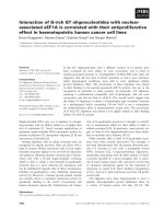
the PDE5A1 DQGD(778–781) site. From the PDE5
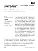
Fig. 8. PDE5 homology model. Homology
model of PDE5A1 based on PDE4B2 crystal
structure showing how the removal of the
C-terminal tail containing Q807 and F810 by
caspase-3 affects the architecture of the cata-
lytic site, and in particular interaction with
Q765.
968 M. J. Frame et al. (Eur. J. Biochem. 270) Ó FEBS 2003
homology model, proteolytic cleavage at DQGD(778–781)
in PDE5A1 might be expected to remove the C-terminal tail
(highlighted in yellow in Fig. 8) containing Q807 and F810,
which are potentially important amino acids required for
substrate binding. Q807 is completely conserved across the
PDE superfamily and, in principle, might accept either the
guanine base of cGMP or the adenine base of cAMP. F810
in PDE5A1 is conserved in PDE4B2 as F446, and this
residue has been shown by site-directed mutagenesis to be
essential for catalytic competence in PDE4 and to play a key
role in the binding of competitive PDE4 inhibitors [36]. The
side chain of this residue may conceivably p-stack with the
purine base of the bound substrate and form hydrophobic
interactions with a number of inhibitors. In PDE4B2 the
sequence QQGD(416–419), corresponding to the PDE5A1
caspase-3 site DQGD(778–781) is identical, except that the
site is disabled by replacement of D for Q at the P4 position.
The site is located on the exposed C-terminal end of helix 14
in the PDE4B2 crystal structure. In conclusion, the caspase-
3-catalyzed cleavage at DQGD(778–781) in PDE5A1 will
very likely remove a key wall from the catalytic site
containing Q807/F810. This might prevent potential inter-
action with critical adjacent amino acid residues present on
the other side of the catalytic pocket, identified by Turko
and colleagues, such as Q765 [13]. The removal of part of
the catalytic pocket explains the inactivity of the engine-
ered protein truncated at D781. Potential cleavage at
DQGD(778–781) by caspase-3 could severely disrupt the
structure of PDE5A1. Interestingly, there is substantial
similarity between the amino acid sequence of the PDE5A1
DQGD(778–781) site and the corresponding region in
PDE2A3,PDE4C,PDE4D,PDE6ab and PDE11A1. D778
at P4 of the caspase-3 consensus site in PDE5A1 is replaced
withEinPDE11A1andPDE6ab, Q in PDE4C, R in
PDE4D and S in PDE2A3. Therefore, the replacement of
the
4
D effectively disables the caspase-3 site in these PDE
isoforms.
Summary
The results presented in this article are consistent with
PDE5A1 acting as a substrate for caspase-3 in intact cells. In
addition, PDE5A1 may be subject to cleavage by a caspase-
3-initiated protease(s) event. These results raise the possi-
bility of a role for PDE5A1 in apoptosis. However, further
investigation is required to establish a causal linkage
between PDE5A1 cleavage and apoptosis.
Acknowledgments
This study was supported by the BBSRC.
References
1. Beavo, J.A. (1995) Cyclic nucleotide phosphodiesterases:
functional implications of multiple isoforms. Physiol. Rev. 75,
725–748.
2. Beavo, J.A. & Brunton, L.L. (2002) Cyclic nucleotide research –
still expanding after half a century. Nat. Rev. Mol. Cell. Biol. 3 (9),
710–718.
3. Houslay, M.D. & Adams, D.R. (2003) PDE4 cAMP phos-
phodiesterases: modular enzymes that orchestrate signalling
cross-talk, desensitisation and compartmentalisation. Biochem.
J. 370, 1–18.
4. Corbin, J.D. & Francis, S.H. (1999) Cyclic GMP phosphodies-
terase-5: target of sildenafil. J. Biol. Chem. 274, 13729–13732.
5. McAllister-Lucas, L.M., Sonnenburg, W.K., Kadlecek, A.,
Seger, D., Trong, H.L., Colbran, J.L., Thomas, M.K., Walsh,
K.A., Francis, S.H., Corbin, J.D. & Beavo, J. (1993) The
structure of a bovine lung cGMP-binding, cGMP-specific phos-
phodiesterase deduced from a cDNA clone. J. Biol. Chem. 268,
22863–22873.
6. Thomas, M.K., Francis, S.H. & Corbin, J. (1990) Characteriza-
tion of a purified bovine lung cGMP-binding cGMP phospho-
diesterase. J. Biol. Chem. 265, 14964–14970.
7. Burns, F., Rodger, I.W. & Pyne, N.J. (1992) The catalytic subunit
of protein kinase A triggers activation of the type V cyclic GMP-
specific phosphodiesterase from guinea-pig lung. Biochem. J. 283,
487–491.
8. Frame, M., Wan, K F., Tate, R., Vandenabeele, P. & Pyne, N.J.
(2001) The gamma subunit of the rod photoreceptor cGMP
phosphodiesterase can modulate the proteolysis of two cGMP
binding cGMP-specific phosphodiesterases (PDE6 and PDE5) by
caspase-3. Cell. Signal. 13, 735–741.
9. Aravind, L. & Ponting, C.P. (1997) The GAF domain: an evolu-
tionary link between diverse phototransducing proteins. Trends
Biochem. Sci. 22, 458–459.
10. Ho, Y.S., Burden, L.M. & Hurley, J.H. (2000) Structure of the
GAF domain, a ubiquitous signaling motif and a new class of
cyclic GMP receptor. EMBO J. 19, 5288–5299.
11. Martinez, S.E., Wu, A.Y., Glavas, N.A., Tang, X.B., Turley, S.,
Hol, W.G. & Beavo, J.A. (2002) The two GAF domains in
phosphodiesterase 2A have distinct roles in dimerization and in
cGMP binding. Proc. Natl. Acad. Sci. USA 99, 13260–13265.
12. McAllister-Lucas, L.M., Haik, T.L., Colbran, J.L., Sonnenburg,
W.K., Seger, D., Turko, I.V., Beavo, J.A., Francis, S.H. & Corbin,
J.D. (1998) An essential aspartic acid at each of two allosteric
cGMP-binding sites of a cGMP-specific phosphodiesterase.
J. Biol. Chem. 270, 30671–30679.
13. Turko, I.V., Francis, S.H. & Corbin, J.D. (1998) Hydropathic
analysis and mutagenesis of the catalytic domain of the cGMP-
binding cGMP-specific phosphodiesterase (PDE5). cGMP versus
cAMP substrate selectivity. Biochemistry 37, 4200–4205.
14. Turko, I.V., Francis, S.H. & Corbin, J.D. (1998) Potential roles of
conserved amino acids in the catalytic domain of the cGMP-
binding cGMP-specific phosphodiesterase. J. Biol. Chem. 273,
6460–6466.
15. Francis,S.H.,Turko,I.V.,Grimes,K.A.&Corbin,J.D.(2000)
Histidine-607 and histidine-643 provide important interactions for
metal support of catalysis in phosphodiesterase-5. Biochemistry
39, 9591–9596.
16. Nunez, G., Benedict, M.A., Hu, Y.M. & Inohara, N. (1998)
Caspases: the proteases of the apoptotic pathway. Oncogene 17,
3237–3245.
17. Alnemri, E.S., Fernandes-Alnemri, T. & Litwack, G. (1995)
Cloning and expression of four novel isoforms of human inter-
leukin-1 beta converting enzyme with different apoptotic activities.
J. Biol. Chem. 270, 4312–4317.
18. Hengartner, M.O., Ells, R.E. & Horvitz, H.R. (1992) Caenor-
habditis elegans gene ced-9 protects cells from programmed cell
death. Nature 356, 494–499.
19. Van De Craen, M., Vandenabeele, P., Declercq, W., Van Den
Brande, I., Van Loo, G., Molemans, F., Schotte, P., Van Criek-
inge, W., Beyaert, R. & Friers, W. (1997) Characterization of
seven murine caspase family members. FEBS Lett. 403, 61–69.
20. Van De Craen, M., Declercq, W., Van Den Brande, I., Friers, W.
& Vandenabeele, P. (1999) The proteolytic procaspase activation
network: an in vitro analysis. Cell Death Differ. 11, 1117–1124.
Ó FEBS 2003 Interaction of caspase-3 with PDE5A1 (Eur. J. Biochem. 270) 969
21. Cardone, M.H., Salvesen, G.S., Widmann, C., Johnson, G. &
Frisch, S.M. (1997) The regulation of anoikis: MEKK-1 activa-
tion requires cleavage by caspases. Cell 90, 315–323.
22. Graves, J.D., Gotoh, Y., Draves, K.E., Ambrose, D., Han, D.K.,
Wright, M., Chernoff. J., Clark, E.A. & Krebs, E.G. (1998) Cas-
pase-mediated activation and induction of apoptosis by the
mammalian Ste20-like kinase Mst1. EMBO J. 17, 2224–2234.
23. Rudel, T. & Bokoch, G.M. (1997) Membrane and morphological
changes in apoptotic cells regulated by caspase-mediated activa-
tion of PAK2. Science 276, 1571–1574.
24. Francois, F. & Grimes, M.L. (1999) Phosphorylation-dependent
Akt cleavage in neural cell in vitro reconstitution of apoptosis.
J. Neurochem. 73, 1773–1776.
25. Frutos, S., Moscat, J. & Diaz-Meco, M.T. (1999) Cleavage of
zetaPKC but not lambda/iotaPKC by caspase-3 during UV-
induced apoptosis. J. Biol. Chem. 274, 10765–10770.
26. Van de Water, B., Nagelkerke, J.F. & Stevens, J.L. (1999)
Dephosphorylation of focal adhesion kinase (FAK) and loss of
focal contacts precede caspase-mediated cleavage of FAK
during apoptosis in renal epithelial cells. J. Biol. Chem. 274,
13328–13337.
27. Baixeras, E., Garcia-Lozano, E. & Martinez, A C. (1996)
Decrease in cAMP levels promoted by CD48–CD2 interaction
correlates with inhibition of apoptosis in B cells. Scan.J.Immunol.
43, 406–412.
28. Tortorella, C., Piazzolla, G., Spaccavento, F. & Antonaci, S.
(1998) Effects of granulocyte-macrophage colony-stimulating
factor and cyclic AMP interaction on human neutrophil apopto-
sis. Med. Inflamm. 7, 391–396.
29. Shimojo, T., Hiroe, M., Ishiyama, S., Ito, H., Nishikawa, T. &
Marumo, F. (1999) Nitric oxide induces apoptotic death of
cardiomyocytes via a cyclic-GMP-dependent pathway. Exp. Cell.
Res. 247, 38–47.
30. Li. Y., Maher, P. & Schubert, D. (1997) Requirement for cGMP in
nerve cell death caused by glutathione depletion. J. Cell. Biol. 139,
1317–1324.
31. Huston, E., Beard, M., McCallum, F., Pyne, N.J., Vandenabeele,
P., Scotland, G. & Houslay, M.D. (2000) The cAMP-specific
phosphodiesterase PDE4A5 is cleaved downstream of its SH3
interaction domain by caspase-3. Consequences for altered
intracellular distribution. J. Biol. Chem. 275, 28063–28074.
32. Stennicke, H.R., Jurgansmeier, J.M., Shin, H., Deveraux, Q.,
Wolf, B.B., Yang, X., Zhou, Q., Ellerby, H.M., Bredesen, D.,
Green, D.R., Froelich, C.J. & Salvesen, G.S. (1998) Pro-caspase-3
is a major physiologic target of caspase-8. J. Biol. Chem. 273,
27084–27090.
33. Thompson, J. & Appleman, M. (1971) Multiple cyclic nucleotide
phosphodiesterase activities from rat brain. Biochemistry 10, 311–
316.
34. Xu, R.Y., Hassell, A.M., Vanderwall, D., Lambert, M.H., Hol-
mes, W.D., Luther, M.A., Rocque, W.J., Milburn, M.U., Zhao,
Y., Ke, H. & Nolte, R.T. (2000) Atomic structure of PDE4:
insights into phosphodiesterase mechanism and specificity. Science
288, 1822–1825.
35. Brophy, V.A., Tavare, J.M. & Rivett, A.J. (2002) Treatment of
COS-7 cells with proteasome inhibitors or gamma-interferon
reduces the increase in caspase 3 activity associated with
staurosporine-induced apoptosis. Arch. Biochem. Biophys. 397,
199–205.
36. Richter, W., Unciuleac, L., Hermsdorf, T., Kronbach, T. &
Dettmer, D. (2001) Identification of inhibitor binding sites of the
cAMP-specific phosphodiesterase 4. Cell. Signall. 13, 287–297.
970 M. J. Frame et al. (Eur. J. Biochem. 270) Ó FEBS 2003