Báo cáo khoa học: Human proteoglycan testican-1 inhibits the lysosomal cysteine protease cathepsin L pdf
Bạn đang xem bản rút gọn của tài liệu. Xem và tải ngay bản đầy đủ của tài liệu tại đây (275.61 KB, 8 trang )
Human proteoglycan testican-1 inhibits the lysosomal cysteine
protease cathepsin L
Jeffrey P. Bocock
1
, Cora-Jean S. Edgell
2
, Henry S. Marr
2
and Ann H. Erickson
1
1
Department of Biochemistry and Biophysics and
2
Department of Pathology and Laboratory Medicine, The University of North
Carolina, Chapel Hill, NC, USA
Testican-1, a secreted proteoglycan enriched in brain, has a
single thyropin domain that is highly homologous to
domains previously shown to inhibit cysteine proteases. We
demonstrate that purified recombinant human testican-1 is a
strong competitive inhibitor of the lysosomal cysteine pro-
tease, cathepsin L, with a K
i
of 0.7 n
M
, but it does not inhibit
the structurally related lysosomal cysteine protease cathep-
sin B. Testican-1 inhibition of cathepsin L is independent of
its chondroitin sulfate chains and is effective at both pH 5.5
and 7.2. At neutral pH, testican-1 also stabilizes cathepsin L,
slowing pH-induced denaturation and allowing the protease
to remain active longer, although the rate of proteolysis is
reduced. These data indicate that testican-1 is capable of
modulating cathepsin L activity both in intracellular vesicles
and in the extracellular milieu.
Keywords: cathepsin L; proteoglycan; protease; testican;
thyropin.
Testican is a proteoglycan first identified in human seminal
plasma [1]. The cDNA was subsequently cloned from the
human testis [2], hence the name testican, and from human
vascular endothelial cells [3,4] and mouse brain [5]. In both
human and mouse, testican mRNA is prominent in brain
and absent in certain other tissues. Two additional human
homologues have been identified, testican-2 [6] and testican-
3 [7]. The amino acid sequences of human and mouse
testican-1 are 94% identical, which argues for a significant
function for this proteoglycan [5].
Testican is a multidomain protein (Fig. 1), including
three domains that have homology to inhibitors of three
different classes of proteases. An N-terminal region of
testican-1 has been shown to inhibit membrane-type 1
matrix metalloproteinase activation of matrix metallopro-
teinase-2 [7]. Adjacent to this domain is a follistatin-like
domain that includes a six-cysteine pattern with similarity to
Kazal domains found in serine protease inhibitors such as
pancreatic secretory trypsin inhibitor [8,9]. The next domain
has homology to EF-hands and has been shown to bind
calcium when expressed as an independent domain [10].
Finally, near the C-terminus is a 64-amino acid domain
highly homologous to protein sequences shown to inhibit
cysteine proteases. Such protease inhibition domains have
collectively been called thyropins [11] due to their homology
with a domain repeated 11 times in thyroglobulin, a
precursor of thyroid hormones [12].
The cysteine protease inhibitory function of thyropin
domains was established when a fragment of the class II
invariant chain, that is normally part of the major
histocompatibility complex (MHC), was isolated from
human kidney bound to cathepsin L [13]. The class II
invariant chain exists in two alternatively spliced forms, p31
and p41. The latter form has a region which shares
significant homology with the thyropin domain of thyro-
globulin. This domain of the p41 invariant chain was shown
to inhibit cathepsin L and to stabilize the active protease at
a pH which would normally denature the enzyme [13].
Crystallography of this p41 domain complexed with cath-
epsin L revealed that the domain assumes a wedge-shape
conformation comprised of three loops stabilized by three
disulfide bonds and is lodged in the active site of cathepsin L
[14]. Saxiphilin, a bullfrog serum protein that binds a
neurotoxin [15], and equistatin, from a sea anemone [16],
also have one or more thyropin domains. Like p41, these
proteins inhibit cathepsin L proteolytic activity [15,16], but
a mammalian proteoglycan has not been demonstrated to
serve this role.
Cathepsin L is a ubiquitously expressed protease that is
normally efficiently segregated into lysosomes, where low
pH allows for optimal activity [17]. When expression levels
are increased, however, either during specific developmental
stages, by cell transformation, or by ectopic expression from
a transfected plasmid, the proenzyme is secreted in signifi-
cant amounts [18,19]. In response to signaling events, active
enzyme can also be released [20,21]. In addition to
mediating housekeeping proteolysis in the lysosome, the
protease participates in developmental processes and anti-
gen processing [22–24]. Many studies also implicate extra-
cellular cathepsin L in tumor biology [23,25], where the
major role ascribed to secreted lysosomal proteases is
degradation of extracellular matrix [26–30].
Correspondence to A. H. Erickson, Department of Biochemistry and
Biophysics, CB 7260, Mary Ellen Jones Building, The University of
North Carolina, Chapel Hill, NC 27599–7260, USA.
Fax: + 1 919 966 2852, Tel.: +1 919 966 4694,
E-mail:
Abbreviations: BCIP, 5-bromo-4-chloro-3-indolylphosphate; MHC,
major histocompatibility complex; HEK 293, human embryonic
kidney (cells).
(Received 18 June 2003, revised 31 July 2003,
accepted 12 August 2003)
Eur. J. Biochem. 270, 4008–4015 (2003) Ó FEBS 2003 doi:10.1046/j.1432-1033.2003.03789.x
Little is known about the function of testicans. We have
determined that testican-1, which includes a thyropin
domain, is a competitive inhibitor of cathepsin L but not
of the related cysteine protease cathepsin B. Inhibition is
independent of the chondroitinase ABC-sensitive glycos-
aminoglycan chains associated with this proteoglycan. This
establishes a new role for testican-1 and provides the first
evidence that the protein backbone of a proteoglycan can
regulate lysosomal protease activity, thus expanding our
understanding of the role proteoglycans play in modulating
extracellular events.
Experimental procedures
Materials
Human embryonic kidney 293 (HEK 293) cells were
obtained from ATCC (Manassas, VA, USA). Alkaline
phosphatase-conjugated goat antibodies to mouse immu-
noglobulins were purchased from Jackson Immuno-
Research, and mouse monoclonal antibodies specific for
the Myc epitope tag, Lipofectamine and Geneticin were
obtained from Invitrogen. Centriprep concentrators were
from Millipore and Ni-nitrilotriacetic acid agarose was from
Qiagen. Rainbow molecular mass markers were from
Amersham and Gelcode Blue Staining Reagent was
purchased from Pierce (Rockford, IL, USA). 5-Bromo-4-
chloro-3-indolylphosphate (BCIP)/nitro blue tetrazolium
Color Development Substrate was obtained from Promega.
Z-Phe-Arg-4-methyl-7-coumarin (Z-Phe-Arg-NHMec), E64
and Chondroitinase ABC were from Sigma-Aldrich. Fluo-
trac 96-well microtiter plates were from Greiner Bio-One
(Longwood, FL, USA). Human cathepsin L, purified from
liver, was obtained from Athens Research, (Athens, GA,
USA) and human cathepsin B, purified from liver, was
from Calbiochem.
Recombinant testican-1
A complete open reading frame cDNA for human testican-
1 less its last amino acid was assembled from several cDNA
clones and inserted between EcoRV and XhoI sites in the
Invitrogen expression plasmid, pcDNA3.1/MycHis, keep-
ing the Myc epitope tag and the His
6
encoding DNA from
the vector in frame at the 3¢-end of the testican-1 open
reading frame. The plasmid construct was cloned in
Escherichia coli DH5a, and the intended cDNA insert was
verified by sequencing. This plasmid was transfected into
HEK 293 cells using Lipofectamine according to the
manufacturer’s recommendations. Cells that had incorpor-
ated plasmid DNA were selected in the presence of
Geneticin at 250 lgÆmL
)1
. Expression of the recombinant
gene was indicated by detecting the Myc epitope in culture
fluid from the Geneticin-resistant cells by ELISA.
Chondroitinase ABC treatment and purification
of recombinant testican-1
Conditioned Opti-MEM culture fluid was collected after
24 h from 810 cm
2
of confluent HEK 293 cells expressing
recombinant testican-1. After pelleting cellular debris, the
conditioned culture fluid was concentrated to 1 mL using a
Centriprep concentrator designed to retain molecules larger
than 10 kDa. Half of the concentrated culture fluid was
adjusted to basic pH by addition of pH 8 Tris/HCl to
40 m
M
and sodium acetate to 40 m
M
and treated with
Chondroitinase ABC at 2 UÆmL
)1
for 40 min at 37 °C.
Recombinant testican-1 was then purified by His
6
binding
and elution from Ni-nitrilotriacetic acid agarose, as recom-
mended by the manufacturer. For molecular mass analyses,
samples were reduced and denatured in the presence of
1m
M
dithiothreitol and 2% SDS at 100 °Cfor5minand
then resolved by standard PAGE using 12% acrylamide
with 0.1% SDS. Most of the full-length recombinant
testican-1 expressed by HEK 293 cells possessed significant
amounts of chondroitin sulfate that prevented the majority
of the protein from entering a 12% polyacrylamide gel.
Treatment with chondroitinase ABC reduced the effective
mass, enabling the use of polyacrylamide gels stained with
Gelcode Blue Staining Reagent to assess the purity of the
recombinant protein isolated by Ni-nitrilotriacetic acid-
affinity chromatography. The size of the testican-Myc-
His
6
product was determined by probing gel blots with
monoclonal antibodies specific for the recombinant, using
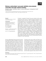
Fig. 1. Alignment of the cathepsin-inhibitory domain of mouse p41 invariant chain with homologous domains of mouse and human testican-1. Identical
residues are shown on a shaded background. The location of the thyropin domain within testican-1 is illustrated relative to the other known
domains of testican-1 (not drawn to scale). Residues 1–21 comprise the signal peptide [45]. The following domain (residues 25–84) is unique to the
three testicans. This region of testican-1 is responsible for the inhibition of a membrane-type metalloproteinase [7]. Residues 86–183 have similarity
to follistatin domains [55], with a six cysteine Kazal-like sequence. Residues 197–312 comprise an extracellular calcium-binding (EC) module [10].
Thyropin domain homology occurs between residues 310 and 379 [11], comprised of exons 9 and 10. Following the thyropin domain is a region
enriched for acidic residues. Twelve of the 13 amino acids within five amino acids of the C-terminus are negatively charged. The serines at 383 and
388 in this domain may have chondroitin or heparan sulfate attached [1], which is designated here as GAG for glycosaminoglycans.
Ó FEBS 2003 Testican-1 inhibits cathepsin L (Eur. J. Biochem. 270) 4009
alkaline phosphatase-conjugated goat antibodies to mouse
immunoglobulins as the secondary antibody, and localizing
the bound alkaline phosphatase activity as a blue precipi-
tate using BCIP/nitro blue tetrazolium Color Development
Substrate as recommended by the manufacturer. The
protein concentrations were determined using Bio-Rad
Protein Assay reagent 500–006 in a microtiter plate assay
using bovine serum albumin for the standard curve.
Cathepsin L active site titration
Cathepsin L was diluted in buffer consisting of 340 m
M
sodium acetate pH 5.5 and 1 m
M
EDTA, and incubated on
ice for 5 min with 5 m
M
dithiothreitol to activate the
enzyme [15]. The concentration of active cathepsin L in
the preparation used for these studies was determined by
titration with increasing amounts of the stoichiometric
inhibitor, E64, at a constant Z-Phe-Arg-NHMec substrate
concentration of 6 l
M
[31]. Liberated fluorophore was
detected by excitation at 355 nm and emission at 460 nm
using a fluorescence microplate reader and
FLUOSTAR
2000
analysis software from BMG Labtechnologies (Durham,
NC, USA).
Testican-1 inhibition of cathepsin L
Cathepsin L was preactivated in the same buffer utilized for
active site titration, as described above. Active cathepsin L
(0.2 n
M
) and varying concentrations (423 p
M
)100 n
M
)of
recombinant testican-1 were incubated at room temperature
for 20 min to allow for complex formation. The tempera-
ture was reduced to 0 °C to synchronize the reactions and
substrate was added to 6 l
M
. Reaction mixtures were then
incubated at 30 °C for 10 min. The substrate conversion
was monitored as described above. The effect of testican-1
on cathepsin B was similarly assayed at a final enzyme
concentration of 2 n
M
in a reaction buffer consisting of
50 m
M
sodium acetate, pH 5.0, 100 m
M
NaCl, 1 m
M
EDTA, 5 m
M
dithiothreitol, and 6 l
M
Z-Phe-Arg-NHMec
as substrate [15].
Determination of inhibition constant
Two approaches were used to determine inhibition con-
stants. In the first approach, the enzyme and inhibitor were
preincubated in the reaction buffer to allow complex
formation, as above. Nonlinear regression analysis of
testican-1 titration data obtained on assay of residual
enzyme activity was used to determine the inhibition
constant K
i
due to the tight binding of the protease by the
inhibitor and the possibility of modification of the inhibitor
bytheenzyme[32].Thesedatawerefittedtothetheoretical
equation for competitive inhibition:
a ¼1À
ðE
0
ÞþðI
0
ÞþK
i
Àf½ðE
0
ÞþðI
0
ÞþK
i
2
À4ðE
0
ÞðI
0
Þg
1=2
2ðE
0
Þ
where a is the experimentally determined residual
enzyme activity in the presence of inhibitor, E
0
is the
initial concentration of enzyme, and I
0
is the initial
concentration of inhibitor [33]. For these studies,
chondroitinase ABC-treated testican-1 was utilized
because the preparation purity could be assayed readily
by gel electrophoresis.
To compare the ability of testican-1 to inhibit cathepsin L
at pH 5.5 and 7.2, an alternative method for determination
of inhibition constants was necessary to avoid cathepsin L
inactivation that would occur during a preincubation at
neutral pH. The reactions were initiated by addition of
cathepsin L to 0.2 n
M
into buffer containing a final
concentration of 5 m
M
dithiothreitol, varying concentra-
tions of Z-Phe-Arg-NHMec, and varying concentrations of
testican-1. To make it possible to detect any change in
affinity should the enzyme be allosteric, we chose to
emphasize substrate concentrations below the K
m
[34].
The pH 7.2 buffer was 50 m
M
sodium phosphate pH 7.2,
100 m
M
NaCl and 1 m
M
EDTA [15]. Reactions were
monitored fluorometrically every 20 s for up to 20 min. A
lag phase up to 100 s was observed to be required for
the enzyme to react completely with dithiothreitol and the
reaction mixture to warm to assay temperature. The
subsequent linear region of each curve was utilized to create
the Lineweaver–Burk plots. K
i
was determined as the
x-intercept of a plot of the slopes of these lines vs. inhibitor
concentration [34].
Results
Testican-1 purity
Recombinant testican-1 purified by His
6
affinity chromato-
graphy from the conditioned culture fluid of transfected
HEK 293 cells before and after treatment with chondroi-
tinase ABC was resolved by SDS/PAGE and visualized by
Coomassie staining and immunoblotting (Fig. 2 insert). The
most abundant proteins in the conditioned culture fluid
(lane 1) were absent after Ni-nitrilotriacetic acid affinity
chromatography (lane 2). Testican-1 purified after chond-
roitinase treatment migrated with a relative molecular mass
of 50–60 kDa and was shown to contain the Myc epitope by
the Western blot. The mass is consistent with that expected
for the recombinant polypeptide less its signal sequence
(51 kDa), plus varying amounts of O-linked oligosaccharide
that has been reported to be attached in the calcium
binding domain [10]. Testican-1 purified before chond-
roitinase ABC treatment had the same protein profile but
was less intense (data not shown). Treatment with chond-
roitinase increased the amount of protein entering the gel by
2.6-fold, indicating that at least 60% of the testican had
chondroitin sulfate chains removed by the chondroitinase
treatment.
Cathepsin L is inhibited by testican-1
To determine whether purified testican-1 could inhibit
cathepsin L proteolytic activity, the enzyme was preincu-
bated with various concentrations of recombinant testican-1
to allow complex formation prior to assay for cleavage of a
synthetic peptide substrate. Greater than 50% of cathep-
sin L activity was lost at an inhibitor to enzyme ratio of
2 : 1, while nearly 80% was lost at a 10 : 1 ratio (Fig. 2).
This dramatic decrease in enzyme activity at low concentra-
tions of inhibitor indicates that inhibitor binding is tight.
To determine whether the inhibition of cathepsin L was
4010 J. P. Bocock et al. (Eur. J. Biochem. 270) Ó FEBS 2003
affected by chondroitin sulfate chains on testican-1, cath-
epsin L activity was also assayed in the presence of testican-
1 that had not been treated with chondroitinase ABC prior
to purification. There was no change in the efficiency of
cathepsin L inhibition (Fig. 2), indicating that chondroitin
sulfate associated with testican-1 does not mediate or
prevent the inhibition of cathepsin L.
The inhibition constant, K
i
,atpH5.5wasdeterminedto
be 0.7 n
M
using nonlinear regression analysis of enzyme
activity remaining after cathepsin L had been preincubated
with testican-1 to allow enzyme-inhibitor complexes to
form. The data fit the theoretical equation for competitive
inhibition [33] with an R
2
value of greater than 0.9. The K
m
at pH 5.5 was calculated to be 8.5 l
M
, which is consistent
with the reported value of 7 l
M
[31] for this substrate,
although others have reported a lower K
m
[35].
Testican-1 does not inhibit cathepsin B
Certain thyropin domain-containing proteins have been
found to inhibit the endopeptidase activity of cysteine
proteases other than cathepsin L [15,16]. Therefore, to
determine whether testican-1 could inhibit cathepsin B, the
enzyme was assayed at 2 n
M
in the presence of up to 200 n
M
testican-1. The mean residual activity for cathepsin B was
91.8 ± 8.9%, n ¼ 34. Thus, no significant inhibition of
cathepsin B by testican-1 was observed.
Testican-1 inhibition of cathepsin L is competitive
The thyropins thus far characterized have been found to act
as competitive inhibitors of cathepsin L [15,16,36], consis-
tent with detection by X-ray crystallography of the p41
thyropin domain in the active site of cathepsin L [14]. To
confirm that testican-1 is a competitive inhibitor of cathep-
sin L, kinetic assays were performed to measure the rate of
cleavage of varying concentrations of substrate in the
presence and absence of testican-1. The intersection of the
Lineweaver–Burk plots on the y-axis above the origin
indicates that cathepsin L is competitively inhibited by
testican-1 at pH 5.5 (Fig. 3A).
Testican-1 inhibition of cathepsin L at neutral pH
Although lysosomal enzymes are assayed commonly at
pH 5.5, where the enzymes are most stable, we also assayed
cathepsin L inhibition by testican-1 near neutral pH, as
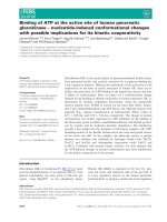
Fig. 3. Testican-1 is a competitive inhibitor of cathepsin L at pH 5.5
and pH 7.2. Testican-1 was added at the indicated concentrations to
cathepsin L incubated at pH 5.5 or at pH 7.2 with concentrations of
Z-Phe-Arg-NHMec between 154 n
M
and 7.7 l
M
. The Lineweaver–
Burk plots show the lines representing reactions at different testican-1
concentrations that all intercept at the y-axis, as expected for com-
petitive inhibition. The error bars represent the standard deviation of
at least three replicates. Obvious outliers were discarded. For each
replicate, the reaction velocity was determined from the linear region of
the curve as the rate of change of fluorescence over a period of at least
200 s. Linear fits for the data at each testican-1 concentration were
generated by linear regression, and all R
2
correlation coefficients were
>0.92.
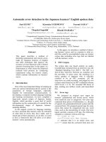
Fig. 2. Testican-1 with or without chondroitin sulfate inhibits cathep-
sin L proteolytic activity. Cathepsin L was incubated with increasing
amounts of testican-1 in either its native form (m) or following treat-
ment with chondroitinase ABC (n). The inhibitory activity is
expressed as residual activity of the enzyme compared to the control
reaction without testican-1 set as 100% activity. Residual activity is
graphed as a function of the molar ratio of testican-1 added to active
cathepsin L present. Each point represents the mean of three repli-
cates; error bars represent the standard deviation of each set of repli-
cates and the line represents the theoretical curve fit. Testican-1
purified with chondroitin sulfate chains intact and testican-1 purified
after chondroitinase ABC digestion were used at the same protein
concentration in the enzymatic assays shown. (Inset) Purification of
recombinant testican-1. A Coomassie-stained SDS/polyacrylamide gel
and a nitrocellulose blot of a parallel gel immunostained for recom-
binant testican-Myc-His
6
show its purification from the culture fluid of
transfected 293 cells. The first lanes show the unfractionated culture
fluid. The second lanes show testican-1 after treatment with chond-
roitinase ABC and purification by Ni-nitrilotriacetic acid affinity. The
migration distances of Rainbow protein molecular mass markers in
these gels are indicated in kDa.
Ó FEBS 2003 Testican-1 inhibits cathepsin L (Eur. J. Biochem. 270) 4011
testican-1 is a secreted proteoglycan. As cathepsin L
denatures rapidly at neutral pH and above [37], data
collection was initiated immediately after addition of
enzyme. Lineweaver–Burk plots of the data established that
testican-1 also inhibits cathepsin L competitively at pH 7.2
(Fig. 3B). The K
m
at pH 7.2 was 1.7 l
M
.TheV
max
was 385
and 72 fluorescence units RFU per second at pH 5.5 and
7.2, respectively.
K
i
was determined at both pH values from the Line-
weaver–Burk plots, as described in Experimental proce-
dures, but as such analysis is thought to produce a K
i
that is
less accurate than nonlinear regression analysis for tight-
binding inhibitors [33], these values are only presented to
compare testican-1¢s inhibitory activity at pH 5.5 to that at
pH 7.2. The K
i
values derived by this method were 13 n
M
at
pH 5.5 and 8 n
M
at pH 7.2. The linear regression fits for
these data had R
2
values greater than 0.9. Thus, testican-1
is similarly effective at inhibiting cathepsin L at pH 5.5 and
at pH 7.2.
Testican-1 enhances cathepsin L stability at neutral pH
At pH 7.2, cathepsin L proteolytic activity in the absence of
testican-1 begins to decline before 10 min (Fig. 4), consis-
tent with the measurements of others [38]. This is not merely
due to depletion of substrate, as indicated by the progress
curve of the control reaction at pH 5.5 in the absence of
testican-1 (A). When cathepsin L activity was assayed near
neutral pH in the presence of testican-1, the loss of activity
was noticeably slower (B). This increase in enzyme stability
was observed at testican-1 concentrations as low as 5 n
M
,a
25 : 1 inhibitor-to-enzyme ratio. Thus at pH values similar
to those outside cells, the presence of testican-1 allows the
enzyme to remain active longer, at the cost of a reduced rate
of proteolytic activity.
Cathepsin L could potentially cleave testican within the
thyropin domain that would thus act as a competitive
substrate, but no change in enzyme velocity was detected
over 20 min, as might be expected were the protease
degrading the inhibitor (Fig. 4A). This is unlikely to have
affected our K
i
determination (Fig. 2) as these experiments
utilized l
M
concentrations of substrate and n
M
concentra-
tions of testican.
Discussion
Testican-1, a secreted proteoglycan with a thyropin domain,
was determined to be a potent competitive inhibitor of the
lysosomal cysteine protease, cathepsin L. At pH 5.5, the
physiological pH for a lysosomal enzyme, the proteoglycan
inhibited the enzyme with a K
i
of 0.7 n
M
.Usingan
alternative method, we also demonstrated that testican-1
was similarly effective as an inhibitor of cathepsin L at
pH 5.5 and at pH 7.2. The affinity of testican-1 for
cathepsin L is similar to that observed for another physio-
logical inhibitor, cystatin B [39], but is significantly lower
than the affinity of the isolated thyropin domain of the p41
form of the MHC invariant chain for cathepsin L, which
has a K
i
of 1.7 · 10
)3
n
M
[36]. Proteins containing thyropin
domains have been found to inhibit a variety of papain-
related cysteine proteases with K
i
values in the low
picomolar to low nanomolar range [15].
Testican-1 is unusual in having multiple specific protease
inhibitor activities within a single polypeptide. We show that
testican-1 inhibits the cysteine protease cathepsin L, while
the N-terminal domain unique to testicans 1–3 has been
shown to inhibit pro-matrix metalloproteinase-2 activation
by membrane-type 1 and 3 matrix metalloproteinases [7]. In
addition, the protein contains a domain homologous to
inhibitors of a third family of proteases, the serine proteases.
Another human gene family with recognizable homologies
to multiple specific protease inhibitors in a single protein has
been recognized recently by data bank homology searches
and one of the domains similar to Kunitz-type protease
inhibitors has been shown to inhibit trypsin [40–42].
Cathepsin L inhibition is a novel activity for the protein
core of proteoglycans and thus expands our appreciation of
the regulatory role of these molecules. The multidomain
structure characteristic of proteoglycans enables them to
interact with various molecules including growth factors,
cell adhesion proteins, and other extracellular matrix
components.
Fig. 4. Testican-1 increases the stability of cathepsin L at neutral pH.
Cathepsin L at 0.2 n
M
was incubated at 30 °Cwith350n
M
Z-Phe-
Arg-NHMec at pH 5.5 (A) and at 7.2 (B) without testican-1 (d)and
with testican-1 at the indicated concentrations (h, e). Each progress
curve is representative of four replicate curves at the given conditions.
As fluorescence was measured immediately after the enzyme was
added to the dithiothreitol-containing reaction mixture on ice, the
initial lag period represents the time required for the enzyme to react
with dithiothreitol and the reaction mixture to reach 30 °C. The arrow
indicates the time by which human cathepsin L was previously shown
to be inactivated when incubated at pH 7 at 30 °Cwiththesame
substrate [38].
4012 J. P. Bocock et al. (Eur. J. Biochem. 270) Ó FEBS 2003
Testican-1 had no significant inhibitory effect on the
endopeptidase activity of a related lysosomal cysteine
protease, cathepsin B. This is consistent with the finding
that the structurally similar p41 thyropin domain inhibits
cathepsin L but does not inhibit cathepsin B [36]. Equistatin
[16] and saxiphilin [15] both inhibit cathepsin B, but they
bind with lower affinity than to cathepsin L. Cathepsin B
differs from cathepsin L in that it has an additional loop of
approximately 20 amino acids which partially occludes the
active site and thus affects interactions with competitive
inhibitors such as stefins [43].
Our experiments establish that addition of the full-length
testican-1 polypeptide results in inhibition of cathepsin L
activity. Specific fragments of the polypeptide can be found
in cerebral spinal fluid [44], blood [45] and human semen [1],
indicating that testican-1 undergoes maturation or process-
ing which might expose, free, or destabilize the thyropin
domain. We observed that a preparation containing
primarily proteolytic fragments of recombinant testican-1
also inhibited cathepsin L activity (data not shown). This is
consistent with isolation of only the thyropin domain of p41
with cathepsin L purified from kidney [13]. This p41
domain is a competitive inhibitor of cathepsin L after it is
cleaved from the invariant chain by endosomal proteases
[36]. The identification in seminal plasma of testican-1
fragments cleaved within the thyropin domain [1] suggests
that this cathepsin L inhibitor can also eventually be
degraded by proteases that may be present in blood [45].
Testican interaction with proteases could be mediated by
the polypeptide backbone of a proteoglycan, by its glycos-
aminoglycans, or by both. Two glycosaminoglycan attach-
ment sites are localized near the C-terminus of testican-1,
at Ser residues 383 and 388 [2]. Significantly, the two
preparations of testican-1, with and without chondroitin
sulfate, were equally efficient inhibitors of the protease,
suggesting inhibition was mediated by the protein core
and not affected by the large glycosaminoglycan moieties.
The high homology of the testican-1 thyropin domain to the
cathepsin L-inhibitory domain of the p41 variant of the
MHC invariant chain is consistent with the conclusion that
cathepsin L inhibition is primarily mediated by protein–
protein interactions.
While cathepsin L is an intracellular protease localized
within lysosomes under normal conditions, the protease is
secreted when expression levels are elevated by cell trans-
formation [19,46,47], in response to signaling [19–21], or
during specific developmental stages [48,49]. The thyropin
domain in testican-1 could serve merely to reduce the
potentially destructive activity of this secreted cysteine
protease. Alternatively, a thyropin domain presented in the
context of a proteoglycan could alter cathepsin L-mediated
proteolysis. There are ample reports of extracellular pro-
teolysis ascribed to cathepsin L, however, it has not been
clear how a protease unstable at neutral pH mediates
extracellular proteolysis. pH-induced unfolding has been
reported to cause rapid inactivation of mature cathepsin L
[38]. While the presence of testican-1 reduced the rate of
enzymatic cleavage of substrate, it also significantly slowed
the expected loss of cathepsin L activity due to denaturation
at neutral pH. Thus, our data suggest that testican-1 may
actually stabilize the mature cathepsin L protease, so that its
half-life is increased, although its velocity is reduced.
A role for testican-1 in inhibiting, yet also stabilizing,
protease activity is completely consistent with the recent
findings that the p41 alternatively spliced variant of the
MHC invariant chain is not merely an inhibitor of
cathepsin L activity but also serves as a chaperone that
helps to maintain a pool of active protease in late-endocytic
compartments of antigen presenting cells [50]. Precedent
for this role comes from the observation that coexpression
of p41 with p31 modifies endosomal proteolysis of p31 [51].
Cathepsin L activity is also stabilized extracellularly when
this p41–enzyme complex is secreted by activated macro-
phages [52]. Heparin-like glycosaminoglycans have recently
been reported to protect human cathepsin B from
pH-induced inactivation in vitro [53], while heparan sulfate
on ectodomains of cell membrane proteoglycans shed to
wound fluids are known to protect serine proteases from
interaction with their endogenous inhibitors, thus modify-
ing the proteolytic balance of the fluid [54]. This physio-
logical modulation of proteolysis primarily depends on
protease interactions with the glycosaminoglycans of
proteoglycans and does not require specific protein–protein
interaction as occurs between testican-1 and cathepsin L.
Through regulation of testican-1 expression levels, the
more specific protein–protein interaction may spatially and
temporally control the activity of secreted cathepsin L,
allowing the enzyme to serve multiple, specific roles in
different tissues.
Acknowledgements
We thank Dr Tom Traut for his expert advice on enzyme kinetics, Dr
Mike Caplow for helpful suggestions, Susan Jones for assistance with
the fluorescence microplate reader, and Dr Mohammad BaSalamah for
stimulating the initiation of this study. This work was supported in part
by National Institutes of Health RO1 HL55452 to C J. E and by a
University of North Carolina Medical Faculty Award to A. E.
References
1. Bonnet, F., Perin, J P., Maillet, P., Jolles, P. & Alliel, P.M. (1992)
Characterization of a human seminal plasma glycosaminoglycan-
bearing polypeptide. Biochem. J. 288, 565–569.
2. Alliel, P.M., Pedrin, J P., Jolles, P. & Bonnet, F.J. (1993) Testi-
can, a multidomain testicular proteoglycan resembling modulators
of cell social behaviour. Eur. J. Biochem. 214, 347–350.
3. Rieber, A.J., Marr, H.S., Comer, M.B. & Edgell, C.J.S. (1993)
Extent of differentiated gene expression in the human endo-
thelium-derived EA.hy926 cell line. Thrombo. Haemost. 69,
476–480.
4. Marr, H.S., Basalamah, M.A. & Edgell, C J. (1997) Endothelial
cell expression of testican mRNA. Endothelium 5, 209–219.
5. Bonnet, F., Perin, J.P., Charbonnier, F., Camuzat, A., Roussel,
G., Nussbaum, J.L. & Alliel, P.M. (1996) Structure and
cellular distribution of mouse brain testican. J. Biol. Chem. 271,
565–569.
6. Vannahme, C., Schubel, S., Herud, M., Gosling, S., Hulsmann,
H., Paulsson, M., Hartmann, U. & Maurer, P. (1999) Molecular
cloning of testican-2: defining a novel calcium-binding pro-
teoglycan family expressed in brain. J. Neurochem. 73, 12–20.
7. Nakada, M., Yamada, A., Takino, T., Miyamori, H., Takahashi,
T., Yamashita, J. & Sato, H. (2001) Suppression of membrane-
type 1 matrix metalloprinase (MMP) -mediated MMP-2 activa-
tion and tumor invasion by testican 3 and its splicing variant gene
product, N-Tes. Cancer Res. 61, 8896–8902.
Ó FEBS 2003 Testican-1 inhibits cathepsin L (Eur. J. Biochem. 270) 4013
8. Greene, L.J., DiCarol, J.J., Sussman, A.J. & Bartelt, D.C. (1968)
Two trypsin inhibitors from porcine pancreatic juice. J. Biol.
Chem. 243, 1804–1815.
9. Laskowski, M. & Kato, I. (1980) Protein inhibitors of proteinases.
Ann. Rev. Biochem. (Snell, E.E., Boyer, P.D., Meister, A. &
Richardson, C.C., eds), pp. 593–626. Annual Reviews Inc., Palo
Alto, CA.
10.Kohfeldt,E.,Maurer,P.,Vannahme,C.&Timpl,R.(1997)
Properties of the extracellular calcium binding module of the
proteoglycan testican. FEBS Lett. 414, 557–561.
11. Lenarcic, B. & Bevec, T. (1998) Thyropins – new structually
related proteinase inhibitors. Biol. Chem. 379, 105–111.
12. Malthiery, Y. & Lissitzky, S. (1987) Primary structure of human
thyroglobulin deduced from the sequence of its 8448-base com-
plementary DNA. Eur. J. Biochem. 165, 491–498.
13. Ogrinc, T., Dolenc, I., Ritonja, A. & Turk, V. (1993) Purification
of the complex of cathepsin L and the MHC class II-associated
invariant chain fragment from human kidney. FEBS Lett. 336,
555–559.
14. Guncar,G.,Pungercic,G.,Klemencic,I.,Turk,V.&Turk,D.
(1999) Crystal structure of MHC class II-associated p41, Ii frag-
ment bound to cathepsin L reveals the structural basis for differ-
entiation between cathepsins L and S. EMBO J. 18, 793–803.
15. Lenarcic, B., Krishnan, G., Borukhovich, R., Ruck, B., Turk, V.
& Moczydlowski, E. (2000) Saxiphilin, a saxitoxin-binding protein
with two thyroglobulin type 1 domains, is an inhibitor of papain-
like cysteine proteinases. J. Biol. Chem. 275, 15572–15577.
16. Lenarcic, B., Ritonja, A., Strukelj, B., Turk, B. & Turk, V. (1997)
Equistatin, a new inhibitor of cysteine proteinases from Actinia
equina, is structurally related to thyroglobulin type-1 domain.
J. Biol. Chem. 272, 13899–13903.
17. Kornfeld, S. & Mellman, I. (1989) The biogenesis of lysosomes.
Annu. Rev. Cell Biol. 5, 483–525.
18. Yeyeodu, S., Ahn, K., Madden, V., Chapman, R., Song, L. &
Erickson, A. (2000) Procathepsin L self-association as a mechan-
ism for selective secretion. Traffic 1, 724–737.
19. Ahn, K., Yeyeodu, S., Collette, J., Maden, V., Arthur, J., Li, L. &
Erickson, A.H. (2002) An alternate targeting pathway for pro-
cathepsin L in mouse fibroblasts. Traffic 3, 147–159.
20. Andrews, N.W. (2000) Regulated secretion of conventional lyso-
somes. Trends Cell Biol. 10, 316–320.
21. Blott, E.J. & Griffiths, G.M. (2002) Secretory lysosomes. Nat. Rev.
Mol. Cell Biol. 3, 122–131.
22. Chapman, H., Riese, R.J. & Shi, G P. (1997) Emerging roles for
cysteine proteases in human biology. Annu. Rev. Physiol. 59,
63–88.
23. Ishidoh, K. & Kominami, E. (1998) Gene regulation and extra-
cellular functions of procathepsin L. Biol. Chem. 379, 131–135.
24. Turk, B., Turk, D. & Turk, V. (2000) Lysosomal cysteine proteases:
more than scavengers. Biochim. Biophys. Acta. 1477, 98–111.
25. Kane, S.E. & Gottesman, M.M. (1990) The role of cathepsin L in
malignant transformation. Sem. Cancer Biol. 1, 127–136.
26. Briozzo, P., Morisset, M., Capony, F., Rougeot, C. & Rochefort,
H. (1988) In vitro degradation of extracellular matrix with Mr
52,000 cathepsin D secreted by breast cancer cells. Cancer Res. 48,
3688–3692.
27. Ishidoh, K. & Kominami, E. (1995) Procathepsin L degrades
extracellular matrix proteins in the presence of glycosaminogly-
cans in vitro. Biochem. Biophys. Res. Comm. 217, 624–631.
28. Maciewicz, R.A., Etherington, D.J., Kos, J. & Turk, V. (1987)
Collagenolytic cathepsins of rabbit spleen: a kinetic analysis of
collagen degradation and inhibition by chicken cystatin. Collagen
Relat. Res. 7, 295–304.
29. Mason, R.W., Johnson, D.A., Barrett, A.J. & Chapman, H.A.
(1986) Elastinolytic activity of human cathepsin L. Biochem. J.
233, 925–927.
30. Buck, M.R., Karustis, D.G., Day, N.A., Honn, K.V. & Sloane,
B.F. (1992) Degradation of extracellular-matrix proteins by
human cathepsin B from normal and tumour tissues. Biochem. J.
282, 273–278.
31. Barrett, A.J. & Kirschke, H. (1981) Cathepsin B, cathepsin H, and
cathepsin L. Methods Enzymol. 80, 535–561.
32. Bieth, J.G. (1995) Theoretical and practical aspects of protease
inhibition kinetics. Methods Enzymol. 248, 59–84.
33. Bieth, J.G. (1984) In vivo significance of kinetic constants of pro-
tein proteinase inhibitors. Biochem. Med. 32, 387–397.
34. Segel, I.H. (1975) Enzyme Kinetics. John Wiley & Sons, New
York.
35. Kirschke, H., Barrett, A.J. & Rawlings, N.D. (1998) Lysosomal
Cysteine Proteases. 2nd edn. Oxford University Press, Oxford.
36. Bevec, T., Stoka, V., Pungercic, G., Dolenc, I. & Turk, V. (1996)
Major histocompatibility complex class II-associated p41
invariant chain fragment is a strong inhibitor of lysosomal
cathepsin L. J. Exp. Med. 183, 1331–1338.
37. Dehrmann, F.M., Coetzer, T.H.T., Pike, R.N. & Dennison, C.
(1995) Mature cathepsin L is substantially active in the ionic
milieu of the extracellular medium. Arch. Biochem. Biophys. 324,
93–98.
38. Mason, R.W., Green, G.D.J. & Barrett, A.J. (1985) Human liver
cathepsin L. Biochem. J. 226, 233–241.
39. Barrett, A.J., Rawlings, N.D., Davies, M.E., Machleidt, W.,
Salvesen, G. & Turk, V. (1986) Cysteine proteinase inhibitors of
the cystatin superfamily. In Proteinase Inhibitors (Barrett, A.J. &
Salvesen, G., eds), pp. 515–569. Elsevier, Amsterdam.
40. Trexler, M., Banyai, L. & Patthy, L. (2001) A human protein
containing multiple types of protease-inhibitory modules. Proc.
Natl. Acad. Sci. USA 98, 3705–3709.
41. Trexler, M., Banyai, L. & Patthy, L. (2002) Distinct expression
pattern of two related human proteins containing multiple types of
protease-inhibitory modules. Biol. Chem. 383, 223–228.
42. Nagy, A., Trexler, M. & Patthy, L. (2003) Expression, purification
and characterization of the second Kunitz-type protease inhibitor
domain of the human WFIKKN protein. Eur. J. Biochem. 270,
2101–2107.
43. Lenarcic, B., Krizaj, I., Zunec, P. & Turk, V. (1996) Differences in
specificity for the interactions of stefins A, B and D with cysteine
proteinases. FEBS Lett. 395, 113–118.
44. Stark, M., Danielsson, O., Griffiths, W.J., Jornvall, H. &
Johansson, J. (2001) Peptide repertoire of human cerebrospinal
fluid: novel proteoglytic fragments of neuroendocrine proteins.
J. Chromatog. B. 754, 357–367.
45. BaSalamah, M.A., Marr, H.S., Duncan, A.W. & Edgell, C.J.
(2001) Testican in human blood. Biochem. Biophys. Res. Comm.
283, 1083–1090.
46. Gottesman, M.M. (1978) Transformation-dependent secretion of
a low molecular weight protein by murine fibroblasts. Proc. Natl
Acad. Sci. USA 75, 2767–2771.
47. Prence, E.M., Dong, J. & Sahagian, G.G. (1990) Modulation of
the transport of a lysosomal enzyme by PDGF. J. Cell Biol. 110,
319–326.
48. Erickson-Lawrence, M., Zabludoff, S.D. & Wright, W.W.
(1991) Cyclic protein-2, a secretory product of rat Sertoli cells, is
the proenzyme form of cathepsin L. Mol. Endocrinol. 5, 1789–
1798.
49. Jaffe, R.C., Donnelly, K.M., Mavrogianis, P.A. & Verhage, H.G.
(1989) Molecular cloning and characterization of a progesterone-
dependent cat endometrial secretory protein complementary
deoxyribonucleic acid. Mol. Endocrinol. 3, 1807–1814.
50. Lennon-Dumenil, A M., Bryant, A.R., Valentijn, K., Driessen,
C.,Overkleeft,H.S.,Erickson,A.,Peters,P.J.,Bikoff,E.,Ploegh,
H.L. & Bryant, P.W. (2001) The p41 isoform of invariant chain is
a chaperone for cathepsin L. EMBO J. 29, 4055–4064.
4014 J. P. Bocock et al. (Eur. J. Biochem. 270) Ó FEBS 2003
51. Fineschi, B., Arneson, L.S., Naujokas, M.F. & Miller, J. (1995)
Proteolysis of major histocompatibility complex class II-asso-
ciated invariant chain is regulated by the alternatively spliced gene
product, p41. Proc. Natl Acad. Sci. USA 92, 10257–10261.
52. Fiebiger, E., Maehr, R., Villadangos, J., Weber, E., Erickson,
A.H.,Bikoff,E.,Ploegh,H.L.&Lennon-Dumenil,A M.(2002)
Invariant chain controls the activity of extracellular cathepsin L.
J. Exp. Med. 196, 1263–1270.
53. Almeida, P.C., Nantes, I.L., Chagas, J.R., Rizza, C.C., Faljoini-
Alario, A., Carmona, E., Juliano, L., Nader, H.B. & Tersariol, I.L.
(2001) Cathepsin B activity regulation. Heparin-like glycosami-
noglycans protect human cathepsin B from alkaline pH-induced
activation. J. Biol. Chem. 276, 944–951.
54. Kainulainen, V., Wang, H., Schick, C. & Bernfield, M. (1998)
Syndecans, heparan sulfate proteoglycans, maintain the proteo-
lytic balance of acute wound fluids. J. Biol. Chem. 273, 11563–
11569.
55. Hohenester, E., Maurer, P. & Timpl, R. (1997) Crystal structure of
a pair of follistatin-like and EF-hand calcium-binding domains in
BM-40. EMBO J. 16, 3778–3786.
Ó FEBS 2003 Testican-1 inhibits cathepsin L (Eur. J. Biochem. 270) 4015