Báo cáo Y học: Purification, crystallization, NMR spectroscopy and biochemical analyses of a-phycoerythrocyanin peptides pptx
Bạn đang xem bản rút gọn của tài liệu. Xem và tải ngay bản đầy đủ của tài liệu tại đây (271.87 KB, 10 trang )
Purification, crystallization, NMR spectroscopy and biochemical
analyses of a-phycoerythrocyanin peptides
Georg Wiegand
1
, Axel Parbel
2
*, Markus H. J. Seifert
1
, Tad A. Holak
1
and Wolfgang Reuter
1
1
Max-Planck-Institut fu
¨
r Biochemie, Martinsried, Germany;
2
Botanisches Institut der Ludwig-Maximilians-Universita
¨
t, Mu
¨
nchen,
Germany
The a-phycoerythrocyanin subunits of the different phy-
coerythrocyanin complexes of the phycobilisomes from
the cyanobacterium Mastigocladus laminosus perform a
remarkable photochemistry. Similar to phytochromes –
the photoreceptors of higher plants – the spectral pro-
perties of the molecule reversibly change according to the
irradiation wavelength. To enable extensive analyses, the
protein has been produced at high yield by improving
purification protocols. As a result, several comparative
studies on the Z-andE-configurations of the intact
a-subunit, and also on photoactive peptides originating
from nonspecific degradations of the chromoprotein, were
possible. The analyses comprise absorbance, fluorescence
and CD spectroscopy, crystallization, preliminary X-ray
measurements, mass spectrometry, N-terminal amino acid
sequencing and 1D NMR spectroscopy. Intact a-phyco-
erythrocyanin aggregates significantly, due to hydrophobic
interactions between the two N-terminal helices. Removal
of these helices reduces the aggregation but also desta-
bilizes the protein fold. The complete subunit could be
crystallized in its E-configuration, but the X-ray meas-
urement conditions must be improved. Nevertheless,
NMR spectroscopy on a soluble photoactive peptide
presents the first insight into the complex chromophore
protein interactions that are dependent on the light
induced state. The chromophore environment in the
Z-configuration is rigid whereas other regions of the
protein are more flexible. In contrast, the E-configuration
has a mobile chromophore, especially the pyrrole ring D,
while other regions of the protein rigidified compared to
the Z-configuration.
Keywords: phycobilisomes; phycoerythrocyanin; protein
structure; photochemistry; energy transfer.
Phycobiliproteins are a class of chromoproteins bearing
covalently bound linear tetrapyrrole (phycobilins) chro-
mophores. They are predominantly involved in the photo-
synthetic light harvesting process of cyanobacteria and
certain algae. With respect to this function they are
assembled to supramolecular protein pigment complexes,
i.e. phycobilisomes, building up a highly ordered network of
very rigid chromophores which enable an energy transfer
efficiency of almost 100% [1,2].
The phycoerythrocyanin (PEC) complexes located at the
periphery of phycobilisomes are present in only a few
species of cyanobacteria [3]. PEC of the thermophilic
cyanobacterium Mastigocladus laminosus is the best char-
acterized complex of this biliprotein class. Like other
peripheral phycobiliproteins, e.g. phycoerythrin, low light
and high temperature conditions induce a maximum
content of PEC, reaching approximately 30% of total
protein content within the phycobilisomes [4,5]. The X-ray
structure of PEC reveals three heterodimeric a,b substruc-
tures, so called ‘monomers’, which are associated to a ring
shaped disc, designated as ‘trimer’. The b-subunit contains
two phycocyanobilin (PCB) chromophores, whereas the
a-subunit has a single phycoviolobilin (PVB) chromophore
of which the pyrrole ring A is covalently linked via a
thioether bond to Cys84 [6].
Unlike other phycobiliproteins, the PVB chromophores
of the PEC-‘trimers’ present a remarkable reversible
photochemistry which has been reported first by Bjo
¨
rn
[7]. The observation initiated intense investigations into this
unusual spectroscopic behavior, especially regarding the
possible function as a sensor pigment [8–10]. The sensor
function seems to be questionable, however, in particular
because of the high content and extremely reduced
photochemistry of PEC within the phycobilisomes
[4,11,12]. Biochemical and spectral data assign the photo-
chemistry exclusively to the a-PEC subunit [13,14]. Similar
to phytochrome and phytochrome-like photoreceptors of
higher plants and cyanobacteria, the PVB chromophore
undergoes spectral and molecular changes depending on
light quality [15,16]. The phototransformation of a-PEC is
reflected by the reversible shift in the visible absorption
maximum from 505 to 570 nm and the two states were
termed E and Z, respectively. Isomerization can be
performed by irradiation with complementary chromatic
light and the two states are quite stable in the dark. The
molecular mechanism of the photoreaction is similar to
that of the phytochromes and is proposed to exists as a
chromophore twisting around the D15,16 double bond
between the C and D pyrrole rings [9,16]. However, the
Correspondence to W. Reuter, Max-Planck-Institut fu
¨
rBiochemie,
Am Klopferspitz 18 A, 82152 Martinsried, Germany.
Fax: + 49 (0)89 85783516, Tel.: + 49 (0)89 85782707,
E-mail:
Abbreviations: FID, free induction decay; HIC, hydrophobic interac-
tion chromatography; MPD, 2-methyl-2,4-pentanediol; PCB, phyco-
cyanobilin; PEC, phycoerythrocyanin; PVB, phycoviolobilin.
*Present address: Amersham Biosciences, Munzinger Str. 9, 79111
Freiburg, Germany.
(Received 4 June 2002, revised 28 August 2002,
accepted 29 August 2002)
Eur. J. Biochem. 269, 5046–5055 (2002) Ó FEBS 2002 doi:10.1046/j.1432-1033.2002.03221.x
situation may be more complicated in a-PEC, as at least
type I and type II Z/E-isomerizations have been spectrosc-
opically discerned. The ratio of the two types has been
suggested to be controlled by sulfhydryls of Cys98 and
Cys99 in the protein [8,10]. Whereas, the structures of the
PVB isomers are well defined by spectroscopical charac-
terizations, the participitation of the protein in the
phototransformation process of the a-subunit is almost
unknown. Some reasons for this may be the considerable
difficulties in the preparative purification of a-PEC and the
problems arising during analyses of the protein at high
concentrations.
The present study describes a very effective method of
isolation and purification of PEC and its photoactive
a-subunit. Crystallization and preliminary X-ray experi-
ments suggest pronounced conformational alterations of
both the protein and the PVB chromophore, depending on
light quality. N-Terminal amino acid analyses and mass
spectrometry characterized a series of a-PEC peptides
which occurred during storage in formic acid. The Z-and
E-configurations of one chromopeptide with an excellent
solubility and stability were investigated by 1D NMR
spectroscopy. The information presented here are mainly
focused on the preparations, handling and characterizations
of a-PEC peptides which will be a prerequisite for successful
studies on the detailed molecular events during phototrans-
formation.
MATERIALS AND METHODS
Strain, culturing conditions and isolation
of phycobilisomes
The thermophilic cyanobacterium M. laminosus (syn.
Fischerella sp. PCC 7603) was photoautotropically grown
at 48 °C, 10 lEÆm
)2
Æs
)1
andgassedwith2%(v/v)CO
2
enriched air. The cells were harvested when an optical
density of 0.7 at 740 nm was reached. At these conditions
the phycobilisomes are attributed by the maximum content
of PEC [4].
The phycobilisomes were isolated by step-gradient cen-
trifugation principally following the buffer conditions
described by Reuter and Wehrmeyer [17]. Scaling up of
the phycobilisome preparation was necessary for the
development of the effective purification procedure of the
a-subunit. Cells of M. laminosus with a wet weight of 40 g
were disrupted at 17 °C in a self-constructed cell mill. The
glass beads (1 mm) were filtered off and the filtrate of about
180 mL was incubated with 4% (w/v) N,N-dimethyldode-
cylamine N-oxide (Fluka, Buchs, Switzerland) for 1 h at
17 °C. This homogenate was clarified by centrifugation at
48 000 g for 30 min at 17 °C. The resulting supernatant of
approximately 180 mL was directly applied to step-gradi-
ents comprising 10 mL 40% (w/v), 15 mL 30% (w/v),
15 mL 20% (w/v) and 15 mL 10% (w/v) sucrose, respect-
ively. Centrifugation was performed in a Ti45 rotor
(Beckman Coulter, USA) at 40 000 g for 16 h at 17 °C.
The separated phycobilisomes banding between 40% (w/v)
and 30% (w/v) sucrose were eluted and subsequently
precipitated by a final concentration of 2.0
M
potassium
phosphate, pH 7.0. These products are stable at 4 °Cforat
least 2 years without any changes in their protein compo-
sition.
Purification of PEC complexes
About 1000 mg of precipitated phycobilisomes were sedi-
mented by centrifugation at 74 000 g for 30 min at 17 °C.
The sediment was resolved in distilled water and dissoci-
ation was performed by gel filtration on a Sephadex G25
column (Amersham Biosciences, 40 mm · 250 mm) and
equilibrated with 5 m
M
potassium phosphate, pH 7.0. The
eluted phycobilisomes were applied directly to an anion
exchange column (40 mm · 150 mm) packed with DEAE-
Trisacryl M (Serva, Heidelberg, Germany) and equilibrated
with the dissociation buffer. At this pH and ionic strength
the PEC complexes with and without linker polypeptides
eluted completely, whereas all other biliprotein complexes
remained on the column. The resulting PEC fraction of
about 250 mg was precipitated with a final concentration of
2.0
M
potassium phosphate, pH 7.0.
Purification of the a-PEC subunit
Sedimented PEC was resolved in distilled water and the
biliprotein complexes were dissociated by gel filtration on a
PD-10 column (Amersham Biosciences) in 0.3% (v/v)
formic acid. The dissociation into a-andb-subunits is
accompanied by the loss of the excitonic coupling between
the corresponding chromophores. Therefore, the complete-
ness of dissociation can be followed by the color change of
PEC from pink to blue. Separation of the subunits from
each other was obtained by isocratic hydrophobic interac-
tion chromatography in 0.3% (v/v) formic acid on a
column (10 mm · 30 mm) packed with isopropyl-substi-
tuted Sephacryl-S300 (Amersham Biosciences). The proce-
dure of the isopropyl substitution of the gel will be
published elsewhere. During the chromatography, the
a-subunits show negligible interactions with the gel,
whereas the elution of b-subunits and linker polypeptides
is strongly retarded. The eluted homogeneous a-PEC
fraction of at least 80 mg was concentrated by ultrafiltra-
tion up to 20 mgÆmL
)1
.
Figure 1 summarizes the purification steps which are
necessary for the isolation of the intact, photoactive
a-subunit. Some of the different steps are based on previous
methods, e.g. induction of the maximal PEC content [4,5],
anion exchange chromatography on DEAE [18], or use of
formic acid as isolation medium [19].
Nevertheless, some advantages of the preparation meth-
ods should be described. Based on a maximal phycobilisome
concentration of 30 mgÆmL
)1
within the cells [20], the
isolation yield of about 80% of intact phycobilisomes is very
high. The effectiveness of the cell breakage (¼ 95%) and the
complete precipitation of the phycobilisomes without dis-
sociation are responsible for the extraordinary high yield
(results not shown). A previous study described an unusual
fractionation of PEC on DEAE–cellulose [18]. This non-
specific separation was not observed on DEAE-Trisacryl
M, therefore the elution time and dilution of the sample was
considerably reduced. In addition, short-time dissociation
by gel filtration with formic acid and the fast separation by
hydrophobic interaction chromatography (HIC) reduce
the time consumption and the denaturation effects. How-
ever, the most important step was the chromatography on
the isopropyl-substituted Sephacryl-S300 minimizing non-
specific interactions of a-PEC with the gel matrix.
Ó FEBS 2002 Analyses of a-phycoerythrocyanin peptides (Eur. J. Biochem. 269) 5047
Optical spectroscopy
The spectra of a-PEC were recorded after saturated
irradiation with 577 nm light transforming the E-isomer
and 500 nm light transforming the Z-isomer, respectively.
Absorbance spectra were measured with a Lambda 2 UV/
visible spectrophotometer (Perkin-Elmer) and circular
dichroism (CD) was recorded on a Dichrograph VI (ISA).
The spectral band width was 0.25 nm, the scan speed
5nmÆs
)1
. Fluorescence spectra were recorded with 2 nm
resolution on a Spex model 221 fluorimeter. Details of the
measurements are described by Parbel et al. [21].
Crystallization of a-PEC
Unless otherwise stated, all preparations were carried out
under red light with an emittance maximum of 650 nm
(Phillips, the Netherlands; TLD 15). Crystallization growth
was controlled in a modified microscope at a wavelength of
620 nm. Parallel crystallization experiments were conducted
after transforming the a-PEC in the E-andZ-state,
respectively. The state of the two isomers was monitored
by absorbance spectrometry in the range of 250–650 nm.
Using the vapor diffusion hanging-drop method, the
proteins were crystallized in a pH range of 4.0–8.5 because
at these pH values both protein and chromophore show a
high stability. Crystallization of the E-form could only be
observed in the presence of different salts, e.g. ammonium
sulfate, ammonium phosphate, sodium-potassium phos-
phate or Tris phosphate, as precipitants. Other precipitants
like poly(ethylene glycol) or 2-methyl-2,4-pentanediol
(MPD) have not, as yet, been found to be successful.
Crystals of the Z-form have never been obtained, although
the crystallization conditions of both protein states have
been identical. Thin plates with dimensions of approxi-
mately 0.2 · 0.6 · 0.3 mm of the E-isomer grown in the
presence of 1.0
M
dibasic Tris phosphate, pH 8.0, at 18 °C
were analyzed by diffraction studies and mass spectrometry.
For cryo-measurements the crystals were transferred into
3
M
Tris phosphate, pH 8.0, which serves as cryo-protec-
tant. The crystals were frozen directly in liquid nitrogen and
the X-ray diffractions were recorded under white room-light
at temperatures between )140 and )160 °C.
‘Native’ PAGE
PAGE was performed in 3-mm thick 10% (w/v) polyacryl-
amide slab gels with Tris/boric acid (42 m
M
/100 m
M
,
pH 7.9). Gels were polymerized with 0.1% (v/v) tetrameth-
ylethylenediamine and 0.03% (w/v) ammonium peroxodi-
sulfate [17]. Samples of 1.5 mL containing 10–15 mgÆmL
)1
protein were electrophoresed in the Tris/boric acid buffer
system for 2400 VÆh
)1
at a constant power of 18 W, at
10 °C and continuous buffer circulation in a DESAGA
VA-200 apparatus (DESAGA, Germany). After separ-
ation, the protein bands were cut out and the gel slices were
squeezed between two glass plates. The homogeneous gel
pastes were eluted for 3 h under continuous stirring with a
10-fold volume of water. After elution the homogenates
were centrifuged for 1 h at 75 000 g and the supernatants
were filtered through a 0.22-lm poly(vinylidene difluoride)
membrane (Millipore, USA) [22]. The peptides were con-
centrated by ultrafiltration and the photoactivity was tested
by absorbance spectra after alternative irradiation with the
two light qualities.
Mass spectrometry and N-terminal amino acid sequencing
Mass spectrometry of the a-PEC peptides originating from
the preparation of the hydrophobic interaction column, the
crystals and the ‘native’ PAGE was performed by the
electrospray method in a single quadrupol mass spectro-
meter API165 (Applied Biosystems, Langen, Germany).
The spectra were deconvoluted with the
BIOTOOL
software
of the manufacturer. Removal of salts within the samples
was performed by hydrophobic interaction chromato-
graphy on ReproSil-Pur C18-AQ, 3l,1· 150 mm (Dr
A. Maisch, Ammerbuch, Germany). The peptides were
eluted with a gradient from 10% (v/v) trifluoroacetic acid in
H
2
O to 0.08% (v/v) trifluoroacetic acid in acetonitrile.
All sequences were determined with an Applied Biosys-
tems sequencer model 473 A following the manufacturer’s
instructions.
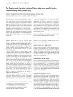
Structural comparison of the intact a-PEC
and the degraded a-PEC peptide 2
The picture comparing the secondary structures of the
intact subunit and peptide 2 was produced with the 3D
Fig. 1. Overview of the isolation and purification steps of a-PEC from
Mastigocladus laminosus. Approximately 250 mg of PEC-linker com-
plexes could be separated from 1000 mg of phycobilisomes. The pre-
cipitated PEC-fraction can be stored without alteration at least for a
period of 2 years. Starting with 250 mg of the linker-PEC complexes,
the purification at dissociating conditions in 0.3% (v/v) formic acid
results in a final preparation of approximately 80 mg of homogeneous
a-PEC.
5048 G. Wiegand et al. (Eur. J. Biochem. 269) Ó FEBS 2002
visualization program ‘WebLab ViewerPro, Version 3.20’
(Molecular Simulations Inc.). The coordinates derived from
the structure analyses of phycoerythrocyanin [6].
Light-dependent 1D NMR spectroscopy
of a-PEC peptide 2
Prior to NMR measurements, peptide 2 in 20 m
M
sodium-
potassium phosphate, pH 7.2 with a protein concentration
of 15–20 mgÆmL
)1
was irradiated with light of 571 and
503 nm inducing the E-andZ-configuration, respectively.
The complete transfer into both configurations was
obtained by an illumination time of 1 h. Continuous
spinning of the NMR tube minimized the self-shadowing
of the highly concentrated sample.
1
H-NMR measurements
were carried out in the dark without spinning on a Bruker
DRX600 spectrometer equipped with a
1
H-
13
C-
15
Ntriple-
resonance probehead including triple-axis gradients. All
spectra were recorded at a temperature of 27 °C. To
suppress the water resonance a jump-return pulse sequence
was used [23]. For each spectrum 512 free induction decays
(FIDs) with 32 k time domain points comprising a sweep
width of 9 kHz were recorded. The interscan delay was set
to 1 s. The 90° pulse was determined to be 8.4 ls. The
spectra were processed by fast Fourier transformation
including a Gaussian window function and digital filtering
of low frequencies in the range of 1.5 p.p.m. to enhance
water suppression. Only 12 k of the recorded 32 k time
domain points were used for transformation to increase
signal-to-noise ratio. The final spectra were processed to
32 k frequency domain points.
RESULTS
Spectral behavior of a-PEC in 0.3% (v/v) formic acid
The steady state absorption, fluorescence and CD spectra of
a-PEC depending on pre-illumination are represented in
Fig. 2. The a-subunit in the E-configuration is characterized
by a long wavelength absorbance maximum at 503 nm with
a pronounced shoulder near 566 nm, an extremely low
fluorescence and a CD minimum near 505 nm. The sharp
peak (arrowhead) in the fluorescence spectrum originates
from the excitation light. The absorbance shoulder near
566 nm, the broad fluorescence maximum at 588 (arrow) and
the minimum in the CD spectrum near 325 nm are typical for
signals of a-PEC in the Z-configuration. Therefore, the
presence of these signals in the spectra of the E-isomer
indicates an incomplete transformation of the molecule or at
least of the chromophore. In contrast, the Z-configuration of
a-PEC reveals uniform maxima at 566 nm (absorbance),
588 nm (fluorescence), 566 nm (CD) and a minimum at
329 nm (CD). The spectral data of the proteins in the E-as
well as the Z-state in 0.3% (v/v) formic acid are nearly equal
to those in conventional buffers near pH 7.0 [9,10,12]. Thus,
the ‘native’ state of the chromoprotein has been assumed.
Crystallization of a-PEC
In order to obtain information about changes of the
polypeptide properties in the Z-andE-state, respectively,
crystallization was performed with the protein in both
configurations. One major problem was the aggregation
Fig. 2. Optical spectroscopy of the E- and Z-configurations of the
a-subunit in 0.3% (v/v) formic acid. The spectral behavior of the
chromoprotein in is nearly identical to that at neutral pH which con-
firms the suitability of the isolation and purification method. It must
be noted that the chromophore cannot completely be transferred into
the E-configuration. This is shown by the arrows in the absorbance,
fluorescence and CD spectra. The fluorescence of a-PEC in the
E-configuration is extremely low, therefore the excitation light, marked
by an arrowhead, is seen in the spectrum.
Ó FEBS 2002 Analyses of a-phycoerythrocyanin peptides (Eur. J. Biochem. 269) 5049
behavior of the protein at pH values near 7.0, especially in
the Z-configuration. Therefore, variations in the protein
concentrations (5–7.5 mgÆmL
)1
) were strongly limited. The
crystallization behavior of the E-isomer is identical in the
dark and in green light (results not shown). This was tested
by parallel crystallization attempts in the dark and under
weak monochromatic green light. All common precipitants
have been used but only different salts at varying ionic
strength and pH values have been successful. Two typical
crystallization conditions comparing the E-andthe
Z-configurations are demonstrated in Fig. 3. Despite the
identical crystallization conditions, only the E-configuration
crystallized (Fig. 3a,b), whereas the Z-configuration always
showed a type of phase separation (Fig. 3c,d). The branch-
ing of the crystals occurred under nearly all conditions,
however, the size of single, homogeneous crystal plates was
sufficient for further analyses.
Unfortunately, X-ray analyses of such plates were
unsuccessful because the diffraction of the crystals decreased
very rapidly during the measurements. This phenomenon
has been observed for different crystals, even at low
temperatures between )140 and )160 °C. Because of the
extreme changes in the diffraction patterns, a unique
determination of the space group and the unit cell was not
possible. Nevertheless, within the limits of the measure-
ments, we tentatively determined an orthorhombic space
group with two molecules in the asymmetric unit. What is
the reason of the strongly decreasing diffractions? The
frozen crystals have been mounted and measured under
white room-light. At this condition, light-induced conform-
ational changes which destroy the well ordered crystal
packing might be possible. The molecular flexibility of
different crystallized phycobiliproteins at temperatures in
the range from )100 to )160 °C has frequently been
observed during the freezing and measuring procedures
(Reuter, unpublished results). In addition, different inter-
mediate chromophore states of PEC were recorded depend-
ing on the measuring temperatures [24,25]. The results
clearly demonstrate the molecular mobility of phycobili-
proteins, even at low temperatures, but the influence of light
on the crystal packing of a-PEC during the measurements
remains uncertain.
Purification and analyses of a-PEC peptides
The storage time of a-PEC in 0.3% (v/v) formic acid at 4 °C
was approximately 6 months. At the end of this time, the
crystals shown in Fig. 3 could not be reproduced. This fact
initiated an analysis of the sample by mass spectrometry
revealing at least seven peptides with molecular weights
between 16 000 and 14 000 Da (results not shown). At
present, the reasons for the degradation are uncertain. A
proteolytic splitting of the a-subunit by proteases may be
possible, although the pH of 2.2 of the formic acid probably
inhibits the activity of most peptidases. Another postulation
is the acid-induced degradation of the a-PEC during long-
term storage. Specific acid-catalyzed degradation reactions
have previously been reported for other proteins [26]. The
most probable explanation would be a nonspecific acid-
catalyzed hydrolysis of a-PEC which is facilitated by a
partial unfolding of the two N-terminal helices (Fig. 4). The
resulting high flexibility of this peptide region may be
responsible for destabilization of favored peptide bonds
within the protein. This view is in line with the variability of
the amino acid sequences for which the degradation occurs.
However, cooperation between the three mechanisms
cannot be excluded. Further studies on the instability of
the isolated a-subunit are in progress and some aspects will
be stressed in the discussion section.
Fig. 3. Crystallization of a-PEC has been successful only with the
molecule in the E-configuration (a,b). In principle, all crystals have been
grown at 17 °C by the hanging-drop method with vapor diffusion
concentration. Only salt precipitation resulted in crystals as shown in
the figure. (a) Potassium phosphate, pH 7.5; (b) Tris phosphate,
pH 8.0; (c) potassium phosphate, pH 7.5; (d) Tris phosphate, pH 8.0.
Crystals of (b) have been tested by X-ray analysis. They diffracted up
to 2.8 A
˚
but structure analysis could not been performed because the
lifetime of the crystals during the measurements was extremely short
even at temperatures between )140 and )160 °C.
Fig. 4. Comparison of the secondary structure of the intact a-PEC and
the peptide 2 obtained by nonspecific degradation. The alignment was
performed with the structure viewer program
WEBLAB VIEWER PRO
and
could be generated concerning the results of mass spectrometry and
N-terminal amino acid sequencing (Table 1). The two N-terminal
helices are responsible for the aggregation of a-PEC in solution. The
mobile D pyrrole ring is marked by an arrow.
5050 G. Wiegand et al. (Eur. J. Biochem. 269) Ó FEBS 2002
The preparative separation of the peptides was achieved
by the high performance ‘native’ PAGE, resolving five
colored bands which have been analyzed by UV/visible
spectroscopy, N-terminal sequencing and mass spectro-
metry (Fig. 5, Table 1). The similarity of absorbance and
fluorescence as well as the complementary phototransfor-
mation of the peptides is indicative for the unchanged
chromophore environment of the peptides. This observation
could be confirmed by the comparative amino acid analyses
and mass spectrometry. The chromopeptides 1–3 showed
different N-terminal degradations, resulting in partially
different charges of the peptides. Nevertheless, the elec-
trophoretic separation cannot be explained solely by the
peptide charges because bands with nearly the same charge
(bands 1B, 1C and 2) migrated quite differently in the gel. It
can be speculated that either structural factors or distinct
aggregations of the peptides are responsible for the individ-
ual migration behavior. The aggregation of the peptides 1A,
1B and 1C is shown from the behavior of these peptides
during concentration by ultrafiltration. As shown in
Table 1, they aggregate at pH 7.0, similar to the intact
a-PEC subunit.
Mass spectrometry of the PEC complexes and purified
a-PEC was performed directly after isolation. The deter-
mined molecular mass of the corresponding a-subunits
agrees exactly with the calculated mass, including amino
acids and the PVB chromophore. In contrast, within the
crystals, two peptide masses differing by 16 Da have been
detected. This fine but significant distinction reproducibly
occurred in the crystal analyses and points to a modifi-
cation of the chromoprotein during crystallization. Within
the error limits, the difference of 16 Da corresponds well
to an addition of oxygen. Although, the site of the
oxidation could not be determined, it is probable that
Cys98 and/or Cys99 of the a-subunit are partially
oxygenated. The reaction mechanisms and conditions
have not been investigated thoroughly, but it is an
interesting result, especially regarding the photochemistry
ofthetypesIandII[8,10].
Structure of peptide 2
The results summarized in the Table 1 clearly show that the
two N-terminal helices are not necessary for the photo-
chromism. Therefore, the molecular events accompanying
the isomerization of the chromophore should be equivalent
within the intact a-PEC and the derived peptides. The
excellent solubility of peptides 2 and 3 at pH 7.0 recom-
mended their employment for further studies such as
crystallization and NMR spectroscopy. Unfortunately,
depending on light, ionic strength and pH, the peptides
are much more sensitive to degradation than the intact
subunit. The reactions and their physical reasons have not
been investigated systematically, however, all crystallization
experiments failed and the peptides often lost their color
Table 1. Comparison of the N-terminal sequences and molecular masses of the a-PEC peptides separated by ‘native’ polyacrylamide gel electrophoresis.
Numbers in parentheses are minor components of the samples.
Peptides Molecular mass N-terminus
Molecular properties
Photoactivity
a-PEC 18 151.6
MKTPLTEAIAÆÆAADLRGSYLSÆÆNTELQAVFGRÆÆFNRARAGLEA Aggregating at pH 7.0
+
Crystals of a-PEC 18 151.6
18 167.8
MKTPLTEAIAÆÆAADLRGSYLSÆÆNTELQAVFGRÆÆFNRARAGLEA Original molecule
Modified molecule
Peptide 1A 15 803.2
(15 473.8)
ÆÆÆÆÆÆÆÆÆÆÆÆÆÆÆÆÆÆÆÆÆÆÆÆÆÆÆÆÆÆÆÆÆÆÆÆÆÆÆÆÆÆÆÆÆÆ ÆÆÆÆÆÆÆÆÆÆÆÆÆÆÆÆÆÆÆÆÆÆÆÆÆÆÆÆÆÆÆÆÆ ÆÆÆÆLQAVFGRÆÆFNRARAGLEA Aggregating at pH 7.0
+
Peptide 1B 15 585.0
ÆÆÆÆÆÆÆÆÆÆÆÆÆÆÆÆÆÆÆÆÆÆÆÆÆÆÆÆÆÆÆÆÆÆÆÆÆÆÆÆÆÆÆÆÆÆ ÆÆÆÆÆÆÆÆÆÆÆÆÆÆÆÆÆÆÆÆÆÆÆÆÆÆÆÆÆÆÆÆÆ ÆÆÆÆÆÆÆÆÆÆÆAVFGRÆÆFNRARAGLEA Aggregating at pH 7.0
+
Peptide 1C 15 157.0
ÆÆÆÆÆÆÆÆÆÆÆÆÆÆÆÆÆÆÆÆÆÆÆÆÆÆÆÆÆÆÆÆÆÆÆÆÆÆÆÆÆÆÆÆÆÆ ÆÆÆÆÆÆÆÆÆÆÆÆÆÆÆÆÆÆÆÆÆÆÆÆÆÆÆÆÆÆÆÆÆ ÆÆÆÆÆÆÆÆÆÆÆÆÆÆÆÆÆÆÆÆÆGRÆÆFNRARAGLEA Aggregating at pH 7.0
+
Peptide 2 14 945.2
(14 797.2)
ÆÆÆÆÆÆÆÆÆÆÆÆÆÆÆÆÆÆÆÆÆÆÆÆÆÆÆÆÆÆÆÆÆÆÆÆÆÆÆÆÆÆÆÆÆÆ ÆÆÆÆÆÆÆÆÆÆÆÆÆÆÆÆÆÆÆÆÆÆÆÆÆÆÆÆÆÆÆÆÆ ÆÆÆÆÆÆÆÆÆÆÆÆÆÆÆÆÆÆÆÆÆÆÆÆÆÆÆÆÆÆ FNRARAGLEA
ÆÆÆÆÆÆÆÆÆÆÆÆÆÆÆÆÆÆÆÆÆÆÆÆÆÆÆÆÆÆÆÆÆÆÆÆÆÆÆÆÆÆÆÆÆÆ ÆÆÆÆÆÆÆÆÆÆÆÆÆÆÆÆÆÆÆÆÆÆÆÆÆÆÆÆÆÆÆÆÆ ÆÆÆÆÆÆÆÆÆÆÆÆÆÆÆÆÆÆÆÆÆÆÆÆÆÆÆÆÆÆÆÆ ÆNRARAGLEA
Soluble at pH 7.0
+
Peptide 3 14 525.6
ÆÆÆÆÆÆÆÆÆÆÆÆÆÆÆÆÆÆÆÆÆÆÆÆÆÆÆÆÆÆÆÆÆÆÆÆÆÆÆÆÆÆÆÆÆÆ ÆÆÆÆÆÆÆÆÆÆÆÆÆÆÆÆÆÆÆÆÆÆÆÆÆÆÆÆÆÆÆÆÆ ÆÆÆÆÆÆÆÆÆÆÆÆÆÆÆÆÆÆÆÆÆÆÆÆÆÆÆÆÆÆÆÆÆÆÆÆÆÆÆ ÆARAGLEA Soluble at pH 7.0
+
Fig. 5. High performance ‘native’ polyacrylamide electrophoresis of the
a-PEC peptides. Cathode (–) is at the top and anode (+) at the bottom
of the picture. All peptides show the ‘normal’ photoactivity suggesting
a nearly unchanged chromophore environment. The peptides were
analyzed by mass spectrometry and N-terminal amino acid sequen-
cing. In both its E-andZ-configurations, peptide 2 was characterized
further by NMR spectroscopy.
Ó FEBS 2002 Analyses of a-phycoerythrocyanin peptides (Eur. J. Biochem. 269) 5051
(results not shown). An explanation for this behavior can be
derived from the structural comparison of intact a-PEC and
peptide 2 (Fig. 4). Within ‘monomeric’ PEC, hydrophobic
interactions between the N-terminal helices of both the
a-andb-subunits stabilize the complex [6]. At pH 7.0,
similar interactions take place in the solutions of isolated
a-PEC and the diffraction data suggest a ‘dimeric’ arrange-
ment of the subunits within the crystals. Consequently, the
association to ‘homodimers’ is proposed to be responsible
for the enhanced stability of the intact a-subunit in contrast
to that of the peptides. The low pH of 2.2 in 0.3% (v/v)
formic acid, or at least the partial degradation of the two
helices, prevents the interactions and reduces the aggrega-
tion. However, complete loss of the helices or even more of
the N-terminus significantly decreases the physical stability
of the chromopeptides. Peptide 2 is characterized by a small
stabilizing section of the second N-terminal helix and a
sufficient solubility. Therefore, providing a good compro-
mise between the two opposite molecular properties, this
chromopeptide enabled light-dependent analysis by 1D
NMR spectroscopy.
Molecular alterations of the a-PEC peptide 2
demonstrated by NMR spectroscopy
The purpose of the NMR study was not the detailed
structural analysis of the two chromopeptide configura-
tions. Moreover, the study should answer some important
questions concerning the methodological knowledge and
the molecular events depending on photochemistry: (a) Is
peptide 2 suitable for further NMR studies? (b) Is the
photochemistry of the chromophores accompanied by
significant changes in the protein structure? (c) Is it possible
to discern between chromophore and protein signals? (d) Is
the photoconversion between the two states of the
chromopeptide complete or incomplete, as indicated by
the spectral data of the E-configuration (Fig. 2).
Initial NMR spectroscopy was performed using the
intact a-PEC, but protein aggregations caused extreme
broadening of the signals. In contrast, the 1D NMR
spectra of peptide 2 in its E-andZ-configuration,
respectively, show the well separated peaks of a monomeric
protein (Fig. 6). A reliable comparison between the spectra
of one sample is possible as the light equipment enabled
complementary irradiation within the NMR tube without
changing the protein environment. For clarity, only the
two important regions (NH and aliphatic) of the spectra
are presented. The main differences between the spectral
data of the E-andZ-configurations are emphasized by the
E/Z-difference spectrum. Multiple spectral deviations in
the height as well as the chemical shifts of the peaks can be
seen. The various differences between nearly all regions of
the spectra are indicative of parallel light-dependent
structural changes of the peptide and the chromophore.
The interpretation of the NMR spectra is rather difficult
because protein and chromophore signals overlap. Obvi-
ously, the presence of two peaks between 10 and 11 p.p.m.
which do not change and their positions within the spectra
suggests that they represent the two tryptophanes, Trp51
and Trp128, in the peptide [13,27], although an unusually
shifted signal from another amino acid residue cannot be
excluded. At least three peaks from the E-configuration
and their slightly shifted negative counterparts from the
Z-configuration are resolved in the aliphatic region of the
difference spectrum. Because of the height and the
sharpness of the peaks, they are assumed to be derived
exclusively from the aliphatic residues of the distinct
chromophore states. These signals probably reflect the
isomerization and mobility of the D pyrrole ring. The
dominant peaks of the peptide in the E-configuration show
the enhanced mobility depending on reduced chromophore
protein interactions in this state. The integration of well
resolved peaks should enable an estimation of the state
populations obtained by complementary illuminations. The
protein peaks at )0.033 p.p.m and )0.099 p.p.m., as well
as the protein peaks at 9.44 p.p.m and 9.38 p.p.m., can be
attributed to the Z-andE-states, respectively. Integration
of both pairs of peaks yields the ratios of state populations
of Z/E ¼ 12%/88% for the E-state and approximately
Z/E ¼ 65%/35% for the Z-state. These estimations are
consistent within the various peaks of the NMR spectra
but are contrary to the optical spectra, where only the
E-form of a-PEC shows a significant amount of the
complementary spectral state [9,10,21].
DISCUSSION
This study presents the purification and molecular analyses
of photoactive a-PEC peptides from phycobilisomes of
M. laminosus. Preliminary results of crystallization and
NMR spectroscopy offer reliable information on the
relations between the protein backbone and the photo-
chemically active chromophore of the peptides.
Fig. 6. 1D NMR spectroscopy of a-PEC peptide 2 in 20 m
M
sodium-potassium phosphate, pH 7.2. The spectra were recorded after
irradiation with light of 571 nm (E-configuration) and 503 nm
(Z-configuration), respectively. To emphasize the spectral deviations
the difference spectrum E-configuration–Z-configuration is presented.
The spectra of the single sample have been recorded three times within
a period of 3 months. Only the last spectrum, recorded after 3 months,
showed significant deviations which could be attributed to a nonspe-
cific degradation of the chromopeptide (results not shown). The
chromophore peaks are marked by arrows and the integrated protein
peaks are labeled by arrowheads.
5052 G. Wiegand et al. (Eur. J. Biochem. 269) Ó FEBS 2002
Methodological aspects
In order to obtain high amounts of a-PEC, the purification
methods have been scaled up without adversely changing
the physical and chemical conditions of previous studies
[18,19,21]. This means that the isolation media are almost
identical, whereas the dissociation conditions for the
purification of PEC complexes and a-PEC subunits, as well
as the time consumption of all steps, have been optimized.
In the ‘native’ PAGE of the isolated PEC fraction (Fig. 1),
only the two naturally occurring PEC-linker complexes are
present, confirming the brief dissociation and separation
conditions [4]. The second important preparation step was
that of hydrophobic interaction chromatography. Dissoci-
ation of PEC and separation of the subunits take place
within 2–3 h, which is extremely shortened in comparison
with established separation methods [12,18,19].
a-PEC from M. laminosus was recently crystallized under
white light, but the photoactive state of the proteins within
the crystals has not been determined [19]. Therefore, it
remains uncertain whether those crystals were composed of
E-, Z-, or possibly both, states of the protein. However, the
X-ray measurements of these crystals, as well as those of
the crystals in this study, failed. Despite cryo-conditions, the
molecular order of the crystals decreased rapidly during
measurements. The reason for this is unknown, although
the occurrence in both studies, as well as the markedly
distinct crystallization behaviors of the E-andZ-states,
point to the influence of light on the protein structure, even
at low temperatures. It may be of interest that no cracks
developed in the crystals during measurement.
The considerable problem of the light sensitivity of
a-PEC in all preparation, crystallization and measuring
steps demands special light equipment. In crystallization,
microscopic control and irradiation for NMR spectrometry,
the light conditions have been optimized. Unequivocally,
the X-ray measurements also need a protection light, and a
long wavelength (650 nm) red light source is favored.
Rapid degradation of a-PEC during all preparations has
often been observed [19]. Certainly, one reason is the
enhanced accessibility of isolated subunits to proteolytic
enzymes. Nevertheless, other factors exist which are
responsible for the degradation (see Results). The analyses
of the peptides revealed various splitting positions of the
amino acid chain. This variability cannot be explained by
specific acid- or protease-catalyzed hydrolyses of the
protein. Additionally, the stability of the chromopeptides
decreases rapidly, depending on the presence and length of
the two N-terminal helices, which has been proven by gel
filtration after the last NMR measurements (3 months) of
peptide 2 at pH 7.2. This sample showed a significant
amount of degraded peptides (results not shown). With
respect to all results, an ‘autolytical’ degradation at prefer-
ential regions of the peptides can be suggested.
Molecular features of a-PEC peptides
The aggregation behavior and the tendency to degrade of
isolated a-PEC strongly limited the investigation methods
elucidating the molecular mechanisms of the photoconver-
sion [10]. The isolation in formic acid enables working with
high protein concentrations, although the influence of low
pH between 2.0 and 2.5 on the molecular structure is not
completely clear. Optical properties as well as photoactivity
are almost equal in the pH range of 2.2–8.5 [9,10,12,19,21],
so that a nearly unchanged protein structure around the
chromophore must be assumed. Aggregation of a-PEC is
assigned exclusively to the two N-terminal helices of the
molecule that bind to each other via hydrophobic patches
deviating from the association of the a-andb-subunits [6].
Subsequently, the dimers unspecifically associate to supra-
molecular particles. Although, the excellent solubility of
peptides 2 and 3 confirms this view, the explanation is not
complete and the influence of low pH values also has to be
considered. Low pH induces a partial and possibly a
temporary unfolding of the N-terminal helices, depressing
dimerization. A rapid degradation of these helices in formic
acid which may be caused by their destabilization support
this hypothesis. Thus, the physical stability of a-PEC is
strongly correlated to the interactions of the N-terminal
helices or at least parts of these helices (Table 1).
The photochemistry of a-PEC peptides
The photoactivity of the a-subunit strongly depends on its
multiple protein interactions within the different association
states of the PEC complexes [11,12,21]. The assembly of
‘monomeric’ and ‘trimeric’ complexes is accompanied by a
decrease of the photochemistry from 100% of the isolated
a-PEC to 8% for the ‘trimers’. Naturally, linker-free
phycoerythrocyanin does not exist. Therefore, the slightly
enhanced photoactivity of 11% of the linker-PEC com-
plexes is of special interest. Structural and spectral results
clearly show that some linker polypeptides are responsible
for an increased flexibility of allophycocyanin and phyco-
cyanin complexes [28; Reuter, unpublished results]. A
similar behavior in the PEC-linker complexes would explain
their relatively high photoactivity.
Optical spectroscopy, as well as theoretical considera-
tions, characterized the changes of the chromophore
configuration on a substructural level [9,10,21,24,29–31].
Strong coupling of excited states within the chromophore
and charge transfer states from the surrounding polar
amino acid residues are assigned either to stabilize the E-and
Z-configurations or to enable the fast photoinduced struc-
tural changes [30]. The chromophore of the protein in the
E-configuration also shows pronounced Z-characteristics
spectrally (Fig. 2), suggesting either a higher mobility or the
existence of different intermediate states of the D pyrrole
ring [24,31]. The role of the apoprotein conformation in the
spectral behavior of the chromophore is unknown because
almost all applications focus on the chromophore and its
neighboring amino acids.
A first indication for considerable structural deviations of
a-PEC in the E-andZ-states can be derived from their
crystallization behavior. The E-state crystallizes under
various conditions whereas crystals, or at least microcrys-
tallization, of the Z-state have never been observed. This
result correlates well with the NMR data, where the protein
peaks of the molecule in the E-conformation are much more
homogenous than that of the Z-conformation. On the other
hand, the mobility of some aliphatic groups of the
E-chromophore are clearly increased compared with those
of the Z-chromophore (Fig. 6). The NMR data can be
interpreted as stabilization of the Z-chromophore configur-
ation by an enhanced protein flexibility. This situation has
Ó FEBS 2002 Analyses of a-phycoerythrocyanin peptides (Eur. J. Biochem. 269) 5053
actually been simulated by molecular dynamics and was
described as oscillation of the chromophore and its
environment [30]. In contrast, the protein in the E-state is
more rigid, although the D pyrrole ring of the chromophore
moves between its E and the Z positions.
At present, the function of the photochemistry in PEC is
uncertain because the analysis in the environment of the
phycobilisomes is not currently possible. According to
evolution studies on the phycobiliproteins of cyanobacteria
and rhodophyceae, PEC is the youngest member of this
protein family [32]. Unequivocally, a-PEC is not a sponta-
neous mutation of a phycocyanin gene because two special
lyases are involved in the synthesis and attachment of the
chromophore [33,34]. Concerning the light harvesting, the
advantage of PEC complexes compared with phycocyanin
complexes is the broadening of the phycobilisome absorb-
ance in the green light gap, whereas the photochemistry of
a-PEC may function in a radiationless energy dissipation.
However, the missing fluorescence of PEC in intact
phycobilisomes and different adapted cells of M. laminosus
support this suggestion [4].
ACKNOWLEDGEMENTS
This work was financially supported by the Deutsche Forschungsg-
emeinschaft, Sonderforschungsbereich 533 (projects A1, A2, A3). The
authors wish to thank K H. Mann for N-terminal amino acid analyses
andF.SiedlerandS.Ko
¨
rner for mass spectrometry.
REFERENCES
1. Glazer, A.N. (1989) Light guides – directional energy transfer in a
photosynthetic antenna. J. Biol. Chem. 264, 1–4.
2. Sidler, W.A. (1994) Phycobilisome and Phycobiliprotein Struc-
tures. In The Molecular Biology of Cyanobacteria (Bryant, D.A.,
ed.), pp. 139–216, Kluwer Academic Publishers, Dortrecht, the
Netherlands.
3. Bryant, D.A. (1982) Phycoerythrocyanin and phycoerythrIn
Properties and occurrence in cyanobacteria. J. Gen. Microbiol.
128, 835–844.
4. Reuter, W. & Nickel-Reuter, C. (1993) Molecular assembly of the
phycobilisome from the cyanobacterium Mastigocladus laminosus.
J. Photochem. Photobiol., B 18, 51–66.
5. Reuter, W. & Mu
¨
ller, C. (1993) Adaptation of the photosynthetic
apparatus of cyanobacteria to light and CO
2
. J. Photochem.
Photobiol., B 21, 3–27.
6. Du
¨
rring, M., Huber, R., Bode, W., Ru
¨
mbeli, R. & Zuber, H.
(1991) Refined three-dimensional structure of phycoerythrocyanin
from the cyanobacterium Mastigocladus laminosus at 2.7 A
˚
.
J. Mol. Biol. 211, 633–644.
7. Bjo
¨
rn, L.O. (1979) Photoreversibly photochromic pigments in
organism: properties and role in physiological light percecption.
Quart. Rev. Biophys. 12, 95–113.
8. Hong, Q., Zhao, K H. & Scheer, H. (1993) Two different types of
photochemistry in phycoerythrocyanin a-subunit. Photochem.
Photobiol. 58, 745–747.
9. Zhao, K H., Haessner, R., Cmiel, E. & Scheer, H. (1995)
Type I reversible photochemistry of phycoerythrocyanin involves
Z/E-isomerization in a-84 phycoviolobilin chromophore. Biochim.
Biophys. Acta 1228, 235–243.
10. Zhao, K H. & Scheer, H. (1995) Type I and type II reversible
photochemistry of phycoerythrocyanin a-subunit from Mastigo-
cladus laminosus both involve Z, E isomerization of phycoviolo-
bilin chromophore and are controlled by sulfhydryls in
apoprotein. Biochim. Biophys. Acta 1228, 244–253.
11. Siebzehnru
¨
bl, S., Fischer, R., Kufer, W. & Scheer, H.
(1989) Photochemistry of phycobiliproteins: reciprocity of
reversible photochemistry and aggregation in phycoerythrocyanin
from Mastigocladus laminosus. Photochem. Photobiol. 49, 753–
761.
12. Parbel, A. (1997) Charakterisierung von Phycobiliprotein-Link-
erkomplexen aus dem Phycobilisom von. Mastigocladus laminosus.
Vergleich nativer und u
¨
berexprimierter Linker.PhDThesis,
Ludwig-Maximilians-Universita
¨
t, Mu
¨
nchen, Germany.
13. Fu
¨
glistaller, P., Suter, F. & Zuber, H. (1983) The complete amino-
acid sequence of both subunits of phycoerythrocyanin from the
thermophilic cyanobacterium Mastigocladus laminosus. Hoppe-
Seyler’s Z. Physiol. Chem. 364, 691–712.
14. Bishop, J.E., Rapoport, H., Klotz, V., Chan, C.F., Glazer, A.N.,
Fu
¨
glistaller, P. & Zuber, H. (1987) Chromopeptides from phy-
coerythrocyanin. Structure and linkage of the three bilin groups.
J. Am. Chem. Soc. 109, 875–881.
15. Hughes, J. & Lamparter, T. (1999) Procaryotes and phytochrome.
The connection to chromophores and signaling. Plant Physiol.
121, 1059–1068.
16. Neff, M.M., Fankhauser, C. & Chory, J. (2000) Light: an
indicator of time and place. Genes Dev. 14, 257–271.
17. Reuter, W. & Wehrmeyer, W. (1988) Core substructure in Mas-
tigocladus laminosus phycobilisomes. I. Microheterogeneity in two
of three allophycocyanin core complexes. Arch. Microbiol. 150,
534–540.
18. Fu
¨
glistaller, P., Widmer, H., Sidler, W., Frank, G. & Zuber, H.
(1981) Isolation and characterization of phycoerythrocyanin and
chromatic adaptation of the cyanobacterium Mastigocladus
laminosus. Arch. Microbiol. 129, 268–274.
19. Zhang, Z Y., Zhou, M., Zhao, K H., Zhang, J P., Chang, W R.
& Liang, D K. (2000) Purification and crystallization of phy-
coerythrocyanin a-subunit from Mastigocladus laminosus. Acta
Biophysica Sinica 16, 667–672.
20. Kume, N., Isono, T. & Katoh, T. (1982) Stability of cyano-
bacterial phycobilisomes in reference to their concentration.
Photobiochem. Photobiophys. 4, 25–37.
21. Parbel, A., Zhao, K H., Breton, J. & Scheer, H. (1997) Chro-
mophore assignment in phycoerythrocyanin from Mastigocladus
laminosus. Photosynth. Res. 54, 25–34.
22. Schnackenberg, J., Than, M.E., Mann, K H., Wiegand, G.,
Huber, R. & Reuter, W. (1999) Amino acid sequence, crystal-
lization and structure determination of reduced and oxidized
cytochrome c
6
from the green alga Scenedesmus obliquus. J. Mol.
Biol. 290, 1019–1030.
23. Plateau, P. & Gueron, M. (1982) Exchangeable proton NMR
without base-line distortion, using new strong-pulse sequences.
J. Am. Chem. Soc. 104, 7311–7317.
24. Kneip, C., Parbel, A., Foerstendorf, H., Scheer, H., Siebert, F. &
Hildebrandt, P. (1998) Fourier transform near-infrared resonance
raman spectroscopic study of the a-subunit of phycoerythrocyanin
and phycocyanin from the cyanobacterium Mastigocladus lami-
nosus. J. Raman Spectrosc. 29, 939–944.
25. Foerstendorf, H., Benda, C., Ga
¨
rtner, W., Storf, M., Scheer, H. &
Siebert, F. (2001) FTIR Studies of phytochrome photoreactions
reveal the C–O bands of the chromophore: consequences for
its protonation states, conformation and protein interaction.
Biochemistry 40, 14952–14959.
26. Jauregui-Adell, J. & Marti, J. (1975) Acidic cleavage of the
aspartyl-proline bond and the limitations of the reaction. Anal.
Biochem. 69, 468–473.
27. Eberlein, M. & Kufer, W. (1990) Genes encoding both subunits of
phycoerythrocyanin, a light-harvesting biliprotein from the cya-
nobacterium Mastigocladus laminosus. Gene 94, 133–136.
28. Reuter, W., Wiegand, G., Huber, R. & Than, M.E. (1999)
Structural analysis at 2.2 A
˚
of orthorhombic crystals present the
asymmetry of the allophycocyanin-linker complex, APÆL
7:8
C
, from
5054 G. Wiegand et al. (Eur. J. Biochem. 269) Ó FEBS 2002
phycobilisomes of Mastigocladus laminosus. Proc. Natl Acad. Sci.
USA 96, 1363–1368.
29. Hucke, M., Schweitzer, G., Holzwarth, A.R., Sidler, W. & Zuber,
H. (1993) Studies of chromophore coupling in isolated phycobi-
liproteins. IV. Femtosecond transient absorption study of ultrafast
excited state dynamics in trimeric phycoerythrocyanin complexes.
Photochem. Photobiol. 57, 76–80.
30. Scharnagel, C. & Fischer, S.F. (1993) Reversible photochemistry
in the a-subunit of phycoerythrocyan: characterization of chro-
mophore and protein by molecular dynamics and quantum
chemical calculations. Photochem. Photobiol. 57, 63–70.
31. Foerstendorf, H., Parbel, A., Scheer, H. & Siebert, F. (1997) Z, E
isomerization of the a-84 phycoviolobilin chromophore of phy-
coerythrocyanin from Mastigocladus laminosus investigated by
fourier-transform infrared difference spectroscopy. FEBS Lett.
402, 173–176.
32. Apt, K.E., Collier, J.L. & Grossman, A.R. (1995) Evolution of the
phycobiliproteins. J. Mol. Biol. 248, 79–96.
33. Zhao, K H., Deng, M G., Zheng, M., Zhou, M., Parbel, A.,
Storf, M., Meyer, M., Strohmann, B. & Scheer, H. (2000) Novel
activity of a phycobiliprotein lyase: both the attachment of phy-
cocyanobilin and the isomerization to phycoviolobilin are cata-
lyzed by the proteins PecE and PecF encoded by the
phycoerythrocyanin operon. FEBS Lett. 469, 9–13.
34. Storf, M., Parbel, A., Meyer, M., Strohmann, B., Scheer, H.,
Deng, M G., Zheng, M., Zhou, M. & Zhao, K H. (2001)
Chromphore attachment to biliproteins: specifity of PecE/PecF, a
lyase-isomeraseforthephotoactive3
1
-Cys-a84-phycoviolobilin
chromophore of phycoerythrocyanin. Biochemistry 40, 12444–
12456.
Ó FEBS 2002 Analyses of a-phycoerythrocyanin peptides (Eur. J. Biochem. 269) 5055