Báo cáo Y học: Amino acids 3–13 and amino acids in and flanking the 23FxxLF27 motif modulate the interaction between the N-terminal and ligand-binding domain of the androgen receptor pdf
Bạn đang xem bản rút gọn của tài liệu. Xem và tải ngay bản đầy đủ của tài liệu tại đây (413.07 KB, 12 trang )
Eur. J. Biochem. 269, 5780–5791 (2002) Ó FEBS 2002
doi:10.1046/j.1432-1033.2002.03276.x
Amino acids 3–13 and amino acids in and flanking the 23FxxLF27
motif modulate the interaction between the N-terminal and
ligand-binding domain of the androgen receptor
Karine Steketee1,*, Cor A. Berrevoets2,*, Hendrikus J. Dubbink1,*, Paul Doesburg1, Remko Hersmus1,
Albert O. Brinkmann2 and Jan Trapman1
1
Department of Pathology, Josephine Nefkens Institute, Erasmus Medical Center, Rotterdam, the Netherlands; 2Department of
Reproduction and Development, Erasmus Medical Center, Rotterdam, the Netherlands
The N-terminal domain (NTD) and the ligand-binding
domain (LBD) of the androgen receptor (AR) exhibit a
ligand–dependent interaction (N/C interaction). Amino
acids 3–36 in the NTD (AR3)36) play a dominant role in this
interaction. Previously, it has been shown that a FxxFF
motif in AR3)36, 23FxxLF27, is essential for LBD interaction.
We demonstrate in the current study that AR3)36 can be
subdivided into two functionally distinct fragments: AR3)13
and AR16)36. AR3)13 does not directly interact with the AR
LBD, but rather contributes to the transactivation function
of the AR.NTD-AR.LBD complex. AR16)36, encompassing
the 23FxxLF27 motif, is predicted to fold into a long
The androgen receptor (AR) is a member of the steroid
receptor subgroup of the nuclear receptor family of
transcription factors. Nuclear receptors have a modular
structure, composed of a moderately conserved carboxyterminal ligand-binding domain (LBD) folded in 12
a-helices, a highly conserved central DNA-binding domain
(DBD) and a nonconserved N-terminal domain (NTD).
Most nuclear receptors contain two transactivation functions: AF-1 in the NTD, and AF-2 in the LBD. Ligandactivated nuclear receptors bind as homo- or heterodimers
to hormone-response elements in the regulatory regions of
their target genes. Together with coactivators, general
transcription factors and RNA polymerase II, they form a
stable transcription initiation complex [1–4].
Upon ligand binding, the LBD acquires a conformation
that facilitates the interaction with coactivators. Best studied
Correspondence to J. Trapman, Department of Pathology,
Josephine Nefkens Institute, Erasmus Medical Center,
PO Box 1738, 3000 DR Rotterdam, the Netherlands.
Fax: +31 10 4089487, Tel.: +31 10 4087933,
E-mail:
Abbreviations: AF, transactivation function; AR, androgen receptor;
DBD, DNA-binding domain; DHT, dihydrotestosterone; E2,
estradiol; ERa, estrogen receptor a; GalAD, Gal4 transactivating
domain; GAlDBD, Gal4 DNA-binding domain; LBD, ligand-binding
domain; N/C interaction, interaction between NTD and LBD; NR,
nuclear receptor; NTD, N-terminal domain; PR, progesterone
receptor; R1881, methyltrienolone.
*Note: These authors contributed equally to this study.
(Received 9 July 2002, revised 18 September 2002,
accepted 23 September 2002)
amphipathic a-helix. A second FxxFF candidate protein
interaction motif within the helical structure, 30VREVI34,
shows no affinity to the LBD. Within AR16)36, amino acid
residues in and flanking the 23FxxLF27 motif are demonstrated to modulate N/C interaction. Substitution of Q24
and N25 by alanine residues enhances N/C interaction.
Substitution of amino acids flanking the 23FxxLF27 motif by
alanines are inhibitory to LBD interaction.
Keywords: androgen receptor; transcription activation
domain; ligand-binding domain; amphipathic a-helix;
FxxLF.
in this regard are the interactions with the p160 coactivators
SRC1, TIF2/GRIP1 and ACTR/RAC3. The nuclear
receptor interaction domains of p160 coactivators contain
LxxLL motifs (NR boxes) which bind to a hydrophobic
cleft in the agonist-activated LBD. Antagonists induce a
different LBD conformation which inhibits the interaction
with coactivators and enables the binding of corepressors
[3,5].
P160 coactivators not only bind to the LBD, but also to
the NTD [6,7]. This interaction is independent of the NR
boxes. As shown for the estrogen receptor a (ERa),
simultaneous NTD and LBD binding by one coactivator
can confer synergism of AF-1 and AF-2 activities, which
might be necessary for optimal functioning [8].
Like shown for other nuclear receptors, p160 coactivators
can bind the AR LBD by their LxxLL motifs, and they
interact with the AR NTD, independent of these motifs
[9–11]. In contrast to AR AF1, which is strong, AF-2 needs
overexpression of a p160 coactivator to become manifest
[9,10,12–15]. Many other proteins with known or unknown
functions have been found to interact with the AR. An
overview of AR-interacting proteins is presented in the AR
mutations database ( [16].
Previously, a ligand-dependent functional interaction
between the AR subdomains NTD and LBD, has been
described [17–19]. This N/C interaction might be intra- or
intermolecular [15,17–19]. In vitro pull-down experiments
indicated that the AR N/C interaction is direct [11]. The
AF-2 core domain in helix 12 of the AR LBD was shown to
be involved in this interaction [11,15]. In the AR NTD, two
regions are involved in the functional interaction with the
AR LBD: AR3)36, including the 23FxxLF27 motif, and
AR370-494, which encompasses a transactivation function
Ó FEBS 2002
Interaction between androgen receptor subdomains (Eur. J. Biochem. 269) 5781
and a presumed supplementary protein interaction domain
[15,20]. In the present study, AR3)36 is subdivided into two
fragments: AR3)13 and AR16)36, which are further characterized.
EXPERIMENTAL PROCEDURES
Materials and plasmid construction
Dihydrotestosterone (DHT) was purchased from Steraloids
(Wilton, NH, USA), R1881 (methyltrienolone) was from
NEN (Boston, MA, USA).
Standard procedures were utilized for PCR and molecular cloning [21]. PCR products were inserted in pGEM-T
Easy (Promega, Madison, WI, USA). All plasmids were
sequenced to verify their correct construction. Primer
sequences are shown in Table 1. AR numbering corresponds to a length of 919 amino acids, as employed by The
Androgen Receptor Gene Mutations Database (http://
www.mcgill.ca/androgendb).
Yeast expression constructs
pGalAD-AR.NTDwt (AR3–503), originally derived from
the yeast expression vector pACT2 (Clontech, Palo Alto,
CA, USA), and pGalDBD-AR.LBD (AR661-919), originally
derived from the yeast expression vector pGBT9 (Clontech),
were previously described as AR.N8 (high) and
pGAL4(DBD)AR(LBD), respectively [15,18]. pGalADAR.NTDD1–13 was obtained by exchange of a 75-bp SmaI
fragment of pGalAD-AR.NTDwt with a corresponding
fragment derived from a PCR product synthesized with
primers pr14 and pr1B, utilizing pSVAR0 [22] as template.
pGalAD-AR.NTDD3–36 was obtained by excision of a
117-bp SmaI fragment from pGalAD-AR.NTDwt. For
generation of pGalAD-AR.NTD23/27RR, pGalADAR.NTD30/33RR, pGalAD-AR.NTD24/25AA and
pGalAD-AR.NTD26/27AA, a 117-bp SmaI fragment of
pGalAD-AR.NTDwt was exchanged with corresponding
fragments containing the indicated mutations, which were
obtained by PCR on the template pGalAD-AR.NTDwt
utilizing primer G4AD1 (Clontech) in combination with
one of the following oligonucleotides: pr23/27RR, pr30/
33RR, pr24/25AA, and pr26/27AA (mutated codons are
underlined in Table 1).
The AR peptide construct pGalAD-AR2–36 was obtained
by insertion of a 117-bp BamHI/EcoRI fragment, which was
synthesized by PCR on the template pSVAR3 [23], utilizing
primers pr2–36sense and pr2–36antisense, into the corresponding sites of pACT2 (Clontech). All other pGalADARpeptide constructs were generated by BamHI/EcoRI in
frame insertion of double-stranded oligonucleotides into
the corresponding sites of pACT2 (Clontech), yielding
pGalAD-AR1)14, pGalAD-AR16)36, pGalAD-AR17)32,
pGalAD-AR24)39, pGalAD-AR17)32 (18/19AA), pGalADAR17)32(20/21AA), pGalAD-AR17)32(23 A), pGalADAR17)32(24/25AA), pGalAD-AR17)32(26/27AA), pGalADAR17)32(28/29AA) and pGalAD-AR17)32(30/31AA).
Oligonucleotides for these AR peptide expression constructs
were: pr1–14sense, pr1–14antisense, pr16–36sense, pr16–
36antisense, pr17–32sense, pr17–32antisense, pr24–39sense,
and pr24–39antisense. Primers pr18/19AA, pr20/21AA,
pr22A, pr24/25AA, pr26/27AA, pr28/29AA, and pr30/
31AA sense and antisense oligonucleotides were modified
pr17–32 sense and antisense oligonucleotides, containing
GCTGCA (sense) and TGCAGC (antisense) as two adjacent
alanine codons at the indicated positions.
Mammalian cell expression constructs
pMMTV-LUC, pSVAR.NTDwt (AR1)503) [originally described as pSVAR(TAD1)494)] and pSVAR.DBD.LBD
(AR537)919) (originally described as pSVAR-104) were
previously published [18,23,24]. Insertion of a 1.9-kb
HindIII fragment from pSVAR3 in HindIII digested
pGAD424 (Clontech) yielded pGAD3. pGAD3.NTDD3–13
Table 1. Primers for construction of plasmids.
Primer name
Primer sequence
pr14
pr1B
pr23/27RR
pr30/33RR
pr24/25AA
pr26/27AA
pr2–36sense
pr2–36antisense
pr1–14sense
pr1–14antisense
pr16–36sense
pr16–36antisense
pr17–32sense
pr17–32antisense
pr24–39sense
pr24–39antisense
pr172B
pr-242
PDsense
PDantisense
5¢-TCTAGATTCCCGGGTCCGCCGTCCAAGACCTACCGAGG-3¢
5¢-CAGCAGCAGCAAACTGGC-3¢
5¢-CTGGGGCCCGGGTTCTGGATCACTTCGCGGACGCTCTGGCGCAGATTCTGGCGAGCTCCT-3¢
5¢-CTGGGGCCCGGGTTCTGGATCCGTTCGCGGCGGCTCTGGAACAGATTCTGGAA-3¢
5¢-CTGGGGCCCGGGTTCTGGATCACTTCGCGGACGCTCTGGAACAGAGCCGCGAAAGCTCC-3¢
5¢-CTGGGGCCCGGGTTCTGGATCACTTCGCGGACGCTCTGGGCCGCATTCTGGAAAGCTCC-3¢
5¢-AATTGGGGATCCGAGAAGTGCAGTTAGGGCTGGGAAGG-3¢
5¢-GATCGAATTCGTTCTGGATCACTTCGCGCACGCTC-3¢
5¢-GATCGAAGTGCAGTTAGGGCTGGGAAGGGTCTACCCTCGGCCGG-3¢
5¢-AATTCCGGCCGAGGGTAGACCCTTCCCAGCCCTAACTGCACTTC-3¢
5¢-GATCTCCAAGACCTACCGAGGAGCTTTCCAGAATCTGTTCCAGAGCGTGCGCGAAGTGATCCAGAACG-3¢
5¢-AATTCGTTCTGGATCACTTCGCGCACGCTCTGGAACAGATTCTGGAAAGCTCCTCGGTAGGTCTTGGA-3¢
5¢-GATCAAGACCTACCGAGGAGCTTTCCAGAATCTGTTCCAGAGCGTGCGCG-3¢
5¢-AATTCGCGCACGCTCTGGAACAGATTCTGGAAAGCTCCTCGGTAGGTCTT-3¢
5¢-GATCCAGAATCTGTTCCAGAGCGTGCGCGAAGTGATCCAGAACCCGGGCCCCG-3¢
5¢-AATTCGGGGCCCGGGTTCTGGATCACTTCGCGCACGCTCTGGAACAGATTCTG-3¢
5¢-CGGAGCAGCTGCTTAAGCCGGGG-3¢
5¢-AAGCTTCTGCAGGTCGACTCTAGG-3¢
5¢-GATCCATATCGATAAGCTTAGATCTGAATTCA-3¢
5¢-AATTCAGATCTAAGCTTATCGATATG-3¢
Ó FEBS 2002
5782 K. Steketee et al. (Eur. J. Biochem. 269)
was obtained by insertion of a 75-bp SmaI fragment
synthesized by PCR on the pSVAR0 template, utilizing
primers pr14 and pr172B, into the XbaI(Klenow-filled)/
SmaI sites of pGAD3. Exchange of a 1.5-kb HindIII/BstEII
fragment of pSVAR.NTDwt with the corresponding fragment of pGAD3.NTDD3–13 yielded pSVAR.NTDD3–13.
pGAD3D3–37 was obtained by excision of a 108-bp
fragment from pGAD3 by XbaI(Klenow-filled)/SmaI
digestion. pSVAR8 was obtained by exchange of a 1.8-kb
HindIII fragment of pSVAR3 with the corresponding fragment of pGAD3D3–37. For construction of
pSVAR.NTDD3-37, a 1.7-kb HindIII/Asp718 fragment
of pSVAR.NTDwt was exchanged with the corresponding fragment of pSVAR8. pSVAR.NTD23/27RR,
pSVAR.NTD30/33RR,
pSVAR.NTD24/25AA
and
pSVAR.NTD26/27AA were obtained by exchange of a
348-bp HindIII/SmaI fragment of pSVAR.NTDwt with
corresponding fragments synthesized by PCR on the
pSVAR0 template, utilizing primer pr-242 and one of the
mutant primers pr23/27RR, pr30/33RR, pr24/25AA or
pr26/27AA.
Pull-down constructs
For pSVAR.NTDwt and pSVAR.NTDmutant see Mammalian cell expression constructs. pCMV-GST-AR.LBD
(AR664)919) was generated as follows: pGEX-2TK-CHB
was obtained by BamHI/EcoRI in frame insertion of a
double-stranded oligonucleotide in the corresponding sites
of pGEX-2TK (Amersham Biosciences, Uppsala, Sweden).
Oligonucleotides were PDsense and PDantisense. Insertion
of the AR.LBD ClaI/BglII fragment from pAR34 [23] into
the corresponding sites of pGEX-2TK-CHB yielded pGSTAR.LBD. Insertion of the AR LBD BamHI/SalI fragment
of pGST-AR.LBD into the corresponding sites of pCMVGST [25] yielded pCMV-GST-AR.LBD.
Yeast growth, transformation and b-galactosidase
assay
Yeast strain Y190 (Clontech), containing an integrated Gal4
driven UASGAL1-lacZ reporter gene, was utilized for twohybrid experiments. Yeast cells were grown in the appropriate selective medium (0.67% w/v yeast nitrogen base
without amino acids, 2% w/v glucose, pH 5.8), supplemented with the required amino acids. Yeast transformation
was carried out according to the lithium acetate method
[26]. A yeast liquid b-galactosidase assay was performed to
quantify the interaction of GalAD-AR.NTDwt, GalADAR.NTDmutant and GalAD-ARpeptide proteins with
GalDBD-AR.LBD. In short, stationary phase cultures of
Y190 yeast transformants grown in selective medium were
diluted in the same medium supplemented with 1 lM DHT
or without hormone, and grown until an OD600 between 0.7
and 1.2. Next, b-galactosidase activity was determined as
described previously [18].
Technologies, Gaithersburg, MD, USA). Cells were plated
in 24-well plates at a density of 2 · 104 cells per well, in a
total volume of 0.5 mL. Cells were transfected with
MMTV-LUC reporter plasmid (50 ngỈwell)1) and
pSVAR.DBD.LBD (10 ngỈwell)1) together with increasing
amounts of pSVAR.NTDwt or pSVAR.NTDmutant
(10, 30, 100, 300 ngỈwell)1), supplemented with pTZ19 as
carrier DNA to a total amount of 300 ngỈwell)1, utilizing
0.5 lL FuGENE transfection reagent (Roche Inc., Mannheim, Germany) per well. After overnight incubation with
or without 1 nM R1881, cells were harvested and luciferase
measurement was performed as described previously [27].
Protein extraction and Western blot analysis
Yeast protein extracts were obtained by direct lysis of yeast
cells in 2 · SDS gel-loading buffer by a freeze/thawing cycle
and boiling, according to Sambrook and Russell (2001) [21].
Western blot analysis for detection of GalAD fusion
proteins was performed as previously described, utilizing a
GAL4AD monoclonal antibody (Clontech) [18].
CHO cells were plated at a density of 1.5 · 106 cells per
80 cm2 flask and the next day were transfected with 1 lg
pSVAR.NTDwt or pSVAR.NTDmutant, utilizing 12 lL
FuGENE transfection reagent. After overnight incubation,
cells were harvested by scraping in 1 mL NaCl/Pi and
centrifugation (5 min, 800 g). Protein extracts were
obtained by lysis of the pelleted cells in 60 lL lysis buffer
A (20 mM Tris, 1 mM EDTA, 0.1% Nonidet P40, 25%
glycerol, 20 mM Na-molybdate, pH 6.8), with addition of
0.3 M NaCl, followed by three cycles of freeze/thawing and
centrifugation (10 min at 400 000 g). Western blot analysis
for detection of AR.NTD proteins was performed as
previously described, utilizing AR antibody SP061 [18,28].
Pull-down assay
CHO cell plating, transfection, harvesting, and protein
extraction were carried out as described in the previous
section, except that 3 lg pCMV-GST-AR.LBD and 1 lg
pSVAR.NTDwt or pSVAR.NTDmutant were utilized, and
that transfection and cell lysis were in the absence or
presence of 100 nM R1881. Protein lysate (5 lL) was
directly applied on a 10% SDS/PAGE gel (10% input).
Lysate (50 lL) was mixed with 150 lL buffer A, with or
without 100 nM R1881, and rotated for 5 h at 4 °C with
25 lL glutathione–agarose beads (Sigma-Aldrich, Deisenhofen, Germany). Next, agarose beads were washed five
times with buffer A supplemented with 0.1 M NaCl with or
without 100 nM R1881, boiled in 30 lL Laemmli sample
buffer and 25 lL supernatant was separated over a 10%
SDS/PAGE gel. After Western blotting, visualization of
input and precipitated AR.NTD proteins was carried out as
described above.
RESULTS
Mammalian cell culture, transfection, and luciferase
assay
Systems for detection of androgen receptor
N/C interaction
Chinese hamster ovary (CHO) cells were maintained in
DMEM/F12 culture medium, supplemented with 5%
dextran-coated charcoal-treated fetal bovine serum (Life
The ligand-dependent interaction between AR NTD and
AR LBD, N/C interaction, was studied in yeast and
mammalian in vivo protein interaction systems, and in
Ó FEBS 2002
Interaction between androgen receptor subdomains (Eur. J. Biochem. 269) 5783
pull-down assays. In the yeast two-hybrid system, vectors
encoding the Gal4 transactivating domain (GalAD) fused
to AR NTDwt, AR NTDmutant or ARpeptides derived
from AR NTD, were transfected to a yeast strain, which
expressed the Gal4 DNA-binding domain (GalDBD) linked
to AR.LBD (Fig. 1A). Upon incubation with DHT, N/C
interaction mediated the expression of an integrated
UASGAL1-lacZ reporter gene, which was assessed in a
b-galactosidase assay. Note that in this assay the transactivating function is provided by both AR NTD and GalAD.
In the mammalian protein interaction system, vectors
encoding wild type or mutated AR NTD, and AR
DBD-LBD were cotransfected to CHO cells (Fig. 1B).
R1881-induced activity of a transiently transfected androgen-inducible MMTV promoter was assessed in a luciferase
assay. Note that in this assay the transactivating function is
solely contributed by AR NTD.
In pull-down assays the fusion protein GST-AR.LBD
and wild type or mutated AR.NTD proteins were transiently expressed in CHO cells.
AR3)13 modulates the androgen receptor
N/C interaction
As assayed in the yeast protein interaction system, deletion
of AR3)36 (GalAD-AR.NTDD3–36) completely abolished
the ligand-dependent functional N/C interaction (Fig. 2A).
Deletion of the N-terminal 13 amino acids (GalAD-
AR.NTDD1-13) resulted in a slightly diminished (approximately 20%) N/C interaction. Because GalADAR.NTDD1-13 was expressed at a higher level than
GalAD-AR.NTDwt (Fig. 2C), the decrease of AR N/C
interaction caused by AR1–13 deletion might actually be
more than observed.
Similar to the yeast assay, in the mammalian protein
interaction assay, deletion of AR3-37 completely prevented
N/C interaction (Fig. 2B). A much more pronounced effect
of AR3)13 deletion on N/C interaction was observed as
compared to the yeast assay. The approximately 90% drop
in activity is indicative of an important role of AR3)13 in
N/C interaction. The diminished interaction was not due to
a lower expression level of AR.NTDD3–13. In fact,
AR.NTDD3–13 expression was higher than AR.NTDwt
expression (Fig. 2C).
To investigate whether AR3)13 directly binds to AR
LBD, pull-down experiments were carried out. The results
are presented in Fig. 3. In the absence of ligand, none of the
AR NTD proteins showed LBD interaction. However, in
the presence of ligand, both AR.NTDwt and AR.NTDD3–
13 bound to AR LBD with similar affinity (Fig. 3). In
contrast, AR.NTDD3–37 did not interact.
AR2)14 cannot autonomously interact with
the androgen receptor LBD
To substantiate the modulating role of AR2)14 in N/C
interaction, as suggested by the experiments described
above, the individual peptides AR2)36, AR2)14 and
AR16)36 coupled to GalAD (Fig. 4A) were assayed in the
yeast protein interaction system (Fig. 4B). No substantial
interaction with AR.LBD was found for GalAD-AR2)14.
Activity was retained for approximately 60% in the GalADAR16)36/AR.LBD complex. Because the GalAD-AR2)36
expression level was lower than that of GalAD-AR16)36
(Fig. 4C), the actual difference in activity between GalADAR2)36 and GalAD-AR16)36, might be larger.
Analysis of
interaction
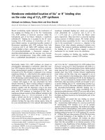
Fig. 1. Schematic representation of in vivo protein interaction systems
utilized in this study. (A) Yeast protein interaction (two-hybrid) system.
DHT-dependent interaction between GalAD-AR.NTD and GalDBD-AR.LBD induces expression of the UASGAL1 regulated lacZ
reporter gene. Cotransfection of pGBT9 and pACT2, which encode
GalDBD and GalAD, respectively, does not induce reporter gene
expression (data not shown). Similarly, individually expressed GalDBD-AR.LBD and GalAD-AR.NTD are not active in this assay. (B)
Mammalian (CHO cells) protein interaction system. R1881-dependent
interaction between AR.NTD and AR.DBD.LBD induces MMTVpromoter driven luciferase expression. Separately expressed
AR.DBD.LBD and AR.NTD are unable to activate the MMTV
promoter (data not shown).
30
VREVI34 in androgen receptor N/C
Prediction programs of protein secondary structures (see
) indicated a long a-helical structure
for AR20)34. A helical wheel drawing of this region
predicted an amphipathic character of this helical structure
(Fig. 5A) [29]. At positions 15 and 37, the putative a-helix is
flanked by proline residues. Within the helix, two candidate
FxxFF protein interaction motifs (F is any hydrophobic
amino acid residue and x is any amino acid residue) are
present: 30VREVI34 and the previously identified
23
FQNLF27 motif (Fig. 5B) [20,30,31]. To investigate
whether like 23FQNLF27, 30VREVI34 could contribute to
N/C interaction, two constructs were generated, expressing
either the complete 30VREVI34 or the complete 23FQNLF27
motif linked to GalAD (Fig. 5B). As expected, in the yeast
protein interaction system, ligand-dependent interaction
with AR LBD could easily be detected for GalAD-AR17–32.
However, the interaction was weak for GalAD-AR24–39
(Fig. 5C). Low activity was not due to decreased protein
expression (Fig. 5D).
In a complementary yeast protein interaction experiment,
the 30VREVI34 motif in GalAD-AR.NTDwt was modified
5784 K. Steketee et al. (Eur. J. Biochem. 269)
Ó FEBS 2002
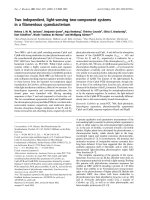
Fig. 2. AR3–13 modulates androgen receptor N/C interaction. (A) Interaction of AR.NTDwt and N-terminal deletion mutants with AR.LBD in the
presence of 1 lM DHT in the yeast protein interaction system. In each experiment the activity of GalAD-AR.NTDwt was set at 100%. Each bar
represents the mean (± SEM) b-galactosidase activity of three independent experiments. (B) Interaction of AR.NTDwt and deletion mutants with
AR.LBD in the presence of 1 nM R1881 in the mammalian protein interaction system. pSVAR.DBD.LBD was cotransfected with increasing
amounts of pSVAR.NTDwt or mutant (see Experimental procedures). In each experiment, carried out in triplicate, the mean of the highest
AR.NTDwt value was set at 100%. Each bar represents the mean (± SEM) luciferase activity of three independent experiments. Fold induction is
shown to the right of each bar and represents the ratio of activities determined in the presence and absence of R1881. (C) Western analysis of
indicated GalAD-AR.NTD proteins in the yeast protein interaction system (left panel) and of indicated AR.NTD proteins in the mammalian
protein interaction system (right panel). See Experimental procedures for details.
by substitution of two hydrophobic amino acids by arginine
residues, resulting in GalAD-AR.NTD30/33RR. These
substitutions might cause steric hindrance in the interaction
with the AR LBD surface, change the charge and disrupt
the proposed amphipathic a-helical structure of AR16)36.
GalAD-AR.NTD23/27RR was utilized as control. Substitution of V30 and V33 partially reduced the interaction,
whereas the F23R,F27R mutation completely abolished
the interaction (Fig. 6A). Expression levels of GalADAR.NTDwt and GalAD-AR.NTD30/33RR were similar
(Fig. 6C).
Results obtained in the mammalian protein interaction
system, utilizing the AR.NTD30/33RR mutant and
AR.NTD23/27RR, were essentially identical to the observations made in the yeast system (Fig. 6B). A partial
inhibition of AR N/C interaction was observed for
AR.NTD30/33RR, and an almost complete inhibition for
AR.NTD23/27RR.
Pull-down experiments confirmed and extended the in vivo
protein interaction experiments (Fig. 6D). AR N/C interaction was diminished due to 30/33RR substitutions, and
completely abolished by 23/27RR substitutions.
Ó FEBS 2002
Interaction between androgen receptor subdomains (Eur. J. Biochem. 269) 5785
Fig. 3. AR3–13 is not involved in direct binding of AR NTD to AR LBD. Interaction of AR.NTDwt and N-terminal deletion mutants with GSTAR.LBD as studied by pull-down assays. Proteins were produced in CHO cells by cotransfection of pCMVAR.LBD and pSVAR.NTDwt or
indicated deletion constructs. CHO cells were cultured in the absence (–) or presence (+) of 100 nM R1881. Input is 1/10th of the lysate utilized in a
pull-down experiment. See Experimental procedures for details.
Amino acid residues flanking F23, L26 and F27
modulate androgen receptor N/C interaction
To study in more detail the role of 24/25QN in the
23
FQNLF27 motif in AR N/C interaction, these amino acids
were substituted by 24/25AA. In both the yeast and
mammalian
protein
interaction
assay,
GalADAR.NTD24/25AA and AR.NTD24/25AA formed even
more active complexes with AR LBD than with wild-type
AR NTD (Fig. 7A,B) (note the low expression levels of the
24/25AA mutants in both systems; Fig. 7C). As expected,
AR.NTD26/27AA was incapable to interact with AR.LBD.
To extend these findings, an alanine scan was carried out
for peptide GalAD-AR17–32 (Fig. 8A). Results of the yeast
protein interaction assay are shown in Fig. 8(B). Substitution of amino acids 23, 26 and 27 completely abolished
interaction with GalDBD-AR.LBD and alanines at positions 24 and 25 increased the interaction capacity. All
alanine substitutions of amino acids flanking 23FQNLF27
reduced the binding to AR LBD. Most prominent inhibitory effects were found for amino acid residues directly
flanking 23FQNLF27. Note that expression levels of the
peptide constructs were similar (Fig. 8C).
DISCUSSION
Previously, we and others demonstrated a ligand–dependent
functional interaction between AR NTD and AR LBD.
Amino acids 3–36 in the NTD (AR3)36), including the
23
FxxLF27 motif, play a pivotal role in N/C interaction
[15,20]. Here we studied the function of the AR3)36
subdomain AR3)13 in N/C interaction and the role of
individual amino acid residues in and flanking the
23
FQNLF27motif in AR16)36 in N/C interaction.
Yeast protein interaction assays indicated that AR3)13
contributed to the ligand-induced transactivation function
of the AR.NTD/AR.LBD complex (Figs 2 and 4). Pulldown experiments provided evidence that AR3)13 does not
directly interact with AR LBD (Fig. 3). On first sight,
conflicting results were obtained in the yeast and mammalian protein interaction assays (Fig. 2). In the yeast
assay, reporter gene activity, which monitored the N/C
interaction, was partly reduced by AR3)13 deletion,
whereas in the mammalian assay almost all reporter gene
activity was lost. The most obvious difference between
both assays is the coupling of AR.NTD to GalAD in the
yeast assay, and the absence of a second transactivation
domain linked to AR NTD in the mammalian assay. The
latter assay completely depends on the intrinsic transactivating function of AR NTD and thus does not allow
discrimination between loss of AR.NTD-AR.LBD binding and loss of AR.NTD transactivating function. In the
yeast assay, loss of transactivation function of AR NTD
mutants, which retain AR LBD interacting capacity, like
AR.NTDD3–13, will be masked by the GalAD transactivating function. So, AR3)13 is not essential but rather
modulates N/C interaction, most probably by affecting
the transactivation function of AR.NTD. Alternative
explanations might be induction of a more favorable
NTD conformation or stabilization of the in vivo N/C
interaction, which are not reflected in the pull-down assays
and peptide interaction experiments. Unfortunately, the
5786 K. Steketee et al. (Eur. J. Biochem. 269)
Ó FEBS 2002
Fig. 4. AR2-14 cannot autonomously interact with AR LBD. (A) AR
peptides utilized in GalAD-ARpeptide fusion proteins in the yeast
protein interaction system. (B) Interaction of indicated GalADARpeptides with GalDBD-AR.LBD in yeast in the presence of 1 lM
DHT. In each experiment the activity of GalAD-AR2-36 was set at
100% (see also legend to Fig. 2A). (C) Western analysis of indicated
GalAD-ARpeptide proteins in yeast. For details, see Experimental
procedures.
primary structure and the predicted secondary structure of
AR3)13 do not give a clue to a more precise description of
its function (data not shown). However, the fact that,
between species, AR3)13 is one of the most conserved
regions of AR NTD, underscores a presumed important
role in AR function [32].
The second domain that was studied, AR16)36, is essential
in N/C interaction. The predicted structure indicated that
AR16)36 can fold in a remarkably long amphipathic
a-helical structure, suggesting an important protein interaction interface [29]. AR16)36 contains two FxxFF putative
protein interaction motifs: 23FxxLF27, which was found to
be pivotal for direct N/C interaction [20, this study], and
30
VxxVI34 (Figs 5 and 6). The latter sequence modulates
N/C interaction. Amino acid residues in this sequence might
contribute to the stability of the predicted a-helix. Alternatively, they might make additional contacts to the LBD
surface. This is also true for other amino acid residues
flanking the 23FxxLF27 motif (Fig. 8). Remarkably, substitution of Q24 and N25 by alanines increased N/C interaction (Figs 7 and 8).
The AR FxxLF motif shows similarities to LxxLL
motifs [5,33,34] present in nuclear receptor interaction
domains (NR boxes) of p160 coactivators. LxxLL motifs
are essential in the interaction with LBDs [33]. They bind
to a hydrophobic cleft in nuclear receptor LBDs, which is
marked by a charged clamp composed of a highly
conserved lysine and glutamate residue in helix 3 and
Fig. 5. Analysis of a predicted long amphipathic a-helix of AR18–35 in
AR N/C interaction. (A) A helical wheel drawing of AR18–35 predicts
a long amphipathic a-helical structure. Gray circles represent hydrophobic amino acids. (B) GalAD-ARpeptide fusion proteins utilized in
the yeast protein interaction system. The FxxFF motifs 23FQNLF27
and 30VREVI34 are underlined. (C) Interaction of GalAD-ARpeptides with GalDBD-AR.LBD in yeast in the presence of 1 lM DHT. In
each experiment the activity of GalAD-AR16–36 was set at 100% (see
also legend to Fig. 2A). (D) Western analysis of indicated GalADARpeptide proteins in the yeast system. For details, see Experimental
procedures.
Ó FEBS 2002
Interaction between androgen receptor subdomains (Eur. J. Biochem. 269) 5787
Fig. 6. 30VREVI34 is not essential for AR
N/C interaction. (A) Interaction of GalADAR.NTDwt and mutants with AR.LBD in
the presence of 1 lM DHT in the yeast protein
interaction system. In each experiment
GalAD-AR.NTDwt activity was set at 100%
(see legend to Fig. 2A). (B) Interaction of
AR.NTDwt and mutants with AR.LBD
in the presence of 1 nM R1881 in the
mammalian protein interaction system.
pSVAR.DBD.LBD was cotransfected with
increasing amounts of pSVAR.NTDwt or
indicated mutants (see Experimental procedures and legend to Fig. 2B). (C) Western
analysis of indicated GalAD-AR.NTD proteins in the yeast system (left panel) and indicated AR.NTD proteins in the mammalian
system (right panel) (see also Experimental
procedures). (D) Pull-down assays showing
interaction of AR.NTDwt and mutants with
GST-AR.LBD (see also legend to Fig. 3).
helix 12 of the LBD, respectively (K720 and E897 in AR)
[35–37]. AR K720 and E897 are both involved in the
ligand–dependent interaction between AR LBD and the
coactivator TIF2 [9,11,15]. However, in the FxxLFmediated AR N/C interaction, E897 is essential, but
K720 can be replaced by many other amino acids, without
5788 K. Steketee et al. (Eur. J. Biochem. 269)
Ó FEBS 2002
Fig. 7. Alanine substitutions of Q24 and N25
stimulate AR N/C interaction. (A) Interaction
of GalAD-AR.NTDwt and mutants with
GalDBD-AR.LBD in the presence of 1 lM
DHT in the yeast protein interaction system.
In each experiment, GalAD-AR.NTDwt
activity was set at 100%. See also legend to
Fig. 2A. (B) Interaction of AR.NTDwt and
mutants with AR.LBD in the presence of
1 nM R1881 in the mammalian protein interaction system. pSVAR.DBD.LBD was
cotransfected with increasing amounts of
pSVAR.NTDwt or mutants (see Experimental procedures and legend to Fig. 2B). (C)
Western analysis of indicated GalADAR.NTD proteins in the yeast protein system
(left panel) and indicated AR.NTD proteins in
the mammalian system (right panel). For
details, see Experimental procedures.
affecting N/C interaction [9,11,15,38]. So, the AR N/C
interaction is similar, but not identical, to LxxLL-mediated coactivator–LBD interaction.
The 3D structures of agonist bound LBD/LxxLL peptide
complexes of several nuclear receptors have been elucidated,
and interactions of the peptide backbone and its amino acid
side chains with the LBD surface have been identified
[5,36,37,39]. It is presumed that upon binding to the LBD
surface, the LxxLL motif adapts a short a-helical structure,
which is stabilized by interaction with the charged clamp
[5,36,37]. The first and last leucine residue in the LxxLL
motif enter the hydrophobic cleft in the LBD, and directly
contact amino acid residues within the cleft. The variable
amino acids (xx) in the LxxLL motif point away from the
cleft and seem not to interact directly with the LBD surface.
Structural data for AR.LBD/LxxLL peptides are not
available but, because AR.LBD/coactivator interaction
also depends on K720 and E897, it might be predicted that
they will be similar to LBD/LxxLL peptide complexes
studied so far [9,11,15]. Because K720 is not essential for
AR23FxxLF27/AR.LBD interaction, the structure of this
complex might be different. A different complex would also
explain the stimulation of AR23FxxLF27/AR.LBD inter-
action by substitution of Q24 and N25 by alanine residues.
Structural analyses of AR.LBD/AR16)36 complexes have to
reveal the function of amino acid residues flanking F23, L26
and F27 and answer the question as to whether or not the
entire long amphipathic AR16)36 a-helix is required for a
stable AR NTD/LBD complex.
The LxxLL-like motifs LxxIL, FxxLL, and L/IxxI/VI,
have been found in LBD binding coactivators or corepressors [40–43]. FxxLF motifs that are able to contact AR
LBD, have only been found in AR NTD and most recently
in the AR coactivators ARA54 and ARA70, suggesting a
specific role of these motifs in AR function [44–47]. The
increasing number of proteins found to interact with the AR
LBD raises the question of the physiological relevance of the
many interactions. It remains to be established whether all
interactions take place in living cells under physiological
conditions, whether interactions with different proteins are
simultaneous or consecutive events, and which interactions
are most stable and most specific. Recently, a start has been
made to identify factors, including the AR, present in the
transcription initiation complex of the prostate specific
antigen enhancer/promoter, using chromatin immunoprecipitation (ChIP) [48].
Ó FEBS 2002
Interaction between androgen receptor subdomains (Eur. J. Biochem. 269) 5789
Fig. 8. Alanine scanning of AR17–32: amino acids flanking F23, L26 and F27 modulate AR N/C interaction. (A) GalAD-ARpeptide fusion proteins
in the yeast protein interaction system. (B) Interaction of GalAD-ARpeptides with AR.LBD in the presence of 1 lM DHT in the yeast protein
interaction system. In each experiment the activity of GalAD-AR17–32 was set at 100%. See also legend to Fig. 2A. (C) Western analysis of
indicated GalAD-ARpeptide proteins in the yeast assay. For details, see Experimental procedures.
Another question concerns the interaction of AR16)36
with other proteins. One candidate might be the TFIID
TATA box-binding protein associated factor 31, TAFII31,
which has been found to interact with FxxFF motifs in
acidic transcription activation domains of p65 (nuclear
factor-kappa B), VP16, p53 and related proteins [31,49–51].
AR NTD has previously been proposed to accommodate
more than one AR LBD interacting domain [9,15,20]. A
candidate second domain is 433WHTLF437, which was
found to modulate 23FxxLF27 function [20]. Another
candidate motif is 179LxxIL183 [9]. However, peptides
containing these motifs were unable to interact with AR
LBD in the yeast protein interaction assay, excluding their
role as a second autonomous interaction motif in AR NTD
(data not shown).
N/C interaction is not unique for the AR, but has also been
described for other nuclear receptors. ERa ligand-dependent
direct N/C interaction has been demonstrated, which was
disrupted by amino acid substitutions that affect receptor
function [52,53]. The ERa N/C interaction could be induced
by the agonist estradiol (E2), but not by the antagonist
ICI164 384 [53]. Recently, it was found that the ERa N/C
5790 K. Steketee et al. (Eur. J. Biochem. 269)
interaction was required for SRC-1-mediated synergism
between AF-1 and AF-2 function [8,53]. The progesterone
receptor (PR) showed direct N/C interaction in the presence
of agonist R5020, but not in the presence of antagonist
RU486 [54]. LxxLL motifs in the PR-B form were most
probably not involved, because the shorter PR-A form,
lacking these motifs, also showed N/C interaction [55].
The role of the N/C interaction in full-length AR function
is not well understood. Ligand-dependent AR N/C interaction affects ligand dissociation [11,20,56]. Whether this is
a direct or an indirect effect is unknown. Disruption of the
N/C interaction by mutation of the 23FxxLF27 motif has a
limited effect on full length AR transactivation function [20,
Steketee, unpublished observation]. However, several AR
LBD mutants with reduced or completely abolished N/C
interaction have been found in androgen insensitivity
patients [11,56,57]. Additionally, both N/C interaction and
the transactivating function of the AR prostate cancer
mutant T877A can be induced by natural low affinity
ligands like progesterone or E2 or the AR antagonist
cyproterone acetate [18].
In conclusion, we propose that AR3)36 is involved in a
dynamic sequence of protein interaction events, including
N/C interaction, in regulation of AR function. Detailed
knowledge on the role of the AR N/C interaction would
require the elucidation of its function under more physiological conditions, including the study of mouse models
carrying AR mutants defective in N/C interaction.
ACKNOWLEDGMENT
This study was supported by a grant from the Dutch Cancer Society
KWF.
REFERENCES
1. Mangelsdorf, D.J., Thummel, C., Beato, M., Herrlich, P., Schutz,
G., Umesono, K., Blumberg, B., Kastner, P., Mark, M. &
Chambon, P. (1995) The nuclear receptor superfamily: the second
decade. Cell 83, 835–839.
2. McKenna, N.J., Lanz, R.B. & O’Malley, B.W. (1999) Nuclear
receptor coregulators: cellular and molecular biology. Endocr.
Rev. 20, 321–344.
3. Glass, C.K. & Rosenfeld, M.G. (2000) The coregulator exchange
in transcriptional functions of nuclear receptors. Genes Dev. 14,
121–141.
4. Lee, K.C. & Lee Kraus, W. (2001) Nuclear receptors, coactivators
and chromatin: new approaches, new insights. Trends Endocrinol.
Metab. 12, 191–197.
5. Darimont, B.D., Wagner, R.L., Apriletti, J.W., Stallcup, M.R.,
Kushner, P.J., Baxter, J.D., Fletterick, R.J. & Yamamoto, K.R.
(1998) Structure and specificity of nuclear receptor–coactivator
interactions. Genes Dev. 12, 3343–3356.
6. Onate, S.A., Boonyaratanakornkit, V., Spencer, T.E., Tsai, S.Y.,
Tsai, M.J., Edwards, D.P. & O’Malley, B.W. (1998) The steroid
receptor coactivator-1 contains multiple receptor interacting and
activation domains that cooperatively enhance the activation
function 1 (AF1) and AF2 domains of steroid receptors. J. Biol.
Chem. 273, 12101–12108.
7. Webb, P., Nguyen, P., Shinsako, J., Anderson, C., Feng, W.,
Nguyen, M.P., Chen, D., Huang, S.M., Subramanian, S.,
McKinerney, E., Katzenellenbogen, B.S., Stallcup, M.R. &
Kushner, P.J. (1998) Estrogen receptor activation function 1
works by binding p160 coactivator proteins. Mol. Endocrinol. 12,
1605–1618.
Ó FEBS 2002
8. Benecke, A., Chambon, P. & Gronemeyer, H. (2000) Synergy
between estrogen receptor alpha activation functions AF1 and
AF2 mediated by transcription intermediary factor TIF2. EMBO
Rep. 1, 151–157.
9. Alen, P., Claessens, F., Verhoeven, G., Rombauts, W. & Peeters,
B. (1999) The androgen receptor amino-terminal domain plays a
key role in p160 coactivator-stimulated gene transcription. Mol.
Cell. Biol. 19, 6085–6097.
10. Bevan, C.L., Hoare, S., Claessens, F., Heery, D.M. & Parker,
M.G. (1999) The AF1 and AF2 domains of the androgen receptor
interact with distinct regions of SRC1. Mol. Cell. Biol. 19, 8383–
8392.
11. He, B., Kemppainen, J.A., Voegel, J.J., Gronemeyer, H. &
Wilson, E.M. (1999) Activation function 2 in the human androgen
receptor ligand binding domain mediates interdomain communication with the NH(2)-terminal domain. J. Biol. Chem. 274,
37219–37225.
12. Jenster, G., van der Korput, H.A., van Vroonhoven, C., van der
Kwast, T.H., Trapman, J. & Brinkmann, A.O. (1991) Domains of
the human androgen receptor involved in steroid binding, transcriptional activation, and subcellular localization. Mol.
Endocrinol. 5, 1396–1404.
13. Simental, J.A., Sar, M., Lane, M.V., French, F.S. & Wilson, E.M.
(1991) Transcriptional activation and nuclear targeting signals of
the human androgen receptor. J. Biol. Chem. 266, 510–518.
14. Palvimo, J.J., Kallio, P.J., Ikonen, T., Mehto, M. & Janne, O.A.
(1993) Dominant negative regulation of trans-activation by the rat
androgen receptor: roles of the N-terminal domain and heterodimer formation. Mol. Endocrinol. 7, 1399–1407.
15. Berrevoets, C.A., Doesburg, P., Steketee, K., Trapman, J. &
Brinkmann, A.O. (1998) Functional interactions of the AF-2
activation domain core region of the human androgen receptor
with the amino-terminal domain and with the transcriptional
coactivator TIF2 (transcriptional intermediary factor2). Mol.
Endocrinol. 12, 1172–1183.
16. Gottlieb, B., Beitel, L.K. & Trifiro, M.A. (2001) Variable
expressivity and mutation databases: the androgen receptor gene
mutations database. Hum. Mutat. 17, 382–388.
17. Langley, E., Zhou, Z.X. & Wilson, E.M. (1995) Evidence for an
anti-parallel orientation of the ligand-activated human androgen
receptor dimer. J. Biol. Chem. 270, 29983–29990.
18. Doesburg, P., Kuil, C.W., Berrevoets, C.A., Steketee, K., Faber,
P.W., Mulder, E., Brinkmann, A.O. & Trapman, J. (1997)
Functional in vivo interaction between the amino-terminal, transactivation domain and the ligand binding domain of the androgen
receptor. Biochemistry 36, 1052–1064.
19. Ikonen, T., Palvimo, J.J. & Janne, O.A. (1997) Interaction
between the amino- and carboxyl-terminal regions of the rat
androgen receptor modulates transcriptional activity and is
influenced by nuclear receptor coactivators. J. Biol. Chem. 272,
29821–29828.
20. He, B., Kemppainen, J.A. & Wilson, E.M. (2000) FXXLF and
WXXLF sequences mediate the NH2–terminal interaction with
the ligand binding domain of the androgen receptor. J. Biol.
Chem. 275, 22986–22994.
21. Sambrook, J. & Russell, D.W. (2001) Molecular Cloning a
Laboratory Manual, 3rd edn. Cold Spring. Harbor Laboratory
Press, Cold Spring Harbor, New York.
22. Brinkmann, A.O., Faber, P.W., van Rooij, H.C., Kuiper, G.G.,
Ris, C., Klaassen, P., van der Korput, J.A., Voorhorst, M.M., van
Laar, J.H. & Mulder, E. (1989) The human androgen receptor:
domain structure, genomic organization and regulation of
expression. J. Steroid Biochem. 34, 307–310.
23. Jenster, G., van der Korput, H.A., Trapman, J. & Brinkmann,
A.O. (1995) Identification of two transcription activation units in
the N-terminal domain of the human androgen receptor. J. Biol.
Chem. 270, 7341–7346.
Ó FEBS 2002
Interaction between androgen receptor subdomains (Eur. J. Biochem. 269) 5791
24. de Ruiter, P.E., Teuwen, R., Trapman, J., Dijkema, R. & Brinkmann, A.O. (1995) Synergism between androgens and protein
kinase-C on androgen-regulated gene expression. Mol. Cell.
Endocrinol. 110, R1–R6.
25. Tsai, R.Y. & Reed, R.R. (1997) Using a eukaryotic GST fusion
vector for proteins difficult to express in E. coli. Biotechniques 23,
794–796,798,800.
26. Gietz, D., St Jean, A., Woods, R.A. & Schiestl, R.H. (1992)
Improved method for high efficiency transformation of intact
yeast cells. Nucleic Acids Res. 20, 1425.
27. Kuil, C.W., Berrevoets, C.A. & Mulder, E. (1995) Ligand-induced
conformational alterations of the androgen receptor analyzed by
limited trypsinization. Studies on the mechanism of antiandrogen
action. J. Biol. Chem. 270, 27569–27576.
28. Zegers, N.D., Claassen, E., Neelen, C., Mulder, E., van Laar, J.H.,
Voorhorst, M.M., Berrevoets, C.A., Brinkmann, A.O., van der
Kwast, T.H. & Ruizeveld de Winter, J.A. (1991) Epitope prediction and confirmation for the human androgen receptor: generation of monoclonal antibodies for multi-assay performance
following the synthetic peptide strategy. Biochim. Biophys. Acta
1073, 23–32.
29. Segrest, J.P., De Loof, H., Dohlman, J.G., Brouillette, C.G. &
Anantharamaiah, G.M. (1990) Amphipathic helix motif: classes
and properties. Proteins 8, 103–117.
30. Nagy, L., Kao, H.Y., Love, J.D., Li, C., Banayo, E., Gooch, J.T.,
Krishna, V., Chatterjee, K., Evans, R.M. & Schwabe, J.W. (1999)
Mechanism of corepressor binding and release from nuclear hormone receptors. Genes Dev. 13, 3209–3216.
31. Uesugi, M. & Verdine, G.L. (1999) The alpha-helical FXXPhiPhi
motif in p53: TAF interaction and discrimination by MDM2.
Proc. Natl. Acad. Sci. USA 96, 14801–14806.
32. Thornton, J.W. & Kelley, D.B. (1998) Evolution of the androgen
receptor: structure-function implications. Bioessays 20, 860–869.
33. Heery, D.M., Kalkhoven, E., Hoare, S. & Parker, M.G. (1997) A
signature motif in transcriptional co-activators mediates binding
to nuclear receptors. Nature 387, 733–736.
34. McInerney, E.M., Rose, D.W., Flynn, S.E., Westin, S., Mullen,
T.M., Krones, A., Inostroza, J., Torchia, J., Nolte, R.T., AssaMunt, N., Milburn, M.V., Glass, C.K. & Rosenfeld, M.G. (1998)
Determinants of coactivator LXXLL motif specificity in nuclear
receptor transcriptional activation. Genes Dev. 12, 3357–3368.
35. Feng, W., Ribeiro, R.C., Wagner, R.L., Nguyen, H., Apriletti,
J.W., Fletterick, R.J., Baxter, J.D., Kushner, P.J. & West, B.L.
(1998) Hormone-dependent coactivator binding to a hydrophobic
cleft on nuclear receptors. Science 280, 1747–1749.
36. Nolte, R.T., Wisely, G.B., Westin, S., Cobb, J.E., Lambert, M.H.,
Kurokawa, R., Rosenfeld, M.G., Willson, T.M., Glass, C.K. &
Milburn, M.V. (1998) Ligand binding and co-activator assembly
of the peroxisome proliferator- activated receptor-gamma. Nature
395, 137–143.
37. Shiau, A.K., Barstad, D., Loria, P.M., Cheng, L., Kushner, P.J.,
Agard, D.A. & Greene, G.L. (1998) The structural basis of
estrogen receptor/coactivator recognition and the antagonism of
this interaction by tamoxifen. Cell 95, 927–937.
38. Slagsvold, T., Kraus, I., Bentzen, T., Palvimo, J. & Saatcioglu, F.
(2000) Mutational analysis of the androgen receptor AF-2 (activation function 2) core domain reveals functional and mechanistic
differences of conserved residues compared with other nuclear
receptors. Mol. Endocrinol. 14, 1603–1617.
39. Gampe, R.T. Jr, Montana, V.G., Lambert, M.H., Miller, A.B.,
Bledsoe, R.K., Milburn, M.V., Kliewer, S.A., Willson, T.M. &
Xu, H.E. (2000) Asymmetry in the PPARgamma/RXRalpha
crystal structure reveals the molecular basis of heterodimerization
among nuclear receptors. Mol. Cell. 5, 545–555.
40. Huang, N., vom Baur, E., Garnier, J.M., Lerouge, T., Vonesch,
J.L., Lutz, Y., Chambon, P. & Losson, R. (1998) Two distinct
nuclear receptor interaction domains in NSD1, a novel SET
41.
42.
43.
44.
45.
46.
47.
48.
49.
50.
51.
52.
53.
54.
55.
56.
57.
protein that exhibits characteristics of both corepressors and
coactivators. EMBO J. 17, 3398–3412.
Li, D., Desai-Yajnik, V., Lo, E., Schapira, M., Abagyan, R. &
Samuels, H.H. (1999) NRIF3 is a novel coactivator mediating
functional specificity of nuclear hormone receptors. Mol. Cell.
Biol. 19, 7191–7202.
Johansson, L., Bavner, A., Thomsen, J.S., Farnegardh, M.,
Gustafsson, J.A. & Treuter, E. (2000) The orphan nuclear receptor
SHP utilizes conserved LXXLL–related motifs for interactions
with ligand-activated estrogen receptors. Mol. Cell. Biol. 20, 1124–
1133.
Gobinet, J., Auzou, G., Nicolas, J.C., Sultan, C. & Jalaguier, S.
(2001) Characterization of the interaction between androgen
receptor and a new transcriptional inhibitor, SHP. Biochemistry
40, 15369–15377.
Yeh, S. & Chang, C. (1996) Cloning and characterization of a
specific coactivator, ARA70, for the androgen receptor in human
prostate cells. Proc. Natl Acad. Sci. USA 93, 5517–5521.
Kang, H.Y., Yeh, S., Fujimoto, N. & Chang, C. (1999) Cloning
and characterization of human prostate coactivator ARA54, a
novel protein that associates with the androgen receptor. J. Biol.
Chem. 274, 8570–8576.
He, B., Minges, J.T., Lee, L.W. & Wilson, E.M. (2002) The
FXXLF Motif Mediates Androgen Receptor–specific Interactions
with Coregulators. J. Biol. Chem. 277, 10226–10235.
Zhou, Z.X., He, B., Hall, S.H., Wilson, E.M. & French, F.S.
(2002) Domain interactions between coregulator ARA (70) and
the androgen receptor (AR). Mol. Endocrinol. 16, 287–300.
Shang, Y., Myers, M. & Brown, M. (2002) Formation of the
androgen receptor transcription complex. Mol. Cell. 9, 601–610.
Burley, S.K. & Roeder, R.G. (1996) Biochemistry and structural
biology of transcription factor IID (TFIID). Annu. Rev. Biochem.
65, 769–799.
Uesugi, M., Nyanguile, O., Lu, H., Levine, A.J. & Verdine, G.L.
(1997) Induced alpha helix in the VP16 activation domain upon
binding to a human TAF. Science 277, 1310–1313.
Yoon, J.W., Liu, C.Z., Yang, J.T., Swart, R., Iannaccone, P. &
Walterhouse, D. (1998) GLI activates transcription through a
herpes simplex viral protein 16-like activation domain. J. Biol.
Chem. 273, 3496–3501.
Kraus, W.L., McInerney, E.M. & Katzenellenbogen, B.S. (1995)
Ligand-dependent, transcriptionally productive association of the
amino- and carboxyl-terminal regions of a steroid hormone
nuclear receptor. Proc. Natl Acad. Sci. USA 92, 12314–12318.
Metivier, R., Penot, G., Flouriot, G. & Pakdel, F. (2001) Synergism between ERalpha transactivation function 1 (AF-1) and
AF-2 mediated by steroid receptor coactivator protein-1:
requirement for the AF-1 alpha-helical core and for a direct
interaction between the N- and C-terminal domains. Mol.
Endocrinol. 15, 1953–1970.
Tetel, M.J., Giangrande, P.H., Leonhardt, S.A., McDonnell, D.P.
& Edwards, D.P. (1999) Hormone-dependent interaction between
the amino- and carboxyl-terminal domains of progesterone
receptor in vitro and in vivo. Mol. Endocrinol. 13, 910–924.
Tung, L., Shen, T., Abel, M.G., Powell, R.L., Takimoto, G.S.,
Sartorius, C.A. & Horwitz, K.B. (2001) Mapping the unique
activation function 3 in the progesterone B-receptor upstream
segment. Two LXXLL motifs and a tryptophan residue are
required for activity. J. Biol. Chem. 276, 39843–39851.
Langley, E., Kemppainen, J.A. & Wilson, E.M. (1998) Intermolecular NH2-/carboxyl–terminal interactions in androgen
receptor dimerization revealed by mutations that cause androgen
insensitivity. J. Biol. Chem. 273, 92–101.
Thompson, J., Saatcioglu, F., Janne, O.A. & Palvimo, J.J. (2001)
Disrupted amino- and carboxyl–terminal interactions of the
androgen receptor are linked to androgen insensitivity. Mol.
Endocrinol. 15, 923–935.