Báo cáo Y học: Guanosine diphosphate-4-keto-6-deoxy-D-mannose reductase in the pathway for the synthesis of GDP-6-deoxy-D-talose in Actinobacillus actinomycetemcomitans potx
Bạn đang xem bản rút gọn của tài liệu. Xem và tải ngay bản đầy đủ của tài liệu tại đây (340.42 KB, 9 trang )
Guanosine diphosphate-4-keto-6-deoxy-
D
-mannose reductase
in the pathway for the synthesis of GDP-6-deoxy-
D
-talose
in
Actinobacillus actinomycetemcomitans
Nao Suzuki
1
, Yoshio Nakano
1
, Yasuo Yoshida
1
, Takashi Nezu
2
, Yoshihiro Terada
2
, Yoshihisa Yamashita
3
and Toshihiko Koga
1
1
Department of Preventive Dentistry and
2
Department of Prosthetic Dentistry I, Kyushu University Faculty of Dental Science,
Fukuoka, Japan;
3
Department of Oral Health, Nihon University School of Dentistry, Tokyo, Japan
The serotype a-specific polysaccharide antigen of Actinoba-
cillus actinomycetemcomitans is an unusual sugar, 6-deoxy-
D
-talose. Guanosine diphosphate (GDP)-6-deoxy-
D
-talose is
the activated sugar nucleotide form of 6-deoxy-
D
-talose,
which has been identified as a constituent of only a few
microbial polysaccharides. In this paper, we identify two
genes encoding GDP-6-deoxy-
D
-talose synthetic enzymes,
GDP-a-
D
-mannose 4,6-dehydratase and GDP-4-keto-6-
deoxy-
D
-mannose reductase, in the gene cluster required for
the biosynthesis of serotype a-specific polysaccharide anti-
gen from A. actinomycetemcomitans SUNYaB 75. Both
gene products were produced and purified from Escherichia
coli transformed with plasmids containing these genes. Their
enzymatic reactants were analysed by reversed-phase
HPLC (RP-HPLC). The sugar nucleotide produced from
GDP-a-
D
-mannose by these enzymes was purified by
RP-HPLC and identified by electrospray ionization-MS,
1
H
nuclear magnetic resonance, and GC/MS. The results
indicated that GDP-6-deoxy-
D
-talose is produced from
GDP-a-
D
-mannose. This paper is the first report on the
GDP-6-deoxy-
D
-talose biosynthetic pathway and the role of
GDP-4-keto-6-deoxy-
D
-mannose reductase in the synthesis
of GDP-6-deoxy-
D
-talose.
Keywords: Actinobacillus actinomycetemcomitans;6-deoxy-
talose; NMR; polysaccharide; serotype-specific antigen.
Capsular polysaccharides are ubiquitous structures found
on the cell surfaces of a broad range of bacterial species. The
polysaccharides often constitute the outermost layer of the
cell, and have been implicated as an important factor in the
virulence of many animal and plant pathogens. These
molecules are prominent structurally, and are serologically
diverse antigens that are involved in pathogenic processes
and in mediating resistance to host defense mechanisms [1].
Actinobacillus actinomycetemcomitans is a nonmotile,
Gram-negative, capnophilic, fermentative coccobacillus
that has been implicated in the aetiology and pathogenesis
of localized juvenile periodontitis [2–4], adult periodontitis
[5], and severe nonoral human infections [6]. Traditionally,
A. actinomycetemcomitans strains were divided into five
serotypes (a, b, c, d and e) [7–9], but recently a new serotype,
f, was reported [10]. The serologic specificity is defined by
the polysaccharides on the surface of the organism [11] and
the serotype-specific polysaccharide antigens (SPAs) are the
immunodominant antigens in the organism [12–16].
Serotype a-specific polysaccharide antigen from A. actino-
mycetemcomitans is a 6-deoxy-
D
-talan composed of
repeating disaccharide units, which are acetylated at the
O-2 position of 1,3-linked 6-deoxy-
D
-talose: )3))6-deoxy-
a-
D
-talose-(1–2))6-deoxy-a-
D
-talose-(1– [17,18]. Bacterial
extracellular polysaccharides consisting solely of 6-deo-
xytalose are rare. Except for the serotype a-specific
polysaccharide antigen from A. actinomycetemcomitans,
the exopolysaccharide isolated from Pseudomonas plantarii
strain DSM 6535 is the only reported homopolysaccharide
of 6-deoxy-
D
-talose [19]. The repeating unit of the
exopolysaccharide from P. plantarii has a different struc-
ture: it is a trisaccharide that is acetylated at the O-2
position of 1,3-linked 6-deoxy-
D
-talose: )3))6-deoxy-a-
D
-
talose-(1–2))6-deoxy-a-
D
-talose-(1–2)-6-deoxy-a-
D
-talose-
(1–. Other SPAs of A. actinomycetemcomitans also contain
rare sugars as constituents of microbial polysaccharides;
examples include
D
-fucose in serotype b-specific polysac-
charide antigen [12] and 6-deoxy-
L
-talose in serotype
c-specific polysaccharide antigen [17].
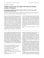
The mechanism for the biosynthesis of GDP-6-deoxy-
D
-
talose, which is the activated sugar nucleotide form of
6-deoxy-
D
-talose, is unknown. It is thought that GDP-6-
deoxy-
D
-talose is formed from a-
D
-mannose-1-phosphate
and GTP in three steps; the first two steps are common to
the GDP-
L
-fucose, GDP-
D
-rhamnose, and GDP-
L
-colitose
synthesis pathways, producing GDP-4-keto-6-deoxy-
D
-
mannose (Fig. 1) [20–22]. a-
D
-Mannose-1-phosphate
guanylyltransferase (ManC) combines a-
D
-mannose-1-
Correspondence to Y. Nakano, Department of Preventive Dentistry,
Kyushu University Faculty of Dental Science,
Fukuoka 812-8582, Japan.
Fax: + 81 92 642 6354, Tel.: + 81 92 642 6423,
E-mail:
Abbreviations: ESI, electrospray ionisation; Gmd, GDP-a-
D
-mannose
4,6-dehydratase; Rmd, GDP-4-keto-6-deoxy-
D
-mannose reductase;
RP-HPLC, reversed-phase HPLC; SPA, serotype-specific polysac-
charide antigen.
Note: This work is dedicated in fondest memory to Prof. T. Koga,
whose influence as a mentor will be greatly missed and without whom
this work would not have been possible.
(Received 4 August 2002, revised 1 October 2002,
accepted 23 October 2002)
Eur. J. Biochem. 269, 5963–5971 (2002) Ó FEBS 2002 doi:10.1046/j.1432-1033.2002.03331.x
phosphate with GTP to produce GDP-a-
D
-mannose. Then,
GDP-a-
D
-mannose is converted into GDP-4-keto-6-deoxy-
D
-mannose by GDP-a-
D
-mannose 4,6-dehydratase (Gmd).
GDP-
D
-rhamnose is then produced from GDP-4-keto-6-
deoxy-
D
-mannose by GDP-4-keto-6-deoxy-
D
-mannose
reductase (Rmd). GDP-6-deoxy-
D
-talose is a stereoisomer
of GDP-
D
-rhamnose at C4. GDP-6-deoxy-
D
-talose can be
synthesized by another GDP-4-keto-6-deoxy-
D
-mannose
reductase and the stereoselectivity of the reduction deter-
mines the direction of synthesis of these two 6-deoxyhex-
oses. However, neither the gene encoding the biosynthesis of
GDP-6-deoxy-
D
-talose nor its corresponding protein has
been found.
Recently, we cloned and characterized a gene cluster
involved in the biosynthesis of SPA from A. actinomyce-
temcomitans SUNYaB 75 (serotype a) (Fig. 2A) [23]. In a
protein database search the ORF9 product shared 52.0%
identity with the gmd gene product of Yersinia pseudotu-
berculosis [24] and the ORF7 product was 28.0% identical
with the rmd gene product of Pseudomonas aeruginosa [25].
We predicted that ORF9 and ORF7 encoded GDP-a-
D
-
mannose 4,6-dehydratase and GDP-4-keto-6-deoxy-
D
-man-
nose reductase in the biosynthesis of GDP-6-deoxy-
D
-talose, respectively. The gmd gene was subcloned into
pIVEX2.3 and the tld gene was subcloned into
pIVEX2.3MCS, and these gene products overproduced in
Escherichia coli were purified and characterized.
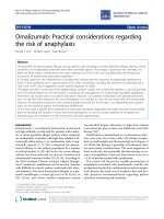
Fig. 2. Restriction map and genetic organization of the gene cluster
responsible for the production of the SPA of A. actinomycetemcomitans
SUNYaB 75 (serotype a) (A) and gel electrophoresis of recombinant
enzymes purified from E. coli strains transformed with the expression
plasmids (B). (A) Closed arrows indicate ORFs. The functions of the
gene products predicted by homology search, the GC content of each
ORF, and the SPA phenotypes caused by specific insertion mutants
are shown in descending order below the restriction map. A flag
indicates the putative promoter. The horizontal lines show the DNA
fragments inserted into pMCL210 used for nucleotide sequencing.
Abbreviations: H, HindIII; E, EcoRI; A, Acc65I; Pa, PacI; Pm, PmeI;
Tld, a putative GDP-4-keto-6-deoxy-
D
-mannose reductase; Ac-TRase,
acetyltransferase; Gmd, GDP-a-
D
-mannose 4,6-dehydratase; XylR,
xylose operon regulatory protein. (B) Approximately 0.5 lgofeach
protein was incubated at 100 °C in a water bath for 5 min with 0.1%
(w/v) SDS and 1% (v/v) 2-mercaptoethanol. Each of the treated
solutions was electrophoresed on a 12.5% SDS-polyacrylamide gel,
which was stained with Coomassie Blue. Lane 1, Purified gmd gene
product (SUNYaB 75); lane 2, purified tld gene product; lane 3,
purified gmd gene product (K12). The positions of molecular mass
markers (kDa) are shown on the left.
Fig. 1. Pathway for the synthesis of GDP-
D
-rhamnose, GDP-
L
-fucose,
and GDP-6-deoxy-
D
-talose from a-mannose-1-phosphate and GTP.
Asterisks above the parentheses indicate the genes encoding the
enzymes in A. actinomycetemcomitans SUNYaB 75.
5964 N. Suzuki et al. (Eur. J. Biochem. 269) Ó FEBS 2002
EXPERIMENTAL PROCEDURES
Bacterial strains, plasmids, and culture conditions
E. coli DH5a (supE44 DlacU169 (/80 lacZDM15) hsdR17
recA1 endA1 gyrA96 thi-1 relA1) [26] was used for the DNA
manipulations and as the host strain for pIVEX2.3 and
pIVEX2.3MCS derivatives (Roche Molecular Biochemi-
cals). E. coli ER2566 (F
–
k
–
fhuA2 (lon) ompT lacZ::T7
gene1 gal sulA11 D(mcrC-mrr) 114::IS10R(mcr-73::mini-
Tn10-TetS)2R(zgb-210::Tn10)(TetS) endA1 (dcm)) (New
England Biolabs) was grown as a host strain when the
IMPACT T7 One-Step Protein Purification System (New
England Biolabs) was used. E. coli strains were grown
aerobically in 2 · TY medium at 37 °C. Ampicillin was
used at a final concentration of 50 lgÆmL
)1
. The DNA
fragments carrying the gmd and tld genes of A. actinomyce-
temcomitans SUNYaB 75 were amplified by PCR using
pSAA212 [23] as a template. pSAA212 contains a 10.6-kb
Acc65I fragment responsible for the biosynthesis of serotype
a-specific polysaccharide antigen in A. actinomycetemcom-
itans SUNYaB 75.
DNA manipulation, PCR, and sequencing techniques
DNA fragment preparation, agarose gel electrophoresis,
DNA labelling, ligation, and bacterial transformation were
performed using the methods described by Sambrook et al.
[26]. PCR amplification was performed using T3 Thermo-
cycler (Biometra, Go
¨
ttingen, Germany). Sequencing was
performed using an ABI 373A or an ABI PRISM 310
DNA sequencer (Applied Biosystems).
Construction of plasmid
Each DNA fragment carrying the gmd and tld genes of
A. actinomycetemcomitans SUNYaB 75 was amplified by
PCR using pSAA212 [23] as a template. To construct
plasmids for gene expression and protein purification, the
following sets of primers were designed to introduce
appropriate restriction sites for subcloning: to subclone
the gmd gene into the vector pIVEX2.3, 5¢-CGCG
CCATGGTGAAAACAGCAATTGTAACT-3¢ (NcoI)
and 5¢-GCGCCCCGGGAAAAGAAAAACC-3¢ (SmaI);
andtosubclonethetld gene into the vector pIVEX2.3MCS,
5¢-GCGCCATATGAAAATCTTAGTA-3¢ (NdeI) and
5¢-GCGCCCCGGGAATCGAAAGCTC-3¢ (SmaI). Each
PCR product was purified using a QIAquick PCR Purifi-
cation Kit (QIAGEN GmbH) and, after double digestion
with the appropriate restriction enzymes, directly ligated
into the vector plasmid, which had been cleaved with the
same enzymes. Plasmids containing the gmd and tld genes
bound to a His
6
-tag were constructed using the vectors
pIVEX2.3 and pIVEX2.3MCS, respectively. The DNA
fragment carrying the gmd gene of E. coli K12 was
amplified by PCR using chromosomal DNA of E. coli
DH5a as a template with the following primers:
5¢-CGCGCATATGTCAAAAGTCGCTCTCATC-3¢ (NdeI)
and 5¢-ATATCCCGGGTGACTCCAGCGCGATCGC-3¢
(SmaI). After purification and double digestion with NdeI
and SmaI, the fragment was directly ligated into NdeI–SmaI
double-digested pTYB2 vector (New England Biolabs).
Enzyme purification
To purify the gmd and tld proteins bound to the His
6
-
tag, E. coli DH5a harbouring the expression plasmids
was grown in 50 mL 2 · TY cultures supplemented with
50 lgÆmL
)1
ampicillin at 37 °C for 16 h. After the cells
had been harvested and disrupted by ultrasonication
(Heat Systems-Ultrasonics Inc., Plainview, USA), cell
extracts were obtained by centrifugation at 20 000 g for
20 min at 4 °C. Purification was based on affinity
chromatography using chelate-absorbent nickel–nitrilotri-
acetic acid resin (Qiagen), which interacted with the His
6
-
tag. To purify the gmd product in E. coli K12, E. coli
ER2566 transformed pTYB2 containing the gmd gene
was grown in 500 mL 2 · TY broth with ampicillin at
37 °C to an optical density of 0.7 at 600 nm. The culture
was induced with 1 m
M
isopropyl-b-thiogalactopyrano-
side. The cells were harvested 4 h after induction and
lysed by ultrasonication. The cell extract was obtained by
centrifugation at 20 000 g for 30 min at 4 °C. Binding
of the fusion proteins to chitin beads via the intein/
chitin binding domain, cleavage of the fusion protein (in
20 m
M
Tris/HCl, pH 8.0, 200 m
M
NaCl, 0.1 m
M
EDTA,
30 m
M
dithiothreitol at 4 °C), and elution of product
were all carried out according to the manufacturer’s
instructions.
Enzyme assay
The conversion of GDP-a-
D
-mannose to GDP-4-keto-6-
deoxy-
D
-mannose or GDP-6-deoxy-
D
-talose was detected
by reversed-phase HPLC (RP-HPLC) (Waters, Milford,
USA). The standard mixture contained 50 m
M
sodium
phosphate buffer, pH 7.2, 12 m
M
MgCl
2
,8m
M
GDP-a-
D
-
mannose, 12 m
M
NADPH, and 2 lg of the purified gene
products per mL. The reactions were performed in the same
Ôone-potÕ assay and incubated at 37 °Cfor3h.
Detection and purification of sugar nucleotides
by RP-HPLC
Sugar nucleotides in the reaction mixtures of the gene
products were identified using RP-HPLC as described by
Albermann et al. [27]. Samples (10 lL) diluted 10-fold with
distilled water were injected onto a TSKgel ODS-80Ts
column (0.46 · 15 cm; Tosoh, Tokyo, Japan) with a
phosphate buffer [30 m
M
potassium phosphate, pH 6.0,
5m
M
tetrabutylammonium hydrogen sulfate, 2% (v/v)
acetonitrile] as the mobile phase at a flow rate of
1.0 mLÆmin
)1
at 40 °C.Theeluatewasmonitoredwitha
UV detector at 254 nm.
ThepredictiveGDP-6-deoxy-
D
-talose was pooled from
repeated RP-HPLC runs on the ODS-80Ts column, as
described by Tonetti et al. [28]. For collection 0.5
M
KH
2
PO
4
was used as the mobile phase to cut down the
running time. The fraction from each run was immediately
cooled on ice to prevent degradation in 0.5
M
KH
2
PO
4
at
room temperature. After removing the excess phosphate by
adding 4 vols cold 100% ethanol, the solution was freeze-
dried using a DC41 freeze dryer (Yamato, Tokyo, Japan)
and lyophilized. The purified sugar nucleotide was stored at
)30 °C.
Ó FEBS 2002 GDP-6-deoxy-
D
-talose synthetic enzyme (Eur. J. Biochem. 269) 5965
Electrospray ionization-MS (ESI/MS)
To remove completely the phosphate and replace the
solvent, RP-HPLC (HP 1100 Series; Hewlett-Packard) was
used. The predictive GDP-6-deoxy-
D
-talose was chroma-
tographed on a TSKgel Super-ODS column (0.46 · 5cm;
Tosoh) with 0.1% formic acid as the mobile phase at a
flow rate of 0.2 mLÆmin
)1
. The eluate was monitored with
a UV detector at 254 nm. The collected fractions were
used for ESI/MS with a Mariner Biospectrometry Work-
station (Perkin-Elmer, Norwalk, USA). The mass was
scanned from m/z 500–700 at a 90-V nozzle potential in
the positive ion mode by manual injection at a rate of
5.0 lLÆmin
)1
.
1
H NMR spectroscopy
Approximately 2-mg samples were dissolved in D
2
Oand
freeze-dried again to remove any H
2
O completely; then
each sample in 0.5 mL of D
2
O was transferred to a 5-mm
NMR tube.
1
H NMR spectra were recorded with a
Bruker AM400 spectrometer (Rheistetten, Germany). The
measurements were made at 298 K. The chemical shifts
were referenced to 3-(trimethylsilyl)propanesulfonic acid at
0.0 p.p.m. The
1
H spectra of 64 scans were recorded with
presaturation of the HOD resonance at 4.72 p.p.m. Two-
dimensional COSY measurement was also performed for
signal assignments.
GC/MS
The predicted GDP-6-deoxy-
D
-talose was obtained by the
method described in ÔDetection and purification of sugar
nucleotides by RP-HPLCÕ. The glycoside of serotype
a-specific polysaccharide antigen, which consists of only
6-deoxy-
D
-talose, was purified from an autoclaved extract
of A. actinomycetemcomitans ATCC 29523 by the
method of Amano et al. [12]. The glycoside of serotype
c-specific polysaccharide antigen, which consists of only
6-deoxy-
L
-talose, was extracted from A. actinomycetem-
comitans NCTC 9710 by the method of Yoshida et al.
[29].
Samples of 2 mg were dissolved in 200 lL0.1
M
HCl.
The ampoules containing the solutions were sealed under
vacuumandheatedat80 °C for 1 h to hydrolyse them; they
were then dried to remove the water and HCl. The pellets
were converted into the corresponding
D
-(+)-2-octylglyco-
side acetate by the method of Leontein et al. [30]. The sugar,
one drop of trifluoroacetic acid, and
D
-(+)-2-octanol
(300 lL) were transferred to an ampoule. After sealing the
ampoule and heating it at 130 °C for 16 h, the ampoule
contents were evaporated at 55 °C. Each product was kept
at 100 °C for 20 min in acetic anhydride-pyridine (1 : 1,
50 lL), and characterized by TurboMass GLC/MS (Per-
kin-Elmer) using a fused silica capillary column (CP Sil-88,
0.25 mm · 50 m; Chrompack Inc., Bridgewater, NJ, USA)
at 200 °C. Approximately 5 lL of sample were injected, and
the split ratio was 1 : 20. Helium was used as the carrier gas
at a flow rate of 0.9 mLÆmin
)1
. Ionization was performed by
electron impact. The fragment ionization peaks were
analysed under an ionization potential of 70 eV. A library
search of mass chromatograms was performed using NIST
Search.
RESULTS
Purifying the enzymes involved in the synthesis
of GDP-6-deoxy-
D
-talose
To characterize the function of the gmd and tld gene
products in A. actinomycetemcomitans SUNYaB 75, the
gene products were purified by affinity chromatography as
described in detail in Experimental procedures. The
molecular masses of the denatured polypeptides, determined
by SDS/PAGE to be 38.9, 33.4, and 42.0 kDa agree with
the predicted His
6
-tagged gmd (SUNYaB 75), His
6
-tagged
tld,andgmd (K12) gene products, respectively (Fig. 2B).
The His
6
-tagged gmd and tld gene products were not
completely homogeneous, as judged by SDS/PAGE. To
determine unequivocally the His
6
-tagged proteins, Western
blotting was performed with RGS–His antibody (Qiagen).
Single bands of the expectative sizes were observed speci-
fically in both (data not shown).
Identifying GDP-6-deoxy-
D
-talose from
GDP-a-
D
-mannose by RP-HPLC and ESI/MS analysis
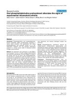
Conversion of GDP-a-
D
-mannose into GDP-sugars was
detected by RP-HPLC (Fig. 3). The elution profile of the
reaction mixture containing GDP-a-
D
-mannose, NADPH,
and the gmd gene product homologue from A. actinomyce-
temcomitans SUNYaB 75 (Fig. 3B) or E. coli K12 (Fig. 3C)
are shown. GDP-4-keto-6-deoxy-
D
-mannose was detected
as a broad peak (42.0 min). Both reactions involving the
gmd gene products halted after consuming some of
the GDP-a-
D
-mannose, regardless of the addition of the
proteins. The peak that appeared at 24.0 min was in
agreement with that of authentic NADP
+
. The reason why
the NADP
+
peak appeared in the gmd gene product
reaction has been unidentified. The retention time of the
putative GDP-6-deoxy-
D
-talose was 36.0 min in the reac-
tion mixture containing GDP-a-
D
-mannose, NADPH, and
the gmd and tld gene products of A. actinomycetemcomitans
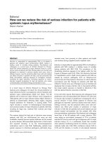
SUNYaB 75 (Fig. 3D). To determine the mass of this final
product, it was purified and analysed by ESI/MS (Fig. 4).
The peak in the ESI/MS spectrum of the product was at
590.1, which corresponds to the [M + H]
+
ion of GDP-6-
deoxy-
D
-talose.
1
H NMR analysis of the structure of the purified
GDP-6-deoxyhexose
Approximately 2 mg of the sugar nucleotide obtained
from the enzyme assay using the gmd and tld gene
products were pooled from several RP-HPLC runs on the
ODS-80Ts. After removing the excess phosphate by
adding ethanol, the solution was concentrated by freeze-
drying. The concentrated solution was lyophilized and
dissolved in D
2
O. The NMR spectra of authentic GTP
and GDP-a-
D
-mannose were also measured. The spectra
of authentic GTP and GDP-a-
D
-mannose, and the sugar
nucleotide are shown in Fig. 5. Assignment of these
resonances was verified in two-dimensional homonuclear
1
H-
1
H COSY experiments (data not shown). The assigned
chemical shifts and coupling constants are summarized in
Table 1. The signals for the nucleotide moieties in the
GDP-sugars were in good agreement with those of GDP.
5966 N. Suzuki et al. (Eur. J. Biochem. 269) Ó FEBS 2002
The signals for H2¢ of the nucleotide moieties overlapped
that of HOD, and the H5¢¢ and H6¢¢ signals of the sugar
moieties of GDP-a-
D
-mannose also overlapped. The
signals for the sugar moiety of the predicted GDP-6-
deoxy-
D
-talose were H6¢¢ (1.18 p.p.m., doublet), H4¢¢
(3.64 p.p.m., double doublet), H5¢¢ (3.88 p.p.m., multi-
plet), H3¢¢ (3.91 p.p.m., double doublet), H2¢¢ (4.03 p.p.m.,
double doublet) and H1¢¢ (5.50 p.p.m., doublet). From the
observed coupling constants J
(1,2)
¼ 5.40 Hz,
J
(2,3)
¼ 3.40 Hz, J
(3,4)
¼ 6.36 Hz and J
(4,5)
¼ 1.96 Hz,
the orientations of H1¢¢,H2¢¢,H3¢¢,H4¢¢ and H5¢¢ are
estimated to be equatorial, equatorial, axial, equatorial
and axial, respectively. This does not conflict with the
structure of GDP-6-deoxy-
D
-talose. Moreover, the con-
formation of –CH
3
was equatorial in this sugar nucleotide.
Since rhamnose has the corresponding coupling constants
of 1.5 (J
(1,2)
),3.5(J
(2,3)
),9.5(J
(3,4)
),and9.5(J
(4,5)
) Hz,
which are totally different from the current ones [31], we
can exclude the possibility that the product was GDP-
D
-
rhamnose and can conclude that GDP-6-deoxy-
D
-talose
was selectively synthesized. In Table 1, the value of J
(1,2)
comes from H2¢¢ , which did not agree with that from H1¢¢ .
The coupling constant J
(1,2)
must be identical, regardless
of whether it comes from H1¢¢ or H2¢¢. It is, however,
possible that the neighbouring phosphate groups affect the
coupling constant [32,33].
Determining the absolute configuration of the talosyl
residue in the GDP-6-deoxytalose by GC/MS
Based on the coupling constants in the
1
H NMR spectra, we
determined that the sugar nucleotide is GDP-6-deoxytalose.
However, the prediction that the absolute configuration of
the talosyl residue in the GDP-6-deoxytalose is
D
was not
supported by direct evidence. GC/MS was performed to
prove the hypothesis. The GDP-6-deoxytalose, the purified
SPAs of A. actinomycetemcomitans ATCC 29523 (serotype
a) and NCTC 9710 (serotype c) were hydrolysed, and then
the talosyl residues were detected as
D
-(+)-2-octylglycoside
acetates. Examination of the mass chromatogram library
produced four fragment ion peaks from 6-deoxytalose for
the talosyl residues of the GDP-6-deoxytalose and the SPA
of ATCC 29523 (Fig. 6A and B). The four peaks were
thought to be two pyranosides and two franosides. The
retention times (13.6, 23.5, 40.8, and 49.0 min) of the
Fig. 3. RP-HPLC profiles during synthesis of GDP-6-deoxy-
D
-talose:
GDP-a-
D
-mannose (1), NADP (2), GDP-4-keto-6-deoxy-
D
-mannose
(3), and GDP-6-deoxy-
D
-talose (4). Samples were injected onto a
TSKgel ODS-80Ts column. (A) No enzyme was added to the reaction
mixture. (B) The purified His
6
-tagged gmd gene product of A. actino-
mycetemcomitans SUNYaB 75 was added to the reaction mixture. (C)
The purified gmd gene product of E. coli K12 were added to the
reaction mixture. (D) The purified His
6
-tagged gmd and tld gene
products were added to the reaction mixture.
Fig. 4. The ESI/MS spectra for the authentic GDP-a-
D
-mannose (A)
and the reaction product GDP-a-
D
-mannose, NADPH, and the gmd and
tld gene products of A. actinom ycetem comitans SUNYaB 75 (B).
Ó FEBS 2002 GDP-6-deoxy-
D
-talose synthetic enzyme (Eur. J. Biochem. 269) 5967
GDP-6-deoxytalose agreed well with those (13.6, 24.0, 41.1,
and 49.3 min) of the SPA of ATCC 29523 (serotype a),
which is 6-deoxy-
D
-talan. Conversely, the retention times
(13.1, 22.0, and 22.7 min) of the fragment ion peaks derived
from 6-deoxy-
L
-talose in the SPA of NCTC 9710 (serotype c)
did not agree with the other retention times (Fig. 6C). Thus,
it was determined that the talose in GDP-6-deoxytalose had
the
D
absolute configuration.
DISCUSSION
Previously, we cloned and characterized the gene clusters
responsible for the biosynthesis of SPAs of A. actinomyce-
temcomitans serotypes a, b, c, d, and e [23,29,34–36]. The
gene cluster associated with the synthesis of SPA in
A. actinomycetemcomitans SUNYaB 75 (serotype a) con-
tains 14 ORFs (Fig. 2A). A protein database search was
performed with the programs
FASTA
[37] and
BLAST
at the
National Institute of Genetics, Mishima, Japan. The
products of 11 genes, ORF2–ORF12, were homologous
to bacterial gene products involved in the biosynthesis of
extracellular polysaccharides. Only the proteins encoded by
ORF3 and ORF4, ABC transport proteins, showed high
identities (64.0 and 73.0%, respectively) to the proteins enco-
ded by ORFs in the clusters responsible for synthesizing
the SPAs in other serotypes of A. actinomycetemcomitans.
The biosynthetic pathway for GDP-6-deoxy-
D
-talose,
Fig. 5.
1
H NMR spectra of GTP (A) and GDP-a-
D
-mannose (B), and
the purified GDP-hexose converted from GDP-a-
D
-mannose by the gmd
and tld gene products (C). The inset shows an expansion of the H4¢–
H4¢¢ region.
Fig. 6. Gas-liquid chromatograph spectra of the acetylated
D
-(+)-2-
octyl glycosides obtained from the hydrolysate of the purified GDP-6-
deoxytalose. (A) Glycosides of the purified serotype a-specific poly-
saccharide antigen (ATCC 29523). (B) Glycosides of the hydrolysate of
the purified GDP-hexose converted from GDP-a-
D
-mannose by the
gmd and tld gene products. (C) Glycosides of the purified serotype
c-specific polysaccharide antigen (NCTC 9710). Arrows indicate the
fragment ion peaks from 6-deoxytalose for the talosyl residues.
5968 N. Suzuki et al. (Eur. J. Biochem. 269) Ó FEBS 2002
which is the activated nucleotide sugar form of 6-deoxy-
D
-
talose, is predicted to be quite different from the pathways
for the precursors of serotype b-, c-, d-, and e-specific
polysaccharide antigens [38]. Insertional inactivation of
ORF2, 3 and ORF7 through ORF12 resulted in loss of
the ability of A. actinomycetemcomitans SUNYaB 75 cells
to produce the polysaccharide. In these genes the ORF2
product shared 58.0% identity with the manC gene
product in E. coli [39]. The manC gene product is a
a-mannose-1-phosphate guanylyltransferase, which con-
verts GTP and a-mannose-1-phosphate into GDP-
a-
D
-mannose. The ORF9 product shared 52.0% identity
with the GDP-a-
D
-mannose 4,6-dehydratase of Y. pseu-
dotuberculosis [24]. In general, GDP-a-
D
-mannose
4,6-dehydratase is an important enzyme converting
GDP-a-
D
-mannose to GDP-4-keto-6-deoxy-
D
-mannose in
the pathway of GDP-
L
-fucose biosynthesis in many
bacteria, plants and mammals [40]. The ORF7 product
had 28.0% homology to the rmd gene product of
P. aeruginosa [25], which reduces GDP-4-keto-6-deoxy-
D
-
mannose to GDP-
D
-rhamnose [20]. GDP-6-deoxy-
D
-talose
is a configrational isomer of GDP-
D
-rhamnose. The rmd
gene product is a reductase that reduces the C4 position of
GDP-4-keto-6-deoxy-
D
-mannose to GDP-
D
-rhamnose,
and we postulated that ORF7 in A. actinomycetemcomi-
tans SUNYaB 75 encodes another reductase producing
GDP-6-deoxy-
D
-talose from GDP-4-keto-6-deoxy-
D
-man-
nose, in spite of sharing low identity (28.0%). Several
consensus domains exist in the tld and rmd gene products.
Among these, the structure YXXXK is an important
conserved structure within the short-chain dehydrogenase/
reductase family [41]. In addition, both the tld and rmd
gene products contain an NAD-binding domain,
GXXGXXG, located near the N-terminus. The tld gene
product can utilize either NADPH or NADH, although
NADPH is used efficiently (data not shown). dTDP-4-
keto-
L
-rhamnose reductase in the biosynthesis of dTDP-6-
deoxy-
L
-talose in A. actinomycetemcomitans NCTC 9710
(serotype c) also preferred NADPH as a cofactor over
NADH [33]. For NCTC 9710, the retention time of the
NADP
+
peak overlapped that of the dTDP-6-deoxy-
L
-
talose peak in RP-HPLC, and NADH was used as the
coenzyme.
The gmd and tld gene products in A. actinomycetem-
comitans SUNYaB 75 were obtained as His
6
-tagged
proteins. The enzymatic activities of the purified His
6
-
tagged gmd and tld gene products were determined by RP-
HPLC analysis. Previously, the gmd gene product with a
His
6
-tag bound at its N terminus was found to be
enzymatically inactive, perhaps because the multiple His-
extender peptide affected its protein structure and altered
the accessibility of the NADP
+
-binding site [27]. Consid-
ering this, we constructed plasmids with the His
6
-tag
bound to the C terminus.
We reported the pathways of dTDP-
D
-fucose (Y4) and
dTDP-6-deoxy-
L
-talose (NCTC 9710) syntheses in
A. actinomycetemcomitans, previously [32,33]. Sugar
nucleotides were detected and collected by RP-HPLC with
0.5
M
KH
2
PO
4
buffer as the mobile phase. In this study, the
retention time (5.1 min) of the GDP-6-deoxy-
D
-talose
profile was close to that (5.0 min) of the GDP-4-keto-6-
deoxy-
D
-mannose profile with 0.5
M
KH
2
PO
4
buffer, in
spite of the different shapes of the two peaks. We could
effectively collect the GDP-6-deoxy-
D
-talose quickly using
0.5
M
KH
2
PO
4
buffer as the mobile phase. By contrast,
for detection, 30 m
M
potassium phosphate (pH 6.0)
containing 5 m
M
tetrabutylammonium hydrogen sulfate
and 2% acetonitrile was used as the mobile phase to
definitely separate the products in the reaction mixture. To
confirm that the intermediate is GDP-4-keto-6-deoxy-
D
-
mannose, the gmd gene product of E. coli K12 was used.
The gmd gene of E. coli has been characterized [39,40].
The retention time of the product profile in the enzyme
assay by the gmd gene product derived from A. actino-
mycetemcomitans SUNYaB 75 was obtained as broad peaks
at 42.0 min, which agree with those from E. coli K12
(Fig. 3B and C).
The conversion of GDP-a-
D
-mannose into GDP-4-
keto-6-deoxy-
D
-mannose stopped when about 50% of the
GDP-a-
D
-mannose was used up. Addition of the protein
was not effective in advancing the reaction. Conversely,
in the Ôone-potÕ assay almost complete conversion
Table 1. NMR spectroscopic identification of GTP, GDP-a-
D
-mannose, and GDP-a-6-deoxy-
D
-talose. (s) Singlet, (d) doublet, (t) triplet, (dd) double
doublet, (m) multiplet. An asterisk indicates that the signal is broad and weakly coupling with H-5¢. ND, not determined.
GTP GDP-a-
D
-mannose GDP-a-6-deoxy-
D
-talose
Proton
Chemical shift
d (p.p.m.)
Chemical shift
d (p.p.m.)
Coupling constant
J (Hz)
Chemical shift
d (p.p.m.)
Coupling constant
J (Hz)
H-8 8.10 (s) 8.09 (s) 8.09 (s)
H-1¢ 5.91 (d) 5.92 (d) 5.91 (d)
H-2¢ ND ND ND
H-3¢ 4.55 (m) 4.50 (dd) 4.48 (dd)
H-4¢ 4.34 (d*) 4.33 (d*) 4.32 (dd*)
H-5¢ 4.22 (m) 4.19 (t) 4.16 (m)
H-1¢¢ 5.50 (d) J
1,2
6.36 5.50 (d) J
1,2
5.40
H-2¢¢ 4.03 (d) J
2,3
2.92 4.03 (dd) J
2,3
3.40
H-3¢¢ 3.90 (dd) J
3,4
9.76 3.91 (dd) J
3,4
6.36
H-4¢¢ 3.66 (t) J
4,5
9.76 3.64 (dd) J
4,5
1.96
H-5¢¢ ND 3.88 (m)
H-6¢¢ ND 1.18 (d)
Ó FEBS 2002 GDP-6-deoxy-
D
-talose synthetic enzyme (Eur. J. Biochem. 269) 5969
occurred. It is possible that feedback inhibition of the
GDP-a-
D
-mannose 4,6-dehydratase occurs via the GDP-
6-deoxy-
D
-talose pathway in A. actinomycetemcomitans
SUNYaB 75. In the enzyme assay using the purified
His
6
-tagged gmd and tld gene products in two successive
steps, the GDP-4-keto-6-deoxy-
D
-mannose was com-
pletely converted into GDP-6-deoxy-
D
-talose, but no
new GDP-4-keto-6-deoxy-
D
-mannose was produced (data
not shown). It is considered that when the tld gene
product was added, the gmd gene product might have
become inactive. However, further detailed analysis has
not been carried out.
In 1973, 6-deoxy-
L
-talose was characterized as an unusual
sugar, and the instability of dTDP-6-deoxy-
L
-talose, which
is the activated sugar nucleotide form of 6-deoxy-
L
-talose,
was reported [42]. Furthermore, we reported that dTDP-6-
deoxy-
L
-talose was degraded in mild alkaline conditions
[33]. GDP-6-deoxy-
D
-talose was more sensitive to alkaline
conditions and heat than dTDP-6-deoxy-
L
-talose. For
example, after GDP-6-deoxy-
D
-talose collected from
ODS-80Ts was evaporated at room temperature using the
same method as for dTDP-6-deoxy-
L
-talose, the peak for
this sugar nucleotide disappeared from the RP-HPLC
elution profile (data not shown). In this study, freeze-drying
was used to concentrate the samples. GDP-4-keto-6-deoxy-
D
-mannose was also unstable. Kneidinger et al. reported
that the product produced from GDP-a-
D
-mannose by the
gmd gene product in A. thermoaerophilus was unstable and
decomposed to form GMP and GDP, as judged by anion
exchange HPLC analysis [20].
The GC contents of the genes essential for SPA
biosynthesis in A. actinomycetemcomitans SUNYaB 75
are lower than the average GC content (47.8%) of the
genes flanking them. The GC contents of ORF2, ORF7,
and ORF9 were 37.2, 30.5 and 35.4%, respectively
(Fig. 2A). It has been reported that genes encoding basic
cellular functions in A. actinomycetemcomitans have an
average GC content of 48.0% [43]. The GC content of the
region essential for the biosynthesis of SPA in the other
serotype strains (b–e) of A. actinomycetemcomitans is also
lower than the average GC content of these genes (48.0%)
[29,34–36]. A lower GC content has been found in gene
clusters involved in the synthesis of various bacterial
polysaccharides [44–46]. These findings suggest the inter-
specific transfer of these genes from other species with a low
GC content to A. actinomycetemcomitans [47].
In conclusion, we identified GDP-4-keto-6-deoxy-
D
-mannose reductase, which converts GDP-4-keto-
6-deoxy-
D
-mannose into GDP-6-deoxy-
D
-talose in
A. actinomycetemcomitans SUNYaB 75 (serotype a), and
revealed the enzymatic process involved in GDP-6-deoxy-
D
-talose synthesis.
ACKNOWLEDGEMENTS
This work was supported in part by a Grant-in-Aid for Encouragement
of Young Scientists 13771265 (Y. N.), 14771185 (Y. Yo.) and a Grant-
in-Aid for Developmental Scientific Research 12557186 (Y. Ya.) from
the Ministry of Education, Culture, Sports, Science and Technology,
Tokyo, Japan, by a research grant from the Takeda Science
Foundation (Y. N.), and by Research Fellowships from the Japan
Society for the Promotion of Science for Young Scientists 13010070
(N. S.).
REFERENCES
1. Roberts, I.S. (1996) The biochemistry and genetics of capsular
polysaccharide production in bacteria. Annu. Rev. Microbiol. 50,
285–315.
2. Asikainen, S., Lai, C H., Alaluusua, S. & Slots, J. (1991) Dis-
tribution of Actinobacillus actinomycetemcomitans serotypes in
periodontal health and disease. Oral Microbiol. Immunol. 6,115–
118.
3. Ebersole, J.L., Cappelli, D. & Sandoval, M N. (1994) Subgingival
distribution of A. actinomycetemcomitans in periodontitis. J. Clin.
Periodontol. 21, 65–75.
4. Meyer, D.H. & Fives-Taylor, P.M. (1997) The role of Actino-
bacillus actinomycetemcomitans in the pathogenesis of periodontal
disease. Trends Microbiol. 5, 224–228.
5. Slots, J., Bragd, L., Wikstro
¨
m, M. & Dahle
´
n, G. (1986) The
occurrence of Actinobacillus actinomycetemcomitans, Bacteroides
gingivalis and Bacteroides intermedius in destructive periodontal
disease in adults. J. Clin. Periodontol. 13, 570–577.
6. Kaplan, A.H., Weber, D.J., Oddone, E.Z. & Perfect, J.R. (1989)
Infection due to Actinobacillus actinomycetemcomitans:15cases
and review. Rev. Infect. Dis. 11, 46–63.
7. Zambon, J.J., Slots, J. & Genco, R.J. (1983) Serology of oral
Actinobacillus actinomycetemcomitans and serotype distribution in
human periodontal disease. Infect. Immun. 41, 19–27.
8. Saarela, M., Asikainen, S., Alaluusua, S., Pyha
¨
la
¨
, L., Lai, C H. &
Jousimies-Somer, H. (1992) Frequency and stability of mono- or
poly-infection by Actinobacillus actinomycetemcomitans serotypes
a, b, c, d or e. Oral Microbiol. Immunol. 7, 277–279.
9. Gmu
¨
r, R., McNabb, H., van Steenbergen, T.J.M., Baehni, P.,
Mombelli, A., van Winkelhoff, A.J. & Guggenheim, B. (1993)
Seroclassification of hitherto nontypeable Actinobacillus actino-
mycetemcomitans strains: evidence for a new serotype e. Oral
Microbiol. Immunol. 8, 116–120.
10. Kaplan, J.B., Perry, M.B., MacLean, L.L., Furgang, D., Wilson,
M.E. & Fine, D.H. (2001) Structural and genetic analyses of O
polysaccharide from Actinobacillus actinomycetemcomitans sero-
type f. Infect. Immun. 69, 5375–5384.
11. Koga, T., Nishihara, T., Amano, K., Takahashi, T., Nakashima,
K., Ishihara, Y. & Shibuya, N. (1991) Chemical and biological
properties of cell-surface components of Actinobacillus actinomy-
cetemcomitans.InPeriodontal Disease: Pathogens and Host
Immune Responses (Hamada, S., Holt, S.C. & McGhee, J.R., eds),
pp. 117–127. Quintessence Publishing Co, Ltd, Tokyo, Japan.
12. Amano, K., Nishihara, T., Shibuya, N., Noguchi, T. & Koga, T.
(1989) Immunochemical and structural characterization of a
serotype-specific polysaccharide antigen from Actinobacillus acti-
nomycetemcomitans Y4 (serotype b). Infect. Immun. 57, 2942–2946.
13. Califano, J.V., Schenkein, H.A. & Tew, J.G. (1991)
Immunodominant antigens of Actinobacillus actinomycetemcomi-
tans serotypes a and c in high-responder patients. Oral Microbiol.
Immunol. 6, 228–235.
14. Page, R.C., Sims, T.J., Engel, L.D., Moncla, B.J., Bainbridge, B.,
Stray, J. & Darveau, R.P. (1991) The immunodominant outer
membrane antigen of Actinobacillus actinomycetemcomitans is
located in the serotype-specific high- molecular-mass carbohydrate
moiety of lipopolysaccharide. Infect. Immun. 59, 3451–3462.
15. Wilson, M.E. & Schifferle, R.E. (1991) Evidence that the serotype
b antigenic determinant of Actinobacillus actinomycetemcomitans
Y4 resides in the polysaccharide moiety of lipopolysaccharide.
Infect. Immun. 59, 1544–1551.
16. Lu, H., Califano, J.V., Schenkein, H.A. & Tew, J.G. (1993)
Immunoglobulin class and subclass distribution of antibodies
reactive with the immunodominant antigen of Actinobacillus
actinomycetemcomitans serotype b. Infect. Immun. 61, 2400–2407.
17. Shibuya, N., Amano, K., Azuma, J., Nishihara, T., Kitamura, Y.,
Noguchi, T. & Koga, T. (1991) 6-Deoxy-
D
-talan and 6-deoxy-
L
-
5970 N. Suzuki et al. (Eur. J. Biochem. 269) Ó FEBS 2002
talan. Novel serotype-specific polysaccharide antigens from
Actinobacillus actinomycetemcomitans. J. Biol. Chem. 266, 16318–
16323.
18. Perry, M.B., MacLean, L.M., Brisson, J R. & Wilson, M.E.
(1996) Structures of the antigenic O-polysaccharides of lipopoly-
saccharides produced by Actinobacillus actinomycetemcomitans
serotypes a, c, d and e. Eur. J. Biochem. 242, 682–688.
19. Za
¨
hringer, U., Rettenmaier, H., Moll, H., Senchenkova, S.N. &
Knirel, Y.A. (1997) Structure of a new 6-deoxy-a-
D
-talan from
Burkholderia (Pseudomonas) plantarii strain DSM 6535, which is
different from the O-chain of the lipopolysaccharide. Carbohydr.
Res. 300, 143–151.
20. Elbein, A.D. & Heath, E.C. (1965) The biosynthesis of cell wall
lipopolysaccharide in Escherichia coli. II. Guanosine diphosphate
4-keto-6-deoxy-
D
-mannose, an intermediate in the biosynthesis of
guanosine diphosphate colitose. J. Biol. Chem. 240, 1926–1931.
21. Sullivan, F.X., Kumar, R., Kriz, R., Stahl, M., Xu, G Y., Rouse,
J., Chang, X.J., Boodhoo, A., Potvin, B. & Cumming, D.A. (1998)
Molecular cloning of human GDP-mannose 4,6-dehydratase and
reconstitution of GDP-fucose biosynthesis in vitro. J. Biol. Chem.
273, 8193–8202.
22.Kneidinger,B.,Graninger,M.,Adam,G.,Puchberger,M.,
Kosma, P., Zayni, S. & Messner, P. (2001) Identification of
two GDP-6-deoxy-
D
-lyxo-4-hexulose reductases synthesizing
GDP-
D
-rhamnose in Aneurinibacillus thermoaerophilus L420–91
T
.
J. Biol. Chem. 276, 5577–5583.
23. Suzuki, N., Nakano, Y., Yoshida, Y., Nakao, H., Yamashita, Y.
& Koga, T. (2000) Genetic analysis of the gene cluster for the
synthesis of serotype a-specific polysaccharide antigen in Actino-
bacillus actinomycetemcomitans. Biochim. Biophys. Acta 1517,
135–138.
24. Skurnik, M., Peippo, A. & Ervela
¨
, E. (2000) Characterization of
the O-antigen gene clusters of Yersinia pseudotuberculosis and the
cryptic O-antigen gene cluster of Yersinia pestis shows that the
plague bacillus is most closely related to and has evolved from
Y. pseudotuberculosis serotype O: 1b. Mol. Microbiol. 37, 316–330.
25. Rocchetta,H.L.,Pacan,J.C.&Lam,J.S.(1998)Synthesisofthe
A-band polysaccharide sugar
D
-rhamnose requires Rmd and
WbpW: identification of multiple AlgA homologues, WbpW and
ORF488. Pseudomonas aeruginosa. Mol. Microbiol. 29, 1419–
1434.
26. Sambrook, J., Fritsch, E.F. & Maniatis, T. (1989) Molecular
Cloning: A Laboratory Manual, Vol. 3, 2nd edn. Cold Spring
Harbor Laboratory Press, Cold Spring Harbor, New York, USA.
27. Albermann, C., Distler, J. & Piepersberg, W. (2000) Preparative
synthesis of GDP-b-
L
-fucose by recombinant enzymes from
enterobacterial sources. Glycobiology 10, 875–881.
28. Tonetti, M., Sturla, L., Bisso, A., Benatti, U. & De Flora, A.
(1996) Synthesis of GDP-
L
-fucose by the human FX protein.
J. Biol. Chem. 271, 27274–27279.
29. Yoshida, Y., Nakano, Y., Yamashita, Y. & Koga, T. (1998)
Identification of a genetic locus essential for serotype b-specific
antigen synthesis in Actinobacillus actinomycetemcomitans. Infect.
Immun. 66, 107–114.
30. Leontein, K., Lindberg, B. & Lonngren, J. (1978) Assignment
of absolute configuration of sugars by g.l.c of their acetylated
glycosides formed from chiral alcohols. Carbohydr. Res. 62,
359–362.
31. Rockey, W.M., Dowd, M.K., Reilly, P.J. & French, A.D. (2001)
Modeling of deoxy- and dideoxyaldohexopyranosyl ring pucker-
ing with MM3 (92). Carbohydr. Res. 335, 261–273.
32. Yoshida, Y., Nakano, Y., Nezu, T., Yamashita, Y. & Koga, T.
(1999) A novel NDP-6-deoxyhexosyl-4-ulose reductase in the
pathway for the synthesis of thymidine diphosphate-
D
-fucose.
J. Biol. Chem. 274, 16933–16939.
33. Nakano, Y., Suzuki, N., Yoshida, Y., Nezu, T., Yamashita, Y. &
Koga, T. (2000) Thymidine diphosphate-6-deoxy-
L
-lyxo-4-hex-
ulose reductase synthesizing dTDP-6-deoxy-
L
-talose from Acti-
nobacillus actinomycetemcomitans. J. Biol. Chem. 275, 6806–6812.
34. Nakano, Y., Yoshida, Y., Yamashita, Y. & Koga, T. (1998) A
gene cluster for 6-deoxy-
L
-talan synthesis in Actinobacillus
actinomycetemcomitans. Biochim. Biophys. Acta 1442, 409–414.
35. Yoshida, Y., Nakano, Y., Suzuki, N., Nakao, H., Yamashita, Y.
& Koga, T. (1999) Genetic analysis of the gene cluster responsible
for synthesis of serotype e-specific polysaccharide antigen in
Actinobacillus actinomycetemcomitans. Biochim. Biophys. Acta
1489, 457–461.
36. Nakano, Y., Yoshida, Y., Suzuki, N., Yamashita, Y. & Koga, T.
(2000) A gene cluster for the synthesis of serotype d-specific
polysaccharide antigen in Actinobacillus actinomycetemcomitans.
Biochim. Biophys. Acta 1493, 259–263.
37. Lipman, D.J. & Pearson, W.R. (1985) Rapid and sensitive protein
similarity searches. Science 227, 1435–1441.
38. Shibaev, V.N. (1986) Biosynthesis of bacterial polysaccharide
chains composed of repeating units. Adv. Carbohydr. Chem. Bio-
chem. 44, 277–339.
39. Stevenson, G., Andrianopoulos, K., Hobbs, M. & Reeves, P.R.
(1996) Organization of the Escherichia coli K-12 gene cluster
responsible for production of the extracellular polysaccharide
colanic acid. J. Bacteriol. 178, 4885–4893.
40. Sturla, L., Bisso, A., Zanardi, D., Benatti, U., De Flora, A. &
Tonetti, M. (1997) Expression, purification and characterization
of GDP-
D
-mannose 4,6-dehydratase from Escherichia coli. FEBS
Lett. 412, 126–130.
41. Jo
¨
rnvall, H., Persson, B., Krook, M., Atrian, S., Gonzalez-
Duarte, R., Jeffery, J. & Ghosh, D. (1995) Short-Chain Dehy-
drogenases/Reductases (SDR). Biochemistry 34, 6003–6013.
42. Gaugler, R.W. & Gabriel, O. (1973) Biological mechanisms
involved in the formation of deoxy sugars. VII. Biosynthesis of
6-deoxy-
L
-talose. J. Biol. Chem. 248, 6041–6049.
43. Kaplan, J.B. & Fine, D.H. (1998) Codon usage in Actinobacillus
actinomycetemcomitans. FEMS Microbiol. Lett. 163, 31–36.
44. Morona, R., Mavris, M., Fallarino, A. & Manning, P.A. (1994)
Characterization of the rfc region of Shigella flexneri. J. Bacteriol.
176, 733–747.
45. Jiang, X M., Neal, B., Santiago, F., Lee, S.J., Romana, L.K. &
Reeves, P.R. (1991) Structure and sequence of the rfb (O antigen)
gene cluster of Salmonella serovar typhimurium (strain LT2). Mol.
Microbiol. 5, 695–713.
46. Arakawa, Y., Wacharotayankun, R., Nagatsuka, T., Ito, H.,
Kato,N.&Ohta,M.(1995)GenomicorganizationoftheKleb-
siella pneumoniae cps region responsible for serotype K2 capsular
polysaccharide synthesis in the virulent strain Chedid. J. Bacteriol.
177, 1788–1796.
47. Reeves, P. (1993) Evolution of Salmonella O antigen variation by
interspecific gene transfer on a large scale. Trends Genet. 9, 17–22.
Ó FEBS 2002 GDP-6-deoxy-
D
-talose synthetic enzyme (Eur. J. Biochem. 269) 5971