Báo cáo khoa học: De-regulation of D-3-phosphoglycerate dehydrogenase by domain removal ppt
Bạn đang xem bản rút gọn của tài liệu. Xem và tải ngay bản đầy đủ của tài liệu tại đây (288.94 KB, 9 trang )
De-regulation of
D
-3-phosphoglycerate dehydrogenase by domain
removal
Jessica K. Bell
1
, Paul J. Pease
1
, J. Ellis Bell
2
, Gregory A. Grant
3
and Leonard J. Banaszak
1
1
Department of Biochemistry, Molecular Biology and Biophysics, University of Minnesota, Minneapolis, MN, USA;
2
Department of Chemistry, University of Richmond, Richmond, Virginia, USA;
3
Department of Molecular Biology
and Pharmacology and the Department of Medicine, Washington University, St Louis, MO, USA
Escherichia coli 3-phosphoglycerate dehydrogenase
(PGDH) catalyzes the first step in serine biosynthesis, and is
allosterically inhibited by serine. Structural studies revealed a
homotetramer in which the quaternary arrangement of
subunits formed an elongated ellipsoid. Each subunit
consisted of three domains: nucleotide, substrate and regu-
latory. In PGDH, extensive interactions are formed between
nucleotide binding domains. A second subunit–subunit
interaction occurs between regulatory domains creating an
extended b sheet. The serine-binding sites overlap this
interface. In these studies, the nucleotide and substrate
domains (NSDs) were subcloned to identify changes in both
catalytic and physical properties upon removal of a subunit–
subunit interface. The NSDs did not vary significantly from
PGDH with respect to kinetic parameters with the exception
that serine no longer had an effect on catalysis. Temperature
dependent dynamic light scattering (DLS) revealed the
NSDs aggregated > 5 °C before PGDH, indicating de-
creased stability. DLS and gel filtration studies showed that
the truncated enzyme formed a tetramer. This result negated
the hypothesis that the removal of the regulatory domain
would create an enzyme mimic of the unregulated, closely
related dimeric enzymes. Expression of the regulatory do-
main, to study conformational changes induced by serine
binding, yielded a product that by CD spectra contained
stable secondary structure. DLS and pulsed field gradient
NMR studies of the regulatory domain showed the presence
of higher oligomers instead of the predicted dimer. We have
concluded that the removal of the regulatory domain is
sufficient to eliminate serine inhibition but does not have the
expected effect on the quaternary structure.
Keywords: domains; enzyme regulation; oxidoreductase;
3-phosphoglycerate dehydrogenase; truncation.
D
-3-Phosphoglycerate dehydrogenase (PGDH) catalyzes
the first committed step in the phosphorylated serine
biosynthetic pathway. During the PGDH reaction, 3-phos-
phoglycerate (GriP), a glycolytic intermediate, is oxidized to
3-phosphohydroxypyruvate (PHP) with the concomitant
reduction of NAD. The pathway, as a branch point off the
glycolysis pathway, is tightly regulated. In prokaryotes and
lower plants, an inhibitory feedback loop utilizes serine to
allosterically regulate the initial step of the pathway, the
PGDH reaction [1–3]. The serine modulation occurs
through rare V
max
-type effects, and may be contrasted with
the more common regulation that directly affects the
binding of substrate(s) by altering K
m
[4].
PGDH belongs to a family of
D
-2-hydroxyacid
dehydrogenases that includes formate dehydrogenase,
D
-glycerate dehydrogenase,
D
-lactate dehydrogenase, ery-
thronate-4-phosphate dehydrogenase,
D
-2-isocaproate
dehydrogenase and vancomycin resistant protein [4]. The
family members share % 22% sequence identity and 50%
sequence similarity. Among the
D
-2-hydroxyacid dehydro-
genases all members are dimeric with the exception of
PGDH, which forms a homotetramer. Crystallographic
studies of four enzymes within this family {2nac (for-
mate,dehydrogenase [5]), 1gdh (
D
-glycerate dehydrogenase
[6]), 2dld (
D
-lactate dehydrogenase), 1psd (3-phosphoglycer-
ate dehydrogenase [7,8]} have revealed a striking similarity
in their conformations, except for the additional regulatory
domaininPGDH.
The crystal structure of the PGDH:NAD:serine complex
[7] is depicted in Fig. 1 and illustrates both the domain and
quaternary arrangements. The 222 symmetric tetramer has
four binding sites for both serine and NADH. The donut-
like appearance of PGDH is similar to the tetrameric form
of glycerol kinase [9], another enzyme that is regulated by
V
max
-type kinetic changes. The interface encompassing
adjacent nucleotide binding domains is labeled I,andthis
subunit:subunit contact is shared among all
D
-2-hydroxy-
acid dehydrogenases. The additional regulatory domain
forms an important new subunit interface, labeled II.The
two serine-binding sites located at each interface are
comprised of residues from both subunits. As will be shown
Correspondence to L. J. Banaszak, 6-155 Jackson Hall,
Department of Biochemistry, Molecular Biology and Biophysics,
University of Minnesota, 321 Church St S.E., Minneapolis,
MN 55455, USA.
Fax: + 1 612 625 2163, Tel.: + 1 612 626 6597,
E-mail:
Abbreviations:PGDH,
D
-3-phosphoglycerate dehydrogenase;
NSD, nucleotide and substrate domains; RBD, regulatory binding
domain; IPTG, isopropyl thio-b-
D
-galactoside; FDH, formate dehy-
drogenase; LDH, lactate dehydrogenase; a-KG, a-ketoglutarate;
PHP, 3-phosphohydroxypyruvate; 3GriP, 3-phosphoglycerate;
DLS, dynamic light scattering; D
t
, translational diffusion
constant; PFG, pulsed-field gradient.
Enzymes:
D
-3-phosphoglycerate dehydrogenase (EC 1.1.1.95).
Note: a website can be found at />(Received 8 May 2002, accepted 25 June 2002)
Eur. J. Biochem. 269, 4176–4184 (2002) Ó FEBS 2002 doi:10.1046/j.1432-1033.2002.03075.x
in this report, the tetrameric PGDH belongs to a family of
dimeric homologues but the differences in quaternary
structure are not explained solely by the presence of the
regulatory domain. Finally a third proposed interface across
the middle of the PGDH toroid near the region labeled III
in Fig. 1 contains essentially no intersubunit contacts except
through the visible loops, residues 160–195. These symmet-
rically related loops could form relatively close hydrophobic
and charge:charge contacts, reinforcing subunit contacts
already stabilized by the interface between nucleotide
binding domains.
The conformational similarity between the family mem-
bers is visible in the stereo-drawing shown in Fig. 2 where a
PGDH subunit and a formate dehydrogenase subunit have
been overlaid by the method of least-squares. The 44-kDa
PGDH subunit is divided as follows: nucleotide binding
domain (residues 108–292), substrate binding domain
(residues 1–102, 304–318), and regulatory or serine-binding
domain (residues 336–410). The interconnecting polypep-
tide segment, residues 103–108, 293–303, and 319–336, may
form hinge-like regions. In fact, the homologous polypep-
tide segments connecting the nucleotide- and substrate-
binding domains in the dimeric family members have been
shown to have conformational variability [5,6,10]. Of equal
relevance, but not shown in Fig. 2, the nucleotide binding
domains of PGDH associate into a dimer interface entirely
homologous with the quaternary structures of dimeric
formate [5], glycerate [6] and
D
-lactate dehydrogenase.
Using this well-defined homology, the potential confor-
mational changes associated with the inhibited vs. the active
form of PGDH were postulated from the crystal structures
of apo- and holo-formate dehydrogenase [5]. These crystal
complexes revealed that the active site cleft, formed by
nucleotide and substrate binding domains, rotated 7.5° into
a more closed conformation when ligand was bound. The
constraints of the tetrameric nature of PGDH suggest that a
similar rotation of the nucleotide and substrate domains
into a more closed conformation at the active site would
require additional relaxation of interactions at the regula-
tory domain interface.
The study of proposed domain movements were exam-
ined by subcloning portions of PGDH to look at the
contribution of the tetrameric structure to catalysis, stability
and potential conformational changes at the serine site upon
ligand binding. Several chimers consisting of the nucleotide
and substrate domains with variable N- and C-termini were
made to resemble counterparts in the 2-hydroxyacid
dehydrogenase family. The kinetic properties and oligo-
meric states of these truncated enzymes were determined
and compared to intact PGDH. In addition, the regulatory
binding domain, RBD, was subcloned to create a smaller
model of the serine-binding pocket that could be manipu-
lated for structural study by NMR and evaluated for
conformational changes upon ligand binding.
MATERIALS AND METHODS
The expression vector, pSAWT containing the serA gene
was described previously [11]. The plasmids, pTrc99A
and pGEX-2T, were from Pharmacia Biotech. PfuDNA
polymerase and the SURE cell line were from Stratagene.
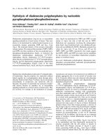
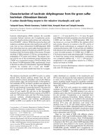
Fig. 2. Stereoview of PGDH and the homologous formate dehydro-
genase. The crystallographic coordinates of formate dehydrogenase
and PGDH have been superimposed by the least-squares methods.
The resulting overlay of the two subunits is shown in stereo with
formate dehydrogenase in red and PGDH in blue. A stick represen-
tation NAD bound to FDH (purple) and PGDH (green) is also shown.
The regulatory domain of PGDH is at the top followed by the sub-
strate binding domain and finally the NAD binding domain at the
bottom of the figure. The overlay of the two coordinate sets illustrates
the close conformational homology between the two enzymes includ-
ing the positioning of the bound coenzyme.
6
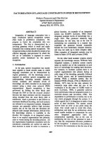
Fig. 1. Structure of PGDH: a summary of structure and mutations. The
cartoon illustrates the crystal structure of the serine-inhibited form of
PGDH. Three of the subunits of the homotetramer are colored gray.
The fourth subunit shows the three component domains, nucleotide-
binding domain (blue), the substrate-binding domain (red) and a
regulatory domain (green). Three arrows mark: (I) the nucleotide-
binding domain interface, and (II) the tetramer interface formed by the
interactions of two regulatory domains and (III) unobserved contact
across the middle of the tetramer. The position of two of the four serine
molecules is shown by van der Waal’s surface at the regulatory inter-
face on the left. Also shown in van der Waal’s surfaces, the NAD
molecule binds within the active site cleft along the top of the nucleo-
tide domain. The numbers 1–7 on the left indicate the Ca positions of
the truncated enzymes. Numbers 1–4 and number 7 describe the NSD
enzymes. Specifically, numbers 1 and 2 show the position of the N
terminus, residue 7 (the first ordered residue in the crystal structure)
and 10, respectively. The blue carbon atoms 3 and 4 indicate residues
314 and 317 at the C-terminus of two of the NSD proteins. Residue
336,usedinboththeNSDandRBDproteins,isindicatedbythegreen
Ca ball.
5
Ó FEBS 2002
D
-3-Phosphoglycerate DH: an active, truncated form (Eur. J. Biochem. 269) 4177
The BLR cell line was from Novagen. Restriction enzymes
and ligase came from either Promega or Boehringer Mann-
heim. Oligonucleotide primers were synthesized by the
Microchemical Facility at the University of Minnesota, or
out-sourced via this facility. DNA gel purification chemicals
were from the Bio-Rad. PCR Cleanup Kit was from
Promega. The Microchemical Facility at the University of
Minnesota confirmed the sequences of DNA inserts. All
other chemicals were from Sigma unless otherwise noted.
Mutagenesis
The nucleotide and substrate domain constructs of residues
1–336 (NSD:336) and 1–317 (NSD:317) were subcloned
from the pSAWT vector using common PCR techniques
into the pTrc99A vector. The NcoIsiteatthe5¢ end of the
serA gene was conserved and a stop codon and unique XbaI
site were introduced at the new 3¢ terminus at residue 336 or
317. The NSD:10–314 and NSD:10–317 mutants were
constructed using the Stratagene Quik Change
TM
mutagen-
esis kit and the NSD:336:pTrc99A vector as the parental
DNA. The RBD:336–410 protein, residues 336–410, was
made using the same technique as the NSD constructs, but
with a BamHI site introduced at the 5¢ end and a HindIII
site at the 3¢ end. The PCR product was ligated into the
pGEX-2T vector. All mutant sequences were confirmed by
DNA sequencing.
Expression and purification
NSD. NSD vectors were transformed into competent SURE
cells. Six 1-L flasks of 2 · YT broth plus 150 lgÆmL
)1
ampicillin were grown at 37 °C until the optical density at
600 nm reached 0.6–0.8. Protein expression was induced
with 1–1.5 m
M
isopropyl thio-b-
D
-galactoside (IPTG). After
induction cells were grown for % 14 h at 22 °C. Cell pellets
were resuspended in 50 m
M
KH
2
PO
4
pH 7.0, 2 m
M
dith-
iothreitol, 1 m
M
EDTA and 0.05% NaN
3
(buffer B) and
lysed by sonication. The remainder of the purification
protocol has been described previously [11]. Purified protein
was concentrated using a Centriprep 10K (Amicon) and
dialyzed into buffer B. Protein concentration was deter-
mined by Bradford assay and/or UV spectra using an
extinction coefficient of 0.67
M
)1
Æcm
)1
. Protein was stored at
4 °C.
RBD:336–410. RBD:336–410 plasmid was transformed
into competent BLR cells. Six 1-L flasks of 2 · YT broth
plus antibiotic were grown at 37 °CtoD
600
¼ 0.6–1.0 and
then induced with 1 m
M
IPTG. Cells were grown for % 14 h
at 22 °C. The cell pellet was resuspended in STE (10 m
M
Tris/HCl pH 8.0, 1 m
M
EDTA, 150 m
M
NaCl) and
incubated on ice with 0.1 mgÆmL
)1
lysozyme for 15 min
The solution was brought to 5 m
M
dithiothreitol, 2% (w/v)
sarkosyl and sonicated. The mixture was stirred at 4 °Cfor
30 min followed by centrifugation at 10 000 g
1
for 30 min.
Polyethyleneamine (0.035%) was added to remove DNA/
RNA, stirred at 4 °C for 30 min and then respun for 30 min
at 10 000 g. The supernatant was concentrated using an
Amicon concentrator with a PM10 membrane (3 kDa cut-
off) and dialyzed into NaCl/P
i
/EDTA (16 m
M
Na
2
HPO
4
,
4m
M
NaH
2
PO
4
, 150 m
M
NaCl, 1 m
M
EDTA pH 7.3). The
dialyzed lysate was respun to remove particulates and
loaded onto a glutathione S-transferase (GST) affinity
column (Novagen). The column was washed with 10
column vols NaCl/P
i
/EDTA and then incubated with
250 U thrombin overnight at room temperature. The
cleaved RBD:336–410 was eluted, concentrated using a
Centriprep 3K (Amicon), and stored at 4 °C. Protein
concentration was calculated from UV spectra using an
extinction coefficient of 0.47
M
)1
Æcm
)1
for RBD:336–410.
The identity of the protein was confirmed by N-terminal
sequencing of the first 10 residues and amino acid analysis
(Microchemical Facility, University of Minnesota, MN,
USA).
Kinetic analysis
The steady-state initial rates were determined by following
either the reduction of 3-PHP or a-ketoglutarate (a-KG).
Thereactionwassetupwithasaturatingconcentrationof
NADH (100–200 l
M
) and varied concentrations of PHP
(1–100 l
M
)ora-KG (10.4–5000 l
M
)at25°C. The enzyme
concentration for the a-KG studies was 1 l
M
and
0.1–0.5 l
M
for the PHP reactions. The assay buffer for
the a-KG reactions was 50 m
M
Tris pH 7.5, 1 m
M
EDTA
and 2 m
M
dithiothreitol. For the 3-PHP reactions, the Tris
concentration was increased to 500 m
M
. The reaction was
initiated by the addition of substrate and the decrease in
D
340
monitored for 10–20 s. The initial rates were deter-
mined by fitting a linear regression to the curve and
calculating the slope using
CARY
50 kinetics software.
Assays were repeated a minimum of five times. The data
were analyzed by Michaelis–Menten or Lineweaver–Burke
plots and kinetic parameters derived using the
SIGMA
PLOT
5.0 software (Jandel Scientific Inc.).
Dynamic light scattering experiments
Dynamic light scattering (DLS) experiments were conducted
in Buffer B for PGDH and the NSD proteins.
RBD:336–410 experiments were done in NaCl/P
i
/EDTA.
For each concentration measured, the protein was spun at
14 000 g for 10 min and passed through a 0.1-l
M
filter. A
12-lL sample was equilibrated by a built-in thermostat at
5 °C increments. Data were collected with a Protein
Solutions DLS system and evaluated with the
DYNAPRO
V
4.0 software. For each temperature 15–20 data points were
collected. Mean values were calculated for the DLS
parameters. Points that were outside 1 SD were excluded.
Data were plotted in
SIGMA PLOT
5.0.
Gel filtration experiments
Gel filtration experiments were performed in Buffer B.
PGDH (2 mgÆmL
)1
), NSD:317 (2 mgÆmL
)1
), or
3
D
-lactate
dehydrogenase (
D
-LDH) (2 mgÆmL
)1
)wererunovera
Sephacryl S200 (Pharmacia) gel filtration column with
both low molecular weight standards (ribonuclease A,
13.7 kDa; chymotrypsinogen A, 25 kDa; ovalbumin,
43 kDa; BSA, 67) (Run 1) and high molecular weight
standards (aldolase, 158 kDa; catalase, 232 kDa; ferritin,
440 kDa; thyroglobulin, 669 kDa) (Run 2). Chromato-
graph profiles were calculated from the absorbance of the
fractions at 280 nm for the molecular mass standards and
activity measurements for PGDH, NSD:317 and
D
-LDH.
4178 J. K. Bell et al. (Eur. J. Biochem. 269) Ó FEBS 2002
The molecular weights of PGDH, NSD:317 and
D
-LDH
were calculated from the linear regression of K
av
[(V
e
) V
o
)/
(V
t
) V
o
), where V
e
is the elution volume, V
o
is the void
volume and V
t
is the total volume] vs. the log of the
molecular weight of the standards.
CD
RBD:336–410 experiments were performed in NaCl/P
i
/
EDTA. CD spectra were collected on protein (0.722ÆmgÆ
mL
)1
) in the presence or absence of 1 m
M
serine. A buffer
blank was completed for both the buffer and buffer plus
1m
M
serine. The spectra were collected on a Jasco 710
instrument at room temperature using a 0.05-mm quartz
cell. Spectra were collected from 250 to % 200 nm with eight
accumulations. The data were averaged over the accumu-
lations, corrected for the buffer blank and random signals
were smoothed using the
JASCO
software package. Data
were exported to
SIGMA PLOT
5.0 for analysis.
Pulsed-field gradient NMR
To corroborate the DLS measurements, pulsed-field gradi-
ent (PFG)-NMR [12,13] was used to give an independent
determination of the translational diffusion constant (D
t
)
for the protein RBD:336–410. NMR was carried out in
collaboration with the Mayo laboratory at the University of
Minnesota. Spectra were collected and analyzed by Shou
Lin Chang of the Mayo laboratory. In the PFG-NMR
experiment, B
o
, constant magnetic field, was superimposed
twice during a short time interval, d, by an additional
inhomogenous gradient (G
z
). The result of the two gradient
pulses is to create an echo. If no motion or relaxation
occurred on the z-axis, the echo would have been identical
to the initial signal. However, the observed echo will be
attenuated by both relaxation and random motion (diffu-
sion), along the z-axis. The attenuation, A(t) can be
described by:
AðtÞ¼Að0Þ
exp
ðÀRðtÞÀc
2
G
2
D
t
d
2
ðd À d=3ÞÞ
where R(t) is attenuation due to relaxation, c is the
magnetogyric ratio, G is the gradient strength, d is the
duration of the gradient pulse, and D is the interval between
the start of the two gradient pulses. To determine an
accurate measurement of D
t
, a series of 12 one-dimensional
PFG spectra were collected at gradient field strengths, 0, 5,
10, 15, 20, 25, 30, 35, 40, 45, 50, 55 and 60 GÆcm
)1
. The data
were then fit to the semi-log of the equation above to
determine the value of D
t
. Experiments were conducted at
both 10 and 25 °C. Protein (%1m
M
)wasin50m
M
KH
2
PO
4
,pH6.5,inD
2
O. Serine, when present, was at
1m
M
. The experiments were carried out on a Varian
UNITY 600 MHz NMR with triple resonance probe and
triple axis gradient unit (High Magnetic Field Facility,
University of Minnesota).
RESULTS AND DISCUSSION
Nucleotide substrate somains from Gri
P
DH
The preparation of a monodisperse form of PGDH
insensitive to the presence of serine but fully active was
designed based on a previously determined crystal structure
(Fig. 1) [7]. Removal of the serine-binding domain was
predicted to eliminate allosteric inhibition by serine and
produce a dimeric enzyme. As shown in the data below,
manipulation of PGDHs quaternary structure was far more
complicated and it was not possible to obtain a dimeric
enzyme. Several variations of the NSD’s were developed
using the standard PCR technique of introducing a stop
codon and unique restriction site at the desired termination
point.
NSD:336 (residues 1–336), the first two-domain protein
to be made, was soluble and yielded % 5–12 mg per 6 L
ferment. However, NSD:336 included a segment of the
linker sequence between the substrate and the regulatory
domains, and tended to form higher oligomeric species
(data not shown). This extended linker may have
decreased stability and provide a site of aggregation,
and was therefore removed in another form NSD:317
(residues 1–317). NSD:317 was monodisperse in solution
and relatively stable (see below), and therefore more
amenable to study. Two other two-domain enzymes were
also created: NSD:10–314 and NSD:10–317. These forms
eliminated the N-terminal segment that was disordered in
crystalline PGDH with serine. The recombinant products
were largely insoluble and further studies were aban-
doned.
Kinetic evaluation of NSDs
As conformational changes had been linked to the catalytic
activity of both PGDH and formate dehydrogenase (FDH)
[5,14] removal of the regulatory domain was hypothesized
to have an effect on the kinetic parameters of PGDH. The
steady-state parameters are reported for two of the chimers
although our primary focus was NSD:317 because the
quaternary structure of this enzyme was definable. The
activities of PGDH and NSD:317 were assayed following
the reduction of PHP, which occurs % 70-fold faster than
the oxidation of GriP [15], or the alternate substrate, a-KG
[16]. Although PHP and a-KG are three- and five-carbon
substrates, respectively, the fourth carbon and 5-carboxyl
of a-KG are similar to the bulky phosphate group in PHP.
The results of the steady-state kinetic studies are summa-
rized in Table 1. For both the PHP and a-KG assays,
substrate inhibition was observed at high concentrations
(Fig. 3), possibly due to the slow release of oxidized
cofactor and leading to an abortive complex of substrate/
NAD. The data from the reduction of PHP and a-KG,
excluding data exhibiting substrate inhibition, were evalu-
ated by Michaelis–Menten plots to derive K
m
and V
max
.
Overall the kinetic parameters of the native and NSD
enzymes do not vary significantly (Table 1). The K
m(PHP)
for PGDH agrees well with the value first published
by Pizer, 1.2 ± 1 vs. 1.3 l
M
[15]. The alternative substrate,
a-KG, shows an 18-fold increase in K
m
over PHP and an
order of magnitude decrease in V
max
/K
m
.Thelower
catalytic efficiency is consistent with the hypothesis that
the 5-carboxyl group in a-KG is not a good substitute for
the phosphate group of PHP. However, both NSD:317
and PGDH behave similarly with respect to this pseudo-
substrate.
The effect of serine on NSD:317 was also tested. Using
saturating concentrations of both substrate and cofactor in
Ó FEBS 2002
D
-3-Phosphoglycerate DH: an active, truncated form (Eur. J. Biochem. 269) 4179
the presence and absence of 5 m
M
serine (IC
50
for native
enzyme ¼ 5 l
M
; [17]), no change in the initial rate of the
catalytic reaction was found (data not shown). Given that
the NSD enzymes were not affected by serine, the purity of
an enzyme preparation, usually contaminated with wild-
type PGDH from Escherichia coli, was routinely determined
by assays in the presence and absence of serine. Because the
kinetic characteristics of NSD:317 are comparable to those
ofthenativeenzyme,thereleaseofthehingedactivesite
from the constraints of the regulatory domain have neither
increased nor decreased its catalytic capabilities. This
reinforces the supposition that the serine-binding domain
evolved solely for regulation, and may explain also why the
mammalian forms of the enzyme, although no longer
regulated by serine [18], have not shed the serine-binding
domain.
Quaternary structure and stability
As shown in Fig. 1, the PGDH tetramer has two major
types of subunit interfaces. Removal of the subunit contacts
formed by the regulatory domains, as in the NSD enzymes,
was predicted to result in a dimeric species. DLS results
from solutions of NSD:336 at micromolar subunit concen-
trations indicated that this enzyme formed higher oligo-
meric species, up to 12-mers (data not shown). The removal
of the C-terminal linker region (residues 318–336) in
the NSD:317 enzyme alleviated the aggregation problem.
An overview of the D
t
for NSD:317 compared to the
native enzyme and the concentration dependence is
shown in Fig. 4. In contrast to NSD:336 protein, this
truncated form gave reproducible measurements at 0.5, 1.0
and 2.0 mgÆmL
)1
(14.7–58.7 l
M
). The D
t
values are slightly
larger than those for the native enzyme up to 30 °C,
consistent with NSD:317 forming a somewhat smaller
molecule.
The D
t
data were analyzed by two different methods,
both of which are summarized in the insert to Fig. 4. Using
the Stokes–Einstein equation, D
t
maybeusedtocalculate
the equivalent hydrodynamic radius, R
h
:
D
t
¼ kT=6pgR
h
where k is the Boltzman constant, T is the absolute
temperature and g is the solvent viscosity. As shown in the
inset, D
t
s of 440 and 520 for PGDH and NSD:317,
respectively, lead to R
h
values of 52 A
˚
and 47 A
˚
.The
corresponding molecular weights of PGDH and NSD:317,
based upon a spherical model, were 157 and 126 kDa
respectively. Given that the subunit molecular mass (m)of
NSD:317 is 34 kDa, these results suggested that the
truncated enzyme was forming a tetramer instead of the
expected dimer.
The second method of evaluating D
t
makes use of the
crystallographic model coordinates of PGDH. If the
coordinates are used to determine a prolate ellipsoid of
equivalent dimensions, R
h
, of a comparable sphere may be
calculated:
R
h
¼ðab
2
Þ
1=3
where a and b are the half lengths of the long and short axis
of the crystallographic prolate ellipsoid, respectively. The
proposed structure of NSD as either a dimer, as expected, or
Table 1. Steady state properties of NSD:336, NSD:317 and
D
-3-phosphoglycerate dehydrogenase. Rates of NADH oxidation were determined by
measuring the decrease in OD at 340 nm. The a-KG assays were completed in 50 m
M
Tris, pH 8.0, 2 m
M
dithiothreitol, 1 m
M
EDTA with
saturating cofactor, 200 l
M
,anda-KG concentrations from 10.4 to 5000 l
M
at 25 °C. The 3-phosphohydroxypyruvate assays were carried out
with a 10-fold higher concentration of Tris, 500 m
M
, and 3-phosphohydroxypyruvate concentrations from 1 to 100 l
M
.
Enzyme form Assay
K
m
a
l
M
V
max
a
s
)1
V
max
/K
m
a
s
)1
Æ
M
)1
PGDH 3-PHP 1.2 ± 1 2.6 ± 0.07 2.2 · 10
6
NSD:336 3-PHP 0.6 ± 0.07 2 ± 0.03 3.3 · 10
6
NSD:317 3-PHP 1.7 ± 0.2 2.3 ± 0.05 1.4 · 10
6
PGDH a-KG 18.5 ± 1 3.5 ± 0.03 1.9 · 10
5
NSD:336 a-KG 21.4 ± 1.8 2.3 ± 0.03 1.1 · 10
5
NSD:317 a-KG 28.3 ± 3.7 2.5 ± 0.05 8.8 · 10
4
a
Parameters derived from fitting the velocity vs. substrate concentration plot to the Michaelis–Menten equation.
Fig. 3. Michaelis–Menten plot of PGDH, NSD:317 and NSD:336 ki-
netic data for the a-KG substrate. The velocity vs. substrate concen-
tration plots of the kinetic data for PGDH (d), NSD:317 (s)
and NSD:336 (m) clearly show that no significant differences between
kinetic parameters are distinguishable. The largest difference occurs in
the value of V
max
but this is less than a twofold difference between
native and truncated enzymes. At a-KG concentrations > 2–3 m
M
,
substrate inhibition was observed. Data points exhibiting inhibition
(shaded in grey) were excluded from calculation of the kinetic
parameters. Experimental conditions are given in Table 1. Similar data
were collected with PHP as the substrate, not shown. The y-axis, v,is
defined as [NADH]/[enzyme] with units of s
)1
.
4180 J. K. Bell et al. (Eur. J. Biochem. 269) Ó FEBS 2002
a tetramer, utilizing contacts of the extended loops across
the ellipsoid, were modeled from the PGDH coordinates by
removing the regulatory domain. The inset of Fig. 4
summarizes the results of these approximations. The
agreement between the observed R
h
and the R
h
calculated
from the crystallographic ellipsoid is consistent with a
tetrameric form of NSD:317.
The unexpected results of the DLS experiments suggest-
ing a tetrameric form of the NSD:317 enzyme was
confirmed by gel filtration. The chromatographs of PGDH
(predicted m 176 kDa) and NSD:317 (predicted dimeric m
68 kDa, predicted tetrameric m 136 kDa) revealed that
both enzymes were eluting before the molecular mass
standard aldolase (m 158 kDa) (Fig. 5). In fact, PGDH
coeluted with the molecular mass standard catalase (m
232 kDa) at a higher than predicted molecular mass,
indicating that the ellipsoidal quaternary structure has
affected its elution pattern. To evaluate the oligomeric state
of NSD:317 while allowing for the overall shape of the
molecule, we compared its elution pattern with that of a
known dimeric
D
-2-hydroxyacid dehydrogenase of similar
fold,
D
-LDH (predicted dimeric m 74 kDa) [19] (1ldh).
The
D
-LDH elution profile indicates that this enzyme
forms both a dimer (majority) and a tetramer [19], with
predicted molecular masses of 74 and 148 kDa, respectively.
NSD:317 elutes slightly after the tetrameric form
D
-LDH
but significantly before the dimeric form of
D
-LDH. The
differences in tetrameric molecular mass of
D
-LDH and
NSD:317 may result from
D
-LDH being slightly larger
(subunit m of 37 kDa vs. 34 kDa) or reflect a tighter
packing of the tetramer form of NSD:317 leading to a more
compact and thus ÔsmallerÕ species. If the elution profiles of
the well characterized PGDH, the tetrameric
D
-LDH and
dimeric
D
-LDH are used to determine a molecular mass
standard curve, the mass of NSD:317 would be calculated
as 141.8 kDa compared to the predicted tetrameric mass of
136 kDa. Therefore, gel filtration results of nonspherical
proteins greatly benefit from evaluation with respect to
proteins of known similar folds and quaternary structure.
The results of the gel filtration studies are consistent with the
DLS data in support of a tetrameric form for NSD:317.
The DLS measurements were also used to evaluate the
stability of NSD:317 in comparison to PGDH by monitor-
ing D
t
as a function of temperature. The D
t
values for the
NSD:317 dropped dramatically above 30 °C compared to
native enzyme, indicative of formation of a larger species. In
addition, the polydispersity, that was negligible below
30 °C, rises considerably. The decreased stability of
NSD:317 and the length dependence of the C-terminus to
determine monodispersity are consistent with the now
exposed substrate:regulatory domain contact potentially
offering a site of aggregation or preliminary unfolding. As
mentioned above, mammalian PGDH retains its regulatory
binding domain although it no longer allosterically regulat-
ed by serine. Perhaps, the RBD has been retained to increase
protein stability and limit aggregation.
Fig. 4. DLS of NSD:317. The DLS experiments were conducted as a
function of both temperature and concentration. D
t
,increases,as
predicted by the Stokes–Einstein equation, with temperature to 30 °C.
At 35 °CtheD
t
value decreases by approximately one-third, sug-
gesting that the protein has begun to aggregate. Native enzyme
is shown as closed circles, mutant as open symbols. The increase in
D
t
for NSD:317 does not appear to be concentration dependent
over this concentration range, 0.5 mgÆmL
)1
(s), 1 mgÆmL
)1
(h)and
2mgÆmL
)1
(n). The inset compares the calculation R
h
,fromthe
experimental D
t
and the Stokes–Einstein equation vs. calculation from
the crystallographic structure and a prolate ellipsoid. The values of
a and b are the length of the two axes of the ellipsoid measured from
the crystal structure, 1psd. DLS measurements were conducted in
50 m
M
KH
2
PO
4
pH 7.0, 2 m
M
dithiothreitol, 1 m
M
EDTA, 0.05%
NaN
3
. At a given temperature the values for each parameter were
averaged for the 0.5, 1.0 and 2.0 mgÆmL
)1
measurements.
Fig. 5. Gel filtration chromatograph of PGDH and NSD:317. The
elution profiles of PGDH (m,176 kDa;d), NSD:317 (j), and
D
-LDH
(m,74 kDadimeric;m, 148 kDa tetrameric; m) are shown with respect
to the profile of molecular weight standards, catalase (m, 232 kDa),
aldolase (m, 158 kDa) and ovalbumin (m,43kDa)depictedbythe
gray line. PGDH elutes with catalase suggesting that the ellipsoidal
shape of the enzyme increases the apparent molecular mass.
D
-LDH
appears to run as a dimer,
D
-LDH 1, and tetramer,
D
-LDH 2, with the
majority seen as a dimer. Both
D
-LDH species elute at a higher than
predicted molecular mass (100 kDa and 220 kDa), again this observed
increase in molecular mass can be attributed to the elongated shape of
the enzyme. The comparison of the NSD:317 elution with the
D
-LDH
pattern suggests that the truncated enzyme is forming a tetramer with a
molecular mass of 196 kDa (predicted m, 136 kDa). Gel filtration
studies were completed in Buffer B on a Sephacryl S200 matrix with
each protein sample at a concentration of 2 mgÆmL
)1
. Note that the
elution of PGDH, NSD:317 and
D
-LDH were determined by activity
measurements to remove ambiguity of elution profiles from absor-
bance measurements at 280 nm.
Ó FEBS 2002
D
-3-Phosphoglycerate DH: an active, truncated form (Eur. J. Biochem. 269) 4181
Regulatory domain
The regulatory or serine-binding domain of PGDH consists
of 76 residues (residues 336–410). In the crystal structure,
the subunit–subunit interface at the regulatory domains (II
in Fig. 1) was shown to consist of an extended b sheet
created by adjacent subunits [7]. Serine binding was
proposed to increase the interactions at this interface
thereby locking the active site into a more open and inactive
conformation. The uninhibited form of the enzyme would
be more flexible at the interface formed by the regulatory
domains allowing more motion at the hinge regions and
permitting the active site to close. To allow this conforma-
tional flexibility, changes at the interface formed by the
regulatory domains were proposed to involve the disruption
of the extended b sheet.
To study the effect of serine binding at this subunit
interface, we attempted to develop a simple dimer of the
regulatory domains. The small size of this domain, 76
residues, would allow for structural studies by NMR or
crystallography. However, polypeptides of this molecular
weight proved difficult to purify from E. coli extracts, so
RBD:336–410 was expressed as a GST fusion protein. After
cell lysis, SDS gels indicated that the majority of the target
protein was in the resulting insoluble pellet. Addition of a
detergent, sarkosyl, solubilized much of the GST-
RBD:336–410. RBD:336–410 could be obtained in pure
form by chromatography on a glutathione column followed
by proteolysis with thrombin to remove the GST tag (data
not shown).
Unlike the NSD proteins, RBD:336–410 could not be
characterized by a catalytic assay. The chemical identity of
this small, purified protein was verified by both amino acid
analysis and N-terminal sequencing of the first 10 residues.
As the protein was solubilized with detergent, CD
measurements were conducted to determine whether stable
secondary structure had formed. The CD measurements
were completed in the presence and absence of serine.
Fig. 6 shows that RBD:336–410 had minima for both
b structure (217 nm) and a helix (222 and 208 nm). The
addition of serine had no significant effect on the
secondary structure. The CD spectra show the presence
of secondary structural elements consistent with the intact
enzyme.
To determine the oligomeric nature of RBD:336–410,
both DLS experiments at 18 and 23 °C(0.5mgÆmL
)1
)
and PFG-NMR studies in collaboration with the Mayo
laboratory at the University of Minnesota were carried
out. If RBD:336–410 was dimeric, this would be
apparent in the D
t
and the corresponding R
h
.The
NMR studies would also be useful for determining
whether the structure of RBD:336–410 could be solved
by NMR. The results of these experiments, shown in
Table 2, indicated that the new protein formed not the
expected dimeric species, but a higher oligomeric mole-
cule.
In Table 2, values of R
h
are based on the Stokes–Einstein
relationship mentioned earlier. The results from the two
experimentally independent methods, NMR and DLS,
agree within 10%. Furthermore, the data in Table 2 indicate
that the addition of serine had no significant effect on the D
t
values. Given the consistency of the data, taking the overall
average appeared justified resulting in an R
h
of 37 A
˚
.Using
a partial specific volume of 0.73 mLÆg
)1
, the molecular mass
of the new aggregate would be 42 kDa. With a monomeric
molecular mass for RBD:336–410 of 8.1 kDa, the regula-
tory domain by itself behaves like either a pentamer or a
Fig. 6. CD spectra of RBD:336–410 in the presence/absence of 1 m
M
serine. CD was performed in 16 m
M
Na
2
HPO
4
,4m
M
NaH
2
PO
4
,
150 m
M
NaCl, 1 m
M
EDTA, pH 7.3 at 0.72 mgÆmL
)1
(% 0.1 m
M
)
protein. Serine, when present, was at 1 m
M
. The RBD:336–410 spectra
are shown as black lines: RBD:336–410 + 1 m
M
Serine are shown as
gray lines. RBD:336–410 contains two minima at 217–222 and 206–
208 nm corresponding to a-helical and b strand content, respectively.
The addition of serine to RBD:336–410 does not have a significant
effect on the secondary structure.
Table 2. DT for RBD:336–410 calculated from DLS and PFG-NMR data. DLS experiments were conducted in 16 m
M
Na
2
HPO
4
,4 m
M
NaH
2
PO
4
,
150 m
M
NaCl, 1 m
M
EDTA pH 7.3 in the presence and absence of 1 m
M
serine as indicated. NMR studies were completed on protein at % 1m
M
under identical conditions. D
t
s are reported in cm
2
Æs
)1
· 10
9
. R
h
(equivalent sphere) was calculated using the Stokes–Einstein model.
DLS NMR
18 °C23°C10°C5°C
D
t
R
h
(A
˚
)D
t
R
h
(A
˚
)D
t
R
h
(A
˚
)D
t
R
h
(A
˚
)
– Ser 641 ± 39 37 698 ± 34 33 426 ± 7 58 615 ± 16 40
+Ser 558 ± 16 43 624 ± 38 37 412 ± 9 60 645 ± 14 38
4182 J. K. Bell et al. (Eur. J. Biochem. 269) Ó FEBS 2002
hexamer. Because of the inherent shape uncertainties in
extrapolating molecular masses from DT, and in spite of the
close agreement between the two independent methods, the
exact oligomeric state and nature of subunit interactions
remains unresolved. The results, however, do clearly
indicate that the RBD does not form the expected dimer.
CONCLUSIONS
The NSD enzymes were developed as an alternative to the
serine regulated native PGDH. Removal of the regulatory
domain had little influence on the enzyme’s catalytic
reaction and kinetic parameters determined from steady-
state studies. The largest differences between the native
enzyme and the NSD proteins occurred in the stability of
the enzymes. The native tetramer retained its predicted DT
upto40°C whereas the NSD protein began to aggregate
and/or denature between 30 and 35 °C. These results are not
surprising as removal of the regulatory or serine-binding
domain exposes a surface area that is partially buried in the
native tetramer and may be susceptible to aggregation or
unfolding. In mammals, serine feedback regulation has been
replaced by transcriptional control [18] yet the alignment of
sequences from a variety of species indicate that the
regulatory domain has been conserved. The retention of
the regulatory domain and thus the subunit:subunit inter-
face may provide additional stability to the quaternary
structure as is observed in the differences between E. coli
PGDH and the NSD enzymes. Mutations within this
domain of the human PGDH lead to loss of or lowered
serine production without a significant decrease in mRNA
production [20]. The work presented here would suggest
that stability studies of these clinically characterized muta-
tions may give insight as to the role of the regulatory
domain in higher eukaryotes.
We predicted that the NSDs would more closely resemble
other dimeric
D
-2-hydroxyacid dehydrogenases. The oligo-
meric structure of NSD:317 was, instead, a tetramer. From
the crystallographic structure of the serine-inhibited en-
zyme, and some preliminary structural results with a mutant
form of PGDH, a model has been formulated. Figure 7A,B
reiterate the subunit contacts of the PGDH–NAD–serine
structure and the proposed conformational change upon
catalysis or release of inhibition. Given that the tetrameric
interface, labeled II in Fig. 7, had been removed, NSD:317
must have formed a new subunit–subunit interface to
remain a tetramer. New structural results from a point
mutation, W139G PGDH, have shown the collapse of the
ellipsoid with extensive interactions being made between the
extended loops (residues 165–190) and the subunits across
the toroid [21]. Based upon this new structural data we
propose that the NSD:317 enzyme has formed a new, or as
yet structurally uncharacterized, tetrameric interface
through the interaction of the extended loops (residues
165–190) (Fig. 7C). Perhaps similar subunit:subunit inter-
actions are important in the uninhibited form of PGDH, in
which the active site cleft has adopted a closed conforma-
tion. Structural studies to investigate that possibility are
currently underway.
The subcloning of the regulatory binding domain offered
a unique opportunity to look at conformational changes
induced by serine as a subset of the whole enzyme.
However, the construct proved poorly soluble unless it
was coupled with a fusion protein and solubilized with
detergents (sarkosyl). The presence of the secondary struc-
ture as assessed by CD spectra suggested that the regulatory
domain could fold independently. However, DLS and
PFG-NMR experiments clearly showed that the protein
aggregated under a variety of conditions. The aggregation
tendency coupled with the small size makes this domain
particularly difficult to analyze with respect to ligand
binding. Nonetheless, the DT values obtained from solu-
tions of RBD were nearly identical whether determined by
DLS or NMR. This establishes the usefulness of both
methods in studying the hydrodynamic properties and
quaternary structures of macromolecules, and demonstrat-
ed that the regulatory domains alone form an even more
complex quaternary structure.
The new enzymes created by recombinant methods
provided a step back in the evolutionary chain. Rather
than stringing together multiple functional units we can
dissect the contribution of individual domains towards the
complex regulation and cooperativity observed within this
enzyme system. The role of the tetrameric PGDH evolved to
provide a means of regulating serine production within
prokaryotes and lower plants. Although at the outset we
predicted, based upon PGDH structural data and homol-
ogous dimeric enzymes, an easily manipulated oligomeric
structure, we were foiled by the complexities of heretofore
unrevealed subunit:subunit contacts. Loss of one of the
obvious tetrameric interfaces still results in a tetrameric
enzyme. We continue our studies of this new subunit
contact by looking at the native enzyme and why this
interface may be beneficial.
Fig. 7. Model of regulatory domain subunit:subunit interface proposed
conformational changes. In this representation of PGDH only half of
the tetramer is depicted. The domains are labeled NAD-BD, nucleo-
tide binding domain; SBD, substrate binding domain; and RBD,
regulatory binding domain. The arrows describe the positions of
twofold rotation axes in the plane of the drawing. The third dyad
associated with the 222 symmetrical tetramer is indicated by the black
ellipse located at the intersection of the dyad arrows. In the inhibited
state of PGDH (A), serine molecules are depicted as black stars, and
the regulatory domains form an extended b sheet with the serine
molecules bridging the two subunits. The crosses (substrate) located
between the SBDs and NAD-BD domains indicate the location of the
active sites. In this schematic model, the uninhibited state of PGDH
(B) differs by the reorientation of all three domains. The new confor-
mational state now contains a more closed conformation at the active
site. The NSDs in (C) lack the RBDs. In this form, new subunit
interfaces form across the dyad perpendicular to the plane of the
drawingandatetramerresults.
Ó FEBS 2002
D
-3-Phosphoglycerate DH: an active, truncated form (Eur. J. Biochem. 269) 4183
ACKNOWLEDGEMENTS
This work was funded by National Science Foundation grants
MCB9318699 to L. J. B. and MCB9986278 to J. E. B. and a grant
from the National Institutes of Health (GM56676) to G. A. G. The
authors are grateful to both M. Lees and J. Bratt of the Banaszak
laboratory for assistance in preparation of DNA constructs and protein
purification. The authors would also like to thank Shou Lin Chang of
the Mayo laboratory at the University of Minnesota for conducting the
PFG-NMR experiments and K. Mayo for use of the Jasco 710 CD
spectrophotometer. We gratefully acknowledge the help of J. Barycki
in the preparation of this report.
REFERENCES
1. Pizer, L. (1963) The pathway and control of serine biosynthesis in
Escherichia coli. J. Biol. Chem. 238, 3934–3944.
2. Slaughter, J.C. & Davies, D.D. (1968) Inhibition of 3-phos-
phoglycerate dehydrotenase by l-serine. Biochem. J. 109, 749–755.
3. Willis, J.E. & Sallach, H.J. (1964) The occurrence of D-3-phos-
phoglycerate dehydrogenase in animal tissues. Biochim. Biophys.
Acta 81, 39–54.
4. Grant, G. (1989) A new family of 2-hydroxyacid dehydrogenases.
Biochem. Biophys. Res. Comm. 165, 1371–1374.
5. Lamzin,V.S.,Dauter,Z.,Popov,V.O.,Harutyunyan,E.H.&
Wilson, K.S. (1994) High resolution structures of holo and apo
formate dehydrogenase. J. Mol. Biol. 236, 759–785.
6. Goldberg, J.D., Yoshida, T. & Brick, P. (1994) Crystal structure of
a NAD-dependent
D
-glycerate dehydrogenase at 2.4 A
˚
resolution.
J. Mol. Biol. 236, 1123–1140.
7. Schuller, D.J., Grant, G.A. & Banaszak, L.J. (1995) The allosteric
ligand site in the V
max
-type cooperative enzyme phosphoglycerate
dehydrogenase. Nat. Struct. Biol. 2, 69–75.
8. Grant, G.A., Schuller, D.J. & Banaszak, L.J. (1996) A model for
the regulation of
D
-3-phosphoglycerate dehydrogenase, a V-max-
type allosteric enzyme. Prot. Sci. 5, 34–41.
9.Hurley,J.H.,Faber,H.R.,Worthylake,D.,Meadow,N.D.,
Roseman, S., Pettigrew, D.W. & Remington, S.J. (1993) Structure
of the regulatory complex of Escherichia coli IIIglc with glycerol
kinase. Science 259, 673–677.
10. Stoll, V.S., Kimber, M.S. & Pai, E.F. (1996) Insights into substrate
binding by
D
-2-ketoacid dehydrogenases from the structure of
Lactobacillus pentosus
D
-lactate dehydrogenase. Structure 4,437–
447.
11. Schuller, D.J., Getter, C.H., Banaszak, L.J. & Grant, G.A. (1989)
Enhanced expression of the Escherichia coli serA gene in a plasmid
vector. J. Biol. Chem. 264, 2645–2648.
12. Tillett, M.L., Lian, L.Y. & Norwood, T.J. (1998) Practical aspects
of the measurement of the diffusion of proteins in aqueous solu-
tion. J. Magn. Reson. 133, 379–384.
13. Karger, J., Pfeifer, H. & Heink, W. (1988) Adv. Magn. Reson. 12,
1–89.
14. Dubrow, R. & Pizer, L. (1977) Transient kinetic and deuterium
isotope effect studies on the catalytic mechanism of phosphogly-
cerate dehydrogenase. J. Biol. Chem. 25, 1539–1551.
15. Sugimoto, E. & Pizer, L. (1968) The mechanism of end product
inhibition of serine biosynthesis I. Purification and kinetics of
phosphoglycerate dehydrogenase. J. Biol. Chem. 243, 2081–2089.
16. Zhao, G. & Winkler, M.E. (1996) A novel alpha-ketoglutarate
reductase activity of the serA encoded 3-phosphoglycerate
dehydrogenase of Escherichia coli K-12 and its possible implica-
tions for human 2-hydroxyglutaric aciduria. J. Bacteriol. 178,232–
239.
17. Sugimoto, E. & Pizer, L. (1968) The mechanism of end product
inhibition of serine biosynthesis II. Optical studies of phos-
phoglycerate dehydrogenase. J. Biol. Chem. 243, 2090–2098.
18. Achouri, Y., Rider, M.H., van Schaftingen, E. & Robbi, M. (1997)
Cloning, sequencing and expression of rat liver 3-phosphoglyce-
rate dehydrogenase. Biochem. J. 323, 365–370.
19. Kochhar, S., Hottinger, H., Chuard, N., Taylor, P.G., Atkinson,
T., Scawen, M.D. & Nicholls, D.J. (1992) Cloning and over-
expression of Lactobacillus helvetic
D
-lactate dehydrogenase gene
in Escherichia coli. Eur. J. Biochem. 208, 799–805.
20. Klomp, L.W., de Konig, T.J., Malingre, H.E., van Beurden,
E.A.,Brink,M.,Opdam,F.L.,Duran,M.,Jaeken,J.,Pineda,
M., van Maldergem, L., Poll-The, B.T., van den Berg, I.E. &
Berger, R. (2000) Molecular characterization of 3-phosphoglyce-
rate dehydrogenase deficiency – a neurometabolic disorder asso-
ciated with reduced
L
-serine biosynthesis. Am.J.Hum.Genet.67,
1389–1399.
21. Bell, J.K., Grant, G.A. & Banaszak, L.J. (2001) Disrupting sub-
unit communication: Point mutations at the subunit interfaces of
D
-3-phosphoglycerate dehydrogenase. FASEB J. 15, A535.
4184 J. K. Bell et al. (Eur. J. Biochem. 269) Ó FEBS 2002