Báo cáo Y học: Disul®de bond formation through Cys186 facilitates functionally relevant dimerization of trimeric hyaluronan-binding protein 1 (HABP1)/p32/gC1qR docx
Bạn đang xem bản rút gọn của tài liệu. Xem và tải ngay bản đầy đủ của tài liệu tại đây (332.15 KB, 9 trang )
Disul®de bond formation through Cys186 facilitates functionally
relevant dimerization of trimeric hyaluronan-binding protein 1
(HABP1)/p32/gC1qR
Babal Kant Jha
1
, Dinakar M. Salunke
2
and Kasturi Datta
1
1
Biochemistry Laboratory, School of Environmental Sciences, Jawaharlal Nehru University, New Delhi, India;
2
Structural Biology Unit, National Institute of Immunology, Aruna Asaf Ali Marg, New Delhi, India
Hyaluronan-binding protein 1 (HABP1), a u biquitous
multifunctional protein, i nteracts with hyaluronan, globu lar
head of complement component 1q (gC1q), and clustered
mannose and has been shown to be involved i n cell sig-
nalling. In vitro, this recombinant p rotein isolated from
human ®broblast exists in dierent oligomeric forms, as is
evident from the results of various independent techniques
in near-physiological conditions. As s hown by s ize-exclu-
sion chromatography under various conditions and glu-
taraldehyde cross-linking, HABP1 exists as a noncovalently
associated trimer in equilibrium with a small fraction of a
covalently linked dimer of trimers, i.e. a hexamer. The
formation of a covalently-linked hexamer of HA BP1
through Cys186 as a dimer of trimers is achieved by thiol
group oxidation, which can be blocked by modi®cation of
Cys186. The gradual structural transition caused by cyste-
ine-mediated disul®de linkage is evident as the ¯uorescence
intensity increases with increasing Hg
2+
concentration until
all the HABP1 trimer is c onverted i nto hexamer. In o rder to
understand the functional implication of these transitions,
we examined the anity of the hexamer for dierent
ligands. The hexamer shows e nhanced anity for hyal-
uronan, gC1q, and mannosylated BSA compared with the
trimeric form. Our data, analyzed with reference to the
HABP1/p32 crystal structu re, suggest that the oligomer-
ization state and th e compactness of i ts structure are factors
that regulate its fun ction.
Keywords: clustered mannose; hyaluronan; hyaluronan-
binding protein 1 (HABP1); oligomerization; p32.
Hyaluronan-binding protein 1 (HABP1), a 68-k Da
protein, was originally puri®ed as a novel receptor of
hyaluronan, an important component of the extracellular
matrix [1]. Subsequently, we characterized the protein and
con®rmed its localization o n the cell surface [2] and i ts
role in cell adhesion and tumour invasion [3], sperm
maturation, and motility [4,5]. T he role of this protein in
hyaluronan-mediated cellular signalling is well document-
ed, as hyaluronan binding to lymphocyte and hyaluronan-
mediated lymphocyte aggregation were inhibited by
pretreatment of the cells with antibodies to HABP1 [ 6].
This is further s trengthened by t he observation of
enhanced phosphorylation o f HABP1 and increased
formation of inositol trisphosphate and phospholipase
C-c in hyaluronan-supplemented cells, which have been
shown to b e inhibited by pretreatment with antibodies to
HABP1 [7]. As a continuation of this study, the cDNA
encoding HABP from human skin ®broblast has been
cloned and sequenced [8]. The p resence of the hyaluronan -
binding motif was con®rmed and the overexpressed
protein s ubsequently shown to bind h yaluronan. T he
gene encoding this protein has been assigned to human
chromosome 17p12-p13 and has been named HABP1 [9].
A computer search of the sequence encoding HABP1
revealed identity with p32, a p rotein copuri®ed with
splicing factor SF2 [10], and with the recep tor for globular
head of complement subcomponent C1q (gC1qR) [11],
and substantial homology (92%) with YL-2, t he HIV-rev
binding murin e homologue [12,13].
Recent studies on p32/HABP1 show its l ocalization in
various cellular compartments including mitochondria,
nucleus, cytosol and the cell surface in different cell types
[14,15]. In addition, interaction of p32/HABP1 with a
number of p roteins, including hepatitis C virus core protein,
which inhibits T-lymphocyte proliferation [16], Staphylo-
coccus aure us protein A [17], Listeria monocytogenes protein
In1B [18], high-molecular-mass kininogen [19], c lustered
mannose [ 20] a nd lamin B receptor [21] give new dimensions
to the actual functional role of p32/HABP1. The crystal
structure of p32/HABP1 shows a solvent-exposed hyaluro-
nan-binding motif in its trimeric assembly [22]. Interaction
of this protein with many d ifferent ligands suggests the
existence of different molec ular forms. In addition to a
tightly coupled receptor±ligand i nteraction, its biological
speci®city and function are regulated by intricate mech-
anisms involving c onformational transitions in several
proteins [23]. However, the structural ¯exibility of HABP1
in solutio n has not been addressed adequately. I n t his s tudy,
we have examined the structural t ransitions of HABP1 and
the effects of these o n af®nity for d ifferent ligands.
Correspondence to K. Datta, Biochemistry Laboratory, School of
Environmental Sciences, Jawaharlal Nehru University,
New Delhi-110 067, India. Fax: + 91 11 6172438 or
+ 9 1 11 6165886, Te l.: + 91 11 6167557 ext. 2327,
E-mail:
Abbreviations: BCIP, 5-bromo-4-chloro-3-indolyl phosphate; gC1q,
globular head of complement component 1q; HABP1, hyaluronan-
binding protein 1; N BT, ni tro blue tetrazolium.
(Received 1 June 2001, revised 25 September 2001, accepted 5
November 2001)
Eur. J. Biochem. 269, 298±306 (2002) Ó FEBS 2002
MATERIALS AND METHODS
Materials
EAH±Sepharose 4B, Superose 6 columns, Sephadex G-25
and molecular mass m arkers were obtained from P har-
macia Biotech Inc. (Uppsala, Sweden). The Protoblot
Western-blot sys tem was purchased from Promega Corp.
(Madison, WI, USA). ImmunoPure Biotin-LC-Hydrazide
was purchased from Pierce (Rockford, IL, USA). Com-
plement component 1q (C1q) and the other chemicals
were obtained from Sigma Chemicals C o. (St Louis, MO,
USA).
Puri®cation of HABP1 and preparation of its polyclonal
antibodies
HABP1 was puri®ed to homogen eity using ion-exchange
chromatography on a Mono Q HR 10/10
TM
column
(Pharmacia), interfaced with a Pharmacia FPLC
TM
system
using a linear gradient of 0 ±1
M
NaCl in 20 m
M
Hepes,
pH 7.5, containing 1 m
M
EDTA, 1 m
M
EGTA, 5 %
glycerol and 0.2% 2-mercaptoethanol, f ollowed by hyal-
uronan±Sepharose af®nity column chromatography as
reported previously [8] and size-exclusion chromatography
in 10 m
M
phosphate-buffered saline c ontaining 150 m
M
NaCl. A ntibodies to puri®ed HABP1 were raised in a New
Zealand White rabbit [8].
Electrophoresis and immunodetection
Gradient or linear polyacrylamide slab gel electrophoresis
was c arried out by the p rocedure o f Laemmli [24].
HABP1 that had undergone different treatments was also
subjected to either 9% nondenaturing PAGE or
pore-limiting gel electrophoresis on polyacrylamide gel
of gradient 7±24% as described previously, with the
modi®cation o f 0.005% SDS in Tris/glycine running
buffer, pH 8.3 [25,26]. S eparated proteins were trans-
ferred to n itrocellulose membrane by applying current at
0.8 mAácm
)2
áh
)1
in a s emidry transfer unit (Pharmacia);
they were immunodetected using r abbit anti-HABP1 I gG
(1 : 1000 dilution) visualized by the nitro blue tetrazolium
(NBT)/5-bromo-4-chloro-3-indolyl phosphate (BCIP)
detection system using alkaline phosphatase conjugated
goat anti-(rabbit IgG) Ig as s econdary antibody (1 : 7500
dilution).
Gel-permeation chromatography of HABP1
Gel-permeation chromatography was carried out on a
Pharmacia Superose 6
TM
analytical column (1 ´ 30 cm)
interfaced with an FPLC
TM
system at a ¯ow rate of
0.3 mLámin
)1
. T he buffer w as 10 m
M
phosphate, p H 7.2,
with or without 0.1% (v/v) 2-mercaptoethanol and/or
0.1% (w/v) sodium lauryl sulfate, keeping the ionic
concentration constant at 150 m
M
NaCl. The standard
molecular m ass m arkers of known m olecular mass
and Stokes radius, alcohol dehydrogenase (150 kDa,
46 A
Ê
); BSA (67 kDa, 35.5 A
Ê
); ovalbumin (43 kDa, 30.5 A
Ê
)
chymotrypsinogen (25 kDa, 20.9 A
Ê
) and ribonuclease A
(13.7 kDa, 16.4 A
Ê
) were independently run in each case.
Chemical modi®cation of HABP1
Chemical modi®cation of t he cysteine residue was carried
out as reported previously [27]. In brief, HABP1
(1 mg ámL
)1
) w as treated w ith iodoacetamide and iodoacetic
acid (1 : 3 molar ratio) i n 50 m
M
Tris/HCl, p H 8.5,
containing 1 m
M
EDTA, 1 m
M
EGTA, 10 m
M
dithiothre-
itol and 8
M
urea at room temperature for 30 min. This
reaction mixture was passed through a Sephadex G-25
column, and the protein fractions were pooled and concen-
trated for use as cysteine-modi®ed HABP1.
Covalent cross-linking of HABP1 subunits
To HABP1 ( 0.2 l
M
)in10m
M
phosphate buffer, pH 7.2,
containing 150 m
M
NaCl, an a liquot of 25% (mass/
volume) glutaraldehyde was added t o a ®nal c oncentra-
tion of 1%. This sample was incubated at 25 °Cfor
5 min; t he cross-linking reaction was then quenched by
adding 30 m
M
1-mercaptoethanol [2 8]. A fter 20 min o f
incubation, 10% (w/v) aqueous sodium deoxycho late
stock was added to the reaction mixture t o a ®nal
concentration of 0.3%. The pH of the reaction mixture (in
10 m
M
phosphate, 150 m
M
NaCl, pH 7.2) was lowered to
2.0±2.5 by the addition of concentrated orthophosphoric
acid, w hich resulted in coprecipitation of cross-link ed
HABP1 w ith sodium d eoxycholate. After ce ntrifugation
(13 327 g,4°C ), the precipitate was redissolved in 0.1
M
Tris/HCl, pH 8 .0, containing 1% SDS and 0.1%
2-mercaptoethanol and heated at 90±100 °Cfor3min.
This sample was separated by SDS/PAGE (12.5% gel),
transferred t o nitrocellulose membrane, immunodetected
using r abbit a nti-HABP1 I gG, a nd visualized by the
NBT/BCIP detection system.
Copper±phenanthroline-induced disul®de linkage
of HABP1
Catalytic oxidation of the thiol group of HABP1 was
achieved with a c opper±phenanthroline complex [29].
Puri®ed HABP1 (1 mgámL
)1
)inNaCl/P
i
was incubated
with one-tenth reaction volume of 2.5 m
M
CuSO
4
á5H
2
O
and 5 m
M
1,10-phenanthroline. The mixture was g ently
vortex-mixed under aerobic conditions, incubated for
10 min at room t emperature, and passed through a
Sephadex G-25 column; p rotein-containing fractions were
pooled and concentrated using a Centricon
TM
membrane
(10-kDa cut-off). The concentrated protein was mixed with
native-PAGE sample buffer and analyzed on a native 9%
polyacrylamide gel. Th e s ame sample w as analyzed f or
copper on a PU2000X Philips atomic a bsorption spectro-
meter with a sensitivity of 0.3 l
M
using copper nitrate as
standard solution.
HgCl
2
-induced disul®de linkage of HABP1
HgCl
2
-induced disul®de linkage of two cysteine residues w as
achieved by a previously described procedure [30]. Puri®ed
HABP1 (1 mgámL
)1
)inNaCl/P
i
was incubated with HgCl
2
at a concentration of 50 l
M
at 30 °C for 10 min. Aliquots
were mixed w ith n ative-PAGE sample buffer and analyzed
by native PAGE (9% gel) or pore-limiting PAGE.
Ó FEBS 2002 Structural transition and ligand anity of HABP1 (Eur. J. Biochem. 269) 299
Estimation of thiol group
The Ellman assay w as performed to d etermine the f ree t hiol
group in HABP1, cysteine-modi®ed and copper±phenan-
throline-oxidized HABP1 as previously described [ 27]. In
brief, 800 lL protein solution (10 l
M
)in10m
M
phosphate
buffer containing 150 m
M
NaCl, 1 m
M
EDTA and 6
M
guanidinium hydrochloride (pH 7.2) was placed in the
sample compartment of a spectrophotometer (Cary100;
Varian Inc.) interfaced w ith a Peltier temperature c ontroller;
the reference compartment contained buffer only. The
absorbance difference at 412 nm was set to zero at 2 5 °C.
Then, 40 lL5,5¢-dithionitrobenzoic acid was added to the
sample and reference compartments of each c uvette, and t he
contents thoroughly mixed. T he absorbance difference at
412 nm was immediately monitored, and the value recorded
when there was no further i ncrease. The thiol molar
concentration w as calculated from the in creased absorbance
caused by 5,5 ¢-dithionitrobenzoic a cid t aking t he mo lar
absorbance of the thionitrobenzoate anion to be
e
412
13 700 in 6
M
guanidinium hydrochloride.
Fluorescence measurement
HgCl
2
-treated HABP1 w as passed t hrough a Sephadex
G-25 column t o remove fr ee Hg
2+
ion a nd concentrated
using a Centricon
TM
membrane (10-kDa cut-off) in 20 m
M
Tris/HCl, pH 7.5, buffer to 0.2 mgámL
)1
so that A
282
£ 0.1
to avoid any inner-®lter e ffect. the sample was excited at
282 nm; the excitation maxima of HABP1 a nd emission
were collected at 347 nm on a PerkinElmer LS50B
¯uorimeter. The background emission intensity was sub-
tracted usin g the buf fer alone.
Biotinylation of hyaluronan,
D
-mannosylated BSA
and HABP1, and their use in binding assays
Hyaluronan was biotinylated by the procedure of Yang
et al. [31]. HABP 1 a nd the polypeptide backbone of
mannosylated BSA were biotinylated according to the
instructions given for protein b iotinylation in the manufac-
turer's (Pierce) guide, and used for the ligand-binding assay.
The bound biotinylated hyaluronan, mannosylated BSA or
HABP1 were probed with horseradish peroxidase-conju-
gated s treptavidin ( 1 : 7500) and visualized with 2,2 ¢-azino-
bis(3-ethylbenzthiazoline-6-sulfonic acid). Alternatively, for
C1q binding, it was coated on a microtitre plate and
incubated with d iffere nt oligomeric forms of H ABP1 for 1 h
at room temperature and probed with anti-HABP1 I gG; it
was detected with horseradish peroxidase-conjugated goat
anti-(rabbit I gG) I g and visualized with 2,2¢-azino-bis
(3-ethylbenzthiazoline-6-sulfonic acid).
RESULTS
Existence of two oligomeric forms of HABP1
The oligomeric states of HABP1 w ere i nvestigated under
native conditions by immunoblot analysis using antibody
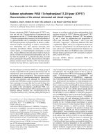
raised against puri®ed rabbit HABP1. As shown in Fig. 1,
different amounts o f puri®ed HABP1 in t he absence o f any
reducing a gent were subjected to native PAGE (9% gel),
transferred to a nitrocellulose membrane, a nd immuno-
detected with anti-HABP1 IgG. It showed two bands, a
broad and relatively prominent lower band (I) and a fairly
sharp minor h igher band (II). The emergence of the higher
band seems to be concentration dependent in vitro,asit
appears only i n lanes 2 and 3 (Fig. 1), in w hich the amount
of protein loaded was 5 and 10 lg, respectively. The
electrophoretic mobility of band II seems to be slightly less
than double that of band I. This is because HABP1 has a
larger than average number of polar amino-acid residues
and therefore it shows anomalous migration on PAGE.
Dimerization of trimers may also change the size and
conformation of the molecule, and this may b e one reason
why a dimer of trimers does not seem to migrate a t t wice the
molecular mass of trimeric HABP1. The concentratio ns of
the two oligomeric populations, estimated by densitometric
comparison of the intensities of the two bands, were
observedtobeintheratioof 12 : 1.
Studies on the oligomeric transitions of HABP1 under
native, r educing, and d enaturing conditions wer e carried o ut
by analysing their relative molecular masses using gel-
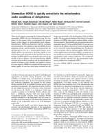
permeation chromatography. Under native conditions
(Fig. 2 A, dotted line), H ABP1 showed a major peak
corresponding to a protein of apparent molecular mass
68 kDa (marked III, Fig. 2A) and a minor peak of protein
corresponding to an apparent molecular mass o f 136 kDa
(marked IV, Fig. 2A). Thus, H ABP1 exists in solution
predominantly as a trimeric (68 kDa) molecule w ith a minor
hexameric (136 kDa) form, assuming a cDNA-derived
Fig. 1. H ABP1 exists i n t wo die rent oligomeric for ms i n solution.
HABP1 (2 lg, lane 1 ; 5 lg, lane 2; 10 lg, lane 3) was electrophoresed
on 9% native polyacrylamide gel in the absence of reducing agents
using a disc ontinuo us buer system, and transferred to nitrocellulose
membrane, probed w ith anti-HABP1 IgG, a nd detected using alkaline
phosphatase conjugate of g oat anti-rabbit IgG a nd an NBT/BCIP
detection system. The two oligomeric forms of HABP1 are marked
I and II. Molecular mass standards are shown on the left.
300 B. K. Jha et al.(Eur. J. Biochem. 269) Ó FEBS 2002
molecular mass of 23801.1 Da for the monomer. HABP1
exists in different oligomeric forms under reducing c ondi-
tions compar ed with native conditions. As s hown in F ig. 2A
(solid line), the protein in the presence of 0.1%
2-mercaptoethanol exhibited a different elution pro®le,
although it showed two peaks, one minor (marked I,
Fig. 2A) and th e other major (marked III, Fig. 2A). The
protein corresponding to the major peak in this case has a
molecular m ass o f 68 kDa, and the protein of the m inor
peak has a molecular m ass of 25 kDa, corresponding to th e
monomeric size of HABP1. Gel-permeation experiments
carried out in the presence of 0 .1% SDS and 0 .1%
2-mercaptoethanol (dotted line, Fig. 2B) showed a single
molecular form corresponding to a molecular mass of
25 kDa (marked I) on the basis of protein standards r un
under i dentical conditions. This clearly implies that the
protein remains in the m onomeric form under reducing and
denaturing conditions. However, in the absence of
2-mercaptoethanol, the 25-kDa protein becomes v ery small
and a new form c orresponding to 46 kDa (marked II,
Fig. 2B) a ppears, suggesting that, under nonreducing
denaturing conditions, the protein predominantly exists in
the dimeric form. On the Superose 6 column, the major
peak (marked III) was eluted with a K
av
of 0.499, which is
equivalent to a Stokes radius of 36.2 A
Ê
, and the m inor peak
(markedIV)waselutedwithaK
av
of 0.412, which is
equivalent to a Stokes r adius of 46.0 A
Ê
.
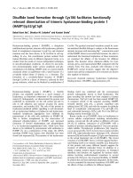
To stabilize the potential oligomeric states of HABP1, the
covalent cross-linker, glutaraldehyde, w as incubated with
HABP1 as described in Materials and methods and
analysed under reducing conditions by SDS/PAGE; this
was followed b y t ransfer to a nitrocellulose membrane and
immunodetection u sing anti-HABP1 IgG (Fig. 3A). It
shows conversion of most of the monomeric band at
34 kDa to a higher species with a r elative m olecular mass of
nearly 70 kDa. However, a smear at 20 ±25 kDa w as
consistently observed, which represents uncross-linked
HABP1 monomer corresponding to the s equence-derived
molecular mass that may arise from modi®ed electrophor-
etic mobility as a result of neutralization of positive charges
of the lysine s ide chain by glu taraldehyde.
Oligomeric transitions
As is evident from the cDNA sequence, each protomer has
only one cysteine (Cys186) in the polypeptide chain o f
HABP1 [8]. T herefore, the trimer has three free cysteine
residues, which can form disul®de bonds by association with
a set of three cysteine residues from another trimer l eading
to formation o f a hexamer. However, i t i s also possible that,
under air oxidation, the cross-linking of these cysteine
residues may lead to the formation of a small proportion of
hexamer. To investigate this, HABP1 was incubated with
the thiol-group-oxidizing agent, Cu
2+
)1,10-phenanthro-
line. There was a 100% shift of band I to band II (Fig. 3B,
lane 1).
To con®rm that the trimer±hexamer transition does
indeed occur through disul®de linkage of cysteine residues,
experiments were c arried out with cysteine-modi®ed
HABP1. Gel-permeation chromatography of cysteine-
modi®ed HABP1 (Fig. 4A) shows a single peak corre-
sponding to 68 kDa. The effect of C u
2+
and Hg
2+
ions on
cysteine-modi®ed HABP1 was e xamined by pore-lim iting
PAGE to examine the role of the metal ion, if any, in
disul®de b ond formation. Native, c ysteine-modi®ed a nd
HgCl
2
-treated HABP1 were separated, transferred to
nitrocellulose membrane, and probed w ith anti-HABP1
IgG. The data indicate dimerization of trimers, which is
inhibited b y cysteine modi®cation (Fig. 4B, lane 4 ). To
examine t he molecular t ransition of H ABP1 by HgCl
2
,
native HABP1 w as treated w ith increasing c oncentrations of
Hg
2+
and subsequently desalted on a Sephadex G-25
column before monitoring of their intrinsic ¯uorescence.
The gradual increase in ¯uorescence intensity with increas-
ing Hg
2+
concentration until all the trimeric HABP1 was
presumably converted into hexameric species indicates a
Fig. 2. Oligomeric states of HABP1 in s olution. (A) Gel-permeation chrom atography of H ABP1 (1.2 mgámL
)1
) on a Superose 6 column
(1 ´ 30 cm) in NaCl/P
i
/0.15
M
NaCl, pH 7.2 (broken line) and N aCl/P
i
/0.15
M
NaCl, pH 7.2, containing 0.1% 2-mercaptoethanol (solid line) at
a ¯ow rate of 0.3 mLámin
)1
. The c olumn was calibrated using molec ular m ass s tandards run under similar conditions: 1, alco hol d eh ydrogen ase;
2, B SA; 3, ova lbumin; 4, chymotrypsinogen; 5, ribonuclease A . The e stimation of the m olecular mass (M
r
) of t he oligomer, i ndicat ed by arrow s, is
shown i n the inset. (B) Gel-permeation c hromatograp hy of H ABP1 (1.2 mgámL
)1
) on a Superose 6 c olumn ( 1 ´ 30 cm) i n N aCl/P
i
/0.15
M
NaCl,
pH 7.2, containing 0.1% SDS ( solid line) and 0.1% SDS and 0.1% 2-mercaptoethanol (broken line). The column was c alibrated using the same
molecular mass standards u nder the above conditions. The molecular mass (M
r
) of s tandards indicated by arrows is shown in the inset and
numbered 1, 2, 3, 4 and 5, respectively. T he peaks marked I, II, III and IV represent monomer, dimer, trimer a nd hexamer, respectively.
Ó FEBS 2002 Structural transition and ligand anity of HABP1 (Eur. J. Biochem. 269) 301
change in the microenvironment around tryptophan as a
result of trimer dimerization (Fig. 4C).
Attempts to gen erate the dimer of trimers using cysteine-
modi®ed HABP1 and copper±phenanthroline as oxidant
also failed; however, the unmodi®ed form was observed to
dimerize after t reatment with 50 l
M
copper±phenanthroline
(data not shown). To examine the role of metal ions in
trimer dimerization, we measured the amount of copper
bound to the d imer of trimers, if any, using a tomic
absorption spectroscopy. A protein c oncentration of
50 l
M
was u sed for detection of t he metal i on. The d ata
show that the c opper content o f this p reparation is less than
15 : 1 (protein to metal ion molar ratio) keeping the
detection limit of the instrument i n the mind. The presence
of any c opper b elow this level is i nsigni®cant a s f ar as trimer
dimerization is concerned. Thus, t he role of cysteine in
trimer dimerization seems t o be unambiguous.
To further establish the role of the cysteine residue in the
generation of dimers of trimers, the free thiols were
determined in HABP1, copper±phenanthroline-induced
dimer of t rimers, and cysteine-modi®ed HABP1. No free
thiol groups wer e available in copper±phenanthroline-
induced dimer of trimers and cysteine-modi®ed HABP1,
but one free thiol group per H ABP1 m onomer was d etected
in reduced unmodi®ed H ABP1.
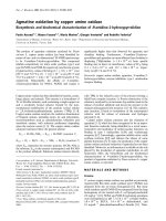
Oligomeric transitions and ligand af®nity
The af®nity of the trimeric and hexameric forms of HABP1
for its various ligands, e.g. hyaluronan,
D
-mannosylated
BSA and gC1q, was analyzed by ELISA. HgCl
2
and copper±
phenanthroline treatment of HABP1 resulted i n trimer to
hexamer conversion, which could be blocked by cysteine
modi®cation. These oligomeric forms of HABP1 were
separated using size-exclusion c hromatography and quanti-
tatively analyzed for binding to biotinylated hyaluronan,
biotinylated
D
-mannosylated BSA, and g C1q. The hexamer
generated by thiol-group oxidation o f native HABP1 had
greater af®nity for its ligand than native a nd cysteine-
modi®ed HABP1 or native HABP1 (Fig 5A,B,C). However,
the cysteine-modi®ed HABP1, which cannot be converted
into hexamer by thiol-group oxidation, showed similar
af®nity for its ligands to the unmodi®ed protein. The trimeric
form of HABP1 h ad less af® nity t han t he hexamer. The
differential binding of trimer and h examer was m ore
pronounced in the case of hyaluronan than gC1q or
mannosylated BSA. Therefore, the dissociation constant
for the hyaluronan±HABP1 i nteraction was calculated b y
Scatchard plot analysis from the data in Fig. 5A, taking the
average molecular mass of hyaluronan to be 10 MDa
(Fig. 5 D). T he apparent dissociation constant of the
hexamer was found to be 0.05 ´ 10
)9
compared with
0.1 ´ 10
)9
for the trimer.
DISCUSSION
In this study, w e demonstrate t he presence of different
oligomeric forms of HABP1: monomer, noncovalently
linked t rimer, and cyste ine-linked dimer of trimers.
Interestingly, all these species of HABP1 h ave differ ent
af®nities for hyaluronan, suggesting a pos sible r ole for
different oligomeric states of HABP1 in hyaluronan
signalling. Th e m ajor peak on gel-®ltration chromatogra-
phy corresponds to the trimer o f HABP1, with an
estimated molecular mass of 68 kDa. This is also identical
with the gel-®ltration-derived molecular mass of HABP1
puri®ed from tissue [3]. A small proportion of the protein
exists in t he h exameric state i n s olution t hrough Cys186-
linked disul®de bonds. However, the crystal structure of
HABP1 i n the presence of 600 m
M
NaCl u nder reducing
conditions suggested that HABP1 is a trimeric p rotein
[22]. This is in agreement with our data in solution under
Fig. 3. Ev idence for oligomeric structural
transition of HABP1. (A) HABP1 was
incubated w ith glutaraldehyde, as described in
Materials and methods. The c ross-linked
samples were analyzed b y SDS/PAGE (12.5%
gel). The electrophoresed gel was transferred
to nitrocellulose membrane, probed with
anti-HABP1 IgG, and detected u sing alkaline
phosphatase conjugate of goat anti-rabbit IgG
andanNBT/BCIPdetectionsystemLane1,
untreated HABP1; lane 2, treated with
glutaraldehyde. The molecular mass standards
are shown on the left. (B) HABP1 was incu-
batedwithCuSO
4
and 1,10-phenanthroline
(1 : 2 molar ratio) for disul®de linkage
following the earlier p rocedure. HABP1 al one
(lane 2), and in the presence of the copper±
phenanthroline comp lex (lan e 1) were
analyzed o n a 9% nondenat uring g el. T h e ge l
was transblotted on a nitrocellulose
membrane and probed with anti-HABP1 IgG.
Molecular mass markers are shown on the left.
302 B. K. Jha et al.(Eur. J. Biochem. 269) Ó FEBS 2002
similar conditions, in which we ®nd the majority o f t he
protein i n t he trimeric form.
There w as a signi®cant change i n shape and size of
HABP1 o n SDS binding as well as in the presence of
2-mercaptoethanol a s observed in gel-®ltration experiments.
It was in the monomeric state under reducing and denatur-
ing conditions, and in the dimeric state (with a m inor
component of monomer) under nonreducing denaturing
conditions. The reason for the predominantly dimeric form
under t hese conditions is the oxidative atmosphere of the
experiment, in which unfolded m onomer b ecomes dimer-
ized through cysteine d isul®de bond formation. On the
other hand, under reducing nondenaturing conditions, it i s
predominantly present as a trimer with a small proportion
of monomer, although u nder native conditions, it predom-
inantly remains as a trimer with a small proportion of
hexamer. This clearly suggests that the trimer is stabiliz ed
through non-covalent i nteractions, and the formation of
hexamer from trimer is facilitated by the disul®de bond
formation between the subunits.
Sequence analysis c on®rms the p resen ce of only one
cysteine (Cys186) residue in HABP1 isolated from human
Fig. 4. Dimerization of trimeric HABP1 through Cys186. (A) Gel-permeation chromatography of cysteine-modi®ed HABP1 (1 mgámL
)1
)ona
Superose 6 colum n (1 ´ 30 cm) in 10 m
M
phosphate buer containing 150 m
M
NaCl,pH7.2,ata¯owrateof0.3mLámin
)1
.Thecolumnwas
calibrated as desc ribed in Fig. 2. (B) P ore-limiting gel electrophoresis of H ABP1 and Cys186-modi®ed HABP1. E qual amounts of H ABP1 treated
with 50 l
M
HgCl
2
(lane 1), native HABP1 (lane 2), modi®ed HABP1 treated with 50 l
M
HgCl
2
(lane 3), modi®ed HABP1 (lane 4), and SDS-treated
HABP1 ( lane 5) were separated on a 7±24 % polyacrylamide gradient g el using 0.005% SDS in Tris/glycine running buer, p H 8.3, a s described in
Materials and methods. The gel was transblotted and probed with anti-HABP1 IgG and detected using goat anti-rabbit IgG and alkaline
phosphatase conjugate. (C) Ch ange in ¯uorescence emission in tensity at 347 nm. HABP1 was treate d with various a mounts of HgCl
2
and then
desalted on a Sephadex G-25 column as described in Materials and metho ds.
Ó FEBS 2002 Structural transition and ligand anity of HABP1 (Eur. J. Biochem. 269) 303
and mouse [22]. O xidation of noncovalent trimer induced
by HgCl
2
or copper±phenanthroline shows the conversion
of HABP1 into the hexameric species in buffer of low ionic
strength. However, cysteine-modi®ed HABP1 remained
trimeric even after treatment with Hg
2+
or Cu
2+
unlike
the native HABP1, clearly establishing the role of cysteine
in trimer to hexamer tran sition. This observation is further
strengthened by the absence of bound copper in the dimer
of trimers of native HABP1 induced by copper±phen-
anthroline. Hence, it may b e postulated that Cu
2+
acts
only as a n oxidant and does not participate d irectly in
dimer formation. In support of this, the higher level of
trimer to hexamer association induced by HgCl
2
was also
evident from the ¯uorescence analysis. HABP1 polypep-
tide sequences of higher eukaryotes (human and mouse)
show three conserved tryptophans, o f which Trp109 and
Trp219 face the relatively hydrophobic s ide of the
molecule and Trp233 resides o n the negatively cha rged
protein surface [22]. The gradual increase in ¯uorescence
intensity of HABP1, s eparated by size-exclusion chroma-
tography after p retreatment with increasing c oncentrations
of Hg
2+
indicates a gradual c hange in the microenviron-
ment around tryptophan as the result of transition from
trimer to hexamer. Dimerization of trimeric HABP1 m ay
lead to exposure o f t ryptophan to a nonpolar/hydrophobic
environment, which in t urn may lead t o an increase in
emission intensity at 347 nm [32]. In contrast with this, t he
crystal structure shows that Cys186 is not easily accessible
for oligomerization induced by an intertrimer disul®de
bond. The c rystal structure o f HABP1/p32 determined in
the presence of 600 m
M
NaCl, 1 m
M
EGTA and 1 m
M
EDTA is compact compared with its form in native
conditions, near pH 7.2 and physiological ionic strength. It
is apparent from our observation t hat t he hydrodynamic
radius of HABP1/p32 near physiological pH and i onic
strength is greater (36.2 A
Ê
)thanthatofthecrystal
structure (34.0 A
Ê
). Such a n increase in hydrodynamic
volume may e xpose C ys186 residues, enabling them to
form S±S bonds.
Under native conditions, at pH 7.2, HABP1 has a
hydrodynamic radius of 36.2 A
Ê
as the major species. T he
Cys186 residue responsible for covalent association of two
trimers t hrough d isul®de bond formation is not accessible in
the crystal structure. The state of HABP1 with the larger
hydrodynamic r adius may be different from the crystal
structure, as the changes associated with the addition of
trace amounts o f b ivalent c ations may represent a s tructural
state in which this cysteine is exposed to the solvent,
Fig. 5. H igher a nity of the hexameric f orm o f HABP1 for its ligand. Dierential ligand anity of HABP1 oligomer puri®ed by size-exclusion
chromatography. Starting f rom 500 ng and using serial dilution, dierent oligomeric for ms of H ABP1 were coated on an ELISA plate in triplicate
and p robed with (A) biotinylated hyaluronan (HA), (B) biotinylated
D
-mannosylated BSA (DMA) and detected with streptavidin±horseradish
peroxidase conjugate; (´)HABP1alone;(d) HABP1 treated with 5 0 l
M
HgCl
2
;(j) c ysteine-modi®ed HABP1 treated with HgCl
2
;(s)BSA.(C)
C1q w as coated o n an ELISA plate starting with 500 ng using serial dilution and incubate d with dierent oligome ric forms of H ABP1; they w ere
then probed with rabbit anti -HABP1 I gG. The b ound HABP1 w as probed with alkaline phosphatase-conjugated goat anti-rabbit I gG (1 : 7500)
and visualiz ed b y the 2,2 ¢-azino-bis(3-ethylbenzthiazoline-6-sulfonic acid) d etection system. ( ´)HABP1alone;(d) H ABP1 treated w ith 50 l
M
HgCl
2
;(j) c ysteine-mo di®ed H ABP1 treated with HgCl
2
;(s) B SA. BSA was u sed as neg ative c ontrol. Each data point is representative of t hree
similar sets of experiment. (D) Scatchard plot analysis of the anity o f dierent oligomeric forms of HABP1 for hyaluronan. ( s)Trimer;(d)
hexamer.
304 B. K. Jha et al.(Eur. J. Biochem. 269) Ó FEBS 2002
facilitating inter-trimeric disul®de bond formation. HABP1
with a larger hydrodynamic r adius and solven t-exposed
cysteine residue under physiological conditions may corre-
spond to the e xpanded structure [33].
The existence of HABP 1 in different oligomeric states
has functional implications. Disul®de-mediated hexamer
formation leads to a c ompact oligomeric structure, wh ich
is shown to have the highest ligand af®nity. The mono-
meric form binds weakly to hyaluronan compared with the
trimeric form. The low af®nity of the HABP1 monomer
may be explained by the presence of a n additional glutamic
acid residue (E127) in the putative hyaluronan-binding
motif. Structural analysis of crystallographic data submit-
tedtotheproteindatabank(PDB)withmolecularID
1P32, a protein that is 100% homologous with HABP1
(synonyms C1QBP, gC1qR, p32 and HABP1), reveals that
the peptide segment K119 to K128 of each monomer in a
trimeric assembly is usually accessible t o the solvent.
However, E127 of each monomer in a t rimeric assembly is
completely b uried, as it is i nvolved in salt bridge formation
with R246 and K174 and the average distances of the two
side chains of R246 and K174 f rom E127 are 3.2 A
Ê
and
2.8 A
Ê
, r espectively. Thus, i n the trimer, there are more
positive charges clustering around the hyaluronan-binding
motif, K119±K128. The dimerization of HABP1 trimers
presumably allows multiple copies of HABP1 t o interact
with its ligand more s trongly. However, the af®nity of the
dimer of trimers for
D
-mannosylated BSA and gC1q is
similar to that of the trimer, suggesting that different
mechanisms are involved in the binding of HABP1 and its
different ligands. P rotomer oligomerization is known t o
have an important role in ligand binding, signal transduc-
tion, and protein function. In the case of serum mannose-
binding protein, its complement-dependent haemolytic
activity is regulated by oligomeric transition [34]. Similarly,
the hyaluronan-binding activity of CD44, another member
of the hyaladherin family, has been linked to cellular
activation. Phorbol 13-myristate 12-acetate is known to
induce clustering o f CD44 f ollowed by d isul®de-mediated
dimerization, which is critical for binding of high levels of
hyaluronan [35,36]. A similar role f or cysteine-mediated
oligomerization in H ABP1 in signal transduction and
hyaluronan binding can be expected as HABP1 is reported
to be involved in hyaluronan-induced signal transduction
[5±7]. So the intricate regulatory mechanism of cellular
signalling by oligomerization of HABP1 may have func-
tional implications in the cell.
ACKNOWLEDGEMENTS
We would like to e xpress our since re thanks to Dr Chandrima Saha o f
NII, New Delhi, India, for providing access to a s pec tro¯uorimeter and
Professor P. Balaram of IISc, Bangalore for useful discussions. The
Department of Science a nd Technology a nd the Department o f
Biotechnology, Government of India, New Delhi have ®nancially
supported this work.
REFERENCES
1. D'Souza, M. & Datta, K. (1985) Evidence of naturally occurring
hyaluronic acid bindin g p rotein in rat liver. Biochem. Int. 10,
43±51.
2. Gupta, S., Babu, B.R. & Datta, K. (1991) Puri®cation, partial
characterization of rat k idn ey hyaluronic acid binding protein and
its localization on cell surface. Eur. J. Cell Biol. 56, 58±67.
3. Gupta, S. & Datta, K. (1991) Possible role of hyaluronectin on c ell
adhesion in rat histiocytoma. Exp. Cell Res. 195, 386±394.
4. Ranganathan, S., Ganguly, A.K. & Datta, K. (1994) Evidence for
presence of hyaluronan binding protein on spe rmatozoa and its
possible involvement in sperm func tion. Mol. Reprod. Dev. 38,
69±76.
5. Ranganathan, S., B haradwaj, A. & Da tta, K. ( 1995) Hyaluronan
mediates sperm motility by enhancing phosphorylation of proteins
including hyaluronan binding protein. Cell. Mol. Biol. Res. 41,
467±476.
6. Rao, C.M., Deb, T.B. & Datta, K. (1996) Hyaluronic acid induced
hyaluronic acid binding protein phosphorylation and inositol tri-
phosphate f ormation in lymphocytes. Bio chem. Mol. Biol. Int. 40 ,
327±337.
7. Rao, C.M., Deb, T.B., Gupta, S. & D atta, K. (1997) Regulation of
cellular phosphorylation of hyaluron an binding prot ein and its
role in the formation of sec ond messenger. Biochim. Bioph ys. Acta
1336, 387±393.
8. Deb, T.B. & Datta, K. (1996) M olecular cloning of human
®broblast hyaluronic a cid binding pr otein con®rms its identify
with P-32, a protein co-puri®ed with splicing f actor SF2. J. Biol.
Chem. 269, 2206±2212.
9. Majumdar, M. & Datta, K. (1998) Assignment of cDNA encoding
hyaluronic ac id binding protein 1 to h uman chromosome 17 p12±
13. Genomics 51, 476±477.
10. Krainer, A.R., Mayeda, A., K o zak, D. & Binns, G. (1991)
Functional expre ssion of cloned human splicing fac tor SF2: h o-
mology to RNA-binding proteins, U1, 70K, and Drosophila
splicing regulators. Cell 66, 383±394.
11. Ghebrehiwet, B ., Lim, B.L., Peerschke, E.I.B., W illis, C .A. &
Reid, K.B.M. (1994) Isolation, cDNA cloning, and overexpression
of a 33-kDa cell surface glycoprotein that binds to the globular
ÔHeadsÕ of C1q. J. Exp. Med. 179, 1809±1821.
12. Luo, Y., Yu, H. & Peterlin, B.M. (1994) Cellular protein modu-
lates eects o f human immunode®ciency virus t ype I R ev. J. Virol.
68, 3850±3856.
13. Das, S., Deb, T.B., Kumar, R. & Datta, K. (1997) M ultifunctional
activities of human ®broblast 34-k Da hyaluron ic acid-bindin g
protein. Gene 190, 223±225.
14. Soltys, B.J., Kang, D. & Gupta, R.S. (2000) Localization o f P32
protein (gC1q-R.) in mitochondria and at speci®c extramito-
chondrial l ocations in normal tissues. Histochem. Cell. Biol. 114,
245±255.
15. Brokstad, K. A., Kalland, K .H., Russell, W.C. & Matthews, D.A.
(2001) Mitochondrial protein p32 can accumulate in the nucleus.
Biochem. Biophys. Res. Commun. 281, 1161±1169.
16. Kittlesen, D.J., Kimberly, A., Chianese-Bullock, K.A., Yao,
Z.Q., Braciale, T.J. & Hahn, Y.S. (2000) Interaction between
complement receptor gC1qR and hepatitis C virus core pro-
tein inhibits T-lymphocyte proliferation. J. Clin. I nvest. 106, 239±
249.
17. Nguyen, T., Ghebrehiwet, B . & Peerschke, E.I.B. (2000) Staphy-
lococcus aureus P rotein A recognizes plate let gc1qR/p33: a novel
mechanism for Staphylococal i nfection with platelets. Inf ect.
Immun. 68, 2061±2068.
18. Braun, L., Ghebrehiwet, B. & Cossart, P. (2000) gC1qR/p32, a
C1q binding p rotein, is a receptor for the In 1B invasion p rotein of
Listeria monocytogenes. EMBO J. 19, 1458±1466.
19. Herwald, H., Dedi o, J., Kellner, R., Loos, M. & Muller-Ester, W .
(1996) Isolation and characterization o f t he k inino gen-bindin g
protein p33 f rom e ndothelial c ells. I dentity w ith t he C1 q receptor.
J. Biol. Chem. 27 1 , 13040±13047.
Ó FEBS 2002 Structural transition and ligand anity of HABP1 (Eur. J. Biochem. 269) 305
20. Kumar, R., Roychoudhary, N., Salunke, D.M. & Datta, K. (2001)
Evidence for clustered mannose as a new ligand for hyaluronan
binding protein (HABP1) from human ®broblast. J. Biosci. 26,
325±332.
21. Simos, G. & Georgatos, S.D. (1994) The lamin B receptor-asso-
ciated protein p 34 sha res se quence homology an d antigenic
determinants with the splicing facto r 2-associated p rotein p32.
FEBS Lett. 346, 225±228.
22. Jiang, J., Zhang, Y., Krainer, A.R. & Xu, R.M. (1999) Crystal
structure of human p32, a doughnut-shaped a cidic m itoc hondrial
matrix protein. Proc. Natl Acad. Sci USA. 96, 3572±3577.
23. Lemmon, M .A. & Schlessinger, J. ( 1994) Regulation of signal
transduction and signal diversity b y receptor o ligomerization.
Trends.Biochem.Sci.19, 459±463.
24. Laemmli, U.K. (1970) Cleavage of structural protein during the
assembly of the head of b acteriophage T4. Nature (London) 227 ,
680±685.
25. Tyagi, R.K., Babu, B.R. & D atta, K. (1993) Simultaneous deter-
mination of native and s ubunit molec ular weight of prote ins by
pore limit electrophoresis and restricted use of sodium dodecyl
sulfate. Electrophoresis 14, 826±828.
26. Hong, K., Ma, D ., Baverley, S.M. & Turco, S.J. (2000) The
Leishmania GDP-Mannose transporter is an aut onomous multi-
speci®c hexameric comple x of LPG2 subunits. B ioc h emi stry 39,
2013±2022.
27. Hollecker, M. (1990) Protein Structure a Practical Approach.
(Creighton, T.E., eds), pp. 146±148. Oxford IRL Press, Oxford,
UK.
28. Ahmad, A., A khtar, M.S. & Bhakuni, V. (2001) Monovalent
cation induced conformational change in glucose oxidase leading
to stablization of enzyme. Biochemi st ry 40, 1945±1955.
29. Kobashi, K. (1968) Catalytic oxidation of sulfhydryl groups
by o-phenanthroline copper c o mplex. Biochim. Biop hys. A cta 158,
239±245.
30. Utschig, L.M., Wright, J.G. & O'Halloran, T.V. (1993) Bio-
chemical and spectroscopic probes of mercury (II) co-ordination
environment in proteins. Methods Enzymol. 226, 71±97.
31.Yang,B.,Yang,B.L.&Goetinck,P.F.(1995)Biotinylated
hyaluronic acid a s a prob e for identifying hyaluronic a cid -binding
proteins. Anal. Biochem. 228, 299±306.
32. Maurice, R.E. (1991) Fluo rescence tech niques for s tudying p rotein
structure. Methods Biochem. Anal. 35, 127±205.
33.Reddy,G.B.,Srinivas,V.R.,Ahmad,N.&Surolia,A.(1999)
Molten globule like state of peanut lectin monomer retains its
carbohydrate speci®city: implications in protein folding and
legume lectin oligomerization. J. Biol. Chem. 274, 4500±4503.
34. Wallis, R. & Drickamer , K. (1999) Molecular d eterminants of
oligomer formation and complement ®xation i n mannose-binding
proteins. J. Biol. Chem. 274, 3580±3589.
35. Sleeman, J., Rudy, W., Hofmann, M., Mo il, J., Herleich, P. &
Ponta, H. (1996) Regulated clustering of variant CD44 proteins
increases their h yaluronate binding capacity. J. Ce ll Biol. 135 ,
1139±1150.
36. Liu, D. & Sy, M. (1997) Phorbol myristate acetate stimulates the
dimerization of CD44 involving cysteine in transmembrane
domain. J. Immunol. 159, 2702±2711.
306 B. K. Jha et al.(Eur. J. Biochem. 269) Ó FEBS 2002