Báo cáo Y học: Role of tyrosine 238 in the active site of Rhodotorula gracilis D-amino acid oxidase A site-directed mutagenesis study docx
Bạn đang xem bản rút gọn của tài liệu. Xem và tải ngay bản đầy đủ của tài liệu tại đây (392.89 KB, 10 trang )
Role of tyrosine 238 in the active site of
Rhodotorula gracilis
D
-amino acid oxidase
A site-directed mutagenesis study
Angelo Boselli, Silvia Sacchi, Viviana Job, Mirella S. Pilone and Loredano Pollegioni
Department of Structural and Functional Biology, University of Insubria, Varese, Italy
Y238, one of the very few conserved residues in the active site
of
D
-amino acid oxidases (DAAO), was mutated to phe-
nylalanine and serine in the enzyme from the yeast Rhodo-
torula gracilis. The mutated proteins are catalytically
competent thus eliminating Tyr238 as an active-site acid/
base catalyst. Y238F and Y238S mutants exhibit a threefold
slower turnover on
D
-alanine as substrate, which can be
attributed to a slower rate of product release relative to the
wild-type enzyme (a change of the rate constants for sub-
strate binding was also evident). The Y238 DAAO mutants
have spectral properties similar to those of the wild-type
enzyme but the degree of stabilization of the flavin
semiquinone and the redox properties in the free form of
Y238S are different. The binding of the carboxylic acid
competitive inhibitors and the substrate
D
-alanine are
changed only slightly, suggesting that the overall substrate
binding pocket remains intact. In agreement with data from
the pH dependence of ligand binding and with the protein
crystal structure, site-directed mutagenesis results emphasize
the importance of residue Y238 in controlling access to the
active site instead of a role in the substrate/ligand interaction.
Keywords: active site lid; function–structure relationships;
flavoprotein; reaction mechanism; substrate recognition.
D
-amino acid oxidase (DAAO; EC 1.4.3.3), an FAD-
containing flavoprotein, catalyses dehydrogenation of the
D
-isomer of amino acids to give the corresponding a-imino
acids and, after subsequent hydrolysis, a-keto acids and
ammonia. The reduced FAD is then reoxidized by molecu-
lar oxygen to yield hydrogen peroxide. The DAAO reaction
has many biotechnological applications. Industrially its
main use is to remove the side chain of cephalosporin c to
give 7-aminocephalosporanic acid, a key intermediate for
the production of semisynthetic cephalosporin antibiotics
[1]. A fundamental question remains within the large class of
flavoprotein oxidases that catalyse the oxidation of amino
or a-hydroxy acids regarding the mechanism by which a
proton and two electrons are transferred from the substrate
a-carbon to the flavin N(5) position during the reductive
half-reaction. The precise mechanism of substrate dehydro-
genation by DAAO is widely debated, even if the crystal
structures of the enzyme purified from pig kidney
(pkDAAO) and of the enzyme from Rhodotorula gracilis
(RgDAAO) (at a resolution of 2.6 A
˚
and 1.2 A
˚
, respect-
ively) have been determined [2–4]. Over the years, three
main but different mechanisms have been proposed for the
reaction catalysed by this flavoenzyme (reviewed in [5]): (a) a
direct hydride-transfer mechanism of a-hydrogen of the
substrate to the N(5) position of the flavin [6]; (b) a
concerted mechanism in which the a-proton abstraction is
coupled with the transfer of a hydride from the amino group
of the substrate [7]; and (c) a carbanion mechanism which
involves the initial formation of a carbanion by subtracting
the a-H of the substrate as a proton [8]. Thus, to
deprotonate the a-proton, the enzyme must have some
highly specific means of removing the proton and stabilizing
the resulting carbanion. Hence, the presence of an enzyme
base for a-proton abstraction is essential for the carbanion
mechanism.
Comparing the primary sequences of the known DAAOs
[9] and the active sites of R. gracilis and mammalian DAAO
[2–4], it is evident that only three residues, among those
identified in or near the active site, are conserved (namely
two tyrosines and one arginine). The crystal structure of
oxidized RgDAAO in complex with the quasi-substrate
CF
3
-
D
-alanine [4] revealed the mode of substrate binding
(Fig. 1A). The a-carboxylic group of the
D
-amino acid
interacts electrostatically with the c-ande-amino groups of
R285 (at % 2.8 A
˚
) and it is hydrogen bonded to the
hydroxyl groups of Y223 and Y238. The substrate a-amino
group is hydrogen bonded symmetrically with the backbone
C@O group of S335 and the active site water molecule
H
2
O72, while the substrate side chain is oriented toward the
hydrophobic binding pocket of the active site (see Fig. 1A).
R285 has been mutated to lysine, glutamine, aspartate, and
alanine [10]. The perturbation of the active site in the R285
mutants modifies the precise substrate alignment: alteration
of the reaction trajectory results in the large change
observed in the reaction velocity. The low stability of the
Correspondence to L. Pollegioni, Dipartimento di Biologia
Strutturale e Funzionale, Universita
`
degli Studi dell’Insubria via
J.H. Dunant 3, 21100 Varese, Italy.
Fax: +39 332 421500, Tel.: +39 332 421506,
E-mail:
Abbreviations:DAAO,
D
-amino acid oxidase; RgDAAO, Rhodotorula
gracilis
D
-amino acid oxidase; pkDAAO, pig kidney
D
-amino acid
oxidase; XO, xanthine oxidase; IP, imino pyruvic acid; EFl
ox
,
oxidized enzyme; EFl
seq
, flavin semiquinone enzyme;
EFl
red
, reduced enzyme.
Enzymes:
D
-amino acid oxidase (DAAO; EC 1.4.3.3); xanthine oxidase
(XO; EC 1.1.3.22).
(Received 16 May 2002, revised 8 July 2002, accepted 9 August 2002)
Eur. J. Biochem. 269, 4762–4771 (2002) Ó FEBS 2002 doi:10.1046/j.1432-1033.2002.03173.x
semiquinone form in the mutants prompted us to propose
that, in the free enzyme form (i.e. in the absence of a ligand),
the side chain of R285 is able to rotate to a distance of $3A
˚
from the N(1)–C(2)@O flavin locus [10]. Mutagenesis of
Y223 in RgDAAO to a phenylalanine and a serine has been
completed [11]. After characterization of the corresponding
mutants we were able to exclude any possibility that Y223
can act as an active-site base. The differences in properties
between Y223F and Y223S mutants suggest that the side
chain at position 223 contributes by fixing the substrate in
the correct orientation for efficient catalysis, mainly by its
shape and less by its hydrogen bonding or electrostatic
properties (the aromatic ring is also important for steric
reasons) [11].
For Y238 a role similar to that inferred for Y223 in
binding and fixation was suggested [4]. This proposal was
changed recently following the investigation of the effect of
pH on benzoate binding in RgDAAO [12] and the
resolution of the three-dimensional structure of RgDAAO
in complex with anthranilate (PDB entry code 1c0l). The
structural data show that the architecture of the active site
can be modified by switching the side chain of Y238 from
the position observed in the structure of DAAO in complex
with
D
-alanine or CF
3
-
D
-alanine (closed form) to the one
adopted in the DAAO–anthranilate complex (opened form)
(compare the position of Y238 in Fig. 1A with that in
Fig. 1B). To obtain an insight of the role of Y238 in
RgDAAO we produced and characterized two single-point
mutations at site Y238 of RgDAAO.
MATERIALS AND METHODS
Reagents
Restriction enzymes and T4 DNA ligase were from
Promega Life Sciences. Site-directed mutagenesis reactions
were made using the Altered Sites
TM
II Kit (Promega Life
Sciences).
D
-amino acids, xanthine, xanthine oxidase, and
all other compounds were purchased from Sigma. Kinetic
experiments were performed in 50 m
M
sodium pyrophos-
phate, pH 8.5, 1% glycerol, 0.3 m
M
EDTA, 0.5 m
M
2-mercaptoethanol and at 25 °C; other experiments were
carried out in 50 m
M
Hepes pH 7.5, 10% glycerol, 5 m
M
2-mercaptoethanol and 0.3 m
M
EDTA at 15 °C, except
where stated otherwise.
Site-directed mutagenesis and enzyme expression
Enzymatic DNA modifications were carried out according
to the manufacturer’s instructions and as described by
Sambrook et al. [13]. The RgDAAO-Y238 mutants were
generated using a dual primer method to simultaneously
introduce ampicillin resistance and a site-directed muta-
tion (Y238F: 5¢-GGCGGGACGTTCGGCGTGGGAG-3¢,
Y238S: 5¢-GGCGGGACG
TCCGGCGTGGGAG-3¢;in
both cases the mutation eliminated a BsiWI restriction site,
shown in italics and, only for Y238S, an AatII site shown in
bold; the codon for the substitution is underlined). Success-
ful mutagenesis was screened by restriction analysis and
confirmed by DNA sequencing of the final plasmid. The
mutant cDNAs were subcloned into the EcoRI restriction
site of the pT7.7A (USB) expression vector (pT7-DAAO
mutants). The Y238F and Y238S DAAO mutants were
expressed and purified as described previously [14].
Activity assay and gel electrophoresis
DAAO activity was assayed with an oxygen electrode at
pH 8.5 and 25 °Cwith28m
MD
-alanine as substrate at air
oxygen saturation ([O
2
] ¼ 0.253 m
M
) [14]. One DAAO unit
is defined as the amount of enzyme that converts 1 lmol
D
-alanine per min, at 25 °C. Substrate specificity was
investigated by means of the same polarographic assay,
using different concentrations of various
D
-amino acids as
substrate. Analytical SDS/PAGE was carried out as
described by Laemmli [15]. The expression of the mutant
enzymes was also determined by Western blot analysis,
using an immunostaining procedure [10,11].
Spectral and ligand binding experiments
The extinction coefficients for the mutant DAAO enzymes
were determined by measuring the change in absorbance
after release of the flavin. The enzymes were heat
denatured by boiling for 5 min in the dark (an extinction
coefficient of 11.3Æm
M
)1
Æcm
)1
at 450 nm for free FAD was
used) [10,11]. Photoreduction experiments were carried
out using an anaerobic cuvette containing % 8 l
M
enzyme,
5m
M
EDTA, and 0.5 l
M
5-deazaflavin. The solution was
made anaerobic and photoreduced with a 250-W lamp,
with the cuvette immersed in a 4 °C water bath [10,16];
the progress of the reaction was followed spectrophoto-
metrically. The thermodynamic stability of the semiqui-
none was determined by the addition of 5 l
M
benzyl
viologen from a side arm of the cuvette after the
photoreduction was complete. Disproportionation of the
semiquinone was then followed until equilibration was
reached(forupto24h)at15°C. Dissociation constants
for ligands were measured spectrophotometrically at
15 °C. The change in absorbance upon adding ligand
was plotted as a function of ligand concentration, after
correction for any volume change.
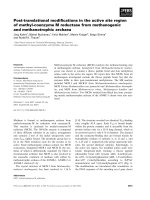
Fig. 1. Active site of R. gracilis DAAO in
complex with (A) CF
3
-
D
-alanine (accession
code 1c0l) and (B) anthranilate (accession code
1c0i). R285, Y223 and Y238 interact in the
structure of RgDAAO complexed with the
substrate with the a-carboxylic group of CF
3
-
D
-alanine [4], and are conserved in all DAAO
sequences. The FAD molecule is shown in
yellow and the ligand molecules in purple.
Ó FEBS 2002 Mutagenesis of RgDAAO (Eur. J. Biochem. 269) 4763
Redox potentials
Redox potentials for the EFl
ox
/EFl
seq
and EFl
seq
/EFl
red
couples of Y238S and Y238F mutants were determined by
the method of dye equilibration using xanthine/xanthine
oxidase (XO) as the source of electrons [17,18]. The enzyme
solution in 50 m
M
Hepes pH 7.5, 10% glycerol, was mixed
in an anaerobic cuvette [18] with 0.2 m
M
xanthine, 5 l
M
benzyl viologen as mediator, and 1–10 l
M
of the appropri-
ate dye, as reported for the wild-type enzyme [19]. The
solution was purged of oxygen, and the reaction was
initiated by adding 10 n
M
XO. The course of the reaction
was followed by recording spectra at various times (typically
3–4 h), at 15 °C. Data were analysed as described by
Minnaert [17]. The amount of oxidized and reduced dye was
determined at a wavelength at which the enzyme has no
absorbance (> 550 nm) and the amount of oxidized and
reduced enzyme was determined at an isosbestic point for
the dye or by subtraction of the dye’s contribution in the
400–470 nm region [19]. The redox potential, E
h
,forthe
system at equilibrium was calculated from the Nernst
equation Eqn (1):
E
h
¼ E
m
þð2:3RT=nFÞ
 logð½oxidized form]/[reduced form]Þð1Þ
where R is the gas constant (8.31441 VÆK
)1
Æmol
)1
), T the
absolute temperature, F the Faraday constant
(9.6485381 · 10
4
CÆmol
)1
), and n is the number of electro-
chemical equivalents. All the potential values are reported
vs. the standard hydrogen electrode. The data were plotted
according to Minnaert [17], in which the log (oxidized/
semiquinone) or the log (semiquinone/reduced) couple for
the enzyme is plotted vs. the log (oxidized/reduced)
concentration ratio for the dye. The separation between
the two single-electron transfers was estimated from the
maximal percentage of the semiquinone form of the enzyme
reached during a reduction experiment in the absence of the
reference dye Eqns (2) and (3) [17,20]:
DE
m
¼ 59 mV Â log K ð2Þ
K ¼½EFl
seq
2
=ð½EFl
red
½EFl
ox
Þ ð3Þ
The semiquinone formation can be determined graphically
by plotting the changes in absorbance at the maximum
wavelength for this form (% 400 nm) and for the oxidized
enzyme (460 nm) and/or using the known extinction
coefficient at the same wavelength [19].
Stopped-flow measurements
The experiments were performed at 25 °C in a thermostated
BioLogic SFM-300 stopped-flow spectrophotometer equip-
ped with a J & M diode array detector. The enzyme-
monitored turnover method was used to assess steady-state
kinetics by mixing 10 l
M
air-saturated enzyme with air-
saturated solutions of
D
-alanine at 25 °C. Traces at 456 nm
were analysed as described previously [10,11,21], using the
KALEIDAGRAPH
program (Synergy Software). For reductive
half-reaction experiments, the stopped-flow instrument was
made anaerobic by overnight equilibration with concentra-
ted sodium dithionite solutions. Prior to use, the instrument
was rinsed well with argon-bubbled buffer to remove the
dithionite. Reaction rates were calculated by extracting
traces at individual wavelengths (456 and 530 nm) and
fitting them to a sum of exponentials equation using
PROGRAM A
(developed in the laboratory of D. P. Ballou,
University of Michigan) or
SPECFIT/32
(Spectrum Software
Assn).
PROGRAM A
wasalsousedtosimulatetheexperi-
mental traces using a three-step kinetic model (with only the
first step reversible), in a manner analogous to that
performed on wild-type DAAO [22].
RESULTS
Enzyme expression and purification
The pT7-Y238F and pT7-Y238S plasmids were used to
transform BL21(DE3)pLysS Escherichia coli cells and the
induction conditions investigated by means of Western blot
analysis and DAAO activity assay. Like the wild-type
RgDAAO [14], the highest level of enzyme expression and
specific activity was obtained for the Y238 mutants by
inducing the cells with 1.0 m
M
isopropyl thio-b-
D
-galacto-
side (IPTG) at saturation (D
600
‡ 2.0) and cultivating them
at 30 °C for an additional 1–3 h (1.6 UÆmg
)1
protein and
2.3 UÆmg
)1
protein for the Y238F and Y238S mutants,
respectively). The Y238 mutants were purified to homo-
geneity according to the standard procedure [14]. Typically,
60–120 mg of pure enzyme was isolated from 10 L bacterial
culture of Y238S and Y238F, a value close to the best
expression (180 mg) obtained for wild-type DAAO [14].
The lower protein recovery of Y238 mutants compared
with wild-type DAAO is due to a twofold decrease in
the overall purification yield. The specific activity of
the purified Y238F and Y238S preparations was
% 37 UÆmg
)1
protein (vs. 104 UÆmg
)1
protein for the wild-
type DAAO) [14].
Spectral properties and redox potentials
The Y238 RgDAAO mutants were purified as holoenzymes
(retaining their FAD prosthetic group). The mutants, in
their oxidized state, show the typical spectrum of the FAD-
containing flavoproteins (line 1 in Fig. 2), an extinction
coefficient at 455 nm of % 12 600Æ
M
)1
Æcm
)1
,andaratio
A
274
/A
455
% 8.7. All of the Y238 mutants of RgDAAO are
competent in catalysis: the anaerobic addition of an excess
of
D
-alanine (trace 3 in Fig. 2) resulted in instantaneous
enzyme reduction of all mutants, with a spectrum like that
of the wild-type. Stabilization of the anionic semiquinone is
typical for
D
-amino acid oxidases and for the family of
flavoprotein oxidases [23]. The amount of semiquinone
form stabilized by each mutant was determined by anaero-
bic photoreduction [16] until the spectrum of the flavin
semiquinone (EFl
seq
) reached a maximum (trace 2 in
Fig. 2); this species represents near-complete formation of
EFl
seq
(% 95%) for both Y238F and Y238S (see Table 1).
The maximal semiquinone formed by photoreduction is a
kinetically stabilized species. Anaerobic addition of benzyl
viologen resulted in dismutation of EFl
seq
to the oxidized
and reduced forms, with the endpoint containing the
thermodynamically stabilized amount of semiquinone.
The Y238S mutant showed a higher percentage of the
thermodynamically stabilized semiquinone form than
the wild-type and Y238F DAAOs (Table 1). The redox
4764 A. Boselli et al.(Eur. J. Biochem. 269) Ó FEBS 2002
potentials of the Y238S DAAO mutant were thus measured
by the dye equilibration method of Minnaert [17], in order
to assess changes in the thermodynamic properties of the
flavin centre caused by the mutation and to explain the
different thermodynamic stability of the semiquinone form
with respect to wild-type and Y238F DAAOs. When the
XO-mediated reduction of Y238S mutant was monitored in
the absence of a reference dye, the percentage of semiqui-
none formed during the reduction was higher (80%) than
that observed for the wild-type enzyme, indicating a larger
separation between the single-electron potentials than in the
wild-type RgDAAO [19]. The potentials of the oxidized/
semiquinone and semiquinone/reduced forms of Y238S
DAAO were determined by using indigo tetrasulfonate and
safranine T as reference dye (data not shown). The redox
potential difference with respect to the dye was calculated by
plotting the log (EFl
ox
/EFl
seq
) or log (EFl
seq
/EFl
red
) flavin
species of the enzyme as a function of log (oxidized/reduced)
of the dye [17,19] (see Table 1). Decreasing the concentra-
tion of XO, and thus slowing the rate at which the reaction
proceeds, had no effect on the potentials measured. The
redox potential E
2
(¼ )257 mV) for Y238S DAAO is
significantly more negative than the corresponding value
determined for the wild-type enzyme. The % 200 mV
separation between the two single-electron transfer poten-
tials of Y238 mutant DAAO is in agreement with the
large amount of stable semiquinone form produced by
photoreduction.
Benzoate is a competitive inhibitor of DAAO and in the
presence of this substrate analogue the two-electron transfer
is the favoured process for wild-type DAAO [19]. In order
to know if the substitution of Y238 with a serine alters the
redox properties even in the enzyme–substrate (or enzyme–
substrate analogue) complex, the Y238S DAAO mutant
was reduced in the presence of benzyl viologen of a
saturating concentration of sodium benzoate (100 m
M
,see
below) using the xanthine/XO system. For wild-type
DAAO, and different from the result obtained for the free
enzyme, the amount of semiquinone form produced under
these experimental conditions is % 20% [19]. When the same
experiment was performed using the Y238S mutant DAAO,
the spectrum of the oxidized enzyme was converted into the
reduced form, lacking the isosbestic points and peak
maxima characteristic of the formation of the semiquinone
intermediate. The amount of semiquinone form produced in
such a way for Y238S was % 22%, corresponding to a
maximal separation between the potentials for each single-
electron transfer of 43 ± 14 mV (36 mV for the wild-type
DAAO). This result indicates that the modification in redox
properties following the substitution of Y238 with a serine
residue is observed only in the free enzyme form, while the
modulation of the redox properties of the Y238S DAAO by
the substrate analogue binding is similar to that observed
for the wild-type DAAO.
Ligand binding
Dissociation constants for several ligands were measured in
order to determine the contribution of residue Y238 to
Table 1. Semiquinone formation and stabilization, and redox potentials of the free forms of wild-type and Y238 mutants of
D
-amino acid oxidase. The
semiquinone form of DAAO was achieved by anaerobic photoreduction, and the percentage of thermodynamically stabilized form was measured
after equilibration with benzyl viologen.
Semiquinone measured (%) E (mV)
Kinetically stabilized Thermodynamically stabilized E°¢
1
E°¢
2
E
m
Wild-type
c
94 40 )43 )177 )109
Y238F ‡ 95 44 ND ND ND
Y238S ‡ 95 82 )60 ± 2.1
a
)257 ± 5.1
b
)160
a,b
The redox potentials were measured at pH 7.5 and 15 °C using
a
indigo tetrasulfonate ()43 mV),
b
safranine T ()276 mV) as redox
standards, and xanthine/xanthine oxidase as the source of reducing equivalents [17–19].
c
[19].
Fig. 2. Spectral properties of wild-type, Y238S, and Y238F RgDAAOs.
(1) Oxidized enzyme in 50 m
M
Hepes buffer pH 7.5, containing 10%
glycerol and 5 m
M
2-mercaptoethanol, at 15 °C; (2) semiquinone form
generated by photo-irradiation in the presence of 5 m
M
EDTA and
0.5 l
M
5-deazaflavin; (3) fully reduced enzyme from the anaerobic
reaction with 5 m
MD
-alanine.
Ó FEBS 2002 Mutagenesis of RgDAAO (Eur. J. Biochem. 269) 4765
substrate/ligand binding. Binding was measured by the
perturbation of the visible spectrum of the FAD upon
formation of the bound complex (see Fig. 3 for Y238F and
anthranilate). With all the compounds tested and for both
Y238 mutants, the spectral modifications were qualitatively
identical to those observed for the binding to the wild-type
DAAO [11,14]. Different from wild-type and Y238S, the
Y238F RgDAAO mutant showed a significant increase in
the intensity of the Ôcharge transferÕ absorbance band at
% 600 nm following the binding of anthranilate (Fig. 3,
inset) and the shoulder at % 500 nm following the binding of
benzoate (De
497nm
of 7500Æ
M
)1
Æcm
)1
vs. a figure of 2000–
4000Æ
M
)1
Æcm
)1
observed with the other DAAO forms) [12].
Anyway, only modest effects (less than fivefold) in binding
were observed for Y238 mutants with the ligands tested
(Table 2). These results indicate that the mode of ligand
binding is retained in the two mutants, and that the
alteration of the spectral effects can be attributed to an
altered polarity of the active site. The formation of a sulfite
covalent adduct to the N(5) flavin position is also marginally
altered by the substitution of Y238 (Table 2).
Steady-state and rapid reaction kinetics with
D
-alanine
The ability of the Y238 mutants to catalyse
D
-alanine/
oxygen catalysis was measured by enzyme-monitored
turnover [21]. Air-saturated solutions of Y238 mutant
enzymes and of
D
-alanine were mixed in the stopped-flow
spectrophotometer and the absorbance spectra were recor-
ded continuously in the 350–650 nm wavelength range at
25 °C. Following absorbance at 455 nm, an initially rapid
decrease in the oxidized flavin absorption was observed,
followed by a steady-state phase, and then by a further
decrease to reach the final reduced state (corresponding to
spectrum 3 in Fig. 2) [24]. During turnover the enzyme is
present largely in the oxidized form, indicating that the
overall process of reoxidation of reduced DAAO with
oxygen is always faster than the reductive half-reaction (see
Fig. 4 for Y238S). The Lineweaver–Burk plots of
D
-alanine/
oxygen turnover show a set of slightly converging lines with
Y238F DAAO mutant, consistent with a ternary complex
mechanism. For Y238S, as well as for wild-type DAAO
[24], a parallel line pattern in the secondary plots was found
instead. Such a behaviour was demonstrated to be consis-
tent with a limiting case of a ternary complex mechanism,
where some specific rate constants (i.e. k
)2
, the reverse of the
reduction rate) are sufficiently small [24]. For Y238F and
Y238S, k
cat
is reduced by about one-third (Table 3). In
comparison with wild-type RgDAAO, the K
m
for
D
-alanine
is increased threefold in the mutants and the K
m
for O
2
is
decreased (up to 10-fold in the Y238S mutant, see Table 3).
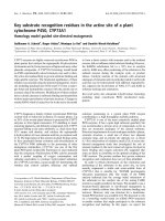
Fig. 3. Effect of anthranilate binding on the spectrum of Y238F
D
-amino
acid oxidase. (––) % 11 l
M
Y238F DAAO in 50 m
M
Hepes buffer
pH 7.5, containing 10% glycerol, and 5 m
M
2-mercaptoethanol; after
the addition of 0.075 m
M
(– ) –) 0.725 m
M
(- - -), 1.45 m
M
(– - –),
5.7 m
M
(– –)and 30m
M
(ÆÆÆ) anthranilate (all final concentrations),
at 15 °C. Inset: difference spectra for anthranilate binding to wild-type
(––), Y238F (ÆÆÆ), and Y238S (– ) –) DAAOs. The difference spectra
were obtained by subtraction of the absorbance spectrum of the
free oxidized form of DAAOs to the spectrum of the same enzyme
after addition of a saturating concentration (% 20 m
M
)ofsodium
anthranilate.
Table 2. Binding of aromatic and aliphatic competitive inhibitors and of
sulfite to wild-type and Y238 mutants of
D
-aminoacidoxidase.All
measurements were made in 50 m
M
Hepes buffer pH 7.5, 10% gly-
cerol, 5 m
M
2-mercaptoethanol, at 15 °C. Wavelengths used to cal-
culate the ligand binding are 497 nm for sodium benzoate and sodium
crotonate, 456 nm for sodium sulfite, 540 nm for sodium anthranilate,
and 345 nm and/or 380 nm for
L
-lactate. The K
d
values were deter-
mined by plotting the change in absorbance upon adding ligand as a
function of ligand concentration [32].
Compound
K
d
(m
M
)
Wild-type
a
Y238F Y238S
Benzoate 0.9 4.4 1.1
Anthranilate 1.9 0.9 2.1
Crotonate 0.4 0.3 0.6
L-Lactate 16.2
b
4.2 5.5
Sulfite 0.12 0.2 0.3
a
[11].
b
[4].
Fig. 4. Time courses of turnover of Y238S mutant RgDAAO followed in
the stopped-flow spectrophotometer. The changes in absorbance were
monitored at 455 nm after mixing 8.7 l
M
mutant enzyme with the
following
D
-alanine concentrations: 0.5 m
M
(1, d), 0.83 m
M
(2),
1.25 m
M
(3, h), 2.5 m
M
(4, r)and5m
M
(5, n). Inset: Lineweaver–
Burk plot of the data determined from the enzyme monitored turnover
traces depicted in the main graph.
4766 A. Boselli et al.(Eur. J. Biochem. 269) Ó FEBS 2002
The ternary complex mechanism shown in the upper loop
of Scheme 1 can be described using the conventions of
Dalziel [25]:
e
t
=v ¼ U
0
þ U
d-Ala
=½d-AlaþU
o
2
=½O
2
þ U
d-Ala;O
2
=½d-Ala½O
2
ð4Þ
where: k
cat
¼ 1=U
0
; K
m;d-Ala
¼U
d-Ala
=U
0
; K
m;O
2
¼U
O
2
=U
0
e
t
v
¼
k
2
þ k
4
k
2
Á k
4
þ
k
À1
þ k
2
k
1
Á k
2
½d-Ala
þ
k
2
þ k
À2
k
2
Á k
3
½O
2
þ
k
À1
þ k
À2
k
1
Á k
2
Á k
3
½d-Ala½O
2
ð5Þ
The reductive half-reaction of Y238 mutants with
D
-alanine
was measured by mixing anaerobically a solution of each
mutant enzyme with solutions containing varying concen-
trations of
D
-alanine, such that a pseudo first-order
condition was maintained with respect to the substrate. In
the absence of oxygen, the oxidized form of each single
mutant was rapidly converted to the reduced enzyme–imino
pyruvate (IP) complex (phase 1, steps k
1
/k
)1
and k
2
/k
)2
),
followed by decay of the spectral intermediate (phase 2, k
5
/
k
)5
) [22,24]. Like the wild-type RgDAAO, no spectral
change has been associated with formation of the EFl
ox
–
D
-alanine complex in the reductive half-reaction of any of
the Y238 mutants. As shown for Y238F in Fig. 5A, the
formation of the spectral intermediate, phase 1, involved a
large extinction decrease at 456 nm and a small extinction
increase at 530 nm, consistent with formation of a EFl
red
–IP
charge-transfer complex [22,24]. Decay of the spectral
intermediate, phase 2, resulted in a decrease in absorbance
at 456 nm and 530 nm, giving a spectrum consistent with
the presence of free, reduced enzyme (Fig. 5A) [24]. In the
case of the Y238F mutant, the increase in absorbance at
530 nm is observable only when the production of the
EFl
red
–IP complex is fast, i.e. at high
D
-alanine concentra-
tions, indicating a fast dissociation of the imino acid from
the reduced enzyme form (see below). The rates of flavin
reduction, k
obs1
, for Y238F and Y238S mutants at different
D
-alanine concentration are close to those determined for
Table 3. Comparison of the steady-state coefficients obtained from stopped-flow experiments of wild-type and Y238 mutants of
D
-amino acid oxidase.
All measurements were made in 50 m
M
sodium pyrophosphate, pH 8.5, 1% glycerol, 0.3 m
M
EDTA and 0.5 m
M
2-mercaptoethanol.
Lineweaver–Burk plot k
cat
(s
)1
) K
m,
D
-Ala
(m
M
) K
m,O
2
(m
M
) F
D
-Ala
(
M
Æs) F
O
2
(
M
Æs) F
D
-Ala,O
2
(
M
2
Æs)
Wild-type
a
parallel 350 2.6 2.3 7.5 · 10
)6
6.7 · 10
)6
Y238F % convergent 125 7.5 0.26 5.9 · 10
)5
5.2 · 10
)6
1.1 · 10
)8
Y238S % parallel 120 7.8 0.96 6.5 · 10
)5
7.9 · 10
)6
a
[24].
Scheme 1. Kinetic scheme of the reaction of RgDAAO with
D
-alanine.
The upper loop shows the ternary complex mechanism, and the lower
loop depicts the ping-pong mechanism. IP, imino pyruvate.
Fig. 5. (A) Spectral courses of anaerobic reduction of Y238F DAAO by
D
-alanine and (B) plot of the dependence of the observed first rate of anaerobic
reduction (k
obs1
) for wild-type (m), Y238F (j), and Y238S (d) DAAOs on the concentration of
D
-alanine. (A) Y238F DAAO (7.5 l
M
)wasmixed
anaerobically with 0.1 m
MD
-alanine in the stopped-flow instrument, at pH 8.5 and 25 °C. From the top at 455 nm: spectrum at 10 ms (is essentially
unreacted enzyme), 50 ms, 108 ms, 195 ms, 310 ms, 510 ms, 1.0 s and 2.24 s after mixing. Inset: time courses of flavin reduction followed at 455 nm
(d)and530nm(j), during the same experiment depicted in the main graph. The solid traces represent fits to the data according to a two sequential
exponentials equation. (B) The reaction rates were determined from experiments as those reported in (A). The line is the best fit obtained for the
values determined for the Y238S DAAO mutant [26].
Ó FEBS 2002 Mutagenesis of RgDAAO (Eur. J. Biochem. 269) 4767
the wild-type DAAO (Fig. 5B). At pH 8.5, wild-type
RgDAAO and Y238 mutants show a hyperbolic depend-
ence of the observed first rate of flavin reduction as a
function of
D
-alanine concentration (see Fig. 5B) [22]: a
saturation is not visible as the reactions at
D
-alanine
concentration > 5 m
M
develop so rapidly that the reaction
rates are at the detection limit of the stopped-flow instru-
ment (¼ 200Æs
)1
). Such a hyperbolic dependence of the
observed reduction rate, as a function of
D
-alanine concen-
tration, describes a first-order reaction of a binary complex,
following a second-order complex formation (Scheme 1)
[26]. As the data are best fit with a rectangular hyperbola
that intersects the origin, these data indicate that the
reduction step is essentially irreversible (k
)2
% 0). A double
reciprocal plot of these data clearly indicates a positive
y-intercept (not shown). Using for the Y238 mutants the
same kinetic model determined for the wild-type DAAO
[22,24], k
2
and K
d,app
values were determined (Table 4).
Numerically, the value of K
d,app
is equal to (k
)1
+ k
2
)/k
1
[26], and its value is similar for the Y238 variants and wild-
type DAAO. As binding never reaches equilibrium, the
thermodynamic representation of substrate binding, K
d
,is
not nearly as important for substrate recognition as the
rate of substrate association, k
1
. To validate the values
determined for rates > 100Æs
)1
, and to estimate lower
limits for the k
1
and k
)1
rate constants, the experimental
traces at 455 nm were simulated using
PROGRAM A
[22].
Simulations were based on the sequential mechanism
described above (i.e. a system including steps k
1
, k
)1
, k
2
and k
5
, and the following extinction coefficients: EFl
ox
and EFl
ox
:
D
-Ala ¼ 12 600Æ
M
)1
cm
)1
; EFl
red
:IP ¼ 4600–
4000Æ
M
)1
Æcm
)1
; EFl
red
¼ 2800Æ
M
)1
Æcm
)1
). Good estimation
of the experimental traces of Y238F and Y238S mutants
at each
D
-alanine concentration can be obtained only
using a k
1
rate constant slightly higher and a k
)1
-value
lower than the corresponding values estimated for the
wild-type one; the rate of flavin reduction was instead
constant for all the DAAO forms. The parameters
obtained from fitting and used for simulations are listed
in Table 4.
The decrease in k
cat
for all Y238 mutants in comparison
to the wild-type RgDAAO resembles the situation observed
for the Y223F mutant of RgDAAO [11]. The fourfold
difference between k
2
and k
cat
couldbeascribedtoa
decrease in k
4
, the rate for IP dissociation from the
reoxidized enzyme form (Scheme 1). Using the measured
values of k
cat
and k
2
, a lower limit for k
4
, ranging from 100
to 150 s
)1
, can be estimated (see Eqn 5).
The second phase in reduction corresponds to k
5
,a
D
-alanine concentration-independent rate constant, and is
changed in Y238 mutants (see Table 4). The IP product
dissociates more slowly from the Y238F (0.9 s
)1
)and
faster from the Y238S (8.3 s
)1
) mutant enzyme than
from the wild-type DAAO (2.8 s
)1
). Because the rate of
product release from the reduced enzyme is very much
slower than k
cat
in Y238S and Y238F (see Table 4), k
5
clearly does not lie within the catalytic cycle, and the
steady-state mechanism must be essentially a ternary
complex. In the case of an irreversible (k
)2
¼ 0) tern-
ary complex mechanism, the steady-state parameter
1/F
O
2
¼ k
2
Æ k
3
/(k
2
+ k
)2
) (see Eqn 5) reduces to k
3
.
For wild-type DAAO, 1/F
O
2
is equivalent to the inde-
pendently measured value of k
3
, within experimental error
[24]. The good correspondence between the F
O
2
parameter
determined with all the Y238 mutants and with wild-type
DAAO (Table 3) indicates that these mutants still largely
follow a ternary complex mechanism and that the oxygen
reactivity (k
3
)oftheEFl
red
–IP complex in the mutant is
not changed.
Substrate specificity
We tested the activity of wild-type and Y238 DAAO
mutants on different
D
-amino acids, measuring the oxygen
consumption with a Clark type electrode at pH 8.5 and
25 °C [14]. The apparent kinetic parameters V
max
and K
m
for the
D
-amino acid determined at fixed (21%) O
2
concentration are reported in Table 5. For both Y238
mutants, and with all the substrates tested, the maximal
activity was lower than the corresponding value determined
for wild-type DAAO. Notwithstanding, the catalytic effi-
ciency expressed by the V
max
/K
m
ratio is frequently similar
(or slightly higher) among the mutants and the wild-type.
This is due to the smaller K
m,app
values determined using the
Y238 DAAO mutants for all
D
-amino acids tested
(Table 5). The decrease in K
m,app
is evident for substrates
with large, hydrophobic side chains (such as cephalospo-
rin C and
D
-phenylalanine), as well as for a small and polar
aminoacidsuchas
D
-serine. The Y238 mutants have a
similar substrate specificity to the with wild-type DAAO:
the highest V
max
/K
m
ratios have been observed with
D
-phe-
nylalanine and
D
-tryptophan. Like the wild-type DAAO,
basic
D
-amino acids are poor substrates for Y238 mutants
(data not shown). The mutants maintain the stereospecifi-
city of the wild-type RgDAAO; they are not reduced by
L
-valine under anaerobic conditions.
Table 4. Kinetic parameters for the reductive half-reaction of wild-type and Y238 mutants of
D
-amino acid oxidase with
D
-alanine as substrate. The
K
d,app
was obtained from the slope divided by the intercept in the double-reciprocal plot of the rate of reduction vs.
D
-alanine concentration. All
measurements were made in 50 m
M
sodium pyrophosphate pH 8.5, 1% glycerol, 0.3 m
M
EDTA, 0.5 m
M
2-mercaptoethanol. The k
1
and k
)1
rate
constants and the k
2
and k
5
values reported in parenthesis are the parameters determined by simulation of the experimental traces using program A
(see text for details).
k
2
(s
)1
)
K
d,app
(m
M
)
Slope (k
2
/K
d,app
)
(
M
Æs) · 10
)5
k
1
(m
M
)1
Æs
)1
)
k
)1
(s
)1
)
k
5
(s
)1
)
Wild-type
a
510 ± 50 (500) 16 ± 3 3.0 30 500 2.3 ± 0.4 (2.8)
Y238F ¼ 400 (500) 11.6 ± 2.8 2.1 60 250 0.9 ± 0.2 (0.8)
Y238S ¼ 400 (500) 14.1 ± 3.5 2.0 40 250 8.3 ± 1.7 (10)
a
[22].
4768 A. Boselli et al.(Eur. J. Biochem. 269) Ó FEBS 2002
DISCUSSION
The Y238 mutants were expressed and purified to homo-
geneity with a good yield using the expression system
constructed to maximize the production in E. coli of wild-
type RgDAAO [14]. The characterization of the kinetic,
substrate specificity and ligand binding properties of Y238F
and Y238S DAAO mutants allows us rule out a main role
of the side chain of this active site residue in substrate/ligand
fixation. The ligand-binding experiments demonstrate that
the overall substrate-binding pocket remains intact, as all
mutants bind the same ligands as the wild-type (Table 2).
The steady state parameters determined with various
D
-amino acids at a fixed O
2
concentration (see Table 5)
indicate that Y238 is not important in determining the
substrate specificity of yeast DAAO. Spectral properties of
the oxidized, semiquinone, and reduced forms of the Y238
mutants are essentially the same as wild-type DAAO
(Fig. 2).
The first significant change observed following the
substitution of Y238 concerned the flavin redox potentials
of Y238S in the free enzyme form: this mutant shows a
larger separation of the single-electron transfer potentials
than the wild-type DAAO, thus a higher stabilization of
the semiquinone form (see Table 1). The stabilization of
the anionic semiquinone form depends on the protein’s
ability to stabilize the negative charge delocalized on the
N(1)-C(2)¼O flavin locus. For free RgDAAO, we previ-
ously proposed that R285 could play such a role through a
conformational change [10]. The higher stabilization of the
semiquinone form observed for the Y238S mutant in the
free form may be the result of a better interaction of R285
with the N(1)-C(2)¼O locus of the reduced flavin following
the substitution of Y238 with a serine (the distance between
the side chains of R285 and Y238 is 4.1 A
˚
), or could be
ascribed to an alteration of the active site polarity.
Anyway, the amount of semiquinone form produced by
the Y238S mutant in the presence of the competitive
inhibitor sodium benzoate resembles that observed for the
wild-type DAAO, thus the change in redox properties is
restricted only to the free enzyme form.
The substitution of Y238 does not alter significantly the
kinetic properties: the rate at which Y238 mutants are
reduced by substrate is similar to that determined for the
wild-type. This result clearly excludes Y238 as a possible
functional group playing a role in acid/base catalysis, e.g.
in the subtraction of the a-carbon proton. The most
striking difference observed for the Y238 mutants in
comparison to the wild-type DAAO is a decrease in the
turnover number. It appears to be a decrease in k
4
,therate
of product dissociation from oxidized enzyme. Other
changes in kinetic properties belong to the rate constant
(k
1
and k
)1
) for substrate binding to the oxidized form,
andtothek
5
rate constant for product release from the
E
red
–IP complex. All of these results point to a role of
the Y238 side chain in substrate/product exchange to the
active site of RgDAAO.
A superimposition of the active sites of yeast and
mammalian DAAO [2–4] shows that the side chain of
Y223 of RgDAAO overlaps with the position occupied by
Y228 in pkDAAO (the residue located on the flexible loop
that adapts its conformation depending on the size of the
ligand side chain) [27] and that Y238 of RgDAAO
Table 5. Substrate specificity of wild-type and Y238 mutants of
D
-amino acid oxidase. All measurements were made in 50 m
M
sodium pyrophosphate, pH 8.5, at air (21%) oxygen saturation, and at 25 °C.
D
-Alanine
D
-Serine
D
-Proline
D
-Tryptophan Cephalosporin C
D
-Valine
D
-Phenylalanine
V
max
(UÆmg
)1
)
K
m
(m
M
) V
max
/K
m
V
max
(UÆmg
)1
)
K
m
(m
M
) V
max
/K
m
V
max
(UÆmg
)1
)
K
m
(m
M
) V
max
/K
m
V
max
(UÆmg
)1
)
K
m
(m
M
) V
max
/K
m
V
max
(UÆmg
)1
)
K
m
(m
M
) V
max
/K
m
V
max
(UÆmg
)1
)
K
m
(m
M
) V
max
/K
m
V
max
(UÆmg
)1
)
K
m
(m
M
) V
max
/K
m
Wild-type 122 0.8 152 61 13.7 4.5 116 21.5 5.4 160 0.3 530 109 5.0 21.8 195 18.9 10.3 144 0.3 480
Y238S 37.7 0.4 94 25.8 2.9 8.9 38.1 12.3 3.1 45.6 0.2 228 21.7 1.9 11.4 42.6 6.1 7.0 33.1 0.07 473
Y238F 37.4 0.4 94 40.7 1.7 24 106.1 13.5 7.9 51.4 0.3 171 11.8 1.9 6.2 62.5 6.0 10.2 27.0 0.04 675
Ó FEBS 2002 Mutagenesis of RgDAAO (Eur. J. Biochem. 269) 4769
overlaps to Y224 of the mammalian enzyme (the residue
interacting with the a-amino group of the substrate and
with a buried water molecule). Y224 in pkDAAO and
Y238 in RgDAAO share the characteristics of being
flexible and adapting their conformation depending on the
size of the ligand side chain [27]. Our results indicate that
the role of Y223 and Y238 in the active site of RgDAAO
is different from that of the tyrosine residues (Y224 and
Y228) of pkDAAO. In fact, and different from the results
obtained with RgDAAO mutants, both Y224F and
Y228F mutants of pkDAAO showed a large decrease in
k
red
(30- and 100-fold lower than in the wild-type) but the
K
d,app
for
D
-alanine was not affected significantly [28].
Furthermore, though these substitutions modified the
interaction of the reduced enzyme with the IP product,
as indicated by the observation that Y228F totally
abolished the formation of the absorbance band centred
at 560 nm during the reduction process, which is typical of
the EFl
red
–IP complex, they did not alter the rates of
product dissociation [28]. Two tyrosine residues are also
present at the active site of other flavoproteins, e.g.
flavocytochrome b
2
[29], glycolate oxidase [30], and lactate
monooxygenase [31]. It has been proposed that these
enzymes work by a carbanion mechanism, and that in
each enzyme these residues play a different role in fine
tuning substrate interactions and enzyme activity. Their
role was also investigated by site-directed mutagenesis
experiments, but only by replacing a phenylalanine (a
nondisruptive mutation). In the case of RgDAAO we also
changed the spatial arrangement in the active site by
introducing a serine.
In conclusion, the results obtained with the Y238
mutant enzymes eliminate this residue as an active site
acid/base catalyst and indicate that this residue is not
important for substrate/ligand fixation. Our results are in
agreement with the different position of Y238 observed in
the structure of DAAO in complex with
D
-alanine or CF
3
-
D
-alanine (closed form) [4] with respect to that occupied in
the DAAO–anthranilate complex (opened form) (Fig. 1).
The movement of Y238 side chain controls substrate
binding and product release, analogously to the role of the
216–228 loop present in pkDAAO [27]. The differences in
properties between the Y223 and Y238 RgDAAO
mutants suggest that the side chain at position 223
contributes to this by fixing the substrate in the correct
orientation for efficient catalysis mainly by its shape and
less by its hydrogen-bonding or electrostatic properties
[11], whereas Y238 essentially controls access to the active
site. These conclusions are also in agreement with the pH-
dependence studies of benzoate binding [12]: for wild-type
and Y238F DAAOs, the binding is pH dependent
(pK
a
¼ 9.8 and 9.1, respectively), whereas no change in
K
d
for benzoate was observed in the 5.5–10.5 pH range
for the Y223F mutant. Thus, the fast release of the imino
acid product observed for the Y238S DAAO can be
speculatively attributed to a lower steric hindrance of the
Ôgate residueÕ in the mutant form with respect to the wild-
type RgDAAO.
ACKNOWLEDGEMENTS
This work was supported by grants from Italian MIUR to Dr M.S.
Pilone (PRIN 2000 Prot. MM05C73482).
REFERENCES
1. Pilone, M.S. & Pollegioni, L. (2002)
D
-amino acid oxidase as an
industrial biocatalyst. Biocatalysis Biotransformation 20, 145–159.
2. Mattevi, A., Vanoni, M.A., Todone, F., Rizzi, M., Teplyakov, A.,
Coda,A.,Bolognesi,M.&Curti,B.(1996)Crystalstructureof
D
-amino acid oxidase: a case of active site mirror-image
convergent evolution with flavocytochrome b
2
. Proc. Natl Acad.
Sci. USA 93, 7496–7501.
3. Mizutani,H.,Miyahara,I.,Hirotsu,K.,Nishina,Y.,Shiga,K.,
Setoyama, C. & Miura, R. (1996) Three-dimensional structure of
porcine kidney
D
-amino acid oxidase at 3.0 A
˚
resolution. J. Bio-
chem. (Tokyo) 120, 14–17.
4. Umhau, S., Pollegioni, L., Molla, G., Diederichs, K., Welte, W.,
Pilone, M.S. & Ghisla, S. (2000) The X-ray structure of
D
-amino
acid oxidase at very high resolution identifies the chemical
mechanism of flavin-dependent substrate dehydrogenation. Proc.
Natl Acad. Sci. USA 97, 12463–12468.
5. Mattevi, A., Vanoni, M.A. & Curti, B. (1997) Structure of
D
-amino acid oxidase: new insights from an old enzyme. Curr.
Opin. Struct. Biol. 7, 804–810.
6. Hersh, L.B. & Schuman Jorns, M. (1975) Use of 5-deazaFAD to
study hydrogen transfer in the
D
-amino acid oxidase reaction.
J. Biol. Chem. 250, 8728–8735.
7. Miura, R. & Miyake, Y. (1988) The reaction mechanism of
D
-amino acid oxidase: concerted or not concerted? Bioorg. Chem.
16, 97–110.
8. Walsh, C.T., Schonbrunn, A. & Abeles, R. (1971) Studies on the
mechanism of action of
D
-amino acid oxidase. Evidence for
removal of substrate a-hydrogen as a proton. J. Biol. Chem. 248,
6855–6866.
9. Faotto, L., Pollegioni, L., Ceciliani, F., Ronchi, S. & Pilone, M.S.
(1995) The primary structure of
D
-amino acid oxidase from
Rhodotorula gracilis. Biotechnol. Lett. 17, 193–198.
10. Molla, G., Porrini, D., Job, V., Motteran, L., Vegezzi, C.,
Campaner, S., Pilone, M.S. & Pollegioni, L. (2000) Role of argi-
nine 285 in the active site of Rhodotorula gracilis
D
-amino acid
oxidase. J. Biol. Chem. 275, 24715–24721.
11. Harris, C.M., Molla, G., Pilone, M.S. & Pollegioni, L. (1999)
Studies on the reaction mechanism of Rhodotorula gracilis
D
-amino acid oxidase: role of the highly conserved Tyr223 on
substrate binding and catalysis. J. Biol. Chem. 274, 36235–36240.
12. Pollegioni, L., Harris, C.M., Molla, G., Pilone, M.S. & Ghisla, S.
(2001) Identification and role of ionizing functional groups at the
active center of Rhodotorula gracilis
D
-amino acid oxidase. FEBS
Lett. 507, 323–326.
13. Sambrook, J., Fritsch, E.P. & Maniatis, T. (1989) Molecular
Cloning: a Laboratory Manual, 2nd edn. Cold Spring. Harbor
Laboratory Press, Cold Spring Harbor, New York.
14. Molla, G., Vegezzi, C., Pilone, M.S. & Pollegioni, L. (1998)
Overexpression in Escherichia coli of a recombinant chimeric
Rhodotorula gracilis
D
-amino acid oxidase. Prot.Express.Purif.
14, 289–294.
15. Laemmli, U.K. (1970) Cleavage of structural proteins during the
assembly of the head of bacteriophage T4. Nature 227, 680–685.
16. Massey, V. & Hemmerich, P. (1978) Photoreduction of flavo-
proteins and other biological compounds catalyzed by deaza-
flavins. Biochemistry 17, 9–16.
17. Minnaert, K. (1965) Measurement of the equilibrium constant of
the reaction between Cytochrome c and Cytochrome a. Biochim.
Biophys. Acta 110, 42–56.
18. Massey, V. (1991) A simple method for the determination of redox
potentials. In: Flavins and Flavoproteins (Curti,B.,Ronchi,S.&
Zanetti, G., eds), pp. 59–66. Walter de Gruyter, Berlin.
19. Pollegioni, L., Porrini, D., Molla, G. & Pilone, M.S. (2000) Redox
potentials and their pH dependence of
D
-amino-acid oxidase of
Rhodotorula gracilis and Trigonopsis variabilis. Eur. J. Biochem.
267, 6624–6632.
4770 A. Boselli et al.(Eur. J. Biochem. 269) Ó FEBS 2002
20. Clark, W.M. (1960) Oxidation-Reduction Potentials of Organic
Compounds, pp. 184–203. Williams & Wilkins, New York.
21. Gibson, Q.H., Swoboda, B.E.P. & Massey, V. (1964) Kinetics and
mechanism of action of glucose oxidase. J. Biol. Chem. 259, 3927–
3934.
22. Harris, C.M., Pollegioni, L. & Ghisla, S. (2001) pH and kinetic
isotope effects in
D
-amino acid oxidase catalysis. Eur. J. Biochem.
268, 1–18.
23. Massey, V. & Gibson, Q.H. (1964) Role of semiquinones in fla-
voprotein catalysis. Fed. Proc. USA 23, 18–29.
24. Pollegioni, L., Langkau, B., Tischer, W., Ghisla, S. & Pilone, M.S.
(1993) Kinetic mechanism of
D
-amino acid oxidase from Rhodo-
torula gracilis and Trigonopsis variabilis. J. Biol. Chem. 268,
13850–13857.
25. Dalziel, K. (1969) The interpretation of kinetic data for enzyme-
catalysed reactions involving three substrates. Biochem. J. 114,
547–556.
26. Strickland, S., Palmer, G. & Massey, V. (1975) Determination of
dissociation constants and specific rate constants of enzyme-sub-
strate (or protein–ligand) interactions from rapid reaction kinetic
data. J. Biol. Chem. 250, 4048–4052.
27. Todone, F., Vanoni, M.A., Mozzarelli, A., Bolognesi, M., Coda,
A., Curti, B. & Mattevi, A. (1997) Active site plasticity in
D
-amino
acid oxidase: a crystallographic analysis. Biochemistry 36, 5853–
5860.
28. Pollegioni, L., Fukui, K. & Massey, V. (1994) Studies on the
kinetic mechanism of pig kidney
D
-amino acid oxidase by site
directed mutagenesis of tyrosine 224 and tyrosine 228. J. Biol.
Chem. 269, 31666–31673.
29. Xia, Z X., Shamala, N., Bethge, P.H., Lim, L.W., Bellamy, H.D.,
Xuong, N.H., Lederer, F. & Mathews, F.S. (1987) Three-dimen-
sional structure of flavocytochrome b
2
from baker’s yeast at 3.0 A
˚
resolution. Proc. Natl Acad. Sci. USA 84, 2629–2633.
30. Lindqvist,Y.&Bra
¨
nde
´
n, C.I. (1989) The active site of spinach
glycolate oxidase. J. Biol. Chem. 264, 3624–3628.
31. Mu
¨
h, U., Massey, V. & Williams, C.H. Jr (1994) Lactate mono-
oxygenase. I. Expression of the mycobacterial gene in Escherichia
coli and site-directed mutagenesis of lysine 266. J. Biol. Chem. 269,
7982–7988.
32. Stinson, R.A. & Holbrook, J.J. (1973) Equilibrium binding of
nicotinamide nucleotides to lactate dehydrogenases. Biochem. J.
131, 719–728.
Ó FEBS 2002 Mutagenesis of RgDAAO (Eur. J. Biochem. 269) 4771