a novel animal model for in vivo study of liver cancer metastasis
Bạn đang xem bản rút gọn của tài liệu. Xem và tải ngay bản đầy đủ của tài liệu tại đây (878.15 KB, 8 trang )
Online Submissions: />
doi:10.3748/wjg.v18.i29.3875
World J Gastroenterol 2012 August 7; 18(29): 3875-3882
ISSN 1007-9327 (print) ISSN 2219-2840 (online)
© 2012 Baishideng. All rights reserved.
BRIEF ARTICLE
A novel animal model for in vivo study of liver cancer
metastasis
Shinsuke Fujiwara, Hikaru Fujioka, Chise Tateno, Ken Taniguchi, Masahiro Ito, Hiroshi Ohishi, Rie Utoh,
Hiromi Ishibashi, Takashi Kanematsu, Katsutoshi Yoshizato
man gastric cancer cells (h-GCCs) and h-hepatocytes
as donor cells in a transgenic mouse line expressing
urokinase-type plasminogen activator (uPA) driven by
the albumin enhancer/promoter crossed with a severe
combined immunodeficient (SCID) mouse line (uPA/
SCID mice). Host mice were divided into two groups (A
and B). Group A mice were transplanted with h-GCCs
alone, and group B mice were transplanted with h-GCCs
and h-hepatocytes together. The replacement index
(RI), which is the ratio of transplanted h-GCCs and
h-hepatocytes that occupy the examined area of a histological section, was estimated by measuring h-AFP
and h-albumin concentrations in sera, respectively, as
well as by immunohistochemical analyses of h-AFP and
human cytokeratin 18 in histological sections.
Shinsuke Fujiwara, Hikaru Fujioka, Ken Taniguchi, Masahiro Ito, Hiromi Ishibashi, Clinical Research Center, National
Hospital Organization Nagasaki Medical Center and Division of
Hepatology, Nagasaki University Graduate School of Biomedical Sciences, Nagasaki 856-8652, Japan
Chise Tateno, Hiroshi Ohishi, Katsutoshi Yoshizato, Liver
Research Laboratory, PhoenixBio Co., Ltd, Hiroshima 739-8511,
Japan
Chise Tateno, Rie Utoh, Katsutoshi Yoshizato, Yoshizato
Project, CLUSTER, Hiroshima Prefectural Institute of Industrial Science and Technology, Hiroshima 739-8511, Japan
Takashi Kanematsu, Division of Surgery Ⅱ, Nagasaki University Graduate School of Biomedical Sciences, Nagasaki
856-8652, Japan
Katsutoshi Yoshizato, Liver Research Center, Osaka City University, Graduate School of Medicine, Osaka 532-0025, Japan
Author contributions: Fujiwara S, Fujioka H and Taniguchi
K designed research; Tateno C, Ohishi H, and Utoh R contributed new agents/analytic tools; Fujiwara S, Fujioka H, Ito M,
Ishibashi H and Kanematsu T analyzed data; and Fujiwara S,
Fujioka H and Yoshizato K wrote the paper.
Supported by CLUSTER-Yoshizato Project and the National
Hospital Organization Nagasaki Medical Center
Correspondence to: Shinsuke Fujiwara, MD, Clinical Research Center, National Hospital Organization Nagasaki Medical
Center and Division of Hepatology, Nagasaki University Graduate School of Biomedical Sciences, 2-1001-1 Kubara, Omura,
Nagasaki 856-8652, Japan.
Telephone: +81-957-523121 Fax: +81-957-536675
Received: November 25, 2011 Revised: January 25, 2012
Accepted: April 21, 2012
Published online: August 7, 2012
RESULTS: The h-GCCs successfully engrafted, repopulated, and colonized the livers of mice in group A (RI
= 22.0% ± 2.6%). These mice had moderately differentiated adenocarcinomatous lesions with disrupted
glandular structures, which is a characteristics feature
of gastric cancers. The serum h-AFP level reached
211.0 ± 142.2 g/mL (range, 7.1-324.2 g/mL). In group
B mice, the h-GCCs and h-hepatocytes independently
engrafted, repopulated the host liver, and developed
colonies (RI = 12.0% ± 6.8% and 66.0% ± 12.3%,
respectively). h-GCC colonies also showed typical adenocarcinomatous glandular structures around the h-hepatocyte-colonies. These mice survived for the full 56
day-study and did not exhibit any metastasis of h-GCCs
in the extrahepatic regions during the observational period. The mice with an h-hepatocyte-repopulated liver
possessed metastasized h-GCCs and therefore could be
a useful humanized liver animal model for studying liver
cancer metastasis in vivo .
Abstract
AIM: To establish an animal model with human hepatocyte-repopulated liver for the study of liver cancer
metastasis.
CONCLUSION: A novel animal model of human liver
cancer metastasis was established using the uPA/SCID
mouse line. This model could be useful for in vivo testing of anti-cancer drugs and for studying the mechanisms of human liver cancer metastasis.
METHODS: Cell transplantation into mouse livers was
conducted using alpha-fetoprotein (AFP)-producing hu-
WJG|www.wjgnet.com
3875
August 7, 2012|Volume 18|Issue 29|
Fujiwara S et al . Animal model for liver cancer metastasis
sion profiles of cytochrome P450, the major xenobioticmetabolizing enzymes, drug-metabolizing capacities, and
hepatitis virus infectivity[11,13-15]. Based on these studies,
which indicate that a chimeric m-liver can appropriately
recapitulate the characteristics of h-liver, we hypothesized that the chimeric mouse as an animal model can be
used to investigate the underlying mechanisms of tumor
metastasis into the liver where the parenchyma is largely
composed of normal and healthy h-hepatocytes.
In the present study, we established a chimeric mouse
as a novel experimental model that sufficiently mimics
the pathophysiological micro-environment in h-liver for
studying liver cancer metastasis.
© 2012 Baishideng. All rights reserved.
Key words: Urokinase-type plasminogen activator/severe combined immunodeficient mouse; Mouse with
humanized liver; Liver cancer metastasis; Alpha-fetoprotein-producing gastric cancer cells
Peer reviewer: Samir Ahboucha, Équipe NPE, Cadi Ayyad
University, Avenue My Abdellah, Marrakesh 40000, Morocco
Fujiwara S, Fujioka H, Tateno C, Taniguchi K, Ito M, Ohishi H,
Utoh R, Ishibashi H, Kanematsu T, Yoshizato K. A novel animal
model for in vivo study of liver cancer metastasis. World J
Gastroenterol 2012; 18(29): 3875-3882 Available from: URL:
DOI:
/>
MATERIALS AND METHODS
This study was approved by the Ethics Committee of
the National Hospital Organization, Nagasaki Medical
Center, the Hiroshima Prefectural Institute of Industrial
Science and Technology Ethics Board, and the PhoenixBio Ethics Board. This study was conducted in accordance with their guidelines.
INTRODUCTION
Tumor metastasis, which is defined by a process in which
tumor cells originating from an organ invade another anatomically distant organ, is the leading cause of cancerrelated mortality[1,2]. One of the major target organs for
cancer metastasis is the liver[1-3], and therefore there is
increasing need for animal models that accurately mimic
the pathophysiological situations in human liver and are
suitable for investigating the mechanisms of hepatic
cancer metastasis. In fact, several studies have attempted
to transplant metastatic h-tumor cells into the livers of
the immuno-compromized mice, such as athymic nude
mice[4], which cannot generate T cells, severe combined
immunodeficient (SCID) mice that lack mature B and
T cells[5-7], and NOD/SCID/cnull (NOG) mice[8,9], which
are deficient in T, B, and natural killer cells, and have impaired dendritic cells. In these animal models, the transplanted h-tumor cells invade the hepatic parenchyma,
which is composed of mouse hepatocytes that are phylogenetically distant from h-hepatocytes and are known
to exhibit biological and pathological features that are
different from the human counterpart.
Heckel et al[10] established transgenic mice expressing
urokinase type plasminogen activator (uPA) under the
control of the albumin (Alb) enhancer/promoter and
found that the m-hepatocytes were constitutively damaged due to constant exposure to the expressed uPA. In
another study, a mouse line possessing a humanized liver
(chimeric mouse) was generated by transplanting healthy
and normal h-hepatocytes into the liver of the immunoand liver-compromized mouse, which was created by
mating the uPA-Tg mouse with the SCID mouse (uPA/
SCID mouse)[10,11].
We previously developed chimeric mice where the
liver was stably and reproducibly replaced with h-hepatocytes and found that the occupancy ratio or replacement
index (RI) in the parenchyma was quite high (> 90%)
in best cases [12]. Human hepatocytes in the chimeric
m-liver have been intensively and extensively characterized based on normal hepatic phenotypes, such as expres-
WJG|www.wjgnet.com
Animals
The uPA/SCID mice were generated and used as transplant hosts once they reached an age of 24-32 d old as
previously described[14,15]. The mice were maintained in
the laboratory in a specific pathogen-free environment
in accordance with the guidelines of the Hiroshima Prefectural Institute of Industrial Science and Technology
Ethics Board as well as the PhoenixBio Ethics Board.
Cancer cells
Human gastric cancer cells (h-GCCs) were purchased
from the Japanese Collection of Research Biosources
(Osaka, Japan) and used as liver metastatic cancer cells.
These cells are adenocarcinoma cells derived from human gastric cancer cells that produce alpha-fetoprotein
(AFP) and have a high affinity for liver tissue[16-18]. The
cells were maintained in Dulbecco’s modified Eagle’s medium (Sigma Chemical Co., St. Louis, MO, United States)
containing 10% fetal bovine serum (Sigma Chemical Co.,
St. Louis, MO, United States) in an atmosphere of 95%
air and 5% CO2 at 37 ℃.
Cell transplantation into the uPA/SCID
Human GCCs were suspended at a concentration of 1
× 107 cells/mL and placed on ice until transplantation.
Cryopreserved h-hepatocytes derived from a 6-year-old
African female were purchased from BD Biosciences (San
Jose, CA, United States), thawed in a 37 ℃ water bath,
rapidly diluted with culture medium at 4 ℃, and washed
twice to remove the cryopreservation solution. The cell
viability was assessed by a trypan blue exclusion test.
The uPA/SCID mice were anesthetized with ether and
then were intrasplenically injected with the h-hepatocytes
as previously described[12]. Blood samples, 5 μL each,
were periodically collected from the host tail-vein for
3876
August 7, 2012|Volume 18|Issue 29|
Fujiwara S et al . Animal model for liver cancer metastasis
Experimental groups
The uPA/SCID mice were divided into two groups (A
and B groups). Four uPA/SCID mice in group A were
each injected with 1 × 10⁶ h-GCCs. Six mice in group B
were co-transplanted with 7.5 × 10⁵ h-hepatocytes and
h-GCCs each. The blood h-Alb and h-AFP concentrations were periodically monitored after cell transplantation. The mice were euthanized at the termination of
the experiments and their livers, spleens, and lungs were
microscopically examined to identify any metastasis of
h-GCCs.
Table 1 Serum concentrations of human albumin and human
alpha-fetoprotein in host mice at 56 d post-transplantation
Experimental Transplanted
groups
cells
Serum concentration
No. of
animals h-Alb (mg/mL) h-AFP (mg/mL)
A
h-GCCs
4
UD
B
h-GCCs and
h-hepatocytes
6
0.03-9.1
(3.1 ± 3.5)
7.1-324.2
(211.0 ± 142.2)
0.3-126.1
(54.3 ± 60.7)
The numerals represent the range of the concentrations and those in the
parentheses indicate the mean ± SD. h-GCCs: Human gastric cancer cells;
h-Alb: Human albumin; h-AFP: Human alpha-fetoprotein; h-hepatocytes:
Human hepatocytes; UD: Undetectable.
RESULTS
Group A experiment
Human GCCs were transplanted into the livers of uPA/
SCID mice and euthanized 56 d after transplantation.
Human GCC colonies were macroscopically distinguishable from the host m-liver cells as brown colored regions
(Figure 1A). Histological examinations showed that
these areas contained h-GCC colonies and host m-liver
cells composed of m-parenchymal and m-nonparenchymal cells (Figure 1B). The whitish or pale regions
observed in Figure 1A were composed of only m-liver
cells. The specimens were also stained for h-AFP to define h-GCCs (Figure 1C and D). Human GCCs formed
colonies with well-developed glandular structures, which
is a characteristic feature of gastric cancer. The serum
concentrations of h-AFP increased to 211.0 ± 142.2
g/mL (range 7.1-324.2 g/mL, Table 1), which reflected
the repopulation of h-GCCs in the liver, since serum
h-AFP was undetectable in uPA/SCID mice without
transplantation of h-GCCs (data; not shown). The MI
of h-GCCs (MIh-GCC) was 22.0% ± 2.6% at the termination of the experiment 56 d post-transplantation.
determining concentrations of human albumin (h-Alb)
and human AFP (h-AFP) using an h-Alb enzyme-linked
immunosorbent assay quantification kit (Bethyl Laboratories Inc., Montgomery, TX) and an h-AFP enzyme
immunoassay test kit (Hope Laboratories, Belmont, CA,
United States), respectively.
Histological and immunohistochemical evaluation of the
m-liver
Liver tissue specimens were removed from the transplanted mice, paraffin-embedded, sectioned at a 4 μm
thickness, and stained with hematoxylin and eosin (H
and E). Human hepatocyte-colonies were identified by
staining the sections with mouse monoclonal antibodies
against human-specific cytokeratin 18 (h-CK18) (DAKO,
Glostrup Denmark). Human GCCs in the m-liver were
identified by h-AFP staining with a polyclonal Ab (Novocastra Laboratories Ltd, United Kingdom). The sections
were treated with a biotinylated, goat anti-rabbit IgG
for h-CK18 and rabbit anti-m-IgG (DAKO, Glostrup
Denmark) for h-AFP. All of the tissue specimens or cells
were counterstained with H and E.
Group B experiment
Both h-hepatocytes and h-GCCs were simultaneously
transplanted into six uPA/SCID mice. The serum concentrations of h-Alb and h-AFP monitored after the cell
transplantation (Figure 2). These protein levels were variable among individual mice, and three mice (No. 1-3) had
substantially elevated h-Alb levels over the 56-d study. In
addition, these mice exhibited RIh-hepatocytes > 70% based
on the correlation graph between h-Alb concentrations
and RIsh-hepatocytes[12]. These hosts also had markedly elevated h-AFP concentrations. In particular, mice No. 1
and 2 showed the highest h-Alb levels (approximately 9.1
mg/mL) and h-AFP concentrations (approximately 126.1
mg/mL) at 56 d post-transplantation (Table 1; Figure 2).
As shown in Figure 3A, mouse 1 had the highest h-Alb
and h-AFP levels, and the liver was composed of brown
and whitish regions indicated by the thick and the thin
arrows, respectively, which corresponded to the colonies
composed of both h-hepatocytes and h-GCCs or m-liver
cells, respectively. The brown region in the liver shown in
Figure 3A was sectioned and stained with H and E (Figure
3B), anti-h-CK18 Abs to identify both h-hepatocytes and
Determination of h-hepatocytes and h-GCCs
repopulation of the uPA/SCID m-liver
Serial liver sections were double immunostained for
h-CK18 and h-AFP to identify h-hepatocytes/h-GCCs
and h-GCCs, respectively. The extent of repopulation
of h-hepatocytes and h-GCCs in the chimeric mouse
liver was determined as the RI, which is the occupational
ratio of the transplanted cells in the examined area of
histological sections, as previously described[12]. The RI
of h-hepatocytes (RIh-hepatocytes) in the uPA/SCID m- liver
was determined using h-CK18 as a maker to histologically identify h-hepatocytes. When appropriate, the RI
for h-GCCs (RIh-GCCs) was referred to as the metastatic
index (MIh-GCCs) in this study. Human hepatocytes and
h-GCCs were identified on histological sections as the
h-CK18-positive (h-CK18 +) and h-AFP-negative (hAFP-) cells and the h-CK18+ and h-AFP+ cells, respectively. The RIh-hepatocytes and MIh-GCC of the m- livers were
calculated as the ratio of the “h-CK18+/h-AFP-” and
“h-CK18+/h-AFP+” areas to the entire examined area
of the sections, respectively.
WJG|www.wjgnet.com
3877
August 7, 2012|Volume 18|Issue 29|
Fujiwara S et al . Animal model for liver cancer metastasis
A
B
H
H
10 mm
M
1 mm
H
H and E
C
D
1 mm
h-AFP
50 μm
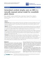
Figure 1 Macro- and microscopic images of the liver from group A mice. A: The urokinase-type plasminogen activator/severe combined immunodeficient mouse
mice were transplanted with human gastric cancer cells (h-GCCs) and euthanized 56 d later, at which time the livers were isolated and photographed; B: The arrows in A
point to concentrated regions of h-GCC colonies, and the sections were stained with hematoxylin and eosin (H and E). H and M in B represent h-GCC colonies and m-liver
cell regions, respectively; C: The sections were stained with anti-human alpha-fetoprotein (h-AFP) antibodies; D: The square region in C is enlarged and shown.
1
10.0
2
2
3
3
4
5
0.1
6
h-AFP (μg/mL)
1
1.0
h-Alb (mg/mL)
100
5
1.0
4
6
Figure 2 Changes in the serum concentrations of human albumin and
human alpha-fetoprotein in group
B-mice. Six mice (No.1-6) were co-transplanted with h-hepatocytes and human
gastric cancer cells. The serum levels of
human albumin (h-Alb) (left panel) and
human alpha-fetoprotein (h-AFP) (right
panel) were periodically monitored after
the cell transplantation.
0.01
0
0
2
4
6
Weeks post-transplantation
8
0
0
2
4
6
8
Weeks post-transplantation
h-GCCs (Figure 3C), and the anti-h-AFP Ab to identify
h-GCCs (Figure 3D). A comparison of Figure 3B and
C showed that most of the section from Figure 3B was
occupied with h-CK18+ cells, which corresponded to the
cells in the less eosinophilic areas of the H and E section. Human CK18- m-liver cells were located in eosinophilic areas in the H and E section, which were sporadically distributed as clusters with variable forms among
large engrafted h-cell colonies. Human-AFP+ h-GCCcolonies were distinguished by comparing Figure 3B-D.
These colonies were surrounded with less eosinophilic
WJG|www.wjgnet.com
h-hepatocytes (Figure 3D) that were swollen and clearer
(Figure 3B and C). Magnified views of the brown area
obtained from another serial sections of the liver shown
in Figure 3A are shown in Figure 4A (H and E) and Figure 4B (h-AFP-stain). Human GCCs formed moderately
differentiated adenocarcinomas with disrupted glandular
structures, which is a characteristic feature of gastric
cancer. Morphometric analyses using these h-CK18and h-AFP-stained serial sections indicated that the RIhhepatocyte and MIh-GCC in group B mice was 66.0% ± 12.3% (n
= 6) and 12.0% ± 6.8% (n = 6), respectively. The mice in
3878
August 7, 2012|Volume 18|Issue 29|
Fujiwara S et al . Animal model for liver cancer metastasis
A
B
H and E
10 mm
C
1 mm
h-AFP
D
h-CK18
1 mm
1 mm
Figure 3 Macroscopic image of the liver of mouse No. 1 from Figure 2 at 56 d post-transplantation. A: The thick and thin white arrows point to h-cells [human
hepatocytes (h-hepatocytes) and human gastric cancer cells (h-GCCs)] and m-liver cell regions, respectively; B: The liver was sectioned and stained with hematoxylin
and eosin (H and E); C: The liver was sectioned and stained with anti-h-CK18; D: The liver was sectioned and stained with anti-human alpha-fetoprotein (h-AFP) antibodies. The h-AFP + (h-GCC) colonies were surrounded by less eosinophilic h-hepatocytes.
H and E
A
H
h-AFP
B
H
H
G
G
M
H
H
M
50 μm
50 μm
Figure 4 Magnified images of hepatic histology from group B mice. A: A serial section of the liver in Figure 3 was subjected to hematoxylin and eosin (H and E); B:
A serial section of the liver in Figure 3 was subjected to human alpha-fetoprotein (h-AFP) staining. H, G and M represent the areas occupied by human-hepatocytes,
human gastric cancer cells (h-GCCs), and host m-liver cells, respectively. h-GCCs composed moderately differentiated adenocarcinoma with disrupted glandular
structures.
cells should possess at least two key features. First, the
transplanted cancer cells need to invade and colonize in
the host liver. Second, the liver of the host model has to
provide the human cells with appropriate pathophysiological microenvironments that recapitulate the h-liver
in vivo. Most of the conventional models to date manifest the first feature, but none of them have been able
to sufficiently recapitulate the microenvironment of the
h-liver [4-6]. In the present study, we established a unique
and novel that possessed both of these features.
In our study, we successfully engrafted the liver with
group B survived for the entire 56 d study. Extrahepatic
sites and organs, such as the peritoneal cavity and kidney, were also examined for the presence of metastatic
h-GCC lesions. The metastatic h-GCCs were not found
in the extrahepatic regions during the observational period, indicating that the cells did not metastasize to any
other regions.
DISCUSSION
An ideal animal model for liver metastasis of h-cancer
WJG|www.wjgnet.com
3879
August 7, 2012|Volume 18|Issue 29|
Fujiwara S et al . Animal model for liver cancer metastasis
of interactions between the invading cancer cells and
the accepting host cells. There seemed to be two groups
of animals within the experimental groups, one that
more easily accepted xenogeneic cells and another that
demonstrated resistance. However, we have consistently
observed similar variances in h-Alb levels among individual mice when we generated h-hepatocyte chimeric
mice[12], though inbred mice were used as hosts. These
variances are accidental in nature and might originate
from some differences in manipulation procedures for
transplantation as well as uncontrollable differences in
the phenotypes of the uPA Tg mice[10]. Despite these
variances at the individual level, experimental group B of
this study clearly demonstrated that we were able to reproducibly create mice whose livers were co-repopulated
with healthy, normal h-hepatocytes and h-GCCs. Both
h-hepatocytes and h-GCCs have high affinities for liver
tissue, which drives engraftment of the liver and results
in the generation of a humanized liver with metastatic
cancer cells. We also found that the RIh-hepatocyte (66.0% ±
12.3%) was significantly higher than MIh-GCC (12.0% ±
6.8%), which may be a reflection of the difference in the
inherent replication rates of the cells and adaptability
to the host liver tissues. Our results indicate that h-hepatocytes are, as a whole, superior to h-GCCs in colony
growth.
Relevant and reproducible animal models are indispensable tools for deducing the mechanisms of liver
metastasis and pharmacokinetics of anti-cancer drugs,
and several models have been developed to meet these
practical needs, though they are quite limited [2,25-30].
Preclinical tests of anti-cancer drugs for their effectiveness and toxicity in relevant animal models are required
prior to application in humans[31]. Toxicity data from
non-primate species have been quite poor at predicting
outcomes in subsequent human clinical trials, since there
are significant differences in the metabolic activities of
the hepatocytes between humans and rodent[32-34]. Therefore, animal models with a humanized liver are more
physiologic and will provide better tools for analyzing
the pharmacokinetics of anti-cancer drugs as well as
studying cancer metastasis[35-37]. To our knowledge, no
intrahepatic metastatic cancer model with a humanized
liver has been available to date[25,30,35-37]. The m-liver in
the present study was chimeric and was composed of
normal h-hepatocytes and m-hepatocytes. Previous studies have reported that the h-hepatocytes in these chimeric livers are functional and secreted a variety of hepatic
proteins, such as Alb, -1 antitrypsin, apolipoprotein A,
apolipoprotein E, several clotting factors, and complement proteins present in h-plasma [38]. Transplanted
h-hepatocytes also retain normal pharmacological responses, which makes the chimeric mouse model useful
for studying the metabolism of compounds that cannot
be easily administered to healthy volunteers[14,15]. In vivo
studies using these mice showed their utility in evaluating
the metabolism of drugs catalyzed by both phase Ⅰ and
phase Ⅱ enzymes[13-15,39,40]. Since the liver functions of
h-GCCs in the group A mice, and the cells formed relatively large colonies, with the MI as high as 25% at 56 d
post-transplantation. However, such a considerably high
MI could be a result of effects from either the donor
or host side of the model. We chose h-AFP+ h-GCCs
as a metastatic cancer cell line, since previous studies
reported that patients with AFP+ gastric cancer showed
a higher liver MI than those with AFP– cells; more than
70% of the patients developed liver metastasis [18,19].
These AFP+ cancer cells express c-Met[19], which is the
receptor for human hepatocyte growth factor (HGF),
and therefore it is plausible that the cells have a high affinity for liver tissues under conditions where the levels
of activated HGF in these tissues become high [20]. In the
present study, we utilized the uPA/SCID mice as hosts,
which possessed a uPA transgene product that continuously damages the hepatocytes. In this model, the host
hepatocytes generate pro-inflammatory environments in
the liver, which stimulates the mobilization and expression of HGF in the liver tissues, including hepatocytes.
The role of uPA is an important aspect in this
model. The host m-hepatocytes express unusually high
levels of uPA, which is thought to induce severe damage in the replicative ability of m-hepatocytes through
the activation of plasminogen, fibrinogen, and other
proteins within the rough endoplasmic reticulum (RER)
involved in proteolysis that lead to functional defects of
the RER[21]. In addition, uPA is secreted from m-hepatocytes into the plasma[10], indicating that it circulates to
liver tissues through sinusoidal capillaries and activates
the conversion of blood plasminogen to plasmin. Therefore, the host liver tissue may provide h-GCCs with a
pro-metastatic-like microenvironment. In fact, previous
studies have indicated that uPA and its receptor (uPAR)
play critical roles in the extravasation of tumors[22-24].
Therefore, the injected h-GCCs are prone to extravasate
liver tissues through the portal vein and sinusoid because
of the uPA-induced fragility of vascular and sinusoidal
endothelia and subsequently engraft liver tissues through
an affinity for c-Met. Once the h-GCCs invade liver tissues, they can relatively easily propagate due to c-Met
signaling in the host parenchyma, and can consequently
replace m-hepatocytes as a result of the uPA-mediated
damage. These conditions are also convenient for engraftment and proliferation of normal, healthy h-hepatocytes, as shown in this study when co-transplanted
with h-GCCs.
The co-transplantation of h-hepatocytes with
h-GCCs also resulted in the development of metastatic
colonies in the mice similar to the transplantation of
h-GCCs alone. In this type of transplantation experiment, large variances in serum concentrations of replacement marker proteins (h-Alb and h-AFP) were
observed. The h-AFP kinetic curves were different from
those of h-Alb and exhibited an increase of the serum
level through “three steps”: initial increase, followed
by a plateau or decline, and then a sharp increase. This
complex h-AFP kinetic pattern suggests the presence
WJG|www.wjgnet.com
3880
August 7, 2012|Volume 18|Issue 29|
Fujiwara S et al . Animal model for liver cancer metastasis
the chimeric mice described in this study have not yet
been characterized, future studies are needed to assess
the model for anti-cancer drug testing. Taking together,
the h-hepatocyte-chimeric mice may provide a useful
bridge for studying human liver-related diseases because
of the similarities with humans in physiological function
and drug kinetics.
In conclusion, we have established a unique and
novel animal model for studying liver cancer metastasis.
The chimeric liver of the uPA/SCID mouse containing both human cancer cells and hepatocytes could be
utilized as an appropriate model for in vivo testing of
the efficacy and human-type metabolisms of candidate
drugs for anti-cancer treatment as well as studying the
mechanisms of liver cancer metastasis.
REFERENCES
1
Yamamoto J, Saiura A, Koga R, Seki M, Ueno M, Oya M,
Azekura K, Seto Y, Ohyama S, Fukunaga S, Yamaguchi T,
Kokudo N, Makuuchi M, Muto T. Surgical treatment for
metastatic malignancies. Nonanatomical resection of liver
metastasis: indications and outcomes. Int J Clin Oncol 2005;
10: 97-102
Ishizu K, Sunose N, Yamazaki K, Tsuruo T, Sadahiro S,
2
Makuuchi H, Yamori T. Development and characterization
of a model of liver metastasis using human colon cancer
HCT-116 cells. Biol Pharm Bull 2007; 30: 1779-1783
3 Leen E, Ceccotti P, Moug SJ, Glen P, MacQuarrie J, Angerson WJ, Albrecht T, Hohmann J, Oldenburg A, Ritz JP, Horgan PG. Potential value of contrast-enhanced intraoperative
ultrasonography during partial hepatectomy for metastases:
an essential investigation before resection? Ann Surg 2006;
243: 236-240
Giavazzi R, Campbell DE, Jessup JM, Cleary K, Fidler IJ.
4
Metastatic behavior of tumor cells isolated from primary
and metastatic human colorectal carcinomas implanted into
different sites in nude mice. Cancer Res 1986; 46: 1928-1933
5 Takamura M, Sakamoto M, Genda T, Ichida T, Asakura H,
Hirohashi S. Inhibition of intrahepatic metastasis of human
hepatocellular carcinoma by Rho-associated protein kinase
inhibitor Y-27632. Hepatology 2001; 33: 577-581
Niedergethmann M, Alves F, Neff JK, Heidrich B, Aramin N,
6
Li L, Pilarsky C, Grützmann R, Allgayer H, Post S, Gretz N.
Gene expression profiling of liver metastases and tumour
invasion in pancreatic cancer using an orthotopic SCID
mouse model. Br J Cancer 2007; 97: 1432-1440
Bosma GC, Custer RP, Bosma MJ. A severe combined im7
munodeficiency mutation in the mouse. Nature 1983; 301:
527-530
Suemizu H, Hasegawa M, Kawai K, Taniguchi K, Monnai
8
M, Wakui M, Suematsu M, Ito M, Peltz G, Nakamura M. Establishment of a humanized model of liver using NOD/Shiscid IL2Rgnull mice. Biochem Biophys Res Commun 2008; 377:
248-252
Suemizu H, Monnai M, Ohnishi Y, Ito M, Tamaoki N,
9
Nakamura M. Identification of a key molecular regulator
of liver metastasis in human pancreatic carcinoma using a
novel quantitative model of metastasis in NOD/SCID/gammacnull (NOG) mice. Int J Oncol 2007; 31: 741-751
10 Heckel JL, Sandgren EP, Degen JL, Palmiter RD, Brinster
RL. Neonatal bleeding in transgenic mice expressing urokinase-type plasminogen activator. Cell 1990; 62: 447-456
11 Mercer DF, Schiller DE, Elliott JF, Douglas DN, Hao C,
Rinfret A, Addison WR, Fischer KP, Churchill TA, Lakey JR,
Tyrrell DL, Kneteman NM. Hepatitis C virus replication in
mice with chimeric human livers. Nat Med 2001; 7: 927-933
12 Tateno C, Yoshizane Y, Saito N, Kataoka M, Utoh R, Yamasaki C, Tachibana A, Soeno Y, Asahina K, Hino H, Asahara
T, Yokoi T, Furukawa T, Yoshizato K. Near completely
humanized liver in mice shows human-type metabolic responses to drugs. Am J Pathol 2004; 165: 901-912
13 Utoh R, Tateno C, Yamasaki C, Hiraga N, Kataoka M, Shimada T, Chayama K, Yoshizato K. Susceptibility of chimeric
mice with livers repopulated by serially subcultured human
hepatocytes to hepatitis B virus. Hepatology 2008; 47: 435-446
14 Yoshizato K, Tateno C. A human hepatocyte-bearing
mouse: an animal model to predict drug metabolism and effectiveness in humans. PPAR Res 2009; 2009: 476217
15 Yoshizato K, Tateno C. In vivo modeling of human liver
for pharmacological study using humanized mouse. Expert
Opin Drug Metab Toxicol 2009; 5: 1435-1446
16 Chang YC, Nagasue N, Abe S, Taniura H, Kumar DD, Nakamura T. Comparison between the clinicopathologic features of AFP-positive and AFP-negative gastric cancers. Am
J Gastroenterol 1992; 87: 321-325
ACKNOWLEDGMENTS
We thank all of our colleagues in CLUSTER-Yoshizato
Project for providing support for the experiment and
preparation of manuscript.
COMMENTS
COMMENTS
Background
One of the major target organs for cancer metastasis is the liver, and therefore,
there has been increasing needs for animal models that can sufficiently mimic
the pathophysiological situation in human liver and that are suitable for investigating the mechanisms of hepatic cancer metastasis.
Research frontiers
An ideal animal model for liver metastasis of human cancer cells should possess at least two key features. First, the transplanted cancer cells need to invade and colonize the liver of the host. Second, the liver of the host model has
to provide the human cells with appropriate pathophysiological microenvironments that recapitulate the human liver in vivo. In the present study, the authors
established a unique and novel animal model with both of these features.
Innovations and breakthroughs
A liver-humanized mouse was generated by transplanting healthy and normal
h-hepatocytes into urokinase type plasminogen activator/severe combined immunodeficient (uPA/SCID) mice (immuno- and liver- compromized mice), and
the liver was stably and reproducibly replaced with human hepatocytes. This is
the first report of a novel experimental model that sufficiently mimics the pathophysiological situation of human liver.
Applications
The chimeric liver of the uPA/SCID mouse containing both human cancer cells
and hepatocytes could be utilized as an appropriate model for the in vivo testing of anti-cancer drugs as well as studying the mechanisms of liver cancer
metastasis.
Terminology
The uPA/SCID mouse is a transgenic mouse line that expressed uPA under the
control of the albumin enhancer/promoter which constitutively damages the hepatocytes due to constant exposure to uPA. A liver- humanized mouse (chimeric
mouse) was generated by transplanting healthy and normal human hepatocytes
into mouse liver of the uPA/SCID mouse (immuno- and liver-compromized
mouse), which had been generated by mating the uPA-Tg mouse with the SCID
mouse. This mouse model sufficiently mimics the pathophysiological situation
in human liver.
Peer review
This study tries to establish an animal model with h-hepatocyte-repopulated liver
for in vivo study of liver cancer using uPA/SCID mouse, which could be useful
for studying liver cancer metastasis. The authors transfected uPA/SCID mouse
either with human gastric cancer cells (h-GCCs) or h-GCCs with h-hepatocytes
and observed that both colonies can repopulate mouse liver. The study is well
conducted, the manuscript is well-written and the figures are of good quality.
WJG|www.wjgnet.com
3881
August 7, 2012|Volume 18|Issue 29|
Fujiwara S et al . Animal model for liver cancer metastasis
17 Sekiguchi M, Fujii Y, Saito A, Suzuki T, Shiroko Y, Nakamura H, Hasumi K. Alpha-fetoprotein-producing gastric
carcinoma: biological properties of a cultured cell line. J
Gastroenterol 1995; 30: 589-598
18 Kamata S, Kishimoto T, Kobayashi S, Miyazaki M, Ishikura
H. Possible involvement of persistent activity of the mammalian target of rapamycin pathway in the cisplatin resistance of AFP-producing gastric cancer cells. Cancer Biol Ther
2007; 6: 1036-1043
19 Amemiya H, Kono K, Mori Y, Takahashi A, Ichihara F, Iizuka H, Sekikawa T, Matsumoto Y. High frequency of c-Met
expression in gastric cancers producing alpha- fetoprotein.
Oncology 2000; 59: 145-151
20 Shanmukhappa K, Matte U, Degen JL, Bezerra JA. Plasminmediated proteolysis is required for hepatocyte growth
factor activation during liver repair. J Biol Chem 2009; 284:
12917-12923
21 Sandgren EP, Palmiter RD, Heckel JL, Daugherty CC,
Brinster RL, Degen JL. Complete hepatic regeneration after
somatic deletion of an albumin-plasminogen activator transgene. Cell 1991; 66: 245-256
22 Van Buren G, Gray MJ, Dallas NA, Xia L, Lim SJ, Fan F,
Mazar AP, Ellis LM. Targeting the urokinase plasminogen
activator receptor with a monoclonal antibody impairs the
growth of human colorectal cancer in the liver. Cancer 2009;
115: 3360-3368
23 Madsen MA, Deryugina EI, Niessen S, Cravatt BF, Quigley
JP. Activity-based protein profiling implicates urokinase activation as a key step in human fibrosarcoma intravasation.
J Biol Chem 2006; 281: 15997-16005
24 Obermajer N, Doljak B, Kos J. Cytokeratin 8 ectoplasmic
domain binds urokinase-type plasminogen activator to
breast tumor cells and modulates their adhesion, growth
and invasiveness. Mol Cancer 2009; 8: 88
25 Desdouets C, Fabre M, Gauthier F, Bréchot C, SobczakThépot J. Proliferation and differentiation of a human hepatoblastoma transplanted in the Nude mouse. J Hepatol 1995;
23: 569-577
26 Leveille-Webster CR, Arias IA. Establishment and serial
quantification of intrahepatic xenografts of human hepatocellular carcinoma in severe combined immunodeficiency
mice, and development of therapeutic strategies to overcome multidrug resistance. Clin Cancer Res 1996; 2: 695-706
27 Miyoshi E, Noda K, Ko JH, Ekuni A, Kitada T, Uozumi N,
Ikeda Y, Matsuura N, Sasaki Y, Hayashi N, Hori M, Taniguchi N. Overexpression of alpha1-6 fucosyltransferase in hepatoma cells suppresses intrahepatic metastasis after splenic
injection in athymic mice. Cancer Res 1999; 59: 2237-2243
28 Kollmar O, Schilling MK, Menger MD. Experimental liver
metastasis: standards for local cell implantation to study
isolated tumor growth in mice. Clin Exp Metastasis 2004; 21:
453-460
29 Hardy B, Morgenstern S, Raiter A, Rodionov G, Fadaeev
L, Niv Y. BAT monoclonal antibody immunotherapy of
human metastatic colorectal carcinoma in mice. Cancer Lett
2005; 229: 217-222
30 Schnater JM, Bruder E, Bertschin S, Woodtli T, de Theije
C, Pietsch T, Aronson DC, von Schweinitz D, Lamers WH,
Köhler ES. Subcutaneous and intrahepatic growth of human
hepatoblastoma in immunodeficient mice. J Hepatol 2006; 45:
377-386
31 Meuleman P, Leroux-Roels G. The human liver-uPA-SCID
mouse: a model for the evaluation of antiviral compounds
against HBV and HCV. Antiviral Res 2008; 80: 231-238
32 Kato R. Characteristics and differences in the hepatic mixed
function oxidases of different species. Pharmacol Ther 1979; 6:
41-98
33 Green CE, LeValley SE, Tyson CA. Comparison of amphetamine metabolism using isolated hepatocytes from five
species including human. J Pharmacol Exp Ther 1986; 237:
931-936
34 Naritomi Y, Terashita S, Kimura S, Suzuki A, Kagayama A,
Sugiyama Y. Prediction of human hepatic clearance from in
vivo animal experiments and in vitro metabolic studies with
liver microsomes from animals and humans. Drug Metab
Dispos 2001; 29: 1316-1324
35 Hata Y, Uchino J, Sato K, Sasaki F, Une Y, Naito H, Manabe
K, Kuwahara T, Kasai Y. Establishment of an experimental
model of human hepatoblastoma. Cancer 1982; 50: 97-101
36 Fuchs J, Wenderoth M, von Schweinitz D, Haindl J, Leuschner I. Comparative activity of cisplatin, ifosfamide, doxorubicin, carboplatin, and etoposide in heterotransplanted
hepatoblastoma. Cancer 1998; 83: 2400-2407
37 Kneteman NM, Mercer DF. Mice with chimeric human livers: who says supermodels have to be tall? Hepatology 2005;
41: 703-706
38 Meuleman P, Libbrecht L, De Vos R, de Hemptinne B, Gevaert K, Vandekerckhove J, Roskams T, Leroux-Roels G.
Morphological and biochemical characterization of a human
liver in a uPA-SCID mouse chimera. Hepatology 2005; 41:
847-856
39 Katoh M, Tateno C, Yoshizato K, Yokoi T. Chimeric mice
with humanized liver. Toxicology 2008; 246: 9-17
40 Tsuge M, Hiraga N, Takaishi H, Noguchi C, Oga H, Imamura M, Takahashi S, Iwao E, Fujimoto Y, Ochi H, Chayama K,
Tateno C, Yoshizato K. Infection of human hepatocyte chimeric mouse with genetically engineered hepatitis B virus.
Hepatology 2005; 42: 1046-1054
S- Editor Gou SX
WJG|www.wjgnet.com
3882
L- Editor A E- Editor Li JY
August 7, 2012|Volume 18|Issue 29|