endoplasmic reticulum stress induced apoptosis in the development of diabetes is there a role for adipose tissue and liver
Bạn đang xem bản rút gọn của tài liệu. Xem và tải ngay bản đầy đủ của tài liệu tại đây (590.09 KB, 11 trang )
Apoptosis (2009) 14:1424–1434
DOI 10.1007/s10495-009-0400-4
DIABETES AND APOPTOSIS
Endoplasmic reticulum stress-induced apoptosis
in the development of diabetes: is there a role
for adipose tissue and liver?
Carla J. H. van der Kallen Ỉ
Marleen M. J. van Greevenbroek Ỉ
Coen D. A. Stehouwer Ỉ Casper G. Schalkwijk
Published online: 16 September 2009
Ó The Author(s) 2009. This article is published with open access at Springerlink.com
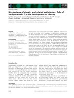
Abstract Diabetes mellitus (DM) is a multifactorial
chronic metabolic disease characterized by hyperglycaemia. Several different mechanisms have been implicated
in the development of the disease, including endoplasmic
reticulum (ER) stress. ER stress is increasingly acknowledged as an important mechanism in the development of
DM, not only for b-cell loss but also for insulin resistance.
Accumulating evidence suggests that ER stress-induced
apoptosis may be an important mode of b-cell loss and
therefore important in the development of diabetes. Recent
data also suggest a role of ER stress-induced apoptosis in
liver and adipose tissue in relation to diabetes, but more
extensive studies on human adipocyte and hepatocyte
(patho)physiology and ER stress are needed to identify the
exact interactions between environmental signals, ER
stress and apoptosis in these organs.
Keywords Diabetes Á Endoplasmic reticulum stress Á
Apoptosis Á Adipose tissue Á Liver
Abbreviations
ASK1
ATF3
ATF4
ATF6
Bcl-2
BiP/GRP78
CHOP/GADD153
DM
EIF2AK3/PERK
EIF2a
ERAD
ERAI
FA
GADD34
GADD153/CHOP
C. J. H. van der Kallen (&) Á M. M. J. van Greevenbroek Á
C. D. A. Stehouwer Á C. G. Schalkwijk
Department of Internal Medicine, Laboratory for Metabolism
and Vascular Medicine, Maastricht University,
Maastricht, The Netherlands
e-mail:
C. J. H. van der Kallen Á M. M. J. van Greevenbroek Á
C. D. A. Stehouwer Á C. G. Schalkwijk
Cardiovascular Research Institute Maastricht (CARIM),
Maastricht, The Netherlands
123
GRP78/BiP
GRP94
HF/HS
HIV
IKK
IL-1b
IL-6
Apoptosis signal-regulating kinase 1
Activated transcription factor 3
Activated transcription factor 4
Activated transcription factor 6
Factor B cell lymphoma-2
Glucose regulated protein 78/binding
immunoglobulin protein
C/EBP-homologous protein/growth
arrest-and DNA damage-inducible
gene GADD153
Diabetes mellitus
ER-resident PKR-like eIF2a kinase/
eukaryotic translation initiation factor
2-alpha kinase 3
Eukaryotic translation initiation factor
2-alpha
ER associated degradation
ER stress activator indicator
Fatty acids
Growth arrest and DNA damage
inducible protein (also known as
PPP1R1A = protein phosphatase 1,
regulatory (inhibitor) subunit 15A)
C/EBP-homologous protein/growth
arrest-and DNA damage-inducible
gene GADD153
Glucose regulated protein 78/binding
immunoglobulin protein
Glucose regulated protein 94
High fat/high sucrose
Human immunodeficiency virus
IjB kinase
Interleukin 1b
Interleukin 6
Apoptosis (2009) 14:1424–1434
IRE1
IRS-1
JNK
MCP-1
NAFLD
NASH
ORP150
PERK/EIF2AK3
PKC
PTP1B
ROS
Ser
mTOR
T1DM
T2DM
TNFa
TRAF2
Tyr
UPR
XBP-1
Inositol requiring 1
Insulin receptor substrate 1
c-Jun N-terminal kinase
Monocyte chemo-attractant protein 1
Nonalcoholic fatty liver disease
Non-alcoholic steatohepatitis
Oxygen regulated protein (150 kD)
ER-resident PKR-like eIF2a kinase/
Eukaryotic translation initiation factor
2-alpha kinase 3
Protein kinase C
Protein tyrosine phosphatase 1B
Reactive oxygen species
Serine
Mammalian target of rapamycin
Type 1 diabetes mellitus
Type 2 diabetes mellitus
Tumor necrosis factor a
TNF receptor-associated factor 2
Tyrosine
Unfolded protein response
X-box binding protein 1
Introduction
Diabetes mellitus (DM) is a multifactorial chronic metabolic disease characterized by hyperglycaemia. Several
different mechanisms have been implicated in the development of the disease. Although the precise molecular
events underlying the different forms of diabetes still
remain unclear, it is generally accepted that the underlying
defects include decreased secretion of insulin, its impaired
signalling or both. Type 1 diabetes (T1DM) is known to
result from an excessive loss of pancreatic b-cells, leading
to insulin deficiency. Among other important causes,
autoimmune and inflammatory processes have been
reported to disrupt b-cells, cause insulin deficiency and
hyperglycaemia and subsequently T1DM. Type 2 diabetes
(T2DM), the most common form of diabetes, is characterized by impaired insulin action (insulin resistance) paralleled by impaired insulin secretion and a progressive
decline in b-cell function. Insulin resistance, often associated with obesity and physical inactivity, is a major factor
in the progression of T2DM. Obesity is a well-known risk
factor for the development of T2DM. Importantly, obesity
is not only associated with lipid accumulation in adipose
tissue, but also in non-adipose tissues, such as liver and
muscle. Lipid accumulation in non-adipose tissue, also
known as ectopic lipid accumulation, has also been associated with the development of insulin resistance. Therefore, muscle, adipose tissue and liver are, beside pancreas,
1425
crucial tissues contributing to the development of insulin
resistance and thus to the development of T2DM.
A relatively new player in the DM field is endoplasmic
reticulum (ER) stress. ER stress and/or ER stress-induced
apoptosis are increasingly acknowledged as important
mechanisms in the development of DM, not only for b-cell
loss but also for insulin resistance. Since the last decade, it
has been generally accepted that ER stress plays an
important role in b-cell function and loss [1]. This is for
instance illustrated in Akita mice [2, 3], and the WolcottRallison syndrome [4, 5]. Akita mice spontaneously
develop diabetes with significant early loss of pancreatic
b-cell mass resulting from a missense mutation (Cys96Tyr)
in the insulin 2 gene that disrupts a disulfide bond between
A and B chains of insulin [6]. The Wolcott-Rallison syndrome is a rare autosomal-recessive disorder characterized
by the association of permanent neonatal or early-infancy
insulin-dependent diabetes, and growth retardation, and
other variable multisystemic clinical manifestations. The
gene responsible for this syndrome is EIF2AK3 (PERK),
the pancreatic eukaryotic initiation factor 2 (eIF2) kinase
[4, 5]. More recently, it was acknowledged that high fatand obesity-induced insulin resistance is also associated
with ER stress in adipose tissue and liver [7, 8]. Remarkably, until now no studies have demonstrated a role for ER
stress in skeletal muscle in relation to (the development of)
obesity or diabetes [7, 9]. Besides, the role of ER stress in
the development of diabetes that will be discussed in this
paper, there is also evidence that diabetes can induce or
aggravate ER stress and thereby affect the complications of
diabetes, such as renal disease, retinopathy and vascular
abnormalities [10–12].
In this review an overview of ER stress, the unfolded
protein response (UPR), and ER stress induced apoptosis is
given (see also refs [13–20]) with a further focus on the
possible role of ER stress-induced apoptosis in the liver
and adipose tissue in the onset of diabetes.
Endoplasmic reticulum stress-unfolded protein
response
The endoplasmic reticulum (ER) is an important organelle
required for cell survival and normal cellular function. In
the ER, nascent proteins are folded with the assistance of
ER chaperones (i.e. molecular chaperones and folding
enzymes). Subsequently, correctly folded proteins are
transported to the Golgi apparatus. Unfolded and misfolded
proteins, on the other hand, are retained in the ER, retrotranslocated to the cytoplasm by the machinery of ER
associated degradation (ERAD), and degraded by the proteasome. As a major intracellular calcium storage compartment, the ER also plays a critical role towards
maintenance of cellular calcium homeostasis. In addition,
123
1426
Apoptosis (2009) 14:1424–1434
the ER also has a role in lipid biosynthesis, e.g. lipid
membrane synthesis and controlling the synthesis of cholesterol and other membrane lipid components.
ER stress is caused by perturbations of any of the three
homeostatic functions of the ER, i.e. functioning as a site
for protein folding, for synthesis of unsaturated fatty acids
(FA), sterols, and phospholipids and for intracellular Ca2?
storage. ER stressors include: (1) disturbances in cellular
redox regulation caused by hypoxia, oxidants, or reducing
agents, which interfere with disulfide bonding of proteins in
the lumen of the ER, (2) glucose deprivation, probably by
interfering with N-linked protein glycosylation in the ER,
(3) disruption of Ca2? metabolism causing impaired functions of Ca2? dependent chaperones such as Grp78, Grp94
and calreticulin, (4) viral infections, which overload the ER
with virus encoded proteins, (5) high fat diet, and (6) protein
mutations that hamper adequate folding [17, 18]. The consequence of ER stress is an overwhelmed or compromised
ability of the ER to properly fold proteins.
Accumulation of unfolded and/or misfolded proteins in
the ER lumen is a hallmark of perturbation of any of the
three functions of the ER and results in activation of the
unfolded protein response (UPR). The UPR is a complex
and coordinated adaptive signalling mechanism to re-establish homeostasis of ER functions (Fig. 1). ER stress sensors
[IRE1 (inositol requiring 1), ATF6 (activated transcription
factor 6) and PERK (ER-resident PKR-like eIF2a kinase)]
detect the accumulation of unfolded and/or misfolded
protein at the onset of ER stress and initiate the UPR. To
re-establish homeostasis and normal ER function, the UPR
initiates a global decrease in protein synthesis while
increasing the production of ER chaperone proteins and
ER-associated degradation (ERAD).
The mammalian UPR with its signalling components is
complex, diverse and flexible as has been described in great
detail in recent reviews [16, 20]. In short, UPR signals
through three pathways, that each utilizes one of the three
Endoplasmic Reticulum Stress
e.g. high glucose, FFA, inflammation
Accumulation of unfolded proteins in the ER
Activation of the Unfolded Protein response (UPR)
Re-establish homeostasis
Failure to restore homeostasis
Normal ER function
Cell death, usually apoptosis
Fig. 1 The relation between ER stress and ER stress induced
apoptosis in the development of diabetes
123
ER stress sensors IRE1, ATF6 and PERK (Fig. 2). IRE1 is a
transmembrane kinase/endoribonuclease (RNAse). Activation of IRE1 initiates the nonconventional splicing of Xbp-1
mRNA. Spliced Xbp-1 mRNA encodes a transcription
activator that drives transcription of genes such as ER
chaperones, whose products directly participate in ER
protein folding. PERK is a transmembrane kinase that
phosphorylates the eukaryotic translation initiation factor 2
subunit (eIF2). This leads to a reduced protein synthesis,
which counteracts ER protein overload. ATF6 is an
ER-resident transmembrane protein. Upon activation, the
cytoplasmic domain of ATF6 is released from its membrane
anchor by regulated proteolysis. The cleaved N-terminal
fragment migrates to the nucleus, acts as an active transcription factor, and increases the expression of the genes
encoding proteins that function to augment the ER protein
folding capacity. The exact mechanism of UPR activation is
unknown. One of the most described models is the competition model, in which the ER chaperone protein glucose
regulated protein (Grp)78/BiP, is an UPR regulator and
plays an essential role in the activation of IRE1, PERK and
ATF6. In the inactive state, i.e. in resting cells, all three ER
stress sensors (IRE-1, PERK and ATF6) are maintained in
an inactive state through their association with the ER
chaperone BiP (Fig. 2a). When the ER homeostasis is
perturbed, i.e. upon ER stress, BiP is sequestered by
unfolded and/or misfolded proteins that accumulate in the
ER lumen (Fig. 2b, c). Dissociation of BiP from de ER
stress sensors triggers the activation of IRE1, PERK and
ATF-6 (Fig. 2d). Other models of UPR activation are the
ligand-binding model in which unfolded and/or misfolded
proteins directly interact with the ER stress-sensing
domains of the ER stress sensors, and the probing model, in
which newly synthesized stress-sensing proteins probe the
efficiency of the ER-resident protein-folding machinery by
presenting themselves as substrates to the folding machinery [20].
Endoplasmic reticulum stress—apoptosis
Under conditions of severe and prolonged ER stress, the
UPR is unable to restore normal cellular function. Subsequently, cell death, usually occurring by apoptosis, is triggered (Fig. 1). Cell death results in loss of cell/tissue
function and may be the primary reason for the manifestation of disease in several ER stress-related disorders. Indeed,
cell death induced by ER stress has been implicated in a
wide variety of diseases including ischemic injury (stroke,
myocardial infarction), heart failure, several neurodegenerative diseases, diabetes and bipolar disorder [17, 18]. The
mechanisms of apoptosis are highly complex, involving an
energy-dependent cascade of molecular events. There are
two main apoptotic pathways: the extrinsic or death receptor
Apoptosis (2009) 14:1424–1434
1427
A
B
Endoplasmic Reticulum
PERK
BiP
ER
Stress
Endoplasmic Reticulum
IRE1
IRE1
BiP
PERK
ATF6
ATF6
C
D
ER
Stress
ER
Stress
Endoplasmic Reticulum
Endoplasmic Reticulum
BiP
BiP
IRE1
PERK
ATF6
= unfolded protein
IRE1
PERK
P P
eiF2α
P
Translation
inhibition
ATF6
P
P
Golgi:
S1P spliced IRE1
S2P XBP1
Nucleus
XBP1
mRNA
UPR-genes
- BiP
- XBP1
-…
Fig. 2 The unfolded proteins response and its signaling components.
A simplified scheme of the initiation of the unfolded protein response
(UPR). In the inactive state, i.e. in resting cells, all three ER stress
sensors (IRE-1, PERK and ATF6) are maintained in an inactive state
through their association with the ER chaperone BiP (a). Upon ER
stress, BiP is recruited by the unfolded and/or misfolded proteins (b).
This results in BiP dissociation from its conformational binding state
to the transmembrane receptor proteins PERK, IRE1 and ATF6 (c).
Dissociation results in activation of IRE1, PERK and ATF6 (d). The
activated cytosolic domain of PERK phosphorylates the eukaryotic
translation initiation factor 2 subunit (eIF2), inhibiting translation.
The activated cytosolic domain of IRE1 initiates the nonconventional
splicing of Xbp-1 mRNA, thereby cleaves a 252 bp intron from
XBP1. Spliced Xbp-1 mRNA encodes a transcription activator that
drives transcription of genes such as ER chaperones, whose products
directly participate in ER protein folding. Activated ATF6 translocates to the Golgi, cleaved by proteases to form an active 50 kDa
fragment. ATF6 p50 and XBP1 bind ER stress-response element
(ERSE) promoters in the nucleus to produce up regulation of the
proteins that function to augment the ER protein folding capacity
pathway and the intrinsic or mitochondrial pathway [21].
Current evidence suggests that these two pathways are
linked and that molecules in one pathway can influence the
other [22]. The extrinsic signalling pathways act via transmembrane receptor-mediated interactions. These involve
death receptors that are members of the tumor necrosis
factor (TNF) receptor gene superfamily [23]. The intrinsic
signalling pathways involve a diverse array of non-receptormediated stimuli that produce intracellular signals that act
directly on targets within the cell and are mitochondrialinitiated events. These non-receptor stimuli include radiation, toxins, hypoxia, hyperthermia, viral infections, and
free radicals but also the absence of certain growth factors,
hormones and cytokines [21].
Signalling through the ER stress sensors can trigger
pro-apoptotic signals during prolonged ER stress. However, the ER stress sensors do not directly cause cell death
but rather initiate the activation of downstream molecules
such as CHOP or JNK, which further push the cell down
the path of death. This results in caspase activation, the
execution phase of ER stress-induced apoptosis, and
finally in the ordered and sequential dismantling of the
cell. Caspases are cysteine proteases that exist within the
cell as inactive pro-forms or zymogens and are cleaved to
form active enzymes following the induction of apoptosis.
ER stress activates both intrinsic and extrinsic apoptotic
pathways [13, 14]. Currently, three main pathways of ER
stress-induced apoptosis are identified (Fig. 3): (1) the
proapoptotic pathway of CHOP/GADD153 transcription
factor which is mainly induced via PERK/eIF2, (2) IRE1mediated activation of apoptosis signal-regulating kinase 1
(ASK1)/c-Jun NH2-terminal kinase (JNK), and (3) activation of the ER localized cysteine protease, caspase 12
[15, 18, 19].
123
1428
Apoptosis (2009) 14:1424–1434
Endoplasmic Reticulum
Endoplasmic
Reticulum
Stress
BiP
PERK
pro-caspase-12
IRE1
TRAF2
ASK1
eiF2α
caspase-12
ATF4
JNK
caspase
cascade
CHOP
APOPTOSIS
= unfolded protein
Fig. 3 ER stress induced apoptosis. Three main pathways of ER
stress-induced apoptosis are identified: (1) the proapoptotic pathway
of CHOP/GADD153 transcription factor which is mainly induced via
PERK/eIF2. CHOP down-regulates the anti-apoptotic factor B cell
lymphoma-2 (Bcl-2), but also upregulates Ero-1, a thiol oxidase that
promotes protein folding in the ER but also generates reactive oxygen
species (ROS), and thereby promotes apoptosis, (2) IRE1-mediated
activation of apoptosis signal-regulating kinase 1 (ASK1)/c-Jun NH2terminal kinase (JNK). IRE1 interacts with TRAF2 (TNF receptorassociated factor-2) and ASK1. This leads to activation of ASK1 and
JNK, followed by apoptosis, and (3) activation of the ER localized
cysteine protease, caspase 12. Caspase 12 is activated by m-Calpain
in the cytoplasm. Activation of m-Calpain is a consequence Ca2?
efflux out of the ER upon ER stress. These three pathways all end in
caspase cascade activation, the execution phase of ER stress-induced
apoptosis
result in misfolding and accumulation of proinsulin in the
ER lumen of b cells. This accumulation can cause b cell
failure [6, 29, 30]. In vivo data show that also pathological
conditions like high glucose, free fatty acids, cytokines,
and nitric oxide induce UPR gene expression and compromise b cell function [25, 31–33]. Moreover, in islets of
T2DM patients, ER stress has been demonstrated by
increased staining for ER chaperones and CHOP along
with increased ER size [34–37].
However, the exact molecular mechanisms for the ER
stress-induced apoptosis in b cells are not entirely clear. The
most recent data support that the PERK-ATF4-CHOP stress
signalling pathway is important in b-cell apoptosis (Fig. 3).
This pathway plays a role in b-cell injury induced by oxidative stress and saturated fatty acids [38–42]. This is
confirmed by the finding that CHOP deletion reduces oxidative stress, improves b cell function, and promotes cell
survival in multiple mouse models of diabetes [39]. However, the PERK-ATF4-CHOP pathway is not the only
pathway inducing apoptosis in b-cells. In contrast to apoptosis by high lipids, the PERK-ATF4-CHOP ER stress–
signalling pathway is not necessary for cytokine-induced
b-cell death [42]. Other data show that also the IRE1-JNK
pathway is associated with the apoptosis in b cells [41]
(Fig. 3). This pathway is also involved in ER stress-induced
apoptosis caused by chronic high glucose, fatty acids, and
Il-1b induced depletion of Ca2? [41, 43–45].
ER stress, UPR and apoptosis in adipose tissue
ER stress, UPR and apoptosis in different organs
and the development of diabetes
ER stress, UPR and apoptosis in the pancreas
b-cell loss plays a crucial role in the development of insulin
deficiency and in the onset and/or progression of diabetes.
Regulation of the b-cell mass involves a balance of b-cell
replication and cell death. Accumulating evidence suggests
that apoptosis may be the main mode of b-cell death in
both types of diabetes. Recent studies point to a role of the
ER in the sensing and transduction of apoptotic signals in
b-cells as recently described in detail in excellent reviews
[19, 24]. We now addressed the most relevant data of the
last year, with a focus on ER stress and apoptosis in the
pancreas, adipose tissue and the liver.
Several studies show evidence for a role of ER stress in
b cell failure. Mutations in the primary sensors of the UPR
or mutations that affect chaperone functions of the UPR,
e.g. EIF2AK3, IRE1, P58IPK (DNAJC3) and EIF2a, impair
b cell health and function [4, 25–28]. Moreover, mutations
in proinsulin causing disruption of disulfide bond pairing
123
The prevalence of obesity is increasing rapidly worldwide,
especially in developing countries. An important consequence
of obesity is an increased risk of developing impaired glucose
tolerance and T2DM. Indeed, along with the increase in
obesity, a parallel increase in the prevalence of T2DM,
impaired glucose tolerance has occurred [46, 47]. The metabolic complications of obesity, usually referred to as the
metabolic syndrome, consist of insulin resistance (often culminating in b-cell failure, impaired glucose tolerance and
T2DM), dyslipidemia, hypertension, and premature heart
disease. Our understanding of the role of adipose tissue in
metabolic syndrome has continued to evolve with the identification of adipose tissue as a potent endocrine organ. Adipose tissue secretes large amounts of adipocyte-generated
factors, such as adipokines, cytokines and complement components. Cells that are specialized for a high secretory
capacity, such as mature B lymphocytes, liver cells and
pancreatic b-cells, are known to expand and adopt their ER
capabilities to meet an increased demand of protein synthesis
[48]. It is, therefore, likely that ER stress plays a role in adipose tissue dysfunction and most probably also in cell death.
Although apoptosis of (pre)adipocytes has not been
extensively studied, there is growing evidence that, under
Apoptosis (2009) 14:1424–1434
specific circumstances, decreases in adipose tissue mass in
humans could result from a loss of fat cells through programmed cell death. The general idea is that in a normal
healthy situation adipocyte number is relatively stable
when the energy intake is less than the energy output. In
this case, the adipose tissue mass only decreases as a result
hypotrophy via mobilization of triglycerides [49]. On the
other hand, conditions of pathological fat wasting can
involve loss of adipocytes through apoptotic mechanisms.
For example, apoptotic events were observed in fat tissue
of patients with tumor cachexia and in the fat remodelling
processes associated with highly active antiretroviral therapy, e.g. ritonavir, in HIV infected patients with lipodystrophy [50–52]. Ritonavir not only induces apoptosis and
inhibits adipocyte differentiation, but also affects inflammatory mediators, ER stress and oxidative stress, as shown
by gene profiling [53, 54].
Recent data suggest that ER stress may, via several
mechanisms, also be involved in apoptosis of (pre)adipocytes in relation to the development of obesity/diabetes. In
obese individuals, adipose tissue is poorly oxygenated [55,
56], which may lead to local hypoxia in adipose tissue. ER
stress may form a link between hypoxia and apoptosis.
Disturbances in cellular redox regulation caused by
hypoxia interfere with disulphide bonding in the lumen of
the ER, leading to unfolded and misfolded proteins. In
3T3-L1 adipocytes, hypoxia is associated with ER stress, as
shown by increased levels of GRP78 and CHOP [57]. Yin
et al. [58] described that hypoxia induces cell death, promotes free FA release and inhibits glucose uptake in adipocytes by inhibition of insulin signalling pathway. These
metabolic effects of hypoxia may also add to the generation
of ER stress, e.g. in addition to hypoxia itself, palmitate, a
saturated fatty acid (FA), also activated UPR and induced
apoptosis in preadipocytes. CHOP was one of the proteins
that were influenced [59]. Moreover, very recently, three
papers for the first time show ER stress in human adipose
tissue [60–62]. Although none of these papers show direct
evidence for a relation between obesity and ER stressinduced apoptosis, the results of Sharma et al. [61], are
very suggestive for this. They used ATF4, GADD43 and
ATF3 as markers of apoptosis pathways, and show a
relation with obesity. Thus, although the data strongly
suggest a role for ER stress in apoptosis of adipose tissue,
experiments are needed to fully explore this pathway.
For all studies performed with adipose tissue biopsies, it
should be emphasized that the precise identity of cells
within adipose tissue that show ER stress, and possibly
related apoptosis, is not clear. Adipocytes generally
account for only 50% of the total number of cells in adipose tissue. Other cells within adipose tissue, e.g. preadipocytes, macrophages and vascular cells, can also secrete
an extensive range of protein signals and factors linked to
1429
inflammatory response and may therefore also be sensitive
for ER stress. This is of special interest since adipose tissue
is more and more recognized as a tissue containing a
molecular network that connects obesity, adipokine secretion, chronic inflammation and insulin resistance. Inflammation of adipose tissue is often observed in obesity and
diabetes and is associated with the infiltration of macrophages into adipose tissue, which may be triggered by
adipocyte death, adipokine secretion e.g. TNF-alpha and
IL-6, and elaboration of chemokines by adipocyte e.g.
monocyte chemo-attractant protein (MCP)-1 [63–65]. The
mechanism via which adipocyte death stimulates macrophage infiltration has been proposed to occur via an alternative death pathway that share features of both necrosis
and apoptosis [66]. This possibility is supported by the
finding that macrophages are located around dead adipocytes in the adipose tissue [67]. Apoptosis of macrophages
in adipose tissue may also be linked to diabetes. It has been
suggested that macrophage cell death in adipose tissue is an
important effect of pioglitazone treatment and this may
play an essential role in the management of diabetes mellitus and metabolic syndrome [68]. Hypoxia and hypoxia
related ER stress may also play a role in apoptosis of
macrophages in adipose tissue. Hypoxia does not only
stimulate the inflammatory response of macrophages [69,
70], but also induced apoptosis and cell cycle arrest at
G0/G1 phase, via AKT and JNK [71]. To our knowledge
no studies have been published on adipose tissue histology
showing ER stress related apoptosis in a specific cell type.
ER stress, UPR and apoptosis in the liver
ER stress has been recognized in various models of liver
injury and human liver diseases (as reviewed in [72]). The
liver plays essential roles in metabolism, biosynthesis,
excretion, secretion and detoxification. Comparable to
adipose tissue, the liver contains a range of different cell
types. The three main liver cell types are hepatocytes,
resident macrophages (i.e. Kupffer cells), and endothelial
cells. Apoptosis in the liver occurs in many forms of liver
injury, e.g. chronic viral liver disease, nonalcoholic and
alcoholic steatohepatitis [73–76].
Nonalcoholic fatty liver disease (NAFLD) results from
metabolic hepatic dysregulation in metabolic syndrome
and T2DM. NAFLD refers to a wide spectrum of liver
disease ranging from simple fatty liver (steatosis), to
nonalcoholic steatohepatitis (NASH), to cirrhosis (irreversible, advanced scarring of the liver). Several studies
have shown that NAFLD predicts future development of
T2DM (reviewed in [77]). The pathogenesis of NAFLD is
thought to be a multiple-hit process involving insulin
resistance, oxidative stress, apoptosis, and adipokines. In
NASH, inflammation of the liver is associated with the
123
1430
accumulation of fat in the liver and additionally to different
degrees of scarring, which may lead to severe liver scarring
and cirrhosis. The general idea is that as consequence of
both hepatic and peripheral insulin resistance, the hepatocellular accumulation of triglycerides, initially leads to an
altered metabolism of glucose and free fatty acids in the
liver. Increased expression of death receptors in response to
this altered hepatic metabolism enhances the hepatocytes’
susceptibility for pro-apoptotic stimuli, thus eliciting
excessive hepatocyte apoptosis and inflammation. Interestingly, hepatocyte apoptosis is significantly increased in
patients with NASH and correlates with disease severity
[75, 78].
Evidence is mounting for an important role for ER
stress-induced apoptosis in NAFLD. In relation to the onset
of diabetes, most in vivo and in vitro studies on the relation
between ER stress-induced apoptosis and fatty liver focus
on saturated FA. Saturated FA induce ER stress and
apoptosis at physiologic concentrations and with a relatively rapid time course in H4IIE liver cells [79, 80], as
illustrated by the induction of ER stress response genes and
apoptosis which occurred after 4 h and between 6 and 16 h,
respectively [79]. Ozcan et al. [7] showed that chronic
excessive nutrient intake activated the UPR both in liver
and in adipose tissue. A very recent study used transgenic
mice carrying the XBP-1-delta-DBD-venus expression
gene, which acts as an ER stress-activated indicator
(ERAI). In these transgenic mice, the gene encoding venus,
a variant of green fluorescent protein, is fused as a reporter
downstream of a partial sequence of human XBP-1
including the ER stress-specific intron. The XBP-1/venus
fusion protein is produced in cells under ER stress conditions, and cells under ER stress can be detected by monitoring the generation of fluorescence. They showed in the
liver of the ERAI transgenic mice that ERAI fluorescence
was observed as early as 4 weeks after treatment with a
high fat, high sucrose (HF/HS) diet, whereas it was not
detected in the fat and muscle, even after 12 weeks of
HF/HS diet treatment [9]. It is important to realize that not
all FA activate the UPR. Only livers and hepatocytes from
rats on a high saturated fat diet, but not high polyunsaturated fat diet, were characterized by the presence of spliced
XBP-1 mRNA and increased GRP78 and CHOP protein
[81]. This not only suggests that the UPR may sense and
respond to the fatty acid environment but also that the ratio
of saturated to unsaturated FA may be an important
determinant of hepatic ER homeostasis. Although not
directly shown in hepatocytes, several mechanisms have
been proposed for fatty acid induced ER stress. One possible mechanism is the rapid incorporation of palmitate into
lipid components of the rough ER followed by disruption
of ER structure and function [82]. Another mechanism of
palmitate-induced ER stress is the generation of reactive
123
Apoptosis (2009) 14:1424–1434
oxygen species (ROS). ROS by itself can induce ER stress.
Prolonged or severe ER stress, which may occur in the
presence of excess palmitate, can lead to further ROS
accumulation, potentially amplifying the apoptotic/cell
death response [83]. Alternatively, as described in b cells,
palmitate
can lead to an early and sustained depletion of ER Ca2?
stores, which may trigger ER stress via impaired protein
folding [41].
ER stress—UPR—insulin resistance
ER stress and the UPR are not only associated with
apoptosis in of b-cells, hepatocytes and adipocytes but also
with metabolic derangements, especially with insulin
resistance. In adipose tissue and liver, the relation of ER
stress with insulin resistance is actually more evident than
its relation with apoptosis. The general idea is that ER
stress interferes with the signalling of the insulin receptor
via JNK (Fig. 4). Therefore JNK can not only be a link
between ER stress and apoptosis (Fig. 3) but also between
ER stress and insulin resistance. A major site of regulation
of insulin signalling, both positive and negative, is phosphorylation of the important insulin receptor docking
protein insulin receptor protein-1 (IRS-1), whereby phosphorylation of the tyrosine (Tyr) residues in IRS-1 induces
phosphorylation of the serine (Ser) residues in IRS-1 and
hampers insulin signal transduction (reviewed in [84]).
Although the exact mechanisms that lead to Ser phosphorylation of IRS-1 are not yet known, it is apparent that
several intracellular serine kinases, e.g. IjB kinase (IKK)
and JNK, mTOR and PKC-h are involved. A wide variety
of factors, including nutrients such as FA and amino acids,
have been found to induce insulin resistance at least in part
through inhibitory IRS-1 Ser phosphorylation. Insulin
resistant states (e.g obesity, T2DM) are associated with
activation of JNK and/or IKK leading to Ser phosphorylation of IRS1 and hence induction of insulin resistance
[85–88]. Activation of JNK in obesity may be a particular
consequence of ER stress since IRE-1 has, apart from endoribonuclease activity, also kinase activity that activates
JNK (Fig. 4). The liver and adipose tissue of genetic and
high-fat diet-induced mouse models of obesity demonstrated increased levels of several ER stress markers as
well as induction of insulin resistance via increased Ser
phosphorylation/decreased Tyr phosporylation IRS-1. It is
of interest that JNK and IKK are also potential links
between ER stress and inflammation [89]. Other evidence
for a link between ER stress and insulin resistance comes
from studies using chaperones, such as 4-phenyl butyric
acid (PBA), trimethylamine N-oxide dihydrate (TMAO),
and dimethyl sulfoxide or oxygen regulated protein 150kD
Apoptosis (2009) 14:1424–1434
Fig. 4 The role of ER stress in
obesity related insulin
resistance. JNK is a link
between ER stress and insulin
resistance. Inflammation and
metabolic stress cause
activation of the UPR.
Activation of IRE1 results in
JNK activation, leading to Ser
phosphorylation of IRS1 and
hence induction of insulin
resistance
1431
Obesity
Metabolic stress
eg high FFA, glucose
Inflammation
eg TNF
IR
IRS1
SER
TYR
P
JNK
Endoplasmic
Reticulum
Stress
Endoplasmic Reticulum
Lipolysis
BiP
IRE1
PERK
Glucose Uptake
P
P
P
ATF6
P
Lipid synthesis
…..
(ORP150). These chaperones protect cells from ER stress,
e.g via stabilization of protein conformation, improvement
of ER folding capacity, and therefore enhance the adaptive
capacity of the ER. Introduction of chaperones increased
insulin sensitivity in the liver of obese diabetic mice
[8, 90]. Moreover, an in vitro model of hepatocytes
(HepG2) shows that triglycerides induce the expression
of endogenous ER stress markers, including GRP 78,
IRE-1alpha, XBP-1, p-eIF2alpha, CHOP, and p-JNK. ER
stress, in turn, leads to the suppression of insulin receptor
signalling through increase in serine phosphorylation and
decrease of tyrosine phosphorylation of insulin receptor
substrate-1 (IRS-1), and therefore insulin resistance [91].
More evidence for a link between insulin resistance and ER
stress is shown in a study using a mouse model that is
hypersensitive to insulin (i.e. liver-specific-PTP1B deficient mice). The livers of these mice are both insulin
sensitive and protected against a high fat diet-induced ER
stress response [92].
Conclusion
Taken together, these data indicate that ER stress plays a
role in diabetes by affecting at least two major events:
b-cell failure and generation of insulin resistance. Although
most of the current understanding of the known mediators
of the ER stress pathway comes from other experimental
systems, it is clear that ER stress-induced apoptosis of b
cells plays a role in the development of diabetes. Data
obtained in liver and adipose tissue suggest that also ER
stress-induced apoptosis in these tissues is important in the
development of diabetes. In contrast to apoptosis of b cells,
which will primarily affect insulin production, ER induced
apoptosis in liver and adipose tissue will rather lead to
increased insulin resistance. More extensive studies with
human adipocytes and hepatocytes are needed to identify
the exact interactions between environmental signals and
ER stress.
Open Access This article is distributed under the terms of the
Creative Commons Attribution Noncommercial License which permits any noncommercial use, distribution, and reproduction in any
medium, provided the original author(s) and source are credited.
References
1. Harding HP, Ron D (2002) Endoplasmic reticulum stress and the
development of diabetes: a review. Diabetes 51:S455–S461. doi:
10.2337/diabetes.51.2007.S455
2. Herbach N, Rathkolb B, Kemter E, Pichl L, Klaften M,
de Angelis MH, Halban PA, Wolf E, Aigner B, Wanke R (2007)
Dominant-negative effects of a novel mutated Ins2 allele causes
early-onset diabetes and severe b-cell loss in Munich Ins2C95S
mutant mice. Diabetes 56:1268–1276. doi:10.2337/db06-0658
3. Oyadomari S, Koizumi A, Takeda K, Gotoh T, Akira S, Araki E,
Mori M (2002) Targeted disruption of the Chop gene delays
endoplasmic reticulum stress-mediated diabetes. J Clin Invest
109:525–532. doi:10.1172/JCI14550
4. Delepine M, Nicolino M, Barrett T, Golamaully M, Lathrop GM,
Julier C (2000) EIF2AK3, encoding translation initiation factor
2-alpha kinase 3, is mutated in patients with Wolcott-Rallison
syndrome. Nat Genet 25:406–409. doi:10.1038/78085
5. Senee V, Vattem KM, Delepine M, Rainbow LA et al (2004)
Wolcott-Rallison syndrome: clinical, genetic, and functional
study of EIF2AK3 mutations and suggestion of genetic heterogeneity. Diabetes 53:1876–1883. doi:10.2337/diabetes.53.7.1876
6. Wang J, Takeuchi T, Tanaka S, Kubo SK, Kayo T, Lu D, Takata
K, Koizumi A, Izumi T (1999) A mutation in the insulin 2 gene
induces diabetes with severe pancreatic beta-cell dysfunction in
the Mody mouse. J Clin Invest 103:27–37. doi:10.1172/JCI4431
7. Ozcan U, Cao Q, Yilmaz E, Lee AH, Iwakoshi NN, Ozdelen E,
Tuncman G, Gorgun C, Glimcher LH, Hotamisligil GS (2004)
Endoplasmic reticulum stress links obesity, insulin action, and type
2 diabetes. Science 306:457–461. doi:10.1126/science.1103160
8. Nakatani Y, Kaneto H, Kawamori D, Yoshiuchi K, Hatazaki M,
Matsuoka TA, Ozawa K, Ogawa S, Hori M, Yamasaki Y,
Matsuhisa M (2005) Involvement of endoplasmic reticulum stress
123
1432
9.
10.
11.
12.
13.
14.
15.
16.
17.
18.
19.
20.
21.
22.
23.
24.
25.
26.
27.
in insulin resistance and diabetes. J Biol Chem 280:847–851. doi:
10.1074/jbc.M411860200
Yoshiuchi K, Kaneto H, Matsuoka T-A, Kohno K, Iwawaki T,
Nakatani Y, Yamasaki Y, Hori M, Matsuhisa M (2008) Direct
monitoring of in vivo ER stress during the development of insulin
resistance with ER stress-activated indicator transgenic mice.
Biochem Biophys Res Commun 366:545–550. doi:10.1016/
j.bbrc.2007.11.182
Oshitari T, Hata N, Yamamoto S (2008) Endoplasmic reticulum
stress and diabetic retinopathy. Vasc Health Risk Manag 4:115–
122
Kitamura M (2008) Endoplasmic reticulum stress in the kidney.
Clin Exp Nephrol 12:317–325. doi:10.1007/s10157-008-0060-7
Lindenmeyer MT, Rastaldi MP, Ikehata M, Neusser MA,
Kretzler M, Cohen CD, Schlondorff D (2008) Proteinuria and
hyperglycemia induce endoplasmic reticulum stress. J Am Soc
Nephrol 19:2225–2236. doi:10.1681/asn.2007121313
Ferri KF, Kroemer G (2001) Organelle-specific initiation of cell
death pathways. Nat Cell Biol 3:E255E263. doi:10.1038/
ncb1101-e255
Schroăder M, Kaufman RJ (2005) ER stress and the unfolded
protein response. Mutat Res 569:29–63. doi:10.1016/j.mrfmmm.
2004.06.056
Szegezdi E, Logue SE, Gorman AM, Samali A (2006) Mediators
of endoplasmic reticulum stress-induced apoptosis. EMBO Rep
7:880–885. doi:10.1038/sj.embor.7400779
Malhotra JD, Kaufman RJ (2007) The endoplasmic reticulum and
the unfolded protein response. Semin Cell Dev Biol 18:716–731.
doi:10.1016/j.semcdb.2007.09.003
Yoshida H (2007) ER stress and diseases. FEBS J 274:630–658.
doi:10.1111/j.1742-4658.2007.05639.x
Kim I, Xu W, Reed JC (2008) Cell death and endoplasmic
reticulum stress: disease relevance and therapeutic opportunities.
Nat Rev Drug Discov 7:1013–1030. doi:10.1038/nrd2755
Scheuner D, Kaufman RJ (2008) The unfolded protein response:
a pathway that links insulin demand with b-cell failure and diabetes. Endocr Rev 29:317333. doi:10.1210/er.2007-0039
Schroăder M (2008) Endoplasmic reticulum stress responses. Cell
Mol Life Sci (CMLS) 65:862–894. doi:10.1007/s00018-0077383-5
Elmore S (2007) Apoptosis: a review of programmed cell death.
Toxicol Pathol 35:495–516. doi:10.1080/01926230701320337
Igney FH, Krammer PH (2002) Death and anti-death: tumour
resistance to apoptosis. Nat Rev Cancer 2:277–288. doi:
10.1038/nrc776
Locksley RM, Killeen N, Lenardo MJ (2001) The TNF and TNF
receptor superfamilies: integrating mammalian biology. Cell
104:487–501. doi:10.1016/S0092-8674(01)00237-9
Eizirik DL, Cardozo AK, Cnop M (2008) The role for endoplasmic reticulum stress in diabetes mellitus. Endocr Rev 29:42–
61. doi:10.1210/er.2007-0015
Lipson KL, Fonseca SG, Ishigaki S, Nguyen LX, Foss E, Bortell
R, Rossini AA, Urano F (2006) Regulation of insulin biosynthesis
in pancreatic beta cells by an endoplasmic reticulum-resident
protein kinase IRE1. Cell Metab 4:245–254. doi:10.1016/
j.cmet.2006.07.007
Inoue H, Tanizawa Y, Wasson J, Behn P et al (1998) A gene
encoding a transmembrane protein is mutated in patients with
diabetes mellitus and optic atrophy (Wolfram syndrome). Nat
Genet 20:143–148. doi:10.1038/2441
Ladiges WC, Knoblaugh SE, Morton JF, Korth MJ, Sopher BL,
Baskin CR, MacAuley A, Goodman AG, LeBoeuf RC, Katze MG
(2005) Pancreatic {beta}-cell failure and diabetes in mice with a
deletion Mutation of the endoplasmic reticulum molecular
chaperone gene P58IPK. Diabetes 54:1074–1081. doi:10.2337/
diabetes.54.4.1074
123
Apoptosis (2009) 14:1424–1434
28. Scheuner D, Vander Mierde D, Song B, Flamez D, Creemers JW,
Tsukamoto K, Ribick M, Schuit FC, Kaufman RJ (2005) Control
of mRNA translation preserves endoplasmic reticulum function
in beta cells and maintains glucose homeostasis. Nat Med
11:757–764. doi:10.1038/nm1259
29. Støy J, Edghill EL, Flanagan SE, Ye H et al (2007) Insulin gene
mutations as a cause of permanent neonatal diabetes. Proc Natl
Acad Sci 104:15040–15044. doi:10.1073/pnas.0707291104
30. Colombo C, Porzio O, Liu M, Massa O et al (2008) Seven
mutations in the human insulin gene linked to permanent neonatal/infancy-onset diabetes mellitus. J Clin Invest 118:2148–
2156. doi:10.1172/JCI33777
31. Kharroubi I, Ladriere L, Cardozo AK, Dogusan Z, Cnop M,
Eizirik DL (2004) Free fatty acids and cytokines induce pancreatic b-cell apoptosis by different mechanisms: role of nuclear
factor-jB and endoplasmic reticulum stress. Endocrinology
145:5087–5096. doi:10.1210/en.2004-0478
32. Cardozo AK, Ortis F, Storling J, Feng Y-M, Rasschaert J, Tonnesen M, Van Eylen F, Mandrup-Poulsen T, Herchuelz A, Eizirik
DL (2005) Cytokines downregulate the sarcoendoplasmic reticulum pump Ca2? ATPase 2b and deplete endoplasmic reticulum
Ca2?, leading to induction of endoplasmic reticulum stress in
pancreatic b-cells. Diabetes 54:452–461. doi:10.2337/diabetes.
54.2.452
33. Oyadomari S, Takeda K, Takiguchi M, Gotoh T, Matsumoto M,
Wada I, Akira S, Araki E, Mori M (2001) Nitric oxide-induced
apoptosis in pancreatic b cells is mediated by the endoplasmic
reticulum stress pathway. Proc Natl Acad Sci 98:10845–10850.
doi:10.1073/pnas.191207498
34. Laybutt DR, Preston AM, Akerfeldt MC, Kench JG, Busch AK,
Biankin AV, Biden TJ (2007) Endoplasmic reticulum stress
contributes to beta cell apoptosis in type 2 diabetes. Diabetologia
50:752–763. doi:10.1007/s00125-006-0590-z
35. Marchetti P, Bugliani M, Lupi R, Marselli L, Masini M, Boggi U,
Filipponi F, Weir G, Eizirik D, Cnop M (2007) The endoplasmic
reticulum in pancreatic beta cells of type 2 diabetes patients.
Diabetologia 50:2486–2494. doi:10.1007/s00125-007-0816-8
36. Hartman MG, Lu D, Kim M-L, Kociba GJ, Shukri T, Buteau J,
Wang X, Frankel WL, Guttridge D, Prentki M, Grey ST, Ron D,
Hai T (2004) Role for activating transcription factor 3 in stressinduced b-cell apoptosis. Mol Cell Biol 24:5721–5732. doi:
10.1128/mcb.24.13.5721-5732.2004
37. Huang C-J, Lin C-Y, Haataja L, Gurlo T, Butler AE, Rizza RA,
Butler PC (2007) High expression rates of human islet amyloid
polypeptide induce endoplasmic reticulum stress mediated b-cell
apoptosis, a characteristic of humans with type 2 but not type 1
diabetes. Diabetes 56:2016–2027. doi:10.2337/db07-0197
38. Ariyama Y, Tanaka Y, Shimizu H, Shimomura K, Okada S, Saito
T, Yamada E, Oyadomari S, Mori M, Mori M (2008) The role of
CHOP messenger RNA expression in the link between oxidative
stress and apoptosis. Metabolism 57:1625–1635. doi:
10.1016/j.metabol.2008.06.019
39. Song B, Scheuner D, Ron D, Pennathur S, Kaufman RJ (2008)
Chop deletion reduces oxidative stress, improves beta cell function, and promotes cell survival in multiple mouse models of
diabetes. J Clin Invest 118:3378–3389. doi:10.1172/JCI34587
40. Diakogiannaki E, Welters HJ, Morgan NG (2008) Differential
regulation of the endoplasmic reticulum stress response in pancreatic beta-cells exposed to long-chain saturated and monounsaturated fatty acids. J Endocrinol 197:553–563. doi:10.1677/
JOE-08-0041
41. Cunha DA, Hekerman P, Ladriere L, Bazarra-Castro A et al (2008)
Initiation and execution of lipotoxic ER stress in pancreatic betacells. J Cell Sci 121:2308–2318. doi:10.1242/jcs.026062
42. Akerfeldt MC, Howes J, Chan JY, Stevens VA, Boubenna N,
McGuire HM, King C, Biden TJ, Laybutt DR (2008) Cytokine-
Apoptosis (2009) 14:1424–1434
43.
44.
45.
46.
47.
48.
49.
50.
51.
52.
53.
54.
55.
56.
induced b-cell death is independent of endoplasmic reticulum
stress signaling. Diabetes 57:3034–3044. doi:10.2337/db07-1802
Martinez SC, Tanabe K, Cras-Meneur C, Abumrad NA, BernalMizrachi E, Permutt MA (2008) Inhibition of Foxo1 protects
pancreatic islet beta-cells against fatty acid and endoplasmic
reticulum stress-induced apoptosis. Diabetes 57:846–859. doi:
10.2337/db07-0595
Lipson KL, Ghosh R, Urano F (2008) The role of IRE1alpha in
the degradation of insulin mRNA in pancreatic beta-cells. PLoS
ONE 3:e1648. doi:10.1371/journal.pone.0001648
Wang Q, Zhang H, Zhao B, Fei H (2009) IL-1beta caused pancreatic beta-cells apoptosis is mediated in part by endoplasmic
reticulum stress via the induction of endoplasmic reticulum
Ca(2?) release through the c-Jun N-terminal kinase pathway.
Mol Cell Biochem 324:183–190. doi:10.1007/s11010-0089997-9
Fourth Joint Task Force of the European Society of Cardiology
and Other Societies on Cardiovascular Disease Prevention in
Clinical Practice M, Graham I, Atar D, Borch-Johnsen K, et al.
(2007) European guidelines on cardiovascular disease prevention
in clinical practice: executive summary: fourth joint task force of
the European society of cardiology and other societies on cardiovascular disease prevention in clinical practice (Constituted by
representatives of nine societies and by invited experts). Eur
Heart J 28:2375–2414. doi: 10.1093/eurheartj/ehm316
Carey VJ, Walters EE, Colditz GA, Solomon CG, Willet WC,
Rosner BA, Speizer FE, Manson JE (1997) Body fat distribution
and risk of non-insulin-dependent diabetes mellitus in women:
the nurses’ health study. Am J Epidemiol 145:614–619
Wu J, Kaufman RJ (2006) From acute ER stress to physiological
roles of the unfolded protein response. Cell Death Differ 13:374–
384
Arner P (1988) Control of lipolysis and its relevance to development of obesity in man. Diabetes Metab Rev 4:507–515. doi:
10.1002/dmr.5610040507
Villarroya F, Domingo P, Giralt M (2005) Lipodystrophy associated with highly active anti-retroviral therapy for HIV infection: the adipocyte as a target of anti-retroviral-induced
mitochondrial toxicity. Trends Pharmacol Sci 26:88–93. doi:
10.1016/j.tips.2004.12.005
Domingo P, Matias-Guiu X, Pujol RM, Francia E, Lagarda E,
Sambeat MA, Vazquez G (1999) Subcutaneous adipocyte apoptosis in HIV-1 protease inhibitor-associated lipodystrophy. AIDS
13:2261–2267
Prins JB, Walker NI, Winterford CM, Cameron DP (1994)
Human adipocyte apoptosis occurs in malignancy. Biochem
Biophys Res Commun 205:625–630. doi:10.1006/bbrc.1994.
2711
Kim RJ, Wilson CG, Wabitsch M, Lazar MA, Steppan CM
(2006) HIV protease inhibitor-specific alterations in human adipocyte differentiation and metabolism. Obesity 14:994–1002.
doi:10.1038/oby.2006.114
Adler-Wailes DC, Guiney EL, Koo J, Yanovski JA (2008) Effects
of ritonavir on adipocyte gene expression: eidence for a stressrelated response. Obesity 16:2379–2387. doi:10.1038/oby.
2008.350
Virtanen KA, Lonnroth P, Parkkola R, Peltoniemi P, Asola M,
Viljanen T, Tolvanen T, Knuuti J, Ronnemaa T, Huupponen R,
Nuutila P (2002) Glucose uptake and perfusion in subcutaneous
and visceral adipose tissue during insulin stimulation in nonobese
and obese humans. J Clin Endocrinol Metab 87:3902–3910. doi:
10.1210/jc.87.8.3902
Fleischmann E, Kurz A, Niedermayr M, Schebesta K, Kimberger
O, Sessler DI, Kabon B, Prager G (2005) Tissue oxygenation in
obese and non-obese patients during laparoscopy. Obes Surg
15:813–819. doi:10.1381/0960892054222867
1433
57. Hosogai N, Fukuhara A, Oshima K, Miyata Y, Tanaka S, Segawa
K, Furukawa S, Tochino Y, Komuro R, Matsuda M, Shimomura I
(2007) Adipose tissue hypoxia in obesity and its impact on
adipocytokine dysregulation. Diabetes 56:901–911. doi:10.2337/
db06-0911
58. Yin J, Gao G, He Q, Zhou D, Guo Z, Ye J (2009) Role of hypoxia
in obesity-induced disorders of glucose and lipid metabolism in
adipose tissue. Am J Physiol Endocrinol Metab 296:E333–E342.
doi:10.1152/ajpendo.90760.2008
59. Guo W, Wong S, Xie W, Lei T, Luo Z (2007) Palmitate modulates intracellular signaling, induces endoplasmic reticulum
stress, and causes apoptosis in mouse 3T3–L1 and rat primary
preadipocytes. Am J Physiol Endocrinol Metab 293:E576–E586.
doi:10.1152/ajpendo.00523.2006
60. Boden G, Duan X, Homko C, Molina EJ, Song W, Perez O,
Cheung P, Merali S (2008) Increase in endoplasmic reticulum
stress-related proteins and genes in adipose tissue of obese,
insulin-resistant individuals. Diabetes 57:2438–2444. doi:
10.2337/db08-0604
61. Sharma NK, Das SK, Mondal AK, Hackney OG, Chu WS, Kern
PA, Rasouli N, Spencer HJ, Yao-Borengasser A, Elbein SC
(2008) Endoplasmic reticulum stress markers are associated with
obesity in nondiabetic subjects. J Clin Endocrinol Metab
93:4532–4541. doi:10.1210/jc.2008-1001
62. Gregor MF, Yang L, Fabbrini E, Mohammed BS, Eagon JC,
Hotamisligil GS, Samuel K (2009) Endoplasmic reticulum stress
is reduced in tissues of obese subjects after weight loss. Diabetes
58:693–700. doi:10.2337/db08-1220
63. Weisberg SP, McCann D, Desai M, Rosenbaum M, Leibel RL,
Ferrante AW Jr (2003) Obesity is associated with macrophage
accumulation in adipose tissue. J Clin Invest 112:1796–1808. doi:
10.1172/JCI19246
64. Xu H, Barnes GT, Yang Q, Tan G, Yang D, Chou CJ, Sole J,
Nichols A, Ross JS, Tartaglia LA, Chen H (2003) Chronic
inflammation in fat plays a crucial role in the development of
obesity-related insulin resistance. J Clin Invest 112:1821–1830.
doi:10.1172/JCI19451
65. Di Gregorio GB, Yao-Borengasser A, Rasouli N, Varma V, Lu T,
Miles LM, Ranganathan G, Peterson CA, McGehee RE, Kern PA
(2005) Expression of CD68 and macrophage chemoattractant
protein-1 genes in human adipose and muscle tissues: association
with cytokine expression, insulin resistance, and reduction by
pioglitazone. Diabetes 54:2305–2313. doi:10.2337/diabetes.
54.8.2305
66. Cinti S, Mitchell G, Barbatelli G, Murano I, Ceresi E, Faloia E,
Wang S, Fortier M, Greenberg AS, Obin MS (2005) Adipocyte
death defines macrophage localization and function in adipose
tissue of obese mice and humans. J Lipid Res 46:2347–2355. doi:
10.1194/jlr.M500294-JLR200
67. Strissel KJ, Stancheva Z, Miyoshi H, Perfield JW II, DeFuria J,
Jick Z, Greenberg AS, Obin MS (2007) Adipocyte death, adipose
tissue remodeling, and obesity complications. Diabetes 56:2910–
2918. doi:10.2337/db07-0767
68. Bodles AM, Varma V, Yao-Borengasser A, Phanavanh B,
Peterson CA, McGehee RE Jr, Rasouli N, Wabitsch M, Kern PA
(2006) Pioglitazone induces apoptosis of macrophages in human
adipose tissue. J Lipid Res 47:2080–2088. doi:10.1194/jlr.M
600235-JLR200
69. Lewis JS, Lee JA, Underwood JC, Harris AL, Lewis CE (1999)
Macrophage responses to hypoxia: relevance to disease mechanisms. J Leukoc Biol 66:889–900
70. Heilbronn LK, Campbell LV (2008) Adipose tissue macrophages,
low grade inflammation and insulin resistance in human obesity.
Curr Pharm Des 14:1225–1230
71. Fong C–C, Zhang Q, Shi Y-F, Wu RSS, Fong W–F, Yang M
(2007) Effect of hypoxia on RAW264.7 macrophages apoptosis
123
1434
72.
73.
74.
75.
76.
77.
78.
79.
80.
81.
and signaling. Toxicology 235:52–61. doi:10.1016/j.tox.2007.
03.006
Ji C (2008) Dissection of endoplasmic reticulum stress signaling
in alcoholic and non-alcoholic liver injury. J Gastroenterol
Hepatol 23:S16–S24. doi:10.1111/j.1440-1746.2007.05276.x
Ribeiro PS, Cortez-Pinto H, Sola S, Castro RE, Ramalho RM,
Baptista A, Moura MC, Camilo ME, Rodrigues CMP (2004)
Hepatocyte apoptosis, expression of death receptors, and activation of NF-jB in the liver of nonalcoholic and alcoholic steatohepatitis patients. Am J Gastroenterol 99:1708–1717. doi:
10.1111/j.1572-0241.2004.40009.x
Natori S, Rust C, Stadheim LM, Srinivasan A, Burgart LJ, Gores
GJ (2001) Hepatocyte apoptosis is a pathologic feature of human
alcoholic hepatitis. J Hepatol 34:248–253. doi:10.1016/S01688278(00)00089-1
Feldstein AE, Canbay A, Angulo P, Taniai M, Burgart LJ, Lindor
KD, Gores GJ (2003) Hepatocyte apoptosis and fas expression
are prominent features of human nonalcoholic steatohepatitis.
Gastroenterology 125:437–443. doi:10.1016/S0016-5085(03)00
907-7
Papakyriakou P, Tzardi M, Valatas V, Kanavaros P, Karydi E,
Notas G, Xidakis C, Kouroumalis E (2002) Apoptosis and
apoptosis related proteins in chronic viral liver disease. Apoptosis
7:133–141. doi:10.1023/A:1014472430976
Kotronen A, Yki-Jarvinen H (2008) Fatty Liver: a novel component of the metabolic syndrome. Arterioscler Thromb Vasc
Biol 28:27–38. doi:10.1161/atvbaha.107.147538
Wieckowska A, Zein N, Yerian L, Lopez A, McCullough A,
Feldstein A (2006) In vivo assessment of liver cell apoptosis as a
novel biomarker of disease severity in nonalcoholic fatty liver
disease. Hepatology 44:27–33. doi:10.1002/hep.21223
Wei Y, Wang D, Pagliassotti MJ (2007) Saturated fatty acidmediated endoplasmic reticulum stress and apoptosis are augmented by trans-10, cis-12-conjugated linoleic acid in liver cells.
Mol Cell Biochem 303:105–113. doi:10.1007/s11010-007-9461-2
Wei Y, Wang D, Topczewski F, Pagliassotti MJ (2006) Saturated
fatty acids induce endoplasmic reticulum stress and apoptosis
independently of ceramide in liver cells. Am J Physiol Endocrinol
Metab 291:E275–E281. doi:10.1152/ajpendo.00644.2005
Wang D, Wei Y, Pagliassotti MJ (2006) Saturated fatty acids
promote endoplasmic reticulum stress and liver injury in rats with
hepatic steatosis. Endocrinology 147:943–951. doi:10.1210/en.
2005-0570
123
Apoptosis (2009) 14:1424–1434
82. Borradaile NM, Han X, Harp JD, Gale SE, Ory DS, Schaffer JE
(2006) Disruption of endoplasmic reticulum structure and integrity in lipotoxic cell death. J Lipid Res 47:2726–2737. doi:
10.1194/jlr.M600299-JLR200
83. Borradaile NM, Buhman KK, Listenberger LL, Magee CJ,
Morimoto ETA, Ory DS, Schaffer JE (2006) A critical role for
eukaryotic elongation factor 1A–1 in lipotoxic cell death. Mol
Biol Cell 17:770–778. doi:10.1091/mbc.E05-08-0742
84. Gual P, Le Marchand-Brustel Y, Tanti J-F (2005) Positive and
negative regulation of insulin signaling through IRS-1 phosphorylation. Biochimie 87:99–109. doi:10.1016/j.biochi.2004.
10.019
85. Yuan M, Konstantopoulos N, Lee J, Hansen L, Li Z-W, Karin M,
Shoelson SE (2001) Reversal of obesity- and diet-induced insulin
resistance with salicylates or targeted disruption of Ikkbeta.
Science 293:1673–1677. doi:10.1126/science.1061620
86. Itani SI, Ruderman NB, Schmieder F, Boden G (2002) Lipidinduced insulin resistance in human muscle is associated with
changes in diacylglycerol, protein kinase C, and IjB-a. Diabetes
51:2005–2011. doi:10.2337/diabetes.51.7.2005
87. Arkan MC, Hevener AL, Greten FR, Maeda S, Li ZW, Long JM,
Wynshaw-Boris A, Poli G, Olefsky J, Karin M (2005) IKK-beta
links inflammation to obesity-induced insulin resistance. Nat Med
11:191–198. doi:10.1038/nm1185
88. Hirosumi J, Tuncman G, Chang L, Gorgun CZ, Uysal KT, Maeda
K, Karin M, Hotamisligil GS (2002) A central role for JNK in
obesity and insulin resistance. Nature 420:333–336. doi:10.1038/
nature01137
89. Zhang K, Kaufman RJ (2008) From endoplasmic-reticulum stress
to the inflammatory response. Nature 454:455–462
90. Ozcan U, Yilmaz E, Ozcan L, Furuhashi M, Vaillancourt E,
Smith RO, Gorgun CZ, Hotamisligil GS (2006) Chemical chaperones reduce ER stress and restore glucose homeostasis in a
mouse model of type 2 diabetes. Science 313:1137–1140
91. Kim D-S, Jeong S-K, Kim H-R, Kim D-S, Chae S-W, Chae H-J
(2007) Effects of triglyceride on ER stress and insulin resistance.
Biochem Biophys Res Commun 363:140–145
92. Delibegovic M, Zimmer D, Kauffman C, Rak K, Hong E–G, Cho
Y-R, Kim JK, Kahn BB, Neel BG, Bence KK (2009) Liverspecific deletion of protein-tyrosine phosphatase 1B (PTP1B)
improves metabolic syndrome and attenuates diet-induced
endoplasmic reticulum stress. Diabetes 58:590–599. doi:10.2337/
db08-0913