lack of group x secreted phospholipase a2 increases survival following pandemic h1n1 influenza infection
Bạn đang xem bản rút gọn của tài liệu. Xem và tải ngay bản đầy đủ của tài liệu tại đây (9.11 MB, 15 trang )
Virology 454-455 (2014) 78–92
Contents lists available at ScienceDirect
Virology
journal homepage: www.elsevier.com/locate/yviro
Lack of group X secreted phospholipase A2 increases survival following
pandemic H1N1 influenza infection
Alyson A. Kelvin a,1, Norbert Degousee b,1, David Banner c, Eva Stefanski b, Alberto J. Leόn c,d,
Denis Angoulvant e, Stéphane G. Paquette c,f, Stephen S.H. Huang c,g, Ali Danesh h,
Clinton S. Robbins c, Hossein Noyan c, Mansoor Husain c,i, Gerard Lambeau j,
Michael Gelb k, David J. Kelvin c,d,f,g,l,n, Barry B. Rubin b
a
Immune Diagnostics & Research, Toronto, Ontario, Canada
Division of Vascular Surgery, Peter Munk Cardiac Centre, Toronto General Hospital, University Health Network and the University of Toronto,
Toronto, Ontario, Canada
c
Division of Experimental Therapeutics, Toronto General Hospital Research Institute, University Health Network, Toronto, Ontario, Canada
d
International Institute of Infection and Immunity, Shantou University Medical College, Shantou, Guangdong, China
e
Division of Cardiology, Trousseau Hospital, Tours University Hospital Center and EA 4245, Francois Rabelais University, Tours, France
f
Institute of Medical Science, Faculty of Medicine, University of Toronto, Toronto, Ontario, Canada
g
Department of Immunology, Faculty of Medicine, University of Toronto, Toronto, Ontario, Canada
h
Blood Systems Research Institute, San Francisco, CA 2-Department of Laboratory Medicine, University of California, San Francisco, CA, USA
i
Heart & Stroke Richard Lewar Centre of Excellence, University of Toronto, University Health Network, Toronto, Ontario, Canada
j
Institut de Pharmacologie Moléculaire et Cellulaire, UMR 7275 CNRS and Université de Nice Sophia Antipolis, IPMC, Sophia Antipolis,
06560 Valbonne, France
k
Departments of Chemistry and Biochemistry, University of Washington, Seattle, Washington, USA
l
Sezione di Microbiologia Sperimentale e Clinica, Dipartimento di Scienze Biomediche, Universita' degli Studi di Sassari, Sassari, Italy
b
art ic l e i nf o
a b s t r a c t
Article history:
Received 9 October 2013
Returned to author for revisions
11 November 2013
Accepted 28 January 2014
Available online 25 February 2014
The role of Group X secreted phospholipase A2 (GX-sPLA2) during influenza infection has not been
previously investigated. We examined the role of GX-sPLA2 during H1N1 pandemic influenza infection in
a GX-sPLA2 gene targeted mouse (GX À / À ) model and found that survival after infection was significantly
greater in GX À / mice than in GX ỵ / ỵ mice. Downstream products of GX-sPLA2 activity, PGD2, PGE2, LTB4,
cysteinyl leukotrienes and Lipoxin A4 were significantly lower in GX À / À mice BAL fluid. Lung microarray
analysis identified an earlier and more robust induction of T and B cell associated genes in GX À / À mice.
Based on the central role of sPLA2 enzymes as key initiators of inflammatory processes, we propose that
activation of GX-sPLA2 during H1N1pdm infection is an early step of pulmonary inflammation and its
inhibition increases adaptive immunity and improves survival. Our findings suggest that GX-sPLA2 may
be a potential therapeutic target during influenza.
& 2014 The Authors. Published by Elsevier Inc. This is an open access article under the CC BY-NC-SA
license ( />
Keywords:
Secreted phospholipase A2
Influenza
Host response
Phospholipids
H1N1 pandemic influenza
Leukotrienes
Prostaglandins
Lipoxin A4
Pathogenesis
Inflammation
Introduction
Influenza is a leading source of morbidity and mortality worldwide that is caused by ever changing and newly emerging influenza
n
Corresponding author at: International Institute of Infection and Immunity,
Shantou University Medical College, Shantou, Guangdong, China.
Tel.: þ 1 416 581 7608; fax: þ 1 416 581 7606.
E-mail address: (D.J. Kelvin).
1
Contributed equally to this study.
viruses including the introduction of the 2009 A/H1N1/2009
(H1N1pdm) and novel avian H7N9 viruses (Fisher et al., 2005;
Groom and Luster, 2011; Widegren et al., 2011). While effective
vaccines and antiviral drugs have been developed for circulating
strains of human influenza (Santone et al., 2008), continued antigenic
drift and shift generate novel virus strains that pose a threat to
immunologically naïve populations. The emergence of pandemic
influenza H1N1pdm in the spring of 2009 led to hundreds of
thousands of hospitalizations with significant numbers of fatalities in
North America (Update: Influenza Activity – United States, 2009–10).
/>0042-6822 & 2014 The Authors. Published by Elsevier Inc. This is an open access article under the CC BY-NC-SA license ( />
A.A. Kelvin et al. / Virology 454-455 (2014) 78–92
Severe cases were characterized by viral pneumonia and uncontrollable pulmonary inflammation, and were similar to the inflammation
observed in severe cases of SARS, H5N1 and Spanish Influenza patients
(Baillie and Digard, 2013; Bermejo-Martin et al., 2010; Cameron et al.,
2012; Curfs et al., 2008) Importantly there is little understood
regarding the pathways driving the pulmonary inflammatory process
for these diseases.
Host-defenses against influenza include anatomic barriers,
mucociliary clearance, anti-microbial secretions and innate and
adaptive immune responses. Early host responses are characterized by the mobilization of leukocytes, such as alveolar and
circulating macrophages, polymorphonuclear leukocytes (PMN)
(World Health Organization, 2012) and NK cells which continues
into the activation of adaptive immune cells such as T-cells, B-cells
and dendritic cells. Importantly, factors that lead to the activation
of these cells and cell networks are increased after H1N1pdm
Infection (Influenza Activity – United States and Worldwide, 2010;
Ohtsuki et al., 2006; Ohtsuki et al., 2006; Paquette et al., 2014).
These include inflammatory mediators such as chemokines, cytokines and lipid mediators like eicosanoids.
The first step in the generation of eicosanoids, inflammatory
mediators that participate in the regulation of the inflammatory
response, is catalyzed by PLA2 enzymes, which release arachidonic
acid (AA) from phospholipids (Del et al., 2007). To date, 11 secreted
PLA2 enzymes (sPLA2; IB, IIA, IIC, IID, IIE, IIF, III, V, X, XIIA and XIIB)
(Murakami et al., 1999), six cytosolic PLA2 enzymes (cPLA2s; α, β, γ,
δ, ε, and ξ) (Kim et al., 2007), nine Ca2 þ -independent PLA2
enzymes (iPLA2s; α, β, γ, δ2, δ, ε, ε2 ξ, θ, and η) (Murakami
et al., 1999), and two lysosomal PLA2 enzymes (Femling et al.,
2005) have been described. The sPLA2 enzymes are structurally
related, Ca2 ỵ -dependent proteins with unique biological properties, enzymatic activities against membrane phospholipids and
tissue and cellular locations, suggest distinct roles for these
enzymes in various pathophysiological events. sPLA2 enzymes
are implicated in lipid mediator release, degranulation, cellular
proliferation, destruction of invading bacteria (Murakami et al.,
1999), viruses (Kennedy et al., 1995; Mazur et al., 2007) and
activation of intracellular signaling cascades (Kim et al., 2007).
GX-sPLA2 is expressed in alveolar macrophages and epithelial cells
in the lungs of patients with pneumonia (Marshall et al., 2000),
neuronal cells (Gaudreault and Gosselin, 2008), male reproductive
organs (Dennis, 1994) and atherosclerotic plaques (Huang et al.,
2011), and is cleaved to its active form in inflamed tissues (Lu et al.,
2006).
Arachidonic acid (AA) is the precursor of prostaglandins,
thromboxanes, leukotrienes and lipoxins, eicosanoids that regulate pulmonary vascular and bronchial responses, leukocyte activation, adhesion and emigration (Guan et al., 2013; Henderson
et al., 1995). Eicosanoids also regulate antigen presenting cell [APC]
function (Degousee et al., 2001; Murakami et al., 2011; Truchetet
et al., 2012), T cell maturation (Myers et al., 2012) and Th17
expansion (Stephenson et al., 1988; Zhao et al., 2011). We found
that IL-17 and Th17 cells are dysregulated during human
H1N1pdm infection (Baillie and Digard, 2013; Bermejo-Martin
et al., 2010; Ohtsuki et al., 2006). Therefore, sPLA2 enzymes and
eicosanoids may play a central role in determining the outcome of
pulmonary viral infection. The role of sPLA2 enzymes in the
immune responses to H1N1pdm infection in vivo has not been
evaluated.
GX-sPLA2 has been highly implicated in the inflammatory
response including pattern recognition receptor function, and
displays the highest activity among all mammalian sPLA2s on
phosphatidylcholine-rich liposomes in vitro (Crooks and Stockley,
1998; Henderson et al., 1995). Recently, GX-sPLA2 has been
suggested as a signal amplifier in TLR4 stimulation which further
suggests a role for GX-sPLA2 in the regulation of the inflammatory
79
response (Schultz-Cherry and Jones, 2010). Considering the potential of GX-sPLA2 in the inflammatory response, sPLA2 enzymes
may play a central role in determining the outcome of pulmonary
viral infections, which cause uncontrolled inflammatory destruction of the respiratory tract (Henderson et al., 1995).
We have developed a robust lethal mouse model of H1N1pdm
infection to study innate host defense mechanisms and antiviral
compound activity (Paquette et al., 2012). We have shown that
H1N1pdm infection in this mouse model leads to pulmonary
inflammation, a histopathological picture similar to what is
observed in fatal human cases, and over 90% lethality within 5–8
days (Paquette et al., 2012). In this study, we document a marked
increase in GX-sPLA2 expression in lung following infection in
GX ỵ / ỵ mice. To specically evaluate the pathophysiological role of
GX-sPLA2 in our lethal influenza mouse model, we subjected
GX ỵ / ỵ and GX / mice (Henderson et al., 1995) to H1N1pdm
infection in vivo. Our results showed that, in two distinct mouse
strains, targeted deletion of GX signicantly increased survival
following H1N1pdm infection in comparison with GX ỵ / ỵ mice.
In addition, eicosanoid accumulation in BAL uid was attenuated
and induction of T cell and B cell associated genes was higher
in GX / than GX ỵ / þ mice after H1N1pdm infection. Taken
together, our data suggests a negative role for GX-sPLA2 in the
immune response to pulmonary infection with H1N1pdm influenza in vivo. Furthermore, these findings implicate GX-sPLA2 as a
potential therapeutic target during severe influenza infection.
Results
GX-sPLA2 is increased in the lungs during H1N1pdm infection
Airway epithelial cells and myeloid cells can both express
GX-sPLA2 (Marshall et al., 2000). Previously, we have investigated
the host immune responses to pulmonary viral infections, including infection with the influenza viruses H5N1 and H1N1pdm
(Baillie and Digard, 2013; Bermejo-Martin et al., 2010; Bhavsar
et al., 2010; Cameron et al., 2008; Cameron et al., 2012; Escoffier
et al., 2010; Huang et al., 2009; Huang et al., 2012; Kudo and
Murakami, 1999). Furthermore, we have also delineated the
biology and molecular regulation of many of the enzymes that
catalyze eicosanoid biosynthesis in vitro and in vivo (De et al.,
2012; Degousee et al., 2006; Degousee et al., 2008; Degousee et al.,
2002; Degousee et al., 2003; Leon et al., 2012; Lu et al., 2006;
Rowe et al., 2010; Saez de et al., 2011). To begin to investigate the
role of GX-sPLA2 during H1N1pdm infection, we evaluated the
pulmonary expression of GX-sPLA2 in our H1N1 pandemic inuenza mouse model.
GX ỵ / ỵ mice were infected intranasally with A/Mexico/4108/
2009 (H1N1pdm) and lung tissues were harvested at baseline and
on day 3, 6 and 14 post infection (pi). Real-time PCR was
performed on the extracted RNA and identified a significant
increase in the ratio of GX-sPLA2 to GAPDH mRNA on day 3 and
day 14, but not day 6 pi (Fig. 1Ai). GX-sPLA2/GAPDH mRNA
increased approximately four fold on day 3 and three fold on
day 14 compared to baseline. Furthermore, we also determined
the regulation of cytosolic PLA2 (cPLA2) (Fig. 1Aii) and the sPLA2
family member GV-PLA2 (Fig. 1Aiii) in both GX ỵ / ỵ and GX / À
mice. There was negligible total protein upregulation of cPLA2 and
GV-PLA2 was not regulated throughout the infection time course.
The absence of the change of total protein of cPLA2 was also
confirmed by immunohistochemistry (data not shown). No statistical differences in mRNA transcripts for cPLA2 and GV-PLA2 were
noted between the GX ỵ / þ or GX À / À mice. These results demonstrated that H1N1pdm influenza infection stimulated a bimodal
increase in pulmonary GX-sPLA2 mRNA expression which was
80
A.A. Kelvin et al. / Virology 454-455 (2014) 78–92
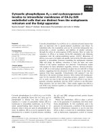
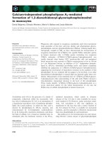
Fig. 1. Infection with H1N1pdm influenza stimulates the expression of GX-sPLA2 in bronchial epithelial cells and inflammatory cells. Mice were infected with H1N1pdm
(A/Mexico/4108/2009) and the lungs were assessed for the mRNA and protein expression and localization of PLAs during a 14 day time course. GX-sPLA2 mRNA (Ai), cPLA2
mRNA (Aii) and GV-sPLA2 mRNA (Aiii) expression quantified by Real-Time RT-PCR was normalized to GAPDH, GX ỵ / ỵ (open bars) and GX / (lled bars) mice (C3H/HeN
background mice). GX ỵ / ỵ and GX À / À mouse lungs were perfusion fixed in situ with 4% paraformaldehyde, sectioned and subject to immunohistochemical analysis with the
IgG fraction of rabbit anti-mouse GX-sPLA2 antiserum (1/100 dilution) (B). GIIA and GX-sPLA2 protein expression determined by immunoblot analysis of lung tissue
homogenates of wild type GX ỵ / þ (lane 1) and knockout GX À / À (lane 2) mice (C). For each blot, the corresponding recombinant sPLA2 enzyme (rec sPLA2) was run alone
(lane 3) as a control. Representative results for five separate experiments are shown. All the mice used in these experiments were genotyped littermates and grouped and
analyzed by their genotype. a, p o 0.05 GX ỵ / ỵ or GX / vs. base; b, p o 0.05 GX ỵ / ỵ vs. GX / À at any time point, ANOVA followed by paired t-test, two tailed, assuming
unequal variance. nZ 8 per group; 400 Â ; scale bar: 50 μm for immunohistochemistry.
A.A. Kelvin et al. / Virology 454-455 (2014) 78–92
specific for this sPLA2 since neither cPLA2 nor GV-sPLA2 was upregulated.
Since GX-sPLA2 mRNA levels increased in response to
H1N1pdm infection, we investigated the spatial and temporal
expression of GX-sPLA2 protein in mouse lungs after inuenza
infection. Lungs from GX ỵ / ỵ and GX / À mice infected with
H1N1pdm were harvested at baseline, 3, 6 and 14 days pi and
subjected to immunohistochemical analysis with anti-mouse
GX-sPLA2 antiserum. Visualization by light microscopy revealed
GX-sPLA2 protein accumulation in the lungs of infected mice
compared to baseline (Fig. 1B). GX-sPLA2 protein was identified
in inflammatory cells that had infiltrated in the alveolar space on
day 3 and 6 in GX ỵ / þ mice infected with H1N1pdm (shown by
arrows). GX-sPLA2 protein was also clearly identified in epithelial
cells lining the bronchioles on day 3, 6 and 14 in GX ỵ / ỵ mice
infected with H1N1pdm (Fig. 1B, upper right panels). No staining
for GX-sPLA2 protein was observed in GX À / À mice at baseline or at
any time point after infection with H1N1pdm (Fig. 1B, lower panel
rows). Similarly, no proteins cross reacting with the secondary
antibody alone was identied in GX ỵ / ỵ or GX À / À mice (Fig. 1B,
left hand panels). We confirmed the loss of GX-sPLA2 in the GX À / À
mice by immunoblot analysis. We indeed observed a specific
depletion of GX-sPLA2 but no change in the expression of GIIAsPLA2 in the GX / mice compared to GX ỵ / ỵ mice (Fig. 1C). Taken
together, these results show that intranasal infection with
H1N1pdm increases GX-sPLA2 RNA and protein expression in the
lung that corresponds to the increase in lung inflammation
associated with influenza infection. This suggests a possible role
for GX-sPLA2 in the pathogenesis of pulmonary H1N1pdm influenza infection.
Depletion of GX-sPLA2 increases host survival following H1N1pdm
infection
Since GX-sPLA2 was upregulated in the lung during H1N1pdm
infection, we explored its role in the host response to pulmonary
infection with H1N1pdm influenza. We first examined the clinical
outcome of GX-sPLA2 deletion by assessing weight loss and
survival of G ỵ / ỵ and GX-sPLA2 gene targeted mice / mice on
two different genetic backgrounds following infection and
assessed weight loss and survival.
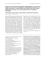
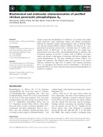
In the rst series of infections, GX ỵ / þ (n¼25), GX þ /– (n¼32),
and GX À /À (n¼24) mice on a C57BL/6J background were infected
intranasally with H1N1pdm influenza A/Mexico/4108/2009 (Fig. 2A).
Mice on this background lack the GIIA-sPLA2 gene (Karabina et al.,
2006). Animals were euthanized if their body weight decreased to
less than 80% of baseline weight, or if the 14-day duration of the
study was completed. Survival 14 days after H1N1pdm influenza
infection was 70% in GX À /À mice (blue line), 48% in GX ỵ / mice
(green line) and 15% in GX ỵ / ỵ mice (red line). The difference in
survival between GX / and GX ỵ / mice, and between GX / and
GX ỵ / ỵ mice after H1N1pdm infection was statistically significant,
pr0.01.
To independently confirm these findings, we evaluated the
survival of GX ỵ / ỵ (nẳ 71) and GX À / À (n ¼ 57) mice on a C3H/HeN
background (Fig. 2B) which have a functional GIIA-sPLA2 gene
(Karabina et al., 2006). As with the studies with the C57BL/6J mice,
animals were infected intranasally with A/Mexico/4108/2009 and
euthanized if their body weight decreased to less than 80% of
baseline weight, or at the end of the study. Survival of GX À / À mice
on the C3H/HeN background was again significantly higher (62%,
blue line) following H1N1pdm infection than survival of GX ỵ / þ
mice on a C3H/HeN background (36%, red line). Together, these
studies showed that targeted deletion of GX-sPLA2 in two different
mouse models led to increased survival following H1N1pdm
infection in vivo. Furthermore, since the C3H/HeN mice expressed
81
endogenous GIIA-sPLA2, these results demonstrate that the ability
to express GIIA-sPLA2 does not compensate for the loss of
GX-sPLA2 during host immune responses to pulmonary H1N1pdm
influenza infection.
Depletion of GX-sPLA2 during H1N1pdm infection leads to a decrease
in downstream phospholipid catalysis (AA) products but no difference
in innate cell recruitment
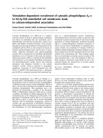
GX ỵ / þ and GX-sPLA2 gene targeted mice À / À on a C3H/HeN
background were infected with A/Mexico/4108/2009, and BAL
fluid was harvested 3 or 6 days post H1N1pdm infection. To assess
the general inflammatory response and lung tissue destruction
that typically occurs during H1N1pdm infection (Ohtsuki et al.,
2006; Paquette et al., 2012), we investigated the histopathology by
H&E staining of lungs isolated from both GX / and GX ỵ / ỵ mice
at baseline, day 3, 6 and 14 pi (Fig. 3A). The pulmonary pathology
peaked quickly by day 3 pi in infected GX ỵ / ỵ and GX / animals.
Bronchiolitis and alveolitis with mononuclear cell and neutrophil
infiltration were observed in several loci of the infected lungs of
both groups. Hemorrhage, edema, and necrotizing respiratory
epithelia were also observed with similar severity among both
groups. Pathology persisted until day 7 pi where mononuclear cell
and neutrophil infiltration were still profound and caused patches
of consolidation in the lung tissue in both groups. It seemed by day
14 pi that the pulmonary pathology was slightly more minimal in
the GX À / À mice with reduced level of leukocyte infiltration.
In contrast, multi foci cell infiltration and tissue consolidation
was still prominent in the GX ỵ / ỵ lungs by day 14 pi.
To further examine the inflammatory cell types that may be
recruited to the lung during H1N1pdm infection we analyzed lung
homogenates from day 0, 3, 6 and 14 pi from both GX ỵ / ỵ and
GX / mice by immunoblot for neutrophil and leukocyte cell
markers, MPO and CD45 respectively. MPO was induced on day
3 and day 6 pi in both the mouse genotypes and returned to
baseline on day 14 and CD45 was induced from baseline for all
time points measured. Neither MPO nor CD45 showed any variation in the lungs between GX / or GX ỵ / ỵ throughout the
infection time course (Fig. 3Bi); densitometry did not reveal any
statistical differences (Fig. 3Bii). Furthermore, we also analyzed
GX À / À and GX ỵ / ỵ lungs for the presence and activation of
macrophages by immunohistochemistry and Real-Time RT-PCR
(Fig. 3Ci, Cii and D). Here we found that the macrophage marker
Mac-3 was significantly increased and peaked at day 6 following
infection as determined by immunohistochemistry staining which
was further confirmed by quantifying the staining and quantification of the signal (Fig. 3Ci and Cii). Furthermore the mRNA for the
inflammatory chemokine CCL2 was also significantly increased
following infection (days 3 and 6) and decreased by day 14
(Fig. 3D). No difference was determined for Mac-3 or CCL2
expression between GX À / or GX ỵ / ỵ mice. Taken together, these
results suggest a similar inflammatory and innate response in both
the GX ỵ / ỵ and GX / mice.
To assess the role of GX-sPLA2 in leukocyte infiltration into the
bronchoalveolar space after H1N1pdm infection, we measured
total leukocyte cell counts and the levels of different leukocyte cell
types in the BAL fluid of H1N1pdm infected in GX ỵ / ỵ and GX À / À
mice (Fig. 4A). No significant difference in total cell counts
(Fig. 4Ai) or the percentage of CD4 ỵ , CD8 ỵ , B or natural killer
cells, or granulocytes were identied in the BAL uid of GX ỵ / ỵ
and GX À / À mice 6 days after H1N1pdm infection (Fig. 4Aii).
In addition, targeted deletion of GX-sPLA2 had no effect on lung
viral titers 3 or 6 days pi (Fig. 4B).
We next determined by ELISA the levels of diferent AA
metabolites including PGD2, LTB4, cysteinyl leukotrienes, PGE2, a
stable PGE metabolite, and Lipoxin A4 which are known to regulate
82
A.A. Kelvin et al. / Virology 454-455 (2014) 78–92
bronchiolar reactivity and inflammatory cell adhesion, migration
and activation (Henderson et al., 1995) were determined by ELISA.
Levels of PGD2 (Fig. 5A), LTB4 (Fig. 5B), cysteinyl leukotrienes
(Fig. 5C), PGE2 (Fig. 5D), the stable PGE metabolite (Fig. 5E), PGE2
plus PGE metabolite (Fig. 5F) and Lipoxin A4 (Fig. 5G) were all
significantly lower in the BAL fluid from GX À / À mice (solid bars)
on day 3 pi compared to GX ỵ / ỵ mice. Conversely, on day 6 pi, the
levels of these metabolites were similar in GX ỵ / ỵ and GX / mice
(Fig. 5A–F). In summary, these results show that deletion of
GX-sPLA2 in mice led to a transient but significant decrease in
the levels of a panel of AA metabolites when mice were infected
with a lethal H1N1pdm influenza virus that was not associated
with alterations in inflammatory cell infiltration or viral clearance.
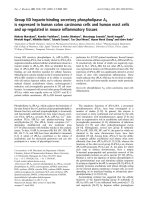
Increased expression of immunoglobulin chain, lymphocyte
differentiation, antigen processing genes and presence of CD3 ỵ
T cells in the lungs of mice lacking GX-sPLA2 after H1N1pdm infection
To increase our understanding of the molecular events leading
to increased survival following H1N1pdm infection in GX À / À mice,
we conducted microarray analysis of RNA extracted from the lungs
of GX þ / þ and GX À / À influenza infected animals. As previously
reported by our group (Kudo and Murakami, 1999; Paquette et al.,
2014; Rowe et al., 2010), influenza infection caused a progressive
increase in the total number of upregulated genes in the lung
tissue of GX ỵ / ỵ mice (1246 genes at 3 days pi and 2469 genes at
6 days pi). Genes that belonged to different functional groups, such
as immune response, inflammatory response and prostaglandin
signaling pathways (Figs. 6 and 7) showed a progressive increase
that was parallel to the global evolution of gene expression.
Conversely, the expression of cytokine-related genes reached
maximal levels 3 days pi and were maintained thereafter (Fig. 6A).
At first sight, lack of GX-sPLA2 did not modify the global
evolution of gene expression in the lungs. Similarly to GX ỵ / ỵ
mice, GX À / À mice showed a progressive increase in the number of
upregulated genes (1578 at 3 days pi and 2469 at 6 days pi).
Further analysis demonstrated that on day 3 pi, GX À / À mice
showed significantly higher levels of the cytokines LTA and LTB,
the chemokines CCL19, CXCL9 and CXCL13 and the chemokine
receptors CXCR3 and CXCR5 (Fig. 6B and C). In contrast, the
expression pattern of cytokines and chemokines showed no
differences between GX ỵ / ỵ and GX / À mice 6 days pi (data not
shown). Interestingly, expression of 21 immunoglobulin chains,
including heavy and light chains, was identified in GX À / À mice
3 days pi, while no expression of immunoglobulin chain genes was
identied in GX ỵ / ỵ mice at this time point. In addition, the
number of immunoglobulin chain related genes was higher in
GX À / À than GX ỵ / ỵ mice 6 days after H1N1pdm infection (Fig. 6B).
No differences were observed in the patterns of interferon regulated genes between GX ỵ / ỵ and GX / À mice after H1N1pdm
infection (data not shown).
To determine which functional pathways are differentially
enriched between GX ỵ / ỵ and GX À / À mice after H1N1pdm infection, we performed intersect analysis of the respective sets of
upregulated genes (Fig. 7). At 3 days pi, expression of interferon
regulated, inflammatory response and innate immune response
genes were common to both GX ỵ / þ and GX À / À mice. A number of
genes related with eicosanoid synthesis and their receptors
were found to be regulated during influenza infection; however,
GX-sPLA2 deficiency did not cause any major alterations in their
expression profiles (Fig. S1).
The set of genes specifically enriched in the GX À / À mice at
3 day pi were those related to adaptive immune responses, such as
immunoglobulin chains, lymphocyte differentiation and antigen
processing and presentation. On the other hand, the set of genes
more enriched in the GX þ / þ mice were genes involved in the
tissue development category at 3 days pi (Fig. 7A). At 6 days pi
(Fig. 7B), the enrichment profiles of upregulated genes in GX þ / þ
and GX À / À mice were nearly identical. While immunoglobulin
chain gene expression was identified in both GX þ / þ and GX À / À
mice, expression of immunoglobulin chain genes remained elevated only in the GX À / À set of genes 6 days pi, while the GX ỵ / ỵ
set of genes continued to show enrichment in the tissue development category at day 6 pi.
To further evaluate the adaptive immune system of the GX À / À
mice infected with H1N1pdm we investigated the T and B cell
responses within the lung during infection. Here we stained lung
sections with anti-CD3 to assess infiltration of T cells using
immunocytochemistry (Fig. 8A and B). We found a significant
increase of CD3 positive T cells in the lung on day 3 pi in the
GX / mice compared to GX ỵ / þ mice (Fig. 8A, upper right panels)
by approximately 2 fold (Fig. 8B). Interestingly, CD3 staining of the
GX ỵ / ỵ animals had increased to similar levels seen in the GX À / À
mice by day 6 and both genotypes had sustained levels of CD3 on
day 14. Moreover, we also investigated CD8A and IgG (IGHG)
mRNA levels in the lungs of the GX À / À mice throughout the time
course and there was a slight trend for increase CD8A levels. Taken
together, the results from the CD3 and IgG analysis supported the
microarray studies where the adaptive immune system of the
GX À / À had a faster and more robust initiation.
Discussion
GX-sPLA2 has been highly implicated in various inflammatory
diseases of the respiratory tract, including Th2 cytokine-driven
asthma (de Jong et al., 2006; Henderson et al., 2007) and lung
injury (Napolitani et al., 2009), but its role during influenza
infection has not been previously investigated. Here we evaluated
the pathophysiological role of GX-sPLA2 during severe influenza A
H1N1pdm infection in the mouse. We found that GX-sPLA2
expression was increased following infection, and that targeted
deletion of GX-sPLA2 led to increased survival in mice. Lack of
GX-sPLA2 resulted in decreased levels of PGD2, LTB4, cysteinyl
leukotrienes, PGE2 and Lipoxin A4 and increased adaptive immune
responses at 3 but not 6 days following H1N1pdm infection. This
demonstrates that GX-sPLA2 plays an important role in the
production of several biologically active inflammatory lipid mediators during the early phase of the inflammatory response that
follows H1N1pdm influenza infection. Human patients with a
severe respiratory disease caused by influenza infection have a
dysregulated inflammatory response that leads to lung pathogenesis associated with hypercytokinemia in most cases (BermejoMartin et al., 2010; Curfs et al., 2008). Taken together with the
previous findings showing a role of GX-sPLA2 in inflammatory
lung diseases, our work supports the further investigation of the
therapeutic potential of attenuating GX-sPLA2 during severe influenza infection as well as the interplay between eicosanoids and
adaptive immunity.
sPLA2 has previously been implicated in pulmonary disease
onset and progression putting it forth as a potential biomarker for
severe respiratory diseases (Henderson et al., 1995; Henderson
et al., 2007). We show that GX-sPLA2 protein and mRNA expression increased in the lungs of GX ỵ / ỵ mice following H1N1pdm
infection, suggesting that GX-sPLA2 may be used as a possible
biomarker of severe influenza infection. This is the first report of
increased GX-sPLA2 expression following influenza virus infection.
Both epithelial cells and leukocytes were found to be sources of
GX-sPLA2 during infection, and GX-sPLA2 expression was detected
in epithelial cells 3 days prior to the infiltration of leukocytes.
In will be interesting to determine in future experiments whether
the specific deletion of GX-sPLA2 expression in epithelial cells vs.
A.A. Kelvin et al. / Virology 454-455 (2014) 78–92
Fig. 2. Increased survival of GX / vs. GX ỵ / or GX ỵ / ỵ mice following A/Mexico/
4108/2009 infection. GX ỵ / ỵ (nẳ 25), GX ỵ / À (n ¼32), and GX À / À (n¼24) mice
(C57BL/6J background, lacks GIIA-sPLA2) were infected intranasally with A/Mexico/
4108/2009 and survival was assessed for a 14 day period (A). GX ỵ / ỵ (nẳ71) and
GX / (nẳ 57) mice (C3H/HeN background, expresses GIIA-sPLA2) were infected
intranasally with A/Mexico/4108/2009 and survival was assessed for a 14 day
period (B). Animals were sacrificed if their body weight decreased to less than 80%
of baseline weight, or if the 14-day duration of the study was completed. Log rank
test, p o0.05 GX À / À vs. GX ỵ / ỵ and GX ỵ / mice or po 0.05, GX / vs. GX ỵ / ỵ
mice. All the mice used in these experiments were genotyped littermates and
grouped and analyzed by their genotype.
infiltrating leukocytes or both is responsible for the increased
survival. Here we observed a bimodal expression pattern of
GX-sPLA2 during the 14 day time course of infection. It is possible
that this occurred due to the protein stability as it is used to
regulate bioactive lipid mediator synthesis. If the protein does not
remain stable throughout the course of infection and recovery, it
may be important to have a second increase in GX-sPLA2 in the
later stages of infection to compensate for the loss of protein. It is
in fact possible that the protein exerts distinct roles in the
clearance of the virus and tissue remodeling in addition to the
regulation of immune cells. Such a scenario would require inductions at specific time points during infection. It will be important
to further explore the local expression in the virus niche and the
stability of protein GX-sPLA2 during influenza infection to better
understand how GX-sPLA2 stability may influence influenza severity in the initiation of the innate immune response, adaptive
maintenance, and recovery. Furthermore, it would also be of value
to investigate the source of GX-sPLA2 by expression analysis of
each cell type and also by investigating the role of hematopoietic
83
GX-sPLA2 compared to epithelial GX-sPLA2. The latter could be
studied by employing bone marrow transplantation experiments
from GX À / À mice into GX ỵ / ỵ and the reverse. While the association between GX-sPLA2 and influenza related complications has
not previously been investigated, LTB4, a downstream product of
GX-sPLA2 has been suggested to be a biomarker for pulmonary
disease and respiratory complications following trauma (Influenza
Activity – United States and Worldwide, 2010; Henderson et al.,
2011; Shridas et al., 2011).
Multiple studies have implicated GX-sPLA2 in the pathophysiology of pulmonary diseases onset and progression, suggesting
GX-sPLA2 might be a suitable therapeutic target in lung
(Henderson et al., 1995; Morita et al., 2013). Deletion of
GX-sPLA2 in a Th2 cytokine-driven mouse asthma model significantly impairs development of asthma (Henderson et al., 1995)
and accordingly, administration of a human GX-sPLA2 selective
inhibitor in a human GX-sPLA2 knock-in mouse model led to a
significant reduction in airway inflammation, mucus hypersecretion and airway hyperresponsiveness (Henderson et al., 2007).
Furthermore, although not specific for human GX-sPLA2, the
indole-based sPLA2 inhibitor varespladib has been shown to
significantly inhibit sPLA2 activity in the BAL fluid of infants with
post-neonatal ARDS (de Jong et al., 2006) during induced asthma,
suggesting the involvement of sPLA2 among other sPLA2s. Our
results showing increased survival of the GX À / À mice after
infection with H1N1pdm further support the notion that
GX-sPLA2 is a therapeutic target in pulmonary diseases due to
viral infection and that infection with H1N1 might be better
controlled by inhibiting this sPLA2.
One of the main functions of GX-sPLA2 is likely the generation
of bioactive lipid mediators which play important roles in lung
inflammatory diseases (Gao et al., 2013; Gaudreault and Gosselin,
2007; Van Elssen et al., 2011). Although we did not see any major
differences in the mRNA analysis of the eicosanoid pathways
between the GX / and GX ỵ / ỵ mice, measuring the mRNA levels
of these genes may have limited value to determine the level of
activation of their signaling pathways. Conversely, we observed
decreased levels of PGD2, LTB4, cysteinyl leukotrienes, PGE2 and
Lipoxin A4 in BAL fluid 3 day pi in the GX À / À mice, indicating that
GX-sPLA2 acts upstream of these bioactive lipid mediators during
influenza infection and thereby suggests a possible role of these
bioactive mediators in pulmonary pathogenesis after influenza
infection. In agreement with our findings, PGD2 has been implicated during influenza A infection as PGD2 expression in the lungs
of older animals inhibits regulatory dendritic cells activity and
T cell responses (Zhang et al., 2000). Other eicosanoids have been
implicated in different lung diseases, and the dysregulation of
leukotrienes and lipoxins have been reported as contributing
factors to the pathogenesis and severity of other respiratory
diseases (Bermejo-Martin et al., 2009). LTB4 has been suggested
to play a destructive inflammatory role in the lung by priming
neutrophils for adhesion, chemotaxis and stimulation of granule
release (Cameron et al., 2007). As well PGD2, PGD receptor,
lipocalin-type PGD synthase and LTB4 have been implicated in
asthma pathogenesis (Arima and Fukuda, 2011; Masuda et al.,
2005; Rusinova et al., 2012). Although asthma and pulmonary
disease due to influenza infection differ in derivation, both are
characterized by hyper-inflammation of the respiratory tract.
Taken together, our data supports a role of GX-sPLA2 signaling
and bioactive mediator production in the regulation of the
pulmonary response to H1N1pdm infection. In the future it would
be important to specifically determine whether PGD2, LTB4,
cysteinly leukotrienes, PGE2, Lipoxin A4 or another AA metabolite
specifically modulates the response to H1N1pdm infection.
The inflammatory response may be simultaneously beneficial
and destructive during lung infection (Baillie and Digard, 2013;
84
A.A. Kelvin et al. / Virology 454-455 (2014) 78–92
Fig. 3. Infection with H1N1pdm influenza induces similar pulmonary inflammation and recruitment of inammatory cells in GX ỵ / ỵ and GX / mice. GX ỵ / ỵ and GX À / À
mice (C3H/HeN background mice) were infected with H1N1pdm (A/Mexico/4108/2009) influenza and the lungs were perfusion fixed in situ with 4% paraformaldehyde on
specific time points following infection, sectioned and subject to hematoxylin and eosin staining (A). MPO protein (neutrophil marker), CD45 protein (leukocyte marker) and
GAPDH protein (loading control) expression levels were determined by immunoblot analysis from lung tissue homogenates of GX ỵ / ỵ and GX / mice over a 14 day time
course of H1N1pdm influenza infection (Bi). Densitometric analysis of MPO (Bii) and CD45 (Biii) protein levels normalized to GAPDH levels in lung tissue of GX þ / þ (open
bars) and GX À / À (filled bars) mice after H1N1pdm influenza infection, are presented. Immunohistochemical analysis with specific rabbit primary antibody against mouse
Mac-3 antigen (marker for macrophages) is shown (Ci). Assessment of Mac-3 positive cells (indicated by -) per high power field before, 3, 6 or 14 days after infection with
H1N1pdm influenza is shown (Cii). CCL2 mRNA expression normalized to GAPDH was determined by quantitative real-time PCR in lung tissue of GX ỵ / ỵ and GX À / À mice
after H1N1pdm influenza infection (D). Representative images ( Â 200) from five independent experiments are shown. Scale bar: 100 μm (A) or 50 μm (C). a, p o 0.05 GX ỵ / ỵ
or GX / vs. base; b, p o0.05 GX ỵ / ỵ vs. GX À / À at any time point, ANOVA followed by paired t-test, two tailed, assuming unequal variance. nZ 8 per group.
Bermejo-Martin et al., 2010). Although destructive killing of
foreign pathogens is imperative for eradication and microbe
clearing, the over production of inflammatory mediators leading
to an overt inflammatory response may accentuate disease
pathology, as is the case during severe influenza H5N1 and
H1N1 infection (Bermejo-Martin et al., 2010; Curfs et al., 2008;
Huang et al., 2009). This illustrates the dual role of proinflammatory mediators, which has also been suggested for some GX-sPLA2
downstream lipid mediators. For instance, LTB4 has been shown to
increase the activity of nasal neutrophil killing of human coronavirus, RSV, and influenza B virus (Van Elssen et al., 2011) and to
induce the release of antimicrobial peptides in vivo in the lungs of
A.A. Kelvin et al. / Virology 454-455 (2014) 78–92
85
Fig. 4. Lung viral titers and cell counts in BAL fluid show similar cell numbers and cell population distributions following infection in GX ỵ / ỵ and GX / mice. GX / and
GX ỵ / ỵ mice infected with A/Mexico/4108/2009 were investigated for lung cell numbers, populations and viral load. BAL uid was harvested from infected GX ỵ / ỵ (open
bars) and GX / (lled bars) mice (C3H/HeN background mice) on day 0 and 6 and the cell numbers (Ai) and cell population distributions (Aii) were assessed by FACS. Viral
load was determined on day 0, 3 and 6 pi of GX ỵ / ỵ (open bars) and GX À / À (filled bars) mice by Real-Time RT-PCR vRNA quantification (B). All the mice used in these
experiments were genotyped littermates and grouped and analyzed by their genotype. a, p o0.05 GX ỵ / ỵ vs. base, ANOVA followed by paired t-test, two tailed, assuming
unequal variance. nZ 7 per group.
mice infected with viruses (Gao et al., 2013; Gaudreault and
Gosselin, 2007). The lipid product protectin D1 has been implicated in influenza therapeutics (Arima and Fukuda, 2011;
Mitsuishi et al., 2006). Although these previous reports seem
to suggest a conflicting role for GX-sPLA2 in consideration
of our data, it may be possible that LTB4 and GX-sPLA2 promote
antiviral activity and are significant during a viral response
but only at moderate levels. Alternatively, it is possible that
GX-sPLA2 prevents H1N1 infection but also triggers excessive
inflammation that is associated with lipid surfactant destruction.
More work is needed to understand how the function of GX-sPLA2
mediates both beneficial and deleterious roles during influenza
infection.
Our survival data from GX gene targeted mice indicated that
the loss of GX-sPLA2 was beneficial to the host during inuenza
infection. The microarray mRNA data from lungs of GX ỵ / ỵ
infected mice were in agreement with our previously published
data on pandemic H1N1 2009 virus infection, in mice including
the progressive increase of immune and inflammatory responses
and of the prostaglandin signaling pathway (Paquette et al., 2014).
Together, the results of microarray analysis, gene expression by
Real-Time RT PCR and immunocytochemistry of the lungs suggested that GX À / À mice exhibited a more robust adaptive immune
response than GX ỵ / ỵ mice. Indeed, we observed significant
differences in lymphocyte gene profiles at day 3 pi, associated
with differences in the levels of lymphotoxin alpha and beta, B cell
chemokines, T cell chemokine receptors and B cell immunoglobulin chains as measured which by immunofluorescence and RT-PCR.
Expression of B cell immunoglobulin chain genes were substantially increased on day 3 pi in the GX À / À mice but not in the
GX ỵ / ỵ . B cell immunoglobulin gene expression was significantly
greater on day 6 pi. and the expression of the T cell, B cell and
dendritic cell chemokines and chemokine receptors, i.e., CCL19,
CXCR3, etc., were significantly higher in the GX / samples than
GX ỵ / ỵ . These results suggest that the downstream products of
GX-sPLA2, such as PGD2, PGE2, LTB4 may inhibit the early adaptive
immune responses of T and B cells during viral infection and this
fits with the fact that aspirin, which attenuates eicosanoid production, can be an effective therapy for patients with influenza
infection (Matsuoka et al., 2000). It would be of value in future
studies to further investigate the effect of GX-sPLA2 on the
proliferation, activation and differentiation of T and B lymphocytes.
Consistent with this notion, the chemokines and chemokine receptors found to be upregulated in the H1N1pdm infected
GX À /À mice are known to play significant roles in T and B cell
migration and localization to the lymph nodes (Goracci et al., 2010;
Muthuswamy et al., 2010). CXCL13/CXCR5 signaling has been shown
to activate B cells (Sadik and Luster, 2012), which may explain the
increased immunoglobulin chain gene expression observed in GX À /À
mice. Our data supports previous findings implicating PGD2 in the
inhibition of cell migration to lymph nodes (Zhang et al., 2000), and
PGE2 in the inhibition of adaptive immune cellular events such as
chemokine production by DCs and the attraction of naïve T cells
(Murakami et al., 2011; Truchetet et al., 2012).
In conclusion, our findings provide new insights into the
molecular pathophysiology of lethal influenza infection, highlighting a new role for GX-sPLA2 during H1N1pdm infection. Overall, the
sPLA2 appears as a negative effector but it may act at several steps
during infection. We found that GX-sPLA2 and its downstream
products may have a role in the inhibition of adaptive immunity
during viral infection in mice thereby contributing to pathogenesis.
Within this mechanism, it is in fact possible that T and B cell
maturation and activation are initiated in mice lacking GX-sPLA2
prior to virus infection, and that a more robust and earlier adaptive
86
A.A. Kelvin et al. / Virology 454-455 (2014) 78–92
Fig. 5. Decreased eicosanoid levels in the BAL fluid 3 but not 6 days after infection with A/Mexico/4108/2009 in GX À / vs. GX ỵ / ỵ mice. The eicosanoid levels in the BAL fluid
of GX À / À and GX þ / þ mice (C3H/HeN background) were investigated at baseline and following infection with A/Mexico/4108/2009. GX ỵ / ỵ (open bars) and GX À / À (filled
bars) mice (C3H/HeN background mice) BAL fluid was harvested by instilling ice cold NaCl (1 ml) five times and pooled. Levels of PGD2 MOX (A), LTB4 (B), cysteinyl
leukotrienes (C), PGE2 (D), stable PGE metabolite (E), PGE2 plus PGE metabolite (F) and Lipoxin A4 (G) were assessed by ELISA. These results are the mean of 5 independent
studies. All the mice used in these experiments were genotyped littermates and grouped and analyzed by their genotype. GX þ / þ (open bars) and GX À / À (filled bars). Results
are expressed in pg/mL. a, p o0.05 GX þ / þ or GX À / À vs. base; b, p o0.05 GX ỵ / ỵ vs. GX / À at any time point, ANOVA followed by paired t-test, two tailed, assuming unequal
variance. n Z7 per group.
immune response increased the survival of GX À / À mice after
H1N1pdm infection. Since GX-sPLA2 may contribute to inflammatory response dysregulation during influenza infection and contribute to the morbidity and mortality associated with hospitalized
influenza patients, this work may shed important insight into the
molecular mechanisms of severe influenza infection. Our findings
further support the notion that GX-sPLA2 is an interesting therapeutic target in lung inflammatory diseases. Whether inhibition or
attenuation of GX-sPLA2 activity during severe influenza infection
has a therapeutic effect remains to be demonstrated.
Materials and methods
Generation of GX-sPLA2 KO mice
To dissect the role of GX-sPLA2 in the molecular regulation of
pulmonary infection with H1N1pdm influenza, mice that lack
GX-sPLA2 (GX À / À mice) on the C57BL/6J background previously
described were used (Matsuoka et al., 2000). This mixed background strain has a naturally occurring mutation in the gene
encoding GIIA-sPLA2 (Karabina et al., 2006), which has been
A.A. Kelvin et al. / Virology 454-455 (2014) 78–92
87
Fig. 6. Effect of GX-sPLA2 deficiency in the mRNA expression levels of cytokines, chemokines and their receptors and immunoglobulin chains in the lung tissue of mice
during inuenza infection. GX ỵ / ỵ and GX À / À mice (C3H/HeN background mice) were infected with influenza A/Mexico/4108/2009 and the gene expression profiles were
analyzed in the lung tissue at day 0, 3, and 6 days after infection by microarray analysis (n¼ 4 per group). Evolution of gene enrichment (Fisher's exact test) of the KEGG
category “cytokine-cytokine receptor interaction” (A). Differences in the expression levels of cytokines (B) and chemokines (C) and their receptors at 3 days pi. The heatmaps
show the genes that are significantly upregulated with respect to the control group and the blue boxes indicate that the expression levels of those genes are significantly
higher in the GX À / À than in the wild type mice at the same time-point. Evolution in the expression levels of immunoglobulin genes: total number of regulated genes (D) and
overview of different experimental groups and time-points (E). All the mice used in these experiments were genotyped littermates and grouped and analyzed by their
genotype.
implicated in bacterial phospholipid hydrolysis (Fang et al., 2011).
Furthermore, we generated GX À / À mice on the C3H/HeN background (Fig. 1C) by backcrossing the C57BL/6J GX À / À mice for 10
generations which had functional GIIA-sPLA2.
Animals maintenance
Mice were maintained on standard animal feed and water ad
libitum in the conventional environmental conditions and controlled temperature and humidity with a 12 h light and dark cycle.
For infection studies, animals were housed in HEPA-ltered cage
racks adherent to ABSL2 ỵ conditions (Toronto General Hospital,
Animal Resource Centre, Toronto, Canada). All animal procedures
were performed in a certified class II biosafety cabinet (Baker
Company, Sanford, NC, USA). Housing and experimental procedures were approved by the Animal Care Committee of the
University Health Network, and were in accordance with the Guide
for the Care and Use of Laboratory Animals Research Statutes,
Ontario (1980).
Viral infection
All infection experiments were conducted with H1N1pdm
strain, A/Mexico/4108/2009 (H1N1pdm), provided by the Centers
for Disease Control and Prevention (Atlanta, GA, USA). Virus was
propagated and titrated in embryonated eggs and titrated prior to
animal challenge. Viral stocks were stored in liquid nitrogen and
thawed prior to use. Mice were weighed and randomly assigned
for sample collection, and were infected through intranasal instillation with 50 mL phosphate-buffered saline (mock infection) or
50 mL A/Mexico/4108/2009 (H1N1pdm) at 1 Â 105 or 1 Â 104 50%
egg infectious dose (EID)50. Virus dosage were 1 Â 104 EID50 and
 105 EID50 for host response profiling in C57BL/6J mice and
1 Â 104 EID50 for comparing disease severity between GX ỵ / ỵ and
GX / mice. Throughout infection experiments, animal survival,
clinical signs, and weights were recorded daily. In accordance with
Animal Care Committee recommendation, mice were euthanized
when recorded body weight fell below 80% of original body
weight.
Viral load measurement
At day 0, 3 and 6 pi, 3 GX ỵ / ỵ and 3 GX / mice were
euthanized and lung homogenates collected for viral load determination by either Madin–Darby Canin Kidney (MDCK) cell
growth determination or Real-time RT-PCR (RNA Analysis methods and Table S1). For MDCK determination lungs were homogenized (10% w/v) in High Glucose (4.5 g/L) Dulbecco's Modified
Eagle Medium (DMEM), supplemented with 1% bovine serum
albumin, 50 mg/mL Gentamycin, 100 U/mL Penicillin, 100 mg/mL
Streptomycin, and 1 mg/mL TPCK-Trypsin (vDMEM). Homogenates
were then serially diluted (0.5 log10) in quadruplicate over Madin–
Darby Canine Kidney cells, cultured at 2.0 Â 104 cells/well in
96-well plates. Cells were incubated for 2 h at 37 1C and 5% CO2.
Homogenates were then removed and replaced with fresh
vDMEM. Cells infected were incubated for 6 days at 37 1C and 5%
88
A.A. Kelvin et al. / Virology 454-455 (2014) 78–92
Fig. 7. Intersect analysis of the genes up-regulated in the lung tissue of GX / and GX ỵ / ỵ mice during influenza infection and functional classification of the resulting gene
subsets. Venn diagrams are representative of the total number of genes that are significantly up-regulated with respected to the uninfected mice. David Annotation tool was
used to classify the genes of each subset, and the fold enrichment is shown for each category. All the mice used in these experiments were genotyped littermates and
grouped and analyzed by their genotype. * The “Immunoglobulin chains” category was manually curated and contains 84 genes. nn The “interferon responses category”.
Fig. 8. GX-sPLA2 deficiency increases T cell recruitment and immunoglobulin heavy chain mRNA expression in the lung tissue of mice during influenza infection. Day 0 and
3 pi with H1N1pdm (A/Mexico/4108/2009) inuenza the lungs from GX ỵ / þ and GX À / À mice (C3H/HeN background mice) were perfusion fixed in situ with 4%
paraformaldehyde, sectioned and subject to immunofluorescence analysis with specific rabbit primary antibody against mouse CD3 antigen (marker for T-cell) (Ai).
Representative images (at  400 with 3.4 zoom factor) from five independent experiments are shown. Assessment of CD3 positive cells per high power field for day 0, 3,
6 and 14 days following infection with H1N1pdm influenza is shown (Aii). IgG (B) and CD8A (C) mRNA expression normalized to GAPDH were determined by quantitative
real-time PCR in lung tissue of GX ỵ / ỵ and GX / À mice after H1N1pdm influenza infection. Scale bar: 10 m. a, po 0.05 GX ỵ / ỵ or GX À / À vs. base; b, p o 0.05 GX þ / þ vs.
GX À / À at any time point, ANOVA followed by paired t-test, two tailed, assuming unequal variance. nZ 8 per group. All the mice used in these experiments were genotyped
littermates and grouped and analyzed by their genotype.
A.A. Kelvin et al. / Virology 454-455 (2014) 78–92
CO2, after which cell culture supernatants were tested for the
presence of virus by hemagglutination assay using 0.5% (v/v)
turkey red blood cells (LAMPIRE Biological Laboratories, Pipersville, PA, USA). Viral loads were determined as the reciprocal of the
dilution at which 50% of wells were positive for viral infection.
Viral loads were reported as TCID50 per gram of lung tissue. Limit
of detection of 101 TCID50/g.
Host gene expression and viral load measurement by Real Time
RT-PCR
Lung tissues from both GX / and GX ỵ / ỵ mice were collected
at 3 and 6 days post infection (pi) and from uninfected controls
(four mice per group). RNA was purified from lung tissue using
TriPure (Roche, Indianapolis, IN, USA). Purified RNA was then
reverse transcribed using ImProm-II Reverse Transcription System
(Promega, Madison, WI, USA). Real Time RT-PCR was performed
using the ABI-Prism 7900HT Sequence Detection Systems (Applied
Biosystems, Foster City, CA, USA). Data was collected with Applied
Biosystems Sequence Detection Systems Version 2.3 software.
Each reaction well contained 4 μL of 0.625 ng/μL cDNA, 0.5 μL
each of forward and reverse primers (final concentration of
200 nM), and 5 μL SYBR Green Master Mix, for a total reaction
volume of 10 μL and run in quadruplicate. Host gene expression
was normalized to the glyceraldehyde-3-phosphate dehydrogenase (GAPDH) housekeeping gene, and quantified by relative
standard curve method. Viral load was quantified by the absolute
standard curve method, normalized to GAPDH housekeeping.
Primer sequences are listed in Table S1.
Histology, immunohistochemistry, immunouorescence
and immunoblotting
ỵ/ỵ
/
GX
and GX
mice were euthanized at baseline, day 3,
day 6 and day 14 pi (n 45 mice) and the mouse whole body was
vascular perfused by cardiac puncture in situ with a fixative
solution of 10% buffered formalin by a continuous release pump
under pressure and volume-controlled conditions. Fixed lung
tissues were paraffin wax embedded for histology and immunohistochemistry. For H&E, tissue slides were then stained with
hematoxylin–eosin for histopathology assessment. Rabbit antimurine GX-sPLA2 (Degousee et al., 2008) was used to assess the
tissue distribution of the GX-sPLA2 protein. Sections were counterstained with hematoxylin and eosin and observed under light
microscope (Accu-scopes, Commack, NY, USA). Images were
captured using a digital camera and SE Premium software (MicrometricsTM, Londonderry, NH, USA) (Degousee et al., 2006). For
Mac-3 tissue expression, rat anti-mouse Mac-3 was used (BD
Biosciences, Mississauga, ON).
For CD3 immunofluorescence, following heat-induced antigen
retrieval, lung tissue sections were blocked with donkey serum
and stained with primary antibodies rabbit anti-CD3 (Dako,
Burlington, ON). Donkey anti-rabbit Cy3 was used as secondary
antibodies (Millipore, Billerica, MA) and DAPI (Sigma) for nuclear
counterstain. Images were recorded with an Olympus Fluo View
1000 confocal laser scanning microscope (Olympus, Tokoyo,
Japan).
Immunoblots for GX-sPLA2 were carried out as described by
our group (Degousee et al., 2008). For Immunoblotting detection
antibodies against MPO (Upstate, Lake Placid), CD45 (BD Biosciences, Mississauga, ON), and GAPDH (Santa Cruz Biotechnology,
Dallas, TX).
89
Eicosanoid analysis
BAL fluid collection
Lungs were lavaged at 0 and 6 days post H1N1 infection with
5 ml of normal saline. The BAL fluid was centrifuged at 250g for
10 min and the supernatant was used for estimation of PGD2,
PGE2, LTB4, cysteinyl leukotriens and Lipoxin A4 content.
Analysis of PGD2 in BAL fluid
0.5 ml of BAL fluid was mixed with 0.5 ml of ice-cold acetone,
incubated on ice for 5 min and centrifuged for 10 min at 3000g at
4 1C. After supernatant aspiration, the pellet was extracted with
1 ml of ice-cold acetone and centrifuged again. The acetone
extracts were combined and the acetone evaporated under nitrogen. All the samples were then methoximated (PGD2-MOX EIA kit
(Cayman Chemical), and purified on Oasis HLB columns (Waters
Corporation) equilibrated with methanol/0.2% formic acid. Methanol eluants were evaporated in a Savant Speed Vac concentrator
and samples dissolved in Cayman EIA buffer before EIA analysis,
according to the manufacturer's instructions.
Analysis of PGE2, LTB4 and cysteinyl leukotriens in BAL fluid
1.2 ml of BAL fluid was mixed with 2.4 ml of methanol containing 0.2% formic acid, incubated on ice for 5 min and centrifuged at
3000g for 10 min at 4 1C. After adjustment of methanol to 15%,
supernatants were loaded on Oasis HLB column equilibrated with
methanol/0.2% formic acid (Waters Corporation). Columns were
processed in a vacuum manifold (Waters Corporation). After wash
with water/0.03% formic acid, the samples were eluted with
methanol/0.2% formic acid, methanol eluants were evaporated in
a Savant Speed Vac concentrator and samples dissolved in Cayman
EIA buffer before EIA analysis for PGE2, LTB4 and cysteinyl
leukotrienes (EIA kits, Cayman Chemical) according to the manufacturer's instructions.
Analysis of Lipoxin A4 in BAL fluid
0.6 ml of BAL fluid was extracted with 1.2 ml of ice-cold
methanol, incubated on ice for 5 min and centrifuged at 3000g
for 10 min at 4 1C. The supernatants were diluted with water to
achieve 11% methanol concentration and adjusted to pH 3.5 with
1N HCl. Samples were purified on C18 Sep-Pak columns (Waters
Corporation) preconditioned with methanol. After column wash
with water followed by hexane, samples were eluted with methyl
formate. The eluants were evaporated under nitrogen and the
samples reconstituted in EIA buffer and assayed for Lipoxin A4
content (Neogen Corporation) according to the manufacturer's
protocol.
Microarray analysis
Lung tissues from both GX À / À and GX þ / þ mice were collected
at 3 and 6 days pi and from uninfected controls (four mice per
group) as with the Real Time RT-PCR. RNA was purified from lung
tissue using TriPure (Roche, Indianapolis, IN, USA) and amplified
with Illumina TotalPrep RNA Amplification Kit (Ambion, Austin,
TX, USA). 1.5 mg of cRNA was labeled and hybridized to Mouse
WG-6 v2.0 Expression BeadChip (Illumina, San Diego, CA, USA) and
scanned on Illumina BeadStation 500GX. Raw data was processed
with Illumina GenomeStudio V2010.3 software. The data sets were
subjected to quantile normalization, variance stabilization and log 2
transformation. Genes were considered significantly regulated if the
expression levels with respect to the uninfected controls were Z1.5fold different and the Student t-test's p value was o0.05. DAVID
Bioinformatics Resource v6.7 ( />(Hicks et al., 2007) was used to perform functional classification of
differentially expressed genes. Additionally, interferon regulated genes
90
A.A. Kelvin et al. / Virology 454-455 (2014) 78–92
were selected by using the Interferome (v2) database (http://interfer
ome.its.monash.edu.au/interferome/home.jspx) (Rubin et al., 2005).
Immunoglobulin chains and prostaglandin-related gene categories
were defined by searching for relevant keywords in the annotated
microarray datasets. MultiExperiment Viewer v4.7.2 (4.
org/mev/) was used to perform complete Hierarchical clustering and
generate heatmap representations of selected genes.
Statistical analysis
Data are presented as mean 7SEM. Analyses of data recorded
at one time point were performed by 2-tailed, unpaired, Student
t-tests. Analyses of data recorded at several time points for two
groups (GX ỵ / ỵ and GX / À mice) were performed by 2-way
ANOVA (to evaluate the effect of group, time and group–time
interactions); if significant, a Bonferroni correction for multiple
comparisons was applied for post-hoc analysis between different
time points or between different groups at the same time point.
Survival after H1N1pdm influenza infection was assessed by a logrank test. A value of p o0.05 was accepted as statistically significant. The authors had full access to and take full responsibility
for the integrity of the data. All authors have read and agree to the
manuscript as written.
Acknowledgments
A/Mexico/4108/2009 was obtained through the Influenza
Reagent Resource, Influenza Division, WHO Collaborating Center
for Surveillance, Epidemiology and Control of Influenza, Centers
for Disease Control and Prevention, Atlanta, GA, USA. We thank the
Li Ka-Shing Foundation of Canada, Immune Diagnostics &
Research, Shantou University Medical College, NIH (Grant R37
HL36235), NIH 1U01AI11598-01 Subaward no. 0038591(123721-3)
to the support of this study and the Canadian Institutes of Health
Research (CIHR) (Grant MOP 126205 (Dr. Rubin)) for the support of
this study. We thank the staff from the Animal Resource Center of
University Health Network for their help with the animal
experiments.
Appendix A. Supporting information
Supplementary data associated with this article can be found in
the online version at />References
Arima, M., Fukuda, T., 2011. Prostaglandin D(2) and T(H)2 inflammation in the
pathogenesis of bronchial asthma. Korean J. Intern. Med. 28, 8–18.
Baillie, J.K., Digard, P., 2013. Influenza – time to target the host? N. Engl. J. Med. 369,
191–193, />Bermejo-Martin, J.F., Ortiz de, L.R., Pumarola, T., Rello, J., Almansa, R., Ramirez, P.,
Martin-Loeches, I., Varillas, D., Gallegos, M.C., Seron, C., Micheloud, D., Gomez, J.M.,
Tenorio-Abreu, A., Ramos, M.J., Molina, M.L., Huidobro, S., Sanchez, E., Gordon, M.,
Fernandez, V., Del, C.A., Marcos, M.A., Villanueva, B., Lopez, C.J., RodriguezDominguez, M., Galan, J.C., Canton, R., Lietor, A., Rojo, S., Eiros, J.M., Hinojosa, C.,
Gonzalez, I., Torner, N., Banner, D., Leon, A., Cuesta, P., Rowe, T., Kelvin, D.J., 2009.
Th1 and Th17 hypercytokinemia as early host response signature in severe
pandemic influenza. Crit. Care 13R201 (doi:cc8208 [pii];101186/cc8208 [doi]).
Bermejo-Martin, J.F., Martin-Loeches, I., Rello, J., Anton, A., Almansa, R., Xu, L., LopezCampos, G., Pumarola, T., Ran, L., Ramirez, P., Banner, D., Ng, D.C., Socias, L., Loza, A.,
Andaluz, D., Maravi, E., Gomez-Sanchez, M.J., Gordon, M., Gallegos, M.C., Fernandez, V., Aldunate, S., Leon, C., Merino, P., Blanco, J., Martin-Sanchez, F., Rico, L.,
Varillas, D., Iglesias, V., Marcos, M.A., Gandia, F., Bobillo, F., Nogueira, B., Rojo, S.,
Resino, S., Castro, C., Ortiz de, L.R., Kelvin, D., 2010. Host adaptive immunity
deficiency in severe pandemic influenza. Crit. Care 14, R167 (doi:cc9259
[pii];10.1186/cc9259 [doi]).
Bhavsar, P.K., Levy, B.D., Hew, M.J., Pfeffer, M.A., Kazani, S., Israel, E., Chung, K.F.,
2010. Corticosteroid suppression of lipoxin A4 and leukotriene B4 from alveolar
macrophages in severe asthma 12. Respir. Res. 1171 (doi:1465-9921-11-71
[pii];101186/1465-9921-11-71 [doi]).
Cameron, C.M., Cameron, M.J., Bermejo-Martin, J.F., Ran, L., Xu, L., Turner, P.V., Ran, R.,
Danesh, A., Fang, Y., Chan, P.K., Mytle, N., Sullivan, T.J., Collins, T.L., Johnson, M.G.,
Medina, J.C., Rowe, T., Kelvin, D.J., 2008. Gene expression analysis of host innate
immune responses during Lethal H5N1 infection in ferrets. J. Virol. 82, 11308–11317 (doi:JVI.00691-08 [pii];10.1128/JVI.00691-08 [doi]).
Cameron, M.J., Ran, L., Xu, L., Danesh, A., Bermejo-Martin, J.F., Cameron, C.M., Muller, M.
P., Gold, W.L., Richardson, S.E., Poutanen, S.M., Willey, B.M., Devries, M.E., Fang, Y.,
Seneviratne, C., Bosinger, S.E., Persad, D., Wilkinson, P., Greller, L.D., Somogyi, R.,
Humar, A., Keshavjee, S., Louie, M., Loeb, M.B., Brunton, J., McGeer, A.J., Kelvin, D.J.,
2007. Interferon-mediated immunopathological events are associated with atypical innate and adaptive immune responses in patients with severe acute
respiratory syndrome. J. Virol. 81, 8692–8706 (doi:JVI.00527-07 [pii];10.1128/
JVI.00527-07 [doi]).
Cameron, M.J., Kelvin, A.A., Leon, A.J., Cameron, C.M., Ran, L., Xu, L., Chu, Y.K.,
Danesh, A., Fang, Y., Li, Q., Anderson, A., Couch, R.C., Paquette, S.G., Fomukong,
N.G., Kistner, O., Lauchart, M., Rowe, T., Harrod, K.S., Jonsson, C.B., Kelvin, D.J.,
2012. Lack of innate interferon responses during SARS coronavirus infection in
a vaccination and reinfection Ferret Model2. PLoS One 7, e45842 (doi:10.1371/
journal.pone.0045842 [doi];PONE-D-12-09714 [pii]).
Crooks, S.W., Stockley, R.A., 1998. Leukotriene B42 30, 173–178 (doi:S1357-2725(97)
00123-4 [pii])Int. J. Biochem. Cell Biol. 30, 173–178 (doi:S1357-2725(97)001234 [pii]).
Curfs, D.M., Ghesquiere, S.A., Vergouwe, M.N., van, d.M., Gijbels, I.M.J., Greaves, D.R.,
Verbeek, J.S., Hofker, M.H., de Winther, M.P., 2008. Macrophage secretory
phospholipase A2 group X enhances anti-inflammatory responses, promotes
lipid accumulation, and contributes to aberrant lung pathology. J. Biol. Chem.
283, 21640–21648 (doi:M710584200 [pii];10.1074/jbc.M710584200 [doi]).
De, L.D., Minucci, A., Piastra, M., Cogo, P.E., Vendittelli, F., Marzano, L., Gentile, L.,
Giardina, B., Conti, G., Capoluongo, E.D., 2012. Ex vivo effect of varespladib on
secretory phospholipase A2 alveolar activity in infants with ARDS. PLoS One 7,
e47066 (doi:10.1371/journal.pone.0047066 [doi];PONE-D-12-14582 [pii]).
Degousee, N., Stefanski, E., Lindsay, T.F., Ford, D.A., Shahani, R., Andrews, C.A.,
Thuerauf, D.J., Glembotski, C.C., Nevalainen, T.J., Tischfield, J., Rubin, B.B., 2001.
p38 MAPK regulates group IIa phospholipase A2 expression in interleukin-1b
stimulated rat neonatal cardiomyocytes. J. Biol. Chem. 276, 43842–43849.
Degousee, N., Ghomashchi, F., Stefanski, E., Singer, A., Smart, B.P., Borregaard, N.,
Reithmeier, R., Lindsay, T.F., Lichtenberger, C., Reinisch, W., Lambeau, G., Arm, J.,
Tischfield, J., Gelb, M.H., Rubin, B.B., 2002. Groups IV, V, and X phospholipases
A2s in human neutrophils: role in eicosanoid production and gram-negative
bacterial phospholipid hydrolysis. J. Biol. Chem. 277, 5061–5073.
Degousee, N., Martindale, J., Stefanski, E., Cieslak, M., Lindsay, T.F., Fish, J.E.,
Marsden, P.A., Thuerauf, D.J., Glembotski, C.C., Rubin, B.B., 2003. MAP kinase
kinase 6-p38 MAP kinase signaling cascade regulates cyclooxygenase-2 expression in cardiac myocytes in vitro and in vivo. Circ. Res. 92, 757–764.
Degousee, N., Angoulvant, D., Fazel, S., Stefanski, E., Saha, S., Iliescu, K., Lindsay, T.F.,
Fish, J.E., Marsden, P.A., Li, R.K., Audoly, L.P., Jakobsson, P.J., Rubin, B.B., 2006. c-Jun
N-terminal kinase-mediated stabilization of microsomal prostaglandin E2
synthase-1 mRNA regulates delayed microsomal prostaglandin E2 synthase-1
expression and prostaglandin E2 biosynthesis by cardiomyocytes. J. Biol. Chem.
281, 16443–16452.
Degousee, N., Fazel, S., Angoulvant, D., Stefanski, E., Pawelzik, S.C., Korotkova, M.,
Arab, S., Liu, P., Lindsay, T.F., Zhuo, S., Butany, J., Li, R.K., Audoly, L., Schmidt, R.,
Angioni, C., Geisslinger, G., Jakobsson, P.J., Rubin, B.B., 2008. Microsomal
prostaglandin E2 synthase-1 deletion leads to adverse left ventricular remodeling after myocardial infarction. Circulation 117, 1701–1710.
Del, P.A., Shao, W.H., Mitola, S., Santoro, G., Sozzani, S., Haribabu, B., 2007.
Regulation of dendritic cell migration and adaptive immune response by
leukotriene B4 receptors: a role for LTB4 in up-regulation of CCR7 expression
and function1. Blood 109, 626–631 (doi:blood-2006-02-003665 [pii];10.1182/
blood-2006-02-003665 [doi]).
Dennis, E.A., 1994. Diversity of group types, regulation, and function of phospholipase A2. J. Biol. Chem. 269, 13057–13060.
de Jong, M.D., Simmons, C.P., Thanh, T.T., Hien, V.M., Smith, G.J., Chau, T.N., Hoang,
D.M., Chau, N.V., Khanh, T.H., Dong, V.C., Qui, P.T., Cam, B.V., Ha, d.Q., Guan, Y.,
Peiris, J.S., Chinh, N.T., Hien, T.T., Farrar, J., 2006. Fatal outcome of human
influenza A (H5N1) is associated with high viral load and hypercytokinemia.
Nat. Med. 12, 1203–1207 (doi:nm1477 [pii];10.1038/nm1477 [doi]).
Escoffier, J., Jemel, I., Tanemoto, A., Taketomi, Y., Payre, C., Coatrieux, C., Sato, H.,
Yamamoto, K., Masuda, S., Pernet-Gallay, K., Pierre, V., Hara, S., Murakami, M.,
De, W.M., Lambeau, G., Arnoult, C., 2010. Group X phospholipase A2 is released
during sperm acrosome reaction and controls fertility outcome in mice. J. Clin.
Invest. 120, 1415–1428 (doi:40494 [pii];10.1172/JCI40494 [doi]).
Fang, Y., Banner, D., Kelvin, A.A., Huang, S.S., Paige, C.J., Corfe, S.A., Kane, K.P.,
Bleackley, R.C., Rowe, T., Leon, A.J., Kelvin, D.J., 2011. Seasonal H1N1 infection
induces cross protective pandemic H1N1 immunity through a CD8 independent, B cell dependent mechanism. J. Virol. 86, 2229–2238 (doi:JVI.05540-11
[pii];10.1128/JVI.05540-11 [doi]).
Femling, J.K., Nauseef, W.M., Weiss, J.P., 2005. Synergy between extracellular group IIA
phospholipase A2 and phagocyte NADPH oxidase in digestion of phospholipids of
Staphylococcus aureus ingested by human neutrophils. J. Immunol. 175, 4653–4661.
Fisher, A.B., Dodia, C., Feinstein, S.I., Ho, Y.S., 2005. Altered lung phospholipid
metabolism in mice with targeted deletion of lysosomal-type phospholipase
A2. J. Lipid Res. 46, 1248–1256 (doi:M400499-JLR200 [pii];10.1194/jlr.
M400499-JLR200 [doi]).
A.A. Kelvin et al. / Virology 454-455 (2014) 78–92
Gao, R., Cao, B., Hu, Y., Feng, Z., Wang, D., Hu, W., Chen, J., Jie, Z., Qiu, H., Xu, K., Xu, X.,
Lu, H., Zhu, W., Gao, Z., Xiang, N., Shen, Y., He, Z., Gu, Y., Zhang, Z., Yang, Y., Zhao, X.,
Zhou, L., Li, X., Zou, S., Zhang, Y., Li, X., Yang, L., Guo, J., Dong, J., Li, Q., Dong, L., Zhu,
Y., Bai, T., Wang, S., Hao, P., Yang, W., Zhang, Y., Han, J., Yu, H., Li, D., Gao, G.F., Wu,
G., Wang, Y., Yuan, Z., Shu, Y., 2013. Human infection with a novel avian-origin
influenza A (H7N9) virus. N. Engl. J. Med. 368, 1888–1897 (doi:10.1056/NEJMoa1304459 [doi]).
Gaudreault, E., Gosselin, J., 2007. Leukotriene B4-mediated release of antimicrobial
peptides against cytomegalovirus is BLT1 dependent. Viral Immunol. 20,
407–420 (doi:10.1089/vim.2006.0099 [doi]).
Gaudreault, E., Gosselin, J., 2008. Leukotriene B4 induces release of antimicrobial
peptides in lungs of virally infected mice. J. Immunol. 180, 6211–6221 (doi:180/
9/6211 [pii]).
Goracci, G., Ferrini, M., Nardicchi, V., 2010. Low molecular weight phospholipases
A2 in mammalian brain and neural cells: roles in functions and dysfunctions4.
Mol. Neurobiol. 41, 274–289 (doi:10.1007/s12035-010-8108-6 [doi]).
Groom, J.R., Luster, A.D., 2011. CXCR3 ligands: redundant, collaborative and
antagonistic functions4. Immunol. Cell Biol. 89, 207–215 (doi:icb2010158
[pii];10.1038/icb.2010.158 [doi]).
Guan, Y., Farooqui, A., Zhu, H., Dong, W., Wang, J., Kelvin, D.J., 2013. H7N9 incident,
immune status, the elderly and a warning of an influenza pandemic. J. Infect.
Dev. Ctries. 7, 302–307.
Henderson, L.M., Banting, G., Chappell, J.B., 1995. The arachidonate-activable,
NADPH oxidase-associated H ỵ channel. Evidence that gp91-phox functions as
an essential part of the channel. J. Biol. Chem. 270, 5909–5916.
Henderson Jr., W.R., Chi, E.Y., Bollinger, J.G., Tien, Y.T., Ye, X., Castelli, L., Rubtsov, Y.P.,
Singer, A.G., Chiang, G.K., Nevalainen, T., Rudensky, A.Y., Gelb, M.H., 2007.
Importance of group X-secreted phospholipase A2 in allergen-induced airway
inflammation and remodeling in a mouse asthma model. J. Exp. Med. 204,
865–877.
Henderson Jr., W.R., Oslund, R.C., Bollinger, J.G., Ye, X., Tien, Y.T., Xue, J., Gelb, M.H.,
2011. Blockade of human group X secreted phospholipase A2 (GX-sPLA2)induced airway inflammation and hyperresponsiveness in a mouse asthma
model by a selective GX-sPLA2 inhibitor. J. Biol. Chem. 286, 28049–28055 (doi:
M111.235812 [pii];10.1074/jbc.M111.235812 [doi]).
Hicks, A., Monkarsh, S.P., Hoffman, A.F., Goodnow Jr., R., 2007. Leukotriene B4
receptor antagonists as therapeutics for inflammatory disease: preclinical and
clinical developments. Expert Opin. Investig. Drugs 16, 1909–1920 (doi:10.1517/
13543784.16.12.1909 [doi]).
Huang, S.S., Banner, D., Fang, Y., Ng, D.C., Kanagasabai, T., Kelvin, D.J., Kelvin, A.A.,
2011. Comparative analyses of pandemic H1N1 and seasonal H1N1, H3N2, and
influenza B infections depict distinct clinical pictures in ferrets. PLoS One 6,
e27512 (doi:10.1371/journal.pone.0027512 [doi];PONE-D-11-09202 [pii]).
Huang, S.S., Banner, D., Degousee, N., Leon, A.J., Xu, L., Paquette, S.G., Kanagasabai, T.,
Fang, Y., Rubino, S., Rubin, B., Kelvin, D.J., Kelvin, A.A., 2012. Differential pathological and immune responses in newly weaned ferrets are associated with mild
clinical outcome of pandemic 2009 H1N1 Infection1. J. Virol. 86, 13187–13701
(doi:JVI.01456-12 [pii];10.1128/JVI.01456-12 [doi]).
Huang, d.W., Sherman, B.T., Lempicki, R.A., 2009. Systematic and integrative
analysis of large gene lists using DAVID bioinformatics resources. Nat. Protoc.
4, 44–57 (doi:nprot.2008.211 [pii];10.1038/nprot.2008.211 [doi]).
Influenza Activity – United States and Worldwide, June 13–September 25, 2010.
Morbidity and Mortality Weekly Report (MMWR). Vol. 59, pp. 1270–1273. (doi:
mm5939a3 [pii]).
Karabina, S.A., Brocheriou, I., Le, N.G., Agrapart, M., Durand, H., Gelb, M., Lambeau, G.,
Ninio, E., 2006. Atherogenic properties of LDL particles modified by human group
X secreted phospholipase A2 on human endothelial cell function. FASEB J. 20,
2547–2549 (doi:fj.06-6018fje [pii];10.1096/fj.06-6018fje [doi]).
Kennedy, B.P., Payette, P., Mudgett, J., Vadas, P., Pruzanski, W., Kwan, M., Tang, C.,
Rancourt, D.E., Cromlish, W.A., 1995. A natural disruption of the secretory group
II phospholipase A2 gene in inbred mouse strains. J. Biol. Chem. 270,
22378–22385.
Kim, J.O., Chakrabarti, B.K., Guha-Niyogi, A., Louder, M.K., Mascola, J.R., Ganesh, L.,
Nabel, G.J., 2007. Lysis of human immunodeficiency virus type 1 by a specific
secreted human phospholipase A2. J. Virol.81, 1444–1450 (doi:JVI.01790-06
[pii];10.1128/JVI.01790-06 [doi]).
Kudo, I., Murakami, M., 1999. Diverse functional coupling of prostanoid biosynthetic enzymes in various cell types. Adv. Exp. Med. Biol. 469, 29–35.
Leon, A.J., Banner, D., Xu, L., Ran, L., Peng, Z., Yi, K., Chen, C., Xu, F., Huang, J., Zhao, Z.,
Lin, Z., Huang, S.H., Fang, Y., Kelvin, A.A., Ross, T.M., Farooqui, A., Kelvin, D.J.,
2012. Sequencing, annotation and characterization of the influenza ferret
infectome1. J. Virol. 87, 1957–1966 (doi:JVI.02476-12 [pii];10.1128/JVI.0247612 [doi]).
Lu, Z., Serghides, L., Patel, S.N., Degousee, N., Rubin, B.B., Krishnegowda, G., Gowda,
D.C., Karin, M., Kain, K.C., 2006. Disruption of JNK2 decreases the cytokine
response to Plasmodium falciparum glycosylphosphatidylinositol in vitro and
confers protection in a cerebral malaria model. J. Immunol. 177, 6344–6352.
Marshall, J., Krump, E., Lindsay, T., Downey, G., Ford, D.A., Zhu, P., Walker, P., Rubin, B.,
2000. Involvement of cytosolic phospholipase A2 and secretory phospholipase A2
in arachidonic acid release from human neutrophils. J. Immunol. 164, 2084–2091.
Masuda, S., Murakami, M., Mitsuishi, M., Komiyama, K., Ishikawa, Y., Ishii, T., Kudo, I.,
2005. Expression of secretory phospholipase A2 enzymes in lungs of humans with
pneumonia and their potential prostaglandin-synthetic function in human lungderived cells. Biochem. J. 387, 27–38.
Matsuoka, T., Hirata, M., Tanaka, H., Takahashi, Y., Murata, T., Kabashima, K., Sugimoto,
Y., Kobayashi, T., Ushikubi, F., Aze, Y., Eguchi, N., Urade, Y., Yoshida, N., Kimura, K.,
91
Mizoguchi, S., Honda, Y., Nagai, H., Narumiya, S., 2000. Prostaglandin D2 as a
mediator of allergic asthma5. Science 287, 2013–2017 (doi:8366 [pii]).
Mazur, I., Wurzer, W.J., Ehrhardt, C., Pleschka, S., Puthavathana, P., Silberzahn, T.,
Wolff, T., Planz, O., Ludwig, S., 2007. Acetylsalicylic acid (ASA) blocks influenza
virus propagation via its NF-kappaB-inhibiting activity. Cell Microbiol. 9,
1683–1694 (doi:CMI902 [pii];10.1111/j.1462-5822.2007.00902.x [doi]).
Mitsuishi, M., Masuda, S., Kudo, I., Murakami, M., 2006. Group V and X secretory
phospholipase A2 prevents adenoviral infection in mammalian cells. Biochem. J.
393, 97–106 (doi:BJ20050781 [pii];10.1042/BJ20050781 [doi]).
Morita, M., Kuba, K., Ichikawa, A., Nakayama, M., Katahira, J., Iwamoto, R.,
Watanebe, T., Sakabe, S., Daidoji, T., Nakamura, S., Kadowaki, A., Ohto, T.,
Nakanishi, H., Taguchi, R., Nakaya, T., Murakami, M., Yoneda, Y., Arai, H.,
Kawaoka, Y., Penninger, J.M., Arita, M., Imai, Y., 2013. The lipid mediator
protectin D1 inhibits influenza virus replication and improves severe influenza.
Cell 153, 112–125 (doi:S0092-8674(13)00216-X [pii];10.1016/j.cell.2013.02.027
[doi]).
Murakami, M., Kambe, T., Shimbara, S., Higashino, K., Hanasaki, K., Arita, H.,
Horiguchi, M., Arita, M., Arai, H., Inoue, K., Kudo, I., 1999. Different functional
aspects of the group II subfamily (Types IIA and V) and type X secretory
phospholipase A(2)s in regulating arachidonic acid release and prostaglandin
generation. Implications of cyclooxygenase-2 induction and phospholipid
scramblase-mediated cellular membrane perturbation57. J. Biol. Chem. 274,
31435–31444.
Murakami, M., Taketomi, Y., Miki, Y., Sato, H., Hirabayashi, T., Yamamoto, K., 2011.
Recent progress in phospholipase A(2) research: from cells to animals to
humans. Prog. Lipid Res. 50, 152–192 (doi:S0163-7827(10)00065-2
[pii];10.1016/j.plipres.2010.12.001 [doi]).
Muthuswamy, R., Mueller-Berghaus, J., Haberkorn, U., Reinhart, T.A., Schadendorf, D.,
Kalinski, P., 2010. PGE(2) transiently enhances DC expression of CCR7 but inhibits
the ability of DCs to produce CCL19 and attract naive T cells11. Blood 116, 1454–1459
(doi:blood-2009-12-258038 [pii];10.1182/blood-2009-12-258038 [doi]).
Myers, R.C., King, R.G., Carter, R.H., Justement, L.B., 2012. Lymphotoxin alpha(1) beta
(2) expression on B cells is required for follicular dendritic cell activation during
the germinal center response1. Eur. J. Immunol. 43, 348–359, />10.1002/eji.201242471 ([doi]).
Napolitani, G., Acosta-Rodriguez, E.V., Lanzavecchia, A., Sallusto, F., 2009. Prostaglandin E2 enhances Th17 responses via modulation of IL-17 and IFN-gamma
production by memory CD4ỵ T cells3. Eur. J. Immunol. 39, 1301–1312, http:
//dx.doi.org/10.1002/eji.200838969 ([doi]).
Ohtsuki, M., Taketomi, Y., Arata, S., Masuda, S., Ishikawa, Y., Ishii, T., Takanezawa, Y.,
Aoki, J., Arai, H., Yamamoto, K., Kudo, I., Murakami, M., 2006. Transgenic
expression of group V, but not group X, secreted phospholipase A2 in mice
leads to neonatal lethality because of lung dysfunction. J. Biol.
Chem.28136420–36433
(doi:M607975200
[pii];101074/jbc.M607975200
[doi]).
Paquette, S.G., Banner, D., Zhao, Z., Fang, Y., Huang, S.S., Leomicronn, A.J., Ng, D.C.,
Almansa, R., Martin-Loeches, I., Ramirez, P., Socias, L., Loza, A., Blanco, J.,
Sansonetti, P., Rello, J., Andaluz, D., Shum, B., Rubino, S., de Lejarazu, R.O., Tran, D.,
Delogu, G., Fadda, G., Krajden, S., Rubin, B.B., Bermejo-Martin, J.F., Kelvin, A.A., Kelvin,
D.J., 2012. Interleukin-6 is a potential biomarker for severe pandemic H1N1
influenza A infection2. PLoS One 7, e38214 (doi:10.1371/journal.pone.0038214
[doi];PONE-D-11-22886 [pii]).
Paquette, S.G., Banner, D., Chi, T.B., le, A.J., Leomicronn, L., Xu, L., Ran, S.S., Huang, A.,
Farooqui, D.J., Kelvin, Kelvin, A.A., 2014. Pandemic H1N1 influenza A directly
induces a robust and acute inflammatory gene signature in primary human
bronchial epithelial cells downstream of membrane fusion. Virology 448,
91–103 (doi:S0042-6822(13)00555-2 [pii];10.1016/j.virol.2013.09.022 [doi]).
Rowe, T., Banner, D., Farooqui, A., Ng, D.C.K., Kelvin, A.A., Rubino, S., Huang, S.S.H.,
Fang, Y., Kelvin, D.J., 2010. In vivo ribavirin activity against severe pandemic
H1N1 influenza A/Mexico/4108/2009. J. Gen. Virol.
Rowe, T., Leon, A.J., Crevar, C.J., Carter, D.M., Xu, L., Ran, L., Fang, Y., Cameron, C.M.,
Cameron, M.J., Banner, D., Ng, D.C., Ran, R., Weirback, H.K., Wiley, C.A., Kelvin, D.J.,
Ross, T.M., 2010. Modeling host responses in ferrets during A/California/07/2009
influenza infection. Virology 401, 257–265 (doi:S0042-6822(10)00129-7
[pii];10.1016/j.virol.2010.02.020 [doi]).
Rubin, B.B., Downey, G.P., Koh, A., Degousee, N., Ghomashchi, F., Nallan, L., Stefanski, E.,
Harkin, D.W., Sun, C., Smart, B.P., Lindsay, T.F., Cherepanov, V., Vachon, E., Kelvin, D.,
Sadilek, M., Brown, G.E., Yaffe, M.B., Plumb, J., Grinstein, S., Glogauer, M., Gelb, M.
H., 2005. Cytosolic phospholipase A2-a is necessary for platelet-activating factor
biosynthesis, efficient neutrophil-mediated bacterial killing, and the innate
immune response to pulmonary infection: cPLA2-a does not regulate neutrophil
NADPH oxidase activity. J. Biol. Chem. 280, 7519–7529.
Rusinova, I., Forster, S., Yu, S., Kannan, A., Masse, M., Cumming, H., Chapman, R.,
Hertzog, P.J., 2012. INTERFEROME v2.0: an updated database of annotated
interferon-regulated genes1. Nucl. Acids Res. 41, D1040–D1046 (doi:gks1215
[pii];10.1093/nar/gks1215 [doi]).
Sadik, C.D., Luster, A.D., 2012. Lipid-cytokine-chemokine cascades orchestrate
leukocyte recruitment in inflammation2. J. Leukoc. Biol. 91, 207–215 (doi:
jlb.0811402 [pii];10.1189/jlb.0811402 [doi]).
Saez de, G.J., Barrio, L., Mellado, M., Carrasco, Y.R., 2011. CXCL13/CXCR5 signaling
enhances BCR-triggered B-cell activation by shaping cell dynamics1. Blood 118,
1560–1569 (doi:blood-2011-01-332106 [pii];10.1182/blood-2011-01-332106
[doi]).
Santone, D.J., Shahani, R., Rubin, B.B., Romaschin, A.D., Lindsay, T.F., 2008. Mast cell
stabilization improves cardiac contractile function following hemorrhagic
shock and resuscitation. Am. J. Physiol. Heart Circ. Physiol. 294, H2456–H2464.
92
A.A. Kelvin et al. / Virology 454-455 (2014) 78–92
Schultz-Cherry, S., Jones, J.C., 2010. Influenza vaccines: the good, the bad, and the eggs.
Adv. Virus Res. 77, 63–84 (doi:B978-0-12-385034-8.00003-X [pii];10.1016/B978-012-385034-8.00003-X [doi]).
Shridas, P., Bailey, W.M., Talbott, K.R., Oslund, R.C., Gelb, M.H., Webb, N.R., 2011.
Group X secretory phospholipase A2 enhances TLR4 signaling in macrophages1.
J. Immunol. 187, 482–489 (doi:jimmunol.1003552 [pii];10.4049/jimmunol.1003552 [doi]).
Stephenson, A.H., Lonigro, A.J., Hyers, T.M., Webster, R.O., Fowler, A.A., 1988.
Increased concentrations of leukotrienes in bronchoalveolar lavage fluid of
patients with ARDS or at risk for ARDS. Am. Rev. Respir. Dis. 138, 714–719.
Truchetet, M.E., Allanore, Y., Montanari, E., Chizzolini, C., Brembilla, N.C., 2012.
Prostaglandin I(2) analogues enhance already exuberant Th17 cell responses in
systemic sclerosis3. Ann. Rheum. Dis. 71, 2044–2050 (doi:annrheumdis-2012201400 [pii];10.1136/annrheumdis-2012-201400 [doi].).
Update: Influenza Activity – United States, 2009–10 season. Morbidity and
Mortality Weekly Report (MMWR). Vol. 59, pp. 901–908. (doi:mm5929a2 [pii]).
Van Elssen, C.H., Vanderlocht, J., Oth, T., Senden-Gijsbers, B.L., Germeraad, W.T., Bos,
G.M., 2011. Inflammation-restraining effects of prostaglandin E2 on natural
killer-dendritic cell (NK-DC) interaction are imprinted during DC maturation1.
Blood 118, 2473–2482 (doi:blood-2010-09-307835 [pii];10.1182/blood-201009-307835 [doi]).
Widegren, H., Andersson, M., Borgeat, P., Flamand, L., Johnston, S., Greiff, L., 2011.
LTB4 increases nasal neutrophil activity and conditions neutrophils to exert
antiviral effects2. Respir. Med. 105, 997–1006doi:S0954-6111(10)00566-4
[pii];10.1016/j.rmed.2010.12.021 [doi].
World Health Organization, 2012. Global Influenza Programme. (Ref Type: Online
Source).
Zhang, P., Summer, W.R., Bagby, G.J., Nelson, S., 2000. Innate immunity and
pulmonary host defense. Immunol. Rev. 173, 39–51.
Zhao, J., Zhao, J., Legge, K., Perlman, S., 2011. Age-related increases in PGD
(2) expression impair respiratory DC migration, resulting in diminished T cell
responses upon respiratory virus infection in mice1. J. Clin. Invest. 121,
4921–4930 (doi:59777 [pii];10.1172/JCI59777 [doi]).