Báo cáo Y học: Regulation of stress-activated protein kinase signaling pathways by protein phosphatases pot
Bạn đang xem bản rút gọn của tài liệu. Xem và tải ngay bản đầy đủ của tài liệu tại đây (171.42 KB, 7 trang )
MINIREVIEW
Regulation of stress-activated protein kinase signaling pathways
by protein phosphatases
Shinri Tamura, Masahito Hanada, Motoko Ohnishi, Koji Katsura, Masato Sasaki and Takayasu Kobayashi
Department of Biochemistry, Institute of Development, Aging and Cancer, Tohoku University, Aoba-ku, Sendai, Japan
Stress-activated protein kinase (SAPK) signaling plays
essential roles in eliciting adequate cellular responses to
stresses and proinflammatory cytokines. SAPK pathways
are composed of three successive protein kinase reactions.
The phosphorylation of SAPK signaling components on
Ser/Thr or Thr/Tyr residues suggests the involvement of
various protein phosphatases in the negative regulation of
these systems. Accumulating evidence indicates that three
families of protein phosphatases, namely the Ser/Thr
phosphatases, the Tyr phosphatases and the dual specif-
icity Ser/Thr/Tyr phosphatases regulate these pathways,
each mediating a distinct function. Differences in substrate
specificities and regulatory mechanisms for these phos-
phatases form the molecular basis for the complex
regulation of SAPK signaling. Here we describe the
properties of the protein phosphatases responsible for the
regulation of SAPK signaling pathways.
Keywords: stress response; stress-activated protein kinase;
protein phosphatase.
INTRODUCTION
Stress-activated p rotein kinases (SAPKs), a subfamily of the
mitogen-activated protein kinase (MAPK) superfamily, are
highly conserved from yeast to mammals. SAPKs relay
signals in response to various e xtracellular stimuli, including
environmental stresses and proinflammatory cytokines. In
mammalian cells, two distinct classes of SAPKs have been
identified, the c-Jun N-terminal kinases (JNK) and the p38
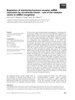
MAPKs [1,2] (Fig. 1).
The activation of SAPKs requires phosphorylation of
conserved tyrosine and threonine residues within the
catalytic domain. This phosphorylation is mediated by dual
specificity protein kinases, members of the MAPK kinase
(MKK) family. MKK3 and MKK6 are specific for p38,
MKK7 selectively phosphorylates JNK, and MKK4
recognizes either class of the stress actived kinases (Fig. 1).
The MKKs are also activated by the phosphorylation of
conserved serine and threonine residues [1,2]. Several
MKK-activating MKK kinases (MKKKs) have been
identified, some of which are activated again by phosphory-
lation [3,4]. In the absence of a signal, the constituents of t he
SAPK cascade return to their inactive, dephosphorylated
state, suggesting an essential role for phosphatases in SAPK
regulation.
Protein p hosphatases are classifi ed into three groups,
Ser/Thr phosphatases, Ser/Thr/Tyr phosphatases and Tyr
phosphatases, depending on their phosphoamino-acid
specificity. The dephosphorylation of SAPK signal p athway
components on either Ser/Thr or Thr/Tyr residues requires
the participation of various p hosphatases. In t his article, we
first review the roles of protein phosphatases in the
regulation of yeast SAPK pathways, then fo cus on the
properties of the protein phosphatases implicated in
the mammalian SAPK systems.
REGULATION OF SAPK SIGNAL
PATHWAYS BY PROTEIN
PHOSPHATASES IN YEAST CELLS
A molecular g enetic analysis of ye ast cells indicated that two
distinct protein phosphatase groups, protein Tyr phospha-
tases (PTP) and protein Ser/Thr phosp hatases of type 2C
(PP2C), act as negative regulators of SAPK pathways [5,6].
In the budding yeast, Saccharomyces cerevisiae,hyper-
osmotic shock activates the SSK2/SSK22 (MKKK)-Pbs2
(MKK)-Hog1 (SAPK) kinases. In the fission yeast,
Schizosaccharomyces pombe, heat shock, oxidative stress,
nutrient stress and osmotic shock all induce the Wik1
(MKKK)-Wis1 (MKK)-Spc1 (SAPK) pathway; the activa-
ted Spc1 in turn changes gene expression through the
activation of the Atf1 transcription factor [7–10].
The PTPs of S. cerevisiae (Ptp2 and Ptp3) and S. pombe
(Pyp1 and Pyp2) suppress the SAPK pathways, as demon-
strated by molecular genetic studies [5,8,10–12]. In S. pombe,
Pyp2 dephosphorylates the tyrosine residue of Spc1 both
in v ivo and in vitro [8,12]. Extracellular stress induces expres-
sion of the pyp2 gene in an Spc1-Atf1-dependent manner
Correspondence to S. Tamura, Department of Biochemistry, Institute
of Development, Aging and Cancer, Tohoku University 4-1
Seiryomachi, Aoba-ku, Sendai 980-8575, Japan.
Fax: + 81 2 2 717 8476, Tel.: + 81 22 717 8471,
E-mail:
Abbreviations: SAPK, stress-activated protein kinase; MAPK,
mitogen-activated protein kinase; JNK, c-Jun N-terminal kinase;
MKK, MAPK kinase; MKKK, MKK kinase; PTP, protein Tyr
phosphatase; PP, protein Ser/Thr phosphatase; DSP, dual specificity
protein phosphatase; MKP, MAPK phosphatase; ERK, extracellu-
lar signal-regulated kinase; TPA, 12-O-tetradecanoylphorbol
13-acetate; TCR, T cell receptor; TGF-b, transforming growth
factor-b;TAK1,TGF-b-activated kinase 1; IL-1, interleukin-1;
KIM, kinase interaction motif; PA, 1,2-dioleoyl-sn-glycero-
3-phosphate.
(Received 6 August 2001, accepted 20 September 2001)
Eur. J. Biochem. 269, 1060–1066 (2002) Ó FEBS 2002
[10,11]. In addition, PP2C (Ptc1 and Ptc3 of S. cerevisiae
and Ptc1 and Ptc3 of S. pombe) acts as a negative regulator
of SAPK pathways [13–15]. In S. pombe, Ptc1 acts upon a
target downstream of SAPK (Spc1) [6]. When Spc1
enhances the expression of Atf1, this up-regulation induces
Ptc1 expression, suppressing Atf1 function. Ptc1 and Ptc3
directly dephosphorylate the threonine of Spc1, but not th e
tyrosine [16]. In a ddition, Ptc1 dephosphorylates Hog1 in S.
cerevisiae both in vivo and in vitro [15].
REGULATION OF SAPK SIGNAL
PATHWAYS BY PROTEIN
PHOSPHATASES IN MAMMALIAN CELLS
In mammalian cells, like yeast cells, both PTP and PP2C
regulate the SAPK signal pathways [17–23]. Mammalian
cells are unique in several r espects as, in addition to PTP and
PP2C, they contain a large family of dual specificity protein
phosphatases (DSP) that negatively influence the SAPK
pathways [24]. Although the participation of a DSP, MSG5,
in the negative regulation of mating hormone-induced
MAPK (Fus3p) activation is well documented [25], the parti-
cipation of such DSPs in the regulation of the yeast SAPK
system has not been observed. In addition, protein phospha-
tase 2A (PP2A) may also function in the regulation of the
mammalian SAPK pathway [26]. In this section, we describ e
the properties of mammalian protein phosphatase mole-
cules involved in the regulation of SAPK signal pathways.
Dual specificity protein phosphatases
The gene products of at least 10 distinct DSP genes share two
unique structural features; they contain a common active site
sequence motif [VXVHCXXGXSRSXTXXX AY(L/I)M]
and two N-terminal CH2 domains, homologous to the cell
cycle regulator Cdc25 [27]. DSP substrate studies indicate
that MAPK phosphatase-3 (MKP-3) specifically dephosph-
orylates extracellular signal-regulated kinase (ERK) but not
JNK or p38 [27,28]. In contrast, both MKP-5 and M3/6
dephosphorylate both JNK and p38 but not ERK (Table 1)
[27,29,30]. The high specificity of MKP-2 for ERK and JNK
(but not for p38) and that of PAC-1 for ERK and p38 (but
not for JNK) has been reported (Table 1) [31]. On the other
hand, MKP-1 and MKP-4 were found to dephosphorylate
ERK, JNK and p38 [31,32]. These facts indicate an
unexpected complexity for the negative regulation of the
MAP kinase signaling. In the forthcoming paragraphs we
present a detailed description of the mammalian DSPs
involved in the regulation of SAPK signaling pathways.
MKP-1 (CL100). MKP-1, a protein of 39.5 kDa, is
expressed upon oxidative stress and heat shock in human
skin cells [33]. MKP-1 mRNA is ubiquitously expressed in
various tissu es, with the protein product localized preferen-
tially to the cell nucleus [34]. This enz yme acts as a DSP,
dephosphorylating both threonine and tyrosine residues of
ERK, JNK and p38 [31,35]. In addition to oxidative stress
and heat shock, MKP-1 is induced by various stimuli such
as, o smotic shock, anisomycin, g rowth factors, UV, 12-O-
tetradecanoylphorbol 13-acetate (TPA), Ca
2+
ionophores
and lipopolysaccharide [33–42]. MKP-1 expression is part
of a feed back mechanism: the activation of MAPKs
induces MKP-1; that in turn inactivates MAPKs. The
details of the regulatory mechanism depend on the cell
lineage. In vascular smooth muscle cells, mesangial cells and
U937 cells, the activation of either ERK, JNK or p38
induces MKP-1; in NIH3T3 cells, the activation of JNK but
not ERK up-regulates MKP-1 expression [35,37,40,43–45].
In addition, activation of p38 but not ERK o r JNK
enhances MKP-1 induction in H4IIE hepatoma cells [36]. In
Rat1 fibroblasts, MKP-1 is induced by Ca
2+
signaling,
independently of MAPK activation [41]. In this context,
Ca
2+
/calmodulin-activated protein phosphatase (PP2B)
participates in the induction of MKP-1 in cardiac myocytes
Fig. 1 . SAPK signaling modules. The p rotein
kinase cascades of SAPK signaling pathways
and the points where the ph osphatases can
interfere with the signals are shown. MKKK,
MKK kinase; MKK, MAPK kinase; MAPK,
MAP kinase; TAK1, T GF-b-activated k inase
1;MEKK,MEKkinase;MLK,mixedlineage
kinase; ASK1, apoptosis signal-regulating
kinase 1; JNK, c-Jun N-terminal kinase.
Ó FEBS 2002 Regulation of SAPK signaling pathways (Eur. J. Biochem. 269) 1061
[46]. MKP-1 binds to C-terminal region of p38, that results
in its activation [34]. The stability of MKP-1 is regulated by
ERK-mediated phosphorylation of two C-terminal serine
residues [47]. This phosphorylation, while not modifying the
intrinsic activity of MKP-1, stabilizes the protein.
MKP-2 (hVH2). MKP-2, a 42.6-kDa nuclear DSP, is
widely expressed in various tissues [48]. This phosphatase is
highly specific f or ERK and JNK, but not p38 [31]. MKP-2
is induced by nerve growth factor, TPA and hepatocyte
growth factor in PC12 cells, peripheral blood T cells and
MDCK cells, respectively [31,49,50]. In MDCK cells,
hepatocyte growth factor-activated ERK induces MKP-2
expression; that inactivates JNK, which has also been
activated by GF, by dephosphorylation [50]. Overexpres-
sion of v-Jun, a constitutively active form of c-Jun, enhances
the expression of MKP-2 mRNA in chick embryo fibro-
blasts [51]. Therefore, the activation of JNK may also
influence in MKP-2 expression.
MKP-4. MKP-4 is a DSP of 41.8 kDa displaying moderate
substrate specificity f or ERK over JNK or p38 [32].
Immunostaining of MKP-4 expressed in either NIH3T3
cells or COS7 cells revealed that MKP-4 is localized mainly
to the cytoplasm; a subset of cells, however, also displays a
punctuate nuclear staining [ 32]. Expression of MKP-4
mRNA is highly restricted to the placenta, kidney a nd
embryonic liver [32]. Phosp hatase activation is mediated by
substrate binding [52].
MKP-5. MKP-5, a widely expressed 52.6-kDa protein,
preferentially dephosphorylates both JNK and p38, and
demonstrates extremely low activity against ERK [29,30].
This enzyme is evenly localized throughout the cytoplasm
and nucleus [29]. In cultured cells, the expression of MKP-5
is elevated by stress stimuli such as an isomycin and osmotic
stress but not by UV irradiation [29]. MKP-5 binds to p38
and JNK, but not ERK [29,30].
MKP-6. MKP-6 (25 kDa) was found as a CD28 (T cell
costimulatory r eceptor) binding protein [53]. In vitro, MKP-
6 d ephosphorylates ERK, JNK and p38. However, expres-
sion of a dominant negative form of MKP-6 in T cells
further stimulates the T cell receptor (TCR)/CD28-
enhanced phosphorylation of both ERK and J NK but
not p38, suggesting that ERK and JNK are the preferred
substrates of MKP-6 in the cells. MKP-6 expression is
up-regulated by CD28 costimulation of T cells. Binding of
the expressed MKP-6 to CD28 is required for the feed back
regulation of ERK and JNK by MKP-6 [53].
M3/6 (hVH5). M3/6 was the first DSP found to
selectively inhibit stress-induc ed activation of JNK and
p38; M3/6 does not, however, affect growth factor-
induced activation of ERK in mammalian cells [27]. In
K562 human leukemia cells, hVH5 (human orthologue of
mouse M3/6) mRNA levels are rapidly enhanced by TPA
treatment [54]. The induction of exogenous M3/6 inhibited
TPA-stimulated phosphorylation of JNK and p38, sug-
gesting a feedback loop governing SAPK activity. The
activation of JNK stimulates the phosphorylation of
M3/6; unlike MKP-1, however, the phosphorylation of
M3/6 does not regulate its half life [54]. An internal motif,
XILPXL(Y/F)LG, homologous to the SAPK binding site
of c-Jun (delta domain), is important for M3/6 activity
[54].
PAC-1. PAC-1 is a DSP of 32 kDa, originally found to be
expressed predominantly in hematopoietic cells [55]. Subse-
quently, induction of PAC-1 mRNA in hippocampus
neurons following forebrain ischemia or kainic acid-induced
seizure has been reported [56,57]. PAC-1 dephosphorylates
both ERK and p38 but not JNK [31]. Activation of ERK
induces the enhanced-expression of PAC-1 and the
expressed PAC-1 the n inactivates ERK in T cells [58].
Protein phosphatase 2C
Protein phosphatase 2C (PP2C) is one of the four major
protein serine/threonine phosphatases (PP1, PP2A, PP2B
and PP2C) in eukaryotes. At least six distinct PP2C gene
products (2Ca,2Cb,2Cc,2Cd,Wip1andCa
2+
/calmodu-
lin-dependent protein kinase phosphatase) operate in
mammalian cells [59–65]. Studies of mammalian PP2C
function indicated that P P2Ca, PP2Cb and Wip1 a re
involved in the negative regulation o f SAPK cascades [20–
23]. In addition, PP2Ca and PP2Cb may regulate cell cycle
progression [66]. PP2Ca is implicated in Wnt signaling
regulation [67]. Here, we describe the properties of PP2C
isoforms regulating the SAPK s ignal pathways.
PP2Ca. PP2Ca, a 42-kDa phosphatase, was first cloned
from a rat kidney c DNA library [59]. The existence of two
distinct human PP2Ca isoforms (a-1 and a-2), differing at
their C-terminal regions, was subsequently reported [20,68].
A cDNA clone encoding PP2Ca-2 was isolated in the
screening of a human cDNA library for genes down-
regulating the yeast Hog1 MAPK pathway [20]. When
expressed in mammalian cells, PP2Ca-2 inhibits stress-
induced activation of p38 and JNK, but does not affect
mitogen-induced activation of ERK. Mouse PP2Ca,cor-
responding to human PP2Ca-1, exhibited a similar inhibi-
tion pattern [21]. PP2Ca-2 dephosphorylates and inactivates
MKK4, MKK6 and p38 both in vivo and in vitro [20].
Table 1. Protein phosphatases involved in regulation of SAPK signal
pathways.
Phosphatase Substrate References
DSP family
MKP-1 (CL100, 3CH134) JNK, p38, ERK [31,35]
MKP-2 (hVH2, Typ-1) JNK, ERK [31]
MKP-4 JNK, p38, ERK [32]
MKP-5 JNK, p38 [29,30]
MKP-6 JNK, ERK [55]
M3/6 (hVH5) JNK, p38 [27]
PAC-1 p38, ERK [31]
PP2C family
PP2Ca-2 MKK4, MKK6, p38 [20]
PP2Cb-1 TAK1 [23]
Wip1 p38 [22]
PTP family
HePTP/LC-PTP p38, ERK [17,18]
PTP-SL/STEP p38, ERK [19,78]
PP2A family
PP2A JNK [26]
1062 S. Tamura et al. (Eur. J. Biochem. 269) Ó FEBS 2002
Furthermore PP2Ca-2 specifically associates with phos-
phorylated p38.
PP2Cb. The PP2Cb gene encodes at least six distinct
isoforms (43 kDa), which are splicing variants of a single
premRNA [60,69–71]. These isoforms differ only at the
C-terminal regions. PP2Cb-1 is expressed ubiquitously in
various tissues, while PP2Cb-2 expression is restricted to th e
brain a nd heart. PP2Cb-3, -4 and -5 transcripts were detec-
ted predominantly in the liver, testes and intestine [69,70]. In
mammalian cells, PP2Cb-1 selectively supp resses the stress-
induced activation of p38 and JNK but has no effect on the
mitogen-induced activation of ERK [21]. Investigation of
the PP2Cb-1-mediated suppression of the SAPK pathway
revealed that PP2Cb-1 dephosphorylates and inactivates
transforming growth factor- b (TGF-b)-activated k inase
(TAK1), a MKKK activated either by stress, TGF-b treat-
ment or interleukin-1 (IL-1) stimulation [23]. In addition,
PP2Cb-1 selectively associates with TAK1 in a stable com-
plex. Expression of a dominant-negative form of PP2Cb-1
enhances the IL-1-induced activation of AP-1 reporter gene,
suggesting PP2Cb-1 ne gatively regulates TAK1 signaling
through the depho sphorylation of TAK1 in vivo [23].
Wip1. Wip1, a 61-kDa Mg
2+
-dependent protein phospha-
tase, is induced by ionizing radiation in a p53-de pendent
manner [64]. It is localized to the nucleus, the nuclear levels
of Wip1 increase in response to the ionizing irradiation.
The expression of Wip1 is also induced by treatment with
methyl methane sulfonate, H
2
O
2
or anisomycin [22].
Functional studies of Wip1 revealed its role in the down-
regulation of p38/p53-induced signaling during the recovery
of damaged cells [22]. Thus, t he induction of Wip1 b y stress
selectively blocks the activation of p 38, and suppresses
subsequent p53 activation. In vitro, Wip1 inactivates p38 by
the specific dephosphorylation of a conserved threonine
residue; however, it does not accept ERK, JNK, MKK4 or
MKK6 as a substrate [22].
Other protein phosphatases
Recently e vidence has emerged suggesting the participation
of okadaic acid-sensitive protein phosphatases and PTPs in
the regulation of mammalian SAPK pathways [17–19,26].
In this section, we describe the roles of protein phosphatase
2A (PP2A) and tyrosine phosphatases, HePTP/LC-PTP
and PTP-SL/STEP, in SAPK signaling.
PP2A. Addition of okadaic acid to the culture medium
enhanced the lipopolysaccharide-induced activation of JNK
in THP-1 cells (a human acute monocytic leukemia ce ll line)
[26]. In addition the regulatory subunit of PP2A, PP2A-Aa,
coprecipitates with JNK [26]. JNK activity was unaffected
by specific pharmacological inhibition of protein phospha-
tase 1 by 1 ,2-dioleoyl- sn-glycero-3-phosphate (PA); the
activation of PP2A by high doses of PA, however, d ecreased
JNK activity [26]. These results suggest that PP2A may
suppress the lipopolysaccharide-induced JNK th rough the
direct dephosphorylation of JNK.
HePTP/LC-PTP. HePTP and LC-PTP are closely related
human cytosolic PTPs, predominantly expressed in hem-
opoietic cells [17,18]. In T lymphocytes, the transcription of
HePTP is enhanced by IL-2 treatment [72]. When expressed
in Jurkat T cells, HePTP/LC-PTP inhibits the TCR-induced
activation of both ERK and p38, but not JNK [17,18]. Both
ERK a nd p38 (but not JNK) associate with the kinase
interaction motif (KIM) in the N-terminal segment of
HePTP/LC-PTP. The phosphorylation of HePTP by PKA
inhibits its association with ERK a nd p38 [73]. Conse-
quently the PKA-mediated r elease o f the phosphatase
activates both ERK and p38.
PTP-SL/STEP. PTP-SL and STEP are non-nuclear
PTPs, which exist in transmembrane and cytosolic forms
and are mainly expressed in n euronal cells [74–77]. PTP-SL
dephosphorylates both ERK and p38 [19,78]. Like HePTP,
PTP-SL associates with ERK and p38 but not with JNK
through its KIM located in the juxtamembrane region [78].
The phosphorylation of PTP-SL by PKA was found to
inhibit its association with ERK and p38, and the
subsequent tyrosine dephosphorylation of these MAPKs
[19].
CONCLUSIONS AND PERSPECTIVES
Numerous phosphatase molecules are capable of negatively
regulating SAPK signaling pathways (summarized in
Table 1 and Fig. 1) including the members of four distinct
groups: DSP, PP2C, PP2A and PTP. Regulation of a single
substrate by multiple protein phosphatases suggests
redundancy. Alternatively, the level of phosp horylation in
each protein component of t he SAPK pathway may be
regulated by multiple upstream s ignals functioning via
distinct protein phosphatases.
We conclude that at least two distinct mechanisms can
operate in the regulation. The expression of phosphatases,
such as MKP-1, MKP-2, MKP-5, M3/6, PTC-1, Wip1
and HePTP, is positively regulated through the activation
of MAP kinases. In addition, some phosphatases are
regulated by direct association with MAPKs. For example
both MKP-1 and MKP-4 are activated via binding to
their MAPK substrates [34,52]. Direct association was
also observed between MKP-5 and p38 or JNK [29,30].
Interestingly, a sequence motif, XILPXL(Y/F)LG, which
is similar to a delta domain consensus motif critical for
binding to JNK and ERK in other proteins, is converved
in all of these DSPs. The delta-like domain is located
N-terminal to the c atalytic consensus sequence of t he
DSPs. The delta-like domain is also conserved in M3/6;
deletion of this sequence bloc ks the ability of M3/6 to
dephosphorylate JNK [54]. These results suggest that the
delta-like domain is involved in the association of
phosphatases with MAPKs. Another association between
MAPKs a nd HePTP/LC-PTP or PTP-SL/STEP phos-
phatases and regulation of this association by PKA is also
of great importance [19,73].
Substrate specificity studies indicated that s everal mem-
bers of the DSP and PTP families dephosp horylate both
ERK and JNK/p38 (Table 1). This suggests that phospha-
tases may mediate the signaling between the ERK and
SAPK pathways. Future studies will certainly clarify the
significance of s uch cross-talk between the ERK and SAPK
pathways via protein phosphatases.
The protein phosphatases that dephosphorylate MKKs
and MKKKs have not been well investigated. The PP2C
Ó FEBS 2002 Regulation of SAPK signaling pathways (Eur. J. Biochem. 269) 1063
family may play a central role in the regulation of these
kinases as PP2Ca-2 dephosphorylates both MKK4 and
MKK6 [20]. In addition, PP2Cb-1 dephosphorylates
TAK1, but not MKK6 [23]. These results suggest that each
isoform of PP2C may have a distinct specificity for
substrates in SAPK pathways. Future studies are required
for identification of phosphatases responsible for dephos-
phorylation of other MKK and MKKK members.
ACKNOWLEDGEMENT
The au thors are gr ateful to Dr Masato Ogata (Osaka University) for
critically revi ewing this a rticle.
REFERENCES
1. Ip, Y.T. & Davis, R.J. (1998) Signal transduction by the c-Jun
N-terminal kinase (JNK) – from i nflammation to development.
Curr. Opin. Cell Biol. 10, 205–219.
2. Garrington, T.P. & Johnson, G.L. (1999) Organization and reg-
ulation of mitogen-activated protein k inase signaling pathways.
Curr. Opin. Cell Biol. 11, 211–218.
3. Davis, R.J. (2000) Signal transduction by the JNK group of MAP
kinases. Cell 103, 239–252.
4. Kishimoto, K., Matsumoto, K. & Ninomiya-Tsuji, J. (2000)
TAK1 mitogen-activated protein kinase kinase kinase is activated
by autophosphorylation within its activation loop. J. Biol. Chem.
275, 7359–7364.
5. Wurgler-Murphy, S.M. & Saito, H. (1997) Two-component signal
transducers and MAPK cascades. Trends Biochem. Sci. 22,172–
176.
6. Gaits, F., Shiozaki, K. & Russell, P. (1997) Protein phosphatase
2C acts independently of stress-activated kinase cascade to reg-
ulate the stress response in fission yeast. J. Biol. Chem. 272, 17873–
17879.
7. Shiozaki, K. & Russell, P. (1995) Cell-cycle control linked to
extracellular environment by MAP kinase pathway in fission
yeast. Nature 378, 739–743.
8. Millar, J.B., Buck, V. & Wilkinson, M.G. (1995) Pyp1 and Pyp2
PTPases dephosphorylate an osmosensing MAP kinase control-
ling cell size at division in fission yeast. Genes Dev. 9, 2117–2130.
9. Shiozaki, K., Shiozaki, M. & Russell, P. (1997) Mcs4 mitotic cata-
strophe suppressor regulates the fission yeast cell cycle through the
Wik1-Wis1-Spc1 kinase cascade. Mol. Biol. Cell. 8, 409–419.
10. Shiozaki, K. & Russell, P. (1996) Conjugation, meiosis, and the
osmotic stress response are regulated by Spc1 kinase through Atf1
transcription factor in fission yeast. Genes Dev. 10 , 2276–2288.
11. Wilkinson, M.G., Samuels, M., Takeda, T., Toone, W.M., Shieh,
J.C., Toda, T., Millar, J.B. & Jones, N. (1996) The Atf1 tran-
scription factor is a target for the Sty1 stress-activated MAP kinase
pathway in fission yeast. Genes Dev. 10, 2289–2301.
12. Degols, G., Shiozaki, K. & Russell, P. (1996) Activation and
regulation of the Spc1 stress-activated protein kinase in Schizo-
saccharomyces pombe. Mol. Cell. Biol. 16, 2870–2877.
13. Maeda, T., Tsai, A.Y. & Saito, H. (1993) Mutations in a protein
tyrosine phosphatase gene (PTP2) and a protein serine/threonine
phosphatase gene (PTC1) cause a synthetic growth defect in
Saccharomyces cerevisiae. Mol. Cell. Biol. 13, 5408–5417.
14. Jacoby,T.,Flanagan,H.,Faykin,A.,Seto,A.G.,Mattison,C.&
Ota, I. (1997) Two protein-tyrosine phosphatases inactivate the
osmotic stress response pathway in yeast by targeting the mitogen-
activated protein kinase, Hog1. J. Biol. Chem. 272, 17749–17755.
15. Warmka, J., Hanneman, J., Lee, J., Amin, D. & Ota, I . (2001)
Ptc1, a type 2C Ser/Thr phosphatase, inactivates the HOG path-
way by d ephosphorylating the mitogen-activated protein k inase
Hog1. Mol. Cell. Biol. 21, 51–60.
16. Nguyen, A.N. & Shiozaki, K. (1999) Heat-shock-induced activa-
tion of stress MAP kinase is regulated by threonine- and tyrosine-
specific pho spha tases. Genes D ev. 13, 1653–1663.
17. Saxena, M., Williams, S., Brockdorff, J., Gilman, J. & Mustelin, T.
(1999) Inhibition of T cell signaling by mitogen-activated protein
kinase-targeted h ematopoietic tyrosine pho sphatase (HePTP).
J. Biol. Chem. 274, 11693–11700.
18. Oh-hora, M., Ogata, M., Mori, Y., Adachi, M., Imai, K., Kosugi,
A. & Hamaoka, T. (1999) Direct suppression of TCR-mediated
activation of extracellular signal-regulated kinase by leukocyte
protein tyrosine p hosphatase, a tyrosine-specific phosphatase.
J. Immunol. 163, 1282–1288.
19. Blanco -Aparicio, C., Torres, J. & P ulido, R. (1999) A novel reg-
ulatory mechanism of MAP kinases activation and nuclear
translocation mediated by PKA and the PTP-SL tyrosine phos-
phatase. J. Cell. Biol. 147, 1129–1136.
20. Takekawa, M., Maeda, T. & Saito, H. (1998) Protein phosphatase
2Ca inhibits the human stress-responsive p38 and JNK MAPK
pathways. EMBO J. 17, 4744–4752.
21. Hanada, M., Ko bayashi, T., Ohnishi, M., Ikeda, S., Wang, H.,
Katsura, K., Yanagawa, Y., Hiraga, A., Kanamaru, R. &
Tamura, S. (1998) Selective suppression of stress-activated protein
kinase pathway by protein phosphatase 2C in mammalian cells.
FEBS Lett. 437, 172–176.
22. Takekawa, M., Adachi, M., Nakahata, A., Nakayama, I., Itoh,
F., Tsukuda, H., Taya, Y. & Imai, K. (2000) p53-inducible wip1
phosphatase mediates a negative feedb ack regulation of p38
MAPK-p53 signaling in response to UV radiation. EMBO J. 19,
6517–6526.
23. Hanada, M., Ninom iya-Tsuji, J., Komaki, K., Ohnishi, M.,
Katsura, K., Kanamaru, R., Matsumoto, K. & Tamura, S. (2001)
Regulation of the TAK1 signaling pathway by protein phospha-
tase 2C. J. Biol. Chem. 276, 5753–5759.
24. Keyse, S.M. (2000) Protein phosphatases and the regulation of
mitogen-activated protein kinase signalling. Curr. Opin. Cell Biol.
12, 186–192.
25. Doi, K., Gartner, A., Ammerer, G., Errede, B., Shinkawa, H.,
Sugimoto, K. & Matsumoto, K. (1994) M SG5, a novel protein
phosphatase promotes adap tation to pheromone r esponse in
S. cerevisiae. EMBO J. 13, 61–70.
26. Shanley, T.P., Vasi, N., Denenberg, A. & Wong, H.R. (2001) The
serine/threonine phosphatase, PP2A: endogenous regulator o f
inflammatory cell signaling. J. Immunol. 166, 966–972.
27. Mud a, M., Theod osiou, A., Rodrigues, N., Bo schert, U., Camps,
M., Gillieron, C., Davies, K., Ashworth, A. & Arkinstall, S. (1996)
The dual specificity phosphatases M3/6 and MKP-3 are highly
selective for inactivatio n of distinct mitogen-activated protein
kinases. J. Biol. Chem . 271, 27205–27208.
28. Groo m, L.A., S neddon, A.A., Alessi, D.R., Dowd, S. & Keyse,
S.M. (1996) Differential regulation of the MAP, SAP and RK/p38
kinases by Pyst1, a novel cytosolic dual-specificity phosphatase.
EMBO J. 15, 3621–3632.
29. Tano ue, T., Moriguchi, T. & Nishida, E. (1999) Molecular cloning
and characterization of a novel dual specificity phosphatase,
MKP-5. J. Biol. Che m. 274, 19949–19958.
30. Theodosiou, A., Smith, A., Gillieron, C., Arkinstall, S. & Ash-
worth, A. (1999) MKP5, a new member of the MAP kinase
phosphatase family, which selectively dephosphorylates stress-
activated kinas es. Oncogene 18, 6981–6988.
31. Chu, Y., Solski, P.A., Khosravi-Far, R., C.J. & Kelly, K. (1996)
The mitogen-activated protein kinase phosphatases PAC1, M KP-
1, and MKP-2 have unique substrate specificities and reduced
activity in vivo toward the ERK2 sevenmak er mutation. J. Biol.
Chem. 271, 6497–6501.
32. Muda, M., Boschert, U., S mith, A., Antonsson, B., Gillieron, C.,
Chabert, C., Camps, M. , M artinou, I ., Ashworth, A . &
Arkinstall, S. (1997) Molecular cloning and functional charac-
1064 S. Tamura et al. (Eur. J. Biochem. 269) Ó FEBS 2002
terization o f a novel mitogen-activated protein kinase phospha-
tase, MKP-4. J. Biol. Chem. 272, 5141–5151.
33. Keyse, S.M. & Emslie, E.A. (1992) Oxidative stress and heat shock
induce a human gene encoding a protein-tyrosine phosphatase.
Nature 359, 644–647.
34. Hutter, D., Chen, P., Barnes, J. & Liu, Y. (2000) C atalytic acti-
vation of m itogen-activated protein (MAP) kinase phosph atase-1
by binding to p38 MAP kinase: critical role of the p38 C-terminal
domain in its negative regulation. Biochem. J. 352, 155–163.
35. Li, C., Hu, Y., Mayr, M. & Xu, Q. ( 1999) Cyclic strain stress-
induced mitogen-activated protein kinase (MAPK) p hosphatase 1
expression in vascular smooth muscle cells is regulated by Ras/
Rac-MAPK pathways. J. Biol. Chem. 274, 25273–25280.
36. Schliess, F., Heinrich, S. & Haussinger, D. (1998) Hyperosmotic
induction of the mitogen-activated protein kinase phosphatase
MKP-1 in H4IIE rat hepatoma cells. Arch. Biochem. Biophys. 351,
35–40.
37. Bokemeyer, D., Sorokin, A. & Dunn, M.J. (1997) Differential
regulation of the dual-specificity protein-tyrosine phosphatases
CL100, B23, and PAC1 in mesangial cells. J. Am. Soc. Nephrol. 8,
40–50.
38. Alessi, D.R., Smythe, C. & Keyse, S.M. (1993) The human CL100
gene encodes a Tyr/Thr-pr otein phosphatase which potently and
specifically inactivates MAP k inase a nd suppresses i ts activa tion by
oncogenic ras in Xenopus oocyte extracts. Oncogene 8, 2015–2020.
39. Liu, Y., Gorospe, M., Yang, C. & Holbrook, N.J. (1995) Role of
mitogen-activated protein kinase phosphatase during the cellular
response to genotoxic stress. Inhibition of c-Jun N-terminal kinase
activity and A P-1-dependent gene activation. J. Biol. Chem. 270,
8377–8380.
40. F ranklin, C.C. & Kraft, A.S. (1997) Conditional expression of the
mitogen-activated protein kinase (MAPK) phosphatase MKP-1
preferentially inhibits p38 MAPK and stress-activated protein
kinase in U937 cells. J. Biol. Chem. 272, 16917–16923.
41. S cimeca, J.C., Servant, M.J., Dyer, J.O. & Meloche, S. (1997)
Essential role of calcium in the regulation of MAP kinase phos-
phatase-1 expression. Oncogene 15, 717–725.
42. Valledor, A.F., Xaus, J., Comalada, M., Soler, C. & Celada, A.
(2000) Protein kinase C epsilon is required for the induction of
mitogen-activated protein kinase phosphatase-1 in lipo po ly-
saccharide-stimulated macrophages. J. Immunol. 164, 29–37.
43. Begum, N., Ragolia, L., Rienzie, J., McCarthy, M. & Duddy, N .
(1998) Regulation mitogen-activated protein kinase phosphatase-1
induction by insulin in vascular smooth muscle cells. Evaluation of
the role of the n itric oxide signaling pathway an d potential defects
in hypertension. J. Biol. Chem. 273, 25164–25170.
44. Bokemeyer,D.,Lindemann,M.&Kramer,H.J.(1998)Regula-
tion of mitogen-activated protein kinase phosphatase-1 in vascular
smooth muscle cells. Hypertension 32, 661–667.
45. B okemeyer, D., Sorokin, A., Yan , M., Ahn, N .G., Templeton,
D.J. & Dunn, M.J. (1996) Induction of mitogen-activated protein
kinase phosphatase 1 by the stress-activated protein kinase sig-
naling p athway but not by extracellular signal-regulated kinase i n
fibroblasts. J. Biol. Chem. 271, 639–642.
46. L im, H.W., New, L., Han, J. & Molkentin, J.D. (2001) Essential
role of calcium in the regulation of MAP kinase phosphatase-1
expression. J. Biol. Chem. 276, 15913–15919.
47. Brondello, J.M., Pouyss egur, J. & McKenzie, F .R. (1999)
Reduced M AP kinase phosphatase-1 degradation after p42/
p44MAPK-dependent phosphorylation. Science 286, 2514–2517.
48. Misra-Press, A., Rim, C.S., Yao, H., Roberson, M.S. & Stork, P.J.
(1995) A novel mitogen-activated protein kinase phosphatase.
Structure, expression, and regulation. J. Biol. Chem. 270, 1 4587–
14596.
49. Hirsch, D.D. & Stork, P.J. (1997) Mitogen-activated protein
kinase phosphatases inactivate stress-activated protein kinase
pathways in vivo. J. Biol. Chem. 272, 4568–4575.
50. Paumelle, R., Tulasne, D., Leroy, C., Coll, J., Vandenbunder, B.
& Fafeur, V. (2000) Sequential activation of ERK and repression
of JNK by scatter factor/hepatocyte growth factor in madin-darby
canine kidney epithelial cells. Mol. Biol. Cell 11, 3751–3763.
51. Fu, S.L., Waha, A. & Vogt, P.K. (2000) Identification and char-
acterization of genes upregulated in cells transformed by v -Jun.
Oncogene 19, 3537–3545.
52. C amps, M., Nich ols, A., G illieron, C., Ant onsson, B., M uda, M.,
Chabert, C., Boschert, U. & Arkinstall, S. (1998) Catalytic acti-
vation of the phosphatase MKP-3 by E RK2 mitogen-activated
protein kinase. Science 280, 1262–1265.
53. Marti, F., Krause, A., Post, N.H., Lyddane, C., Dupont, B.,
Sadelain, M. & King, P.D. (2001) Negative-feedback regulation of
CD28 costimulation by a novel mitogen-activated protein kinase
phosphatase, MKP6. J. Immunol. 166, 197–206.
54. Johnson, T.R., Biggs, J.R., Winbourn, S.E. & Kraft, A.S. (2000)
Regulation of dual-specificity phosphatases M3/6 and hVH5 by
phorbol esters. Analysis of a delta-like domain. J. Biol. Chem. 275,
31755–31762.
55. R ohan, P.J., Davis, P., Moskaluk, C.A., Kearns, M., Krutzsch,
H., Siebenlist, U. & Kelly, K. (1993) PAC-1: a mitogen-induced
nuclear protein tyrosin e phosphat ase. Science 259, 1763–1766.
56. Wiessner, C. (1995) The dual specificity phosphatase PAC-1 is
transcriptionally induced in the rat brain following transient
forebrain ischemia. Brain Res. Mol. Brain Res. 28, 353–356.
57. B oschert, U., Muda, M., Camps, M., Dickinson, R. & Arkinstall,
S. (1997) Induction of the dual specificity phosphatase PAC1 in rat
brain following seizure activity. Neuroreport 8, 3077–3080.
58. Grumont, R.J., Rasko, J.E., Strasser, A. & Gerondakis, S. (1996)
Activation of the mitogen-activated protein kinase pathway
induces transcription of the PAC-1 phosphatase gene. Mol. Cell.
Biol. 16, 2913–2921.
59. Tamura, S., Lynch, K.R., Larner, J., Fox, J., Yasui, A., Kikuchi,
K., Suzuki, Y. & Tsuiki, S. (1989) Molecular cloning of rat type 2C
(IA) protein phosphatase mRNA. Pr oc. N atl Ac ad. Sci. USA 86,
1796–1800.
60. Wenk, J., Trompeter, H.I., Pettrich, K.G., Cohen, P .T.W.,
Campbell, D.G. & Mieskes, G. (1992) Molecular cloning and
primary structure of a protein phosphatase 2C isoform. FEBS
Lett. 297, 135–138.
61. Travis, S.M. & Welsh, M.J. (1997) PP2C gamma: a human protein
phosphatase witha uniqueacidic domain.FEBS Lett. 412, 415–419.
62. Guthridge, M .A., Bellosta, P., Tavoloni, N. & Basilico, C. (1997)
FIN13, a novel growth factor-inducible serine-threonine phos-
phatase which can inhibit cell cycle progression. Mol. Cell. Biol.
17, 5485–5498.
63. T ong, Y., Quirion, R. & Shen, S H. (1998) Clo ning and char-
acterization of a novel mammalian PP2C isozyme. J. Biol. Chem.
273, 35282–35290.
64. F iscella, M., Zhang, H., Fan, S., Sakaguchi, K., Shen, S ., Mercer,
W.E., Vande Woude, G.F., O’Connor, P.M. & Appella, E. (1997)
Wip1, a novel human protein phosphatase that is induced in
response to io nizing radiation in a p53-dependent m anner. Proc.
Natl Acad. Sci. USA 94, 6048–6053.
65. K itani, T., Ishida, A., Ok uno, S., Takeuchi, M ., Kameshita, I. &
Fujisawa, H. (1999) Molecular cloning of Ca
2+
/calmodulin-
dependent proteinkinase phosphatase.J. Biochem. 125, 1022–1028.
66. Cheng, A., Kaldis, P. & Solomon, M.J. (2000) Dephosphorylation
of human cyclin-dependent kinases by protein phosphatase type
2Ca and b2 isoforms. J. Biol. Chem. 275, 34744–34749.
67. Strovel, E.T., Wu, D. & Sussman, D.J. (2000) Protein phosphatase
2Ca dephosphorylates axin and activates LEF-1-dependent tran-
scription. J. Biol. Chem. 275, 2399–2403.
68. M ann, D.J., C ampbell, D.G., McGowan, C.H. & Cohen, P.T.
(1992) Mammalian protein serine/threonine phosphatase 2C:
cDNA cloning and comparative analysis of amino acid sequences.
Biochim. Biophys. Acta 1130, 100–104.
Ó FEBS 2002 Regulation of SAPK signaling pathways (Eur. J. Biochem. 269) 1065
69. Terasawa, T., Kobayashi, T., Murakami, T., Ohnishi, M.,
Kato, S., Tanaka, O., Kondo, H., Yamamoto, H., Takeuchi, T. &
Tamura, S. (1993) Molecular cloning of a n ovel isotype of
Mg
2+
-dependent protein phosphatase b (type 2Cb)enrichedin
brain and heart. Arch. Biochem . Biophys . 307, 342–349.
70. Kato, S., Terasawa, T., Kobayashi, T., Ohnishi, M., Sasahara, Y.,
Kusuda,K.,Yanagawa,Y.,Hiraga,A.,Matsui,Y.&Tamura,S.
(1995) Molecular cloning and expression of mouse Mg
2+
-
dependent protein phosphatase b-4 (type 2Cb-4). Arch. Biochem.
Biophys. 318, 387–393.
71. Marley,A.E.,Kline,A.,Crabtree,G.,Sullivan,J.E.&Beri,R.K.
(1998) The cloning expression and tissue distribution of human
PP2Cb. FEBS Lett. 431, 121–124.
72. Adachi, M., Sekiya, M., Ishino, M., Sasaki, H., Hinoda, Y., Imai,
K. & Yachi, A. (1974) Induction of protein-tyrosine phosphatase
LC-PTP by IL-2 in human T cells. LC-PTP is an early response
gene. FEBS Lett. 338 , 47–52.
73. Saxena, M., Williams, S., Tasken, K. & Mustelin, T. (1999)
Crosstalk between cAMP-dependent kinase and MAP kinase
through a protein tyrosine phosphatase. Nat.CellBiol.1, 305–311.
74. Hendriks, W., Schepens, J., Brugman, C., Zeeuwen, P. &
Wieringa, B. (1995) A novel receptor-type protein tyrosine
phosphatase with a single catalytic domain is specifically expressed
in mouse br ain. Biochem. J. 305, 499–504.
75. Ogata, M., Sawada, M., Fujino, Y. & Hamaoka, T. (1995) cDNA
cloning and characterization of a novel receptor-type protein
tyrosine phosphatase expressed predominantly in the brain. J. Biol.
Chem. 270, 2337–2343.
76. Shiozuka, K., Watanabe, Y., Ikeda, T., Hashimoto, S. & Kawa-
shima, H. (1995) Cloning and expression of PCPTP1 encoding
protein tyrosine phosphatase. Gene 162, 279–284.
77. Ogata, M., Oh-hora, M., Kosugi, A. & Hamaoka, T. (1999)
Inactivation of mitogen-activated protein kinases by a mammalian
tyrosine-specific phosphatase, PTPBR7. Biochem. Biophys. Res.
Commun. 256, 52–56.
78. Zuniga, A., Torres, J., Ubeda, J. & Pulido, R. (1999) Interaction of
mitogen-activated protein kinases with the kinase interaction
motif of the tyrosine phosphatase PTP-SL provides substrate
specificity and r etains ERK2 in the cytoplasm. J. Biol. Chem. 274,
21900–21907.
1066 S. Tamura et al. (Eur. J. Biochem. 269) Ó FEBS 2002