Báo cáo Y học: Amyloid-fibril formation Proposed mechanisms and relevance to conformational disease docx
Bạn đang xem bản rút gọn của tài liệu. Xem và tải ngay bản đầy đủ của tài liệu tại đây (274.63 KB, 10 trang )
REVIEW ARTICLE
Amyloid-fibril formation
Proposed mechanisms and relevance to conformational disease
Eva Z
ˇ
erovnik
Department of Biochemistry and Molecular Biology, Jozˇef Stefan Institute, Ljubljana, Slovenia
The phenomenon of the transformation of proteins into
amyloid-fibrils is of interest, firstly, because it is closely
connected to the so-called conformational diseases, many of
which are hitherto incurable, and secondly, because it
remains to be explained in physical terms (energetically and
structurally). The process leads to fibrous aggregates in the
form of extracellular amyloid plaques, neuro-fibrillary
tangles and other intracytoplasmic or intranuclear inclu-
sions. In this review, basic principles common to the field of
amyloid fibril formation and conformational disease are
underlined. Existing models for the mechanism need to be
tested by experiment. The kinetic and energetic bases of the
process are reviewed. The main controversial issue remains
the coexistence of more than one protein conformation. The
possible role of oligomeric intermediates, and of domain-
swapping is also discussed. Mechanisms for cellular defence
and novel therapies are considered.
Keywords: amyloid fibrils; conformational disease; domain
swapping; kinetics; mechanism of fibrillogenesis.
Protein folding is important for cellular events ranging from
transport, accepting and transmitting signals, regulation at
the gene and RNA levels, cell adhesion, changes in
cytoskeleton, metabolic reactions involving various
enzymes, etc. An active protein conformation is needed
for successful cell functioning, and therefore important in
maintaining health. Several types of disease have been
found where protein misfolding and conformational change
are the main causes of the appearance and progression of
disease [1].
A list of conformational diseases, together with their
associated protein component(s), is shown in Table 1 [2]. In
some cases, more than one protein is involved with a
disorder, coexisting in a plaque or making its formation
easier. Often, proteolytically degraded fragments are more
prone to forming fibrils, e.g. amyloid precursor protein
(APP) where a, b and c secretases [3,4] are responsible for
the initial processing, huntingtin and possibly also a-synuc-
lein [5].
In Alzheimer’s disease, which represents a major problem
in the Western world’s ageing population, the main protein
component is APP, a transmembrane protein of approxi-
mately 700 amino-acid residues [3,4,6,7]. In its normal
processing Ab (1–40) peptide is produced which circulates
extracellularly and usually does not deposit as plaques. It
has been proposed that the peptide may exert an antioxi-
dative function [8]. In sporadic cases, especially when
allele 4 of apolipoprotein E is present, the peptide starts to
form amyloid plaques. In the familial, more severe early-
onset cases, prevalence of the hydrophobic Ab (1–42)
peptide leads to extensive amyloid plaque formation. This
has been linked to mutations in the APP and presenilins 1
and 2 [7], which all increase the production of the more
fibrillogenic Ab (1–42) peptide. Fibrillary tangles of another
protein, sau, are observed in the cell. sau is a microtubule-
associated protein involved in stabilizing axonal
microtubules. Other functions include a role in signal
transduction, and anchoring various kinases and phospha-
tases [9]. Importantly, an anti-amyloidogenic protein,
gelsolin, has been found in plasma and central system fluid
(CSF). This secretory protein is able, by making complexes
with Ab, to inhibit fibril formation and even to break down
already formed fibrils [10]. Recently, it has been found that
the endopeptidase ÔneprilysinÕ degrades Ab peptide. In
neprilysin gene-disrupted mice Ab was found to accumu-
late, with the highest levels in the hippocampus [11].
In Parkinson’s disease, which is the second most common
neurodegenerative disease, several proteins are implicated,
a-synuclein, synphilin (an a-synuclein inteacting protein)
andparkin[12].a-Synuclein is a small (140 amino acid)
acidic protein. It is a naturally unfolded, intracellular and
presynaptic polypeptide that becomes partly helical on
binding to synaptic vesicles [13]. Its function may be, among
others, regulation of synaptic vesicles and neurotransmitter
release [13]. It is interesting that a-synuclein is a target of
serine/threonine [14] as well as tyrosine [15,16] kinases. A
hallmark of Parkinson’s disease is the presence of Lewy
bodies, which are found in sporadic cases of Parkinson’s
disease, in dementia with Lewy bodies and in the Lewy body
variant of Alzheimer’s disease [17]. a-Synuclein is the main
component of the Lewy bodies [18]. Both a-synuclein and
synphilin are required for formation of the Lewy bodies
where ubiquitination of synphilin probably takes place
[12,17]. Parkin is a 465-amino-acid ubiquitin-protein ligase
[17,19]. Mutations in parkin and a-synuclein, in familial
cases of Parkinson’s disease, prevent proper ubiquitination,
Correspondence to E. Z
ˇ
erovnik, Department of Biochemistry and
Molecular Biology, Jozˇ ef Stefan Institute, Jamova 39, 1000 Ljubljana,
Slovenia. E-mail:
Abbreviations: APP, amyloid precursor protein; Ab, amyloid b pep-
tide; CSF, central system fluid; AFM, atomic force microscopy; EM,
electron microscopy.
(Received 28 January 2002, revised 1 May 2002,
accepted 27 May 2002)
Eur. J. Biochem. 269, 3362–3371 (2002) Ó FEBS 2002 doi:10.1046/j.1432-1033.2002.03024.x
so that proteins are not sequestered in the inclusion bodies,
leading to greater toxicity [12].
Even the prion diseases, a range of transmissible spongi-
form encephalophaties (kuru, Creuzfeldt–Jacob disease and
fatal familial insomnia in humans, bovine spongiform
encelopathy in cattle, scrapie in sheep, and chronic wasting
disease in deer) have many features in common with
amyloidoses and most likely are ÔconformationalÕ [20]. No
other agent accompanying the prion protein like virus
(DNA–protein) or bare DNA has convincingly been shown
in the infected tissue [21]. The transmission could be
explained solely by inducing a wrong, irreversible conform-
ational change, resistant to proteolysis, leading to accumu-
lation of harmful protein aggregates. This hypothesis has
recently been confirmed by inducing disease in transgenic
mice inoculated by b rich conformation of mutant P101L
(89–143) peptide of the human prion protein [21], in contrast
to the ones inoculated by non-b-form of the peptide.
The term ÔamyloidÕ was introduced in 1854 by the
German physician R. Virchow, who named it in the belief
that the iodine-staining component was starch-like [22,23].
The first criterion for detecting amyloid ex vivo was
birefringence of the histological dye Congo Red, observed
under polarized light. As the second criterion, electron
microscopy showed that all amyloid deposits exhibited a
similar fibrillar, submicroscopic structure, bundles of
straight, rigid fibrils ranging in width from 60 to 130 A
˚
and in length from 1000 to 16000 A
˚
[23]. In addition to the
fibrillar component of amyloid, nonfibrillar components
were always found, including serum amyloid protein,
heparan sulfate proteoglycans and apolipoprotein E [23].
The importance of the nonprotein and nonfibrillar compo-
nents of amyloid as observed in vivo remains to be
determined. In vitro studies of the disease related proteins,
as well as other amyloidogenic proteins, have been
concerned mostly with the morphology and kinetics of
fibrillogenesis.
It was concluded by Soto [20] that the pathogenesis of all
the conformational diseases, including prion disease,
involves conformational changes leading to aberrantly
folded proteins, rich in b secondary structure that have a
high tendency to form aggregates and are quite resistant to
proteolysis [20,24]. The field is characterized by several
scientific findings that challenge some of the commonly held
dogmas in biology [24]. These findings are that a protein can
exist in more than one conformation with distinct biological
properties, and that biological function is mediated through
changes in protein conformation. Some of the basic
principles underlying protein fibril formation are described
in the following sections.
FIBRIL FORMATION, A GENERAL
PROPERTY OF PROTEINS AND
POLYPEPTIDES?
Several authors have found that proteins that have not been
associated with any disease can form amyloid-like fibrils
[25–31]. Especially surprising was the finding that even
a helical proteins, such as myoglobin [32] or apo-cyto-
chrome c [33] can form fibrils under certain conditions.
These observations led Dobson and coauthors to propose
that amyloid-fibril formation is a generic property of
proteins [27,32,34]. A common observation is that fibrilli-
zation starts from an intermediate state, either partially
unfolded or partially folded, molten globule or native-like
intermediate [35]. In the case of globular proteins such as
phosphoglycerate kinase [25], cystatin C [36], acylphospha-
tase [29] and transthyretin [37], partial unfolding needs to
occur to enable fibril formation and, in the case of unfolded
polypeptides such as a-synuclein [38,39] and islet amyloid
polypeptide, these must partially fold. The parts with the
a helical structure must undergo an a to b transition and the
b strands then associate into a regular fibrillar structure. An
a to b transformation is well characterized with peptides,
like poly(
L
-lysine). It has more recently been observed with
proteins which are initially unfolded or predominantly
b sheet [40–42] and which fold through an a helical
intermediate [43–46].
In vitro, variation of solvent conditions by changing pH
or adding organic solvents [47] can lead to partial unfolding
Table 1. Protein fibrillar inclusions in neurodegenerative and other types of diseases. Data from [2,100]. TSE, transmissible spongiform
encephalopathies.
Disease Protein component Cellular inclusion
Neurodegenerative
Alzheimer’s sau, A42b peptide Neurofibrillary tangles
Pick’s sau Pick bodies/cytoplasmic
Progressive supranuclear palsy (PSP) sau, heat shock proteins Neurofibrillary tangles
Dementia with Lewy bodies a-Synuclein Lewy bodies/cytoplasmic
Parkinson’s a-Synuclein, crystallins Neurofilaments/cytoplasmic
Huntington’s Expanded Glu repeats of Intranuclear inclusion
huntingtin
Spinocerebellar ataxias (SCA) Expanded Glu repeats of Intranuclear inclusion
ataxins 1,3,7
TSE Prion protein, cathepsin B Endosome-like organelles
System amyloidosis
Diabetes type 2 Amylin
Haemodialysis related A b-2 Microglobulin
Reactive amyloidosis Amyloid A
Cystic fibrosis CFTR protein
Ó FEBS 2002 Amyloid-fibrils and conformational disease (Eur. J. Biochem. 269) 3363
and subsequent protein fibril formation [29,48]. With
unfolded polypeptides, partial folding can be obtained by
lowering pH or by heating [39]. In vivo, partial unfolding
may happen as a consequence of lowered protein stability
due to mutation, local change in pH at membranes,
oxidative and heat stress, whereas partial folding may
happen on exposure to environmental hydrophobic sub-
stances, such as pesticides [39].
AMYLOIDOGENIC CONFORMATION
ANDCOMMONSTRUCTURAL
TRAITS OF THE FIBRILS
Amyloid fibrils cannnot be observed in solution whereas the
preamyloidogenic conformation can be trapped in crystal-
line or soluble form. NMR data exist on the native-like acid
intermediate of transthyretin [49] where the authors have
used hydrogen exchange in conjunction with NMR to trace
structural features of the preamyloidogenic conformation.
Similarly, by using hydrogen exchange in the native state,
the labile parts of the prion peptide have been determined
[50]. NMR has been used in combination with electrospray
ionization mass spectrometry (ESI MS) to enable the
population of the intermediate to be seen [51]. Recently,
crystal structures of the domain-swapped dimers of human
cystatin C [52] and prion peptide [53] have been determined.
A solution structure of human stefin A (type I cystatin)
dimer is also available [54] and confirms the main features
observedincrystalstructureofcystatinC.
Ordered fibrillar aggregates and the amyloid-fibrils
themselves can be studied at lower resolution by trans-
mission electron microscopy, atomic force microscopy
(AFM) [55,56], cryo-electron mycroscopy [57], X-ray
diffraction [58] and solid state NMR [59]. The fibrils
appear long, of indefinite length, unbranched, with repeats
that reflect the twisting of the component ÔfilamentsÕ
around one another [60–62]. Common features of the
fibrils are [58], b strands (separated by 4.7 A
˚
) running
perpendicular to the long axis of the fibrils and b sheets
extending parallel to this axis. The bstrands form a
b helical twist with the usual repeat at every 115 or 250 A
˚
[56,58]. There are two main types of the fibrils, type 2
fibrils are built from two intertwined filaments, with a
diameter from 80 to 130 A
˚
. Type 1 fibrils are thinner and
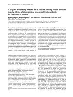
are formed from one filament only. There are other types
of fibrils [62]; for example, a fibril and untwisted filaments
of human stefin B [31] (type I cystatin) are illustrated in
Fig. 1.
ENERGETIC AND KINETIC BASIS
OF FIBRILLOGENESIS
The molecular and energetic basis of protein misfolding and
amyloid fibrillogenesis is still largely unknown [20,63]. In the
conclusion to their review, Rochet & Lansbury [35] propose
that future research should be directed towards understand-
ing the mechanism of amyloid-fibril formation, including
environmental factors, such as temperature, ionic strength,
pH and oxidation potential. Proteins have been treated as
an ensemble of rapidly interconverting conformational
substates. In contrast, recent studies have shown that
interconversion between different conformations may be
slow (taking hours to days). For certain proteins the folding
appears to be determined by kinetic rather than thermody-
namic factors [64]. The free-energy barriers can be quite
high [64–66], leading to persistence of parallel states, which
possibly exhibit different biological functions. The forces
involved are nonspecific, e.g. hydrophobic and repulsive
electrostatic, and specific, e.g. hydrogen bonding and salt-
bridges. As cooling causes reversible disaggregation of Ab
fibrils, a significant contribution to stability must come from
entropy-driven hydrophobic interactions. This led to trials
of various hydrophobic compounds that should be effective
in destabilizing and disaggregating amyloid fibrils.
Fig. 1. Transmission electron micrographs. (A) Amyloid fibrils of
human recombinant stefin B (cystatin B) prepared in vitro at pH 4.8,
showing a b helical repeat. (B) Porous fibrillar aggregate and fine
structure of a fibril (made from four filaments) resulting from the
addition of trifluoroethanol.
3364 E. Z
ˇ
erovnik (Eur. J. Biochem. 269) Ó FEBS 2002
Despite enormous efforts, description of the process of
fibrillogenesis is only qualitative at the moment. Various
morphologic species are described in the literature on
protein fibril formation. Fibrillogenesis often starts with
dimers as initial building blocks [3,56]. These further
oligomerize to tetramers, octamers, etc. The oligomeric
species constitute Ôprefibrillar aggregatesÕ composed of fluid
(micelle-like) nuclei [67]. From these, the ÔprotofibrilsÕ grow
up to 200 nm in length and are slightly curved [67,68]. All
these species accumulate in the so-called Ôlag-phaseÕ char-
acteristic for the kinetics of fibril growth. The lag-phase ends
with an exponential growth when proto-fibrils merge into
ÔfilamentsÕ. Fully grown fibrils are then made from one or
more filaments added laterally or, end by end [69]. The
events in the lag-phase are especially important and some
results have been obtained by real time AFM [70,71].
Presence of prefibrillar (oligomeric) intermediates is an
emerging theme [68,72].
The kinetics of fibrillogenesis have been studied by light
scattering [67,72]. Teplow and coauthors [67] have detec-
ted the following steps: (a) peptide micelles form above a
certain critical concentration, (b) fibrils nucleate within
these micelles or on heterogenous nuclei (seeds), and (c)
fibrils grow by irreversible binding of monomers to the
fibril ends. Simpler, colorimetric methods exist for detect-
ing amyloid fibrils. Use of histological dyes Congo Red
[73] and Thioflavin T [74] is widespread. In fact, both dyes
may actually label the filaments better than the fibrils
(E. Z
ˇ
erovnik, unpublished observation). Thioflavin T
fluorescence is a suitable method to follow the kinetics
of fibril formation in an interrupted manner, whereas
interference with the process on longer standing would be
expected. Whether Congo Red is fibril specific has been
questioned [75]. Substances based on Congo Red dye
structure have been used to inhibit fibril formation in vivo
[76] and others based on Thioflavin dye structure to label
the amyloid plaques in brain imaging [77].
Teplow and coworkers [40] have recently reported that an
intermediate with additional a helix structure was shown to
be a key step in Ab fibrillogenesis. The a helical content (as
revealed by CD) was observed immediately prior to the
appearance of b structure, suggesting a precursor role for
the intermediate. It was not until a helix formation had
begun that fibrils were detected by electron microscopy. The
occurrence of an a helical intermediate that associates into
oligomers is not limited to Ab peptide. It has also been
observed in insulin [41] and helix-turn-helix peptide [42].
The a helical intermediate is reminiscent of several cases
reported in the field of protein folding [43–46]. The same
authors [40] have studied the effect of various substitutions
ontherateofa helical appearance. To test the hypothesis
that aspartic acid and histidine residues control the kinetics
of a helix formation, mutations were made in Ab peptide
where Asp and His were replaced by neutral residues.
Specific influence of Asp23 and His13 was observed.
Substitution of His13 by Ala dramatically inhibited fibril
formation and altered fibril morphology. Similarly, substi-
tution of Asp23 by Asn delayed a helix formation and fibril
formation. This was explained with salt-bridges, which form
in pH range from 4 to 5.5, where Asp is negatively and His
positively charged.
A mechanism for amyloid fibril formation was proposed
by Massi & Straub [78] based on the energy landscape
description. The authors predict that temperature and
denaturants would initially increase the rate of fibril
elongation with a turnover at higher temperatures or
denaturant concentrations. In his study, Friedhoff [9] has
shown that polyanions stimulate filament growth whereas
phosphorylation retards growth.
In a study based on statistical mechanics by Aggeli et al.
[69], the kinetics of fibril-growth of two rationally designed
peptides have been compared. One peptide was made more
hydrophobic by replacing Glu by Phe and Trp residues. At
100 l
M
concentration this peptide formed b sheet ribbons
and at a concentration of > 600 l
M
the ribbons were
transformed into rigid fibrils. Due to the balance of weak
forces, fibril and fibre formation is characterized by slow
kinetics. In the particular case [69], fibril formation takes up
to several weeks to complete, as monitored by CD and
TEM.
Serio et al. [79] have studied the yeast prion, sup 35.
Detailed kinetics showed that seeding accelerated the fibril
growth while, with no seeds present, a lag phase was
observed. During this phase, smaller fibrils (seeds) form that
allow rapid assembly. The lag time should decrease
exponentially with increasing soluble protein concentration
if the nucleated polymerization model were applicable,
which was not the case. They have therefore proposed a new
model, termed the nucleated conformational conversion
(NCC) model, which states that oligomers lacking a
conformation leading to fibril formation accumulate and
associate with the nuclei where conformational conversion
takes place as a rate-determining step.
Several other mechanistic models, in addition to the NCC
model, have been proposed: the monomer-derived conver-
sion (MDC) model [60], which is similar to the template
assisted (TA) model [24,60], the nucleated polymerization
(NP) model proposed by Teplow and coauthors [67,72],
and, lately, a mathematical model by Pallito & Murphy [80],
which is termed here the off-pathway folding (OFF) model.
It is difficult to judge which of the models best describes a
Ôgeneral processÕ of amyloid fibril formation. It may even be,
similarly to protein folding, that several mechanisms apply
to different specific cases. More studies of the influence of
protein concentration, temperature and seeding on the rate
of amyloid fibril formation are needed. A description of the
two most recent models follows.
Nucleated conformational conversion (NCC) model
This model states that oligomers lacking a fibril-competent
conformation accumulate and associate into a ÔnucleusÕ
where conformational conversion takes place as a rate-
determining step. Fluid oligomeric complexes appear to be
crucial intermediates in forming the amyloid nucleus. When
these complexes undergo a conformational change on
association with the nuclei, rapid assembly follows [79].
Off-pathway folding (OFF) model
In the initial refolding step, an amyloidogenic intermediate,
I, forms (A-state) in a parallel reaction [80]. The step is
practically irreversible, in contrast to the normal folding
phase where monomer (M) and dimer (D) are in equilib-
rium (equivalent to S-state). Nucleus formation follows the
initial partitioning of Ôfibril competentÕ and noncompetent
Ó FEBS 2002 Amyloid-fibrils and conformational disease (Eur. J. Biochem. 269) 3365
conformations. Filament formation then takes place,
followed by filament elongation by end-to-end addition of
the intermediate I (equivalent to A-state). Fibrils form by
lateral and end-to-end association of the filaments [80]. We
believe that it would be possible to include irreversible
domain-swapped dimers (A-state dimers) in the model,
inplace of monomeric I.
ROLE OF DOMAIN SWAPPING
IN FIBRILLOGENESIS
It is to be noted that several amyloidogenic proteins form
domain swapped-dimers. Such is the case with prion protein
[53], human cystatin C [36,52] and human stefin A [54,82], a
type 1 cystatin. It remains to be seen if these irreversible
transitions, due to high energetic barriers [81,82], have
relevance to amyloidogenesis.
Eisenberg and coworkers have proposed a method by
whichdomain-swappeddimerscouldleadtohigher
oligomerization and amyloid fibrillization [30,81]. If the
exchange of secondary structure elements is not recipro-
cated but propagated along multiple polypeptide chains,
higher order assemblies may form. In principle, any protein
is capable of oligomerization by 3D domain-swapping [83].
By designing an a helical structure that could domain swap,
Eisenberg et al. [84] have shown that it was possible to
design a sequence that permits a reciprocated swap and
another that promotes a propagated swap. Indeed, domain-
swapped dimer and fibrils resulted, as expected. An
interesting observation was also made with ribonuclease
where pair of domain-swapped structures involving N- and
C-terminal parts can coexist. This suggests another possible
mechanism for propagated domain swapping [30].
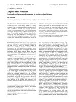
Staniforth et al. [54] discuss ways in which the domain-
swapped dimer of cystatin could propagate into a fibrillar
structure. It is assumed that open ends on the N- and
C-termini would allow further interactions. The electronic
density of a Ôgeneric fibrilÕ could be fitted by two rows of
dimers, each row extending in both directions indefinitely
(Fig. 2). Janowski et al. who determined the crystal struc-
ture of human cystatin C domain-swapped dimer [52],
believe that the dimers most probably represent a Ôdead endÕ
to further amyloidogenesis or, at least, hinder the process.
If domain-swapped dimers were rate-limiting for fibril
formation, a high energetic barrier would be expected; this
could be deduced from the influence of temperature on
the process. In the case of plant monnelin, a structurally
analogous protein to cystatins, the authors [85] did not
look for existence of the dimer. It has been found that
heating was needed for the prenucleus stage of fibril
growth and that maturation of the nucleus proceeded at
lower temperature. A similar observation has been made
with human stefin A (cystatin A), which demonstrates a
high activation energy of 99 kcalÆmol
)1
for domain
swapping [82] and forms dimers when heated to 85 °C
for 1 h. A preheated sample can make fibrils at ambient
temperature if the structure is additionally destabilized by
lowering pH to 2.4 (E. Z
ˇ
erovnik, unpublished observa-
tion). More importantly, the disease-causing variant of
human cystatin C (L68Q) forms dimers under physiolo-
gical conditions [36].
It has been suggested by Bergdoll et al.[86]and
confirmed by Itzhaki and coauthors [87] that a proline in
the linker region might facilitate domain swap. It could
rigidify the hinge region and keep it extended [83]. Parallel
reactions in folding have largely been attributed to the
difference in peptide bond configuration at some critical
proline [88] in the denatured state ensemble. This option,
too, should be considered in searching for an explanation
for slow formation of domain-swapped dimers and fibrils.
The energy of activation determined for the lag and growth
phases in a-synuclein fibrillization [39] was 20 kcalÆmol
)1
,
which would be consistent with a proline isomerization
reaction. Of course, there may be other slow events with
high activation energy. It has been found that a slow rate of
unfolding (a high E barrier) prevents amyloid fibril forma-
tion [89] and that fast unfolding leads to increased rate of
fibrillization.
CONNECTION OF PROTEIN FIBRIL
FORMATION TO PATHOPHYSIOLOGY
AND DISEASE
So far, about 20 human proteins have been found in
proteinaceous deposits in various conformational diseases.
These do not demonstrate any sequence or structural
homology. The common event is thought to be a
conformational change, leading to lack of biological
function or gain of toxic activity, and possibly, formation
of amyloid fibrils.
It is a matter of debate as to whether the fibrillar
aggregates and amyloid plaques are the side-product of
some other pathology or whether they are the main cause of
the disease. Co-localization of protein aggregates with
degenerating tissue and association of their presence with
disease symptoms are a strong indication of the involvement
of amyloid deposition in the pathogenesis of conformation-
al diseases [20]. In familial cases of some neurodegenerative
diseases (Table 1), evidence has been obtained for a direct
link between the ability of mutated protein to form fibrils
and the appearance of signs of the pathology [2,90]. Studies
with transgenic animals have also confirmed the contribu-
tion of the mutation in the amyloidogenic protein and
disease pathogenesis [20,91,92].
Whether the fibrils or the prefibrillar aggregates are the
dangerous species for the cell metabolism is still disputed. In
animal studies it has been shown that significant tissue
damage and clinical symptoms appear before any protein
aggregates are detected, implicating an intermediate on the
amyloidogenic pathway, which could be the real cause of
the pathogenesis [3,4,6,7]. It was proposed that protein
aggregation into fibrils could even represent a protective
event that depletes the cell of the toxic prefibrillar species [3].
Careful usage of fibril inhibitors is indicated as they may
cause accumulation of the toxic precursor [68].
Evidence has been obtained in studies on Alzheimer’s
disease that fibrils are not the most neurotoxic form of Ab
[6]. The peptide also assembles into soluble proto-fibrils and
smaller oligomers. The proto-fibril of Ab was shown by
AFM to be a slightly curved, of 4–11 nm diameter and
< 200 nm long [56]. Isolated protofibrils were found to be
toxic, causing oxidative stress and, eventually, neural death
[72,93]. The smaller oligomers can interfere with signal
transduction, possibly binding a tyrosine kinase important
for memory formation (long-term synaptic potentiation)
and sau phosphorylation [6].
3366 E. Z
ˇ
erovnik (Eur. J. Biochem. 269) Ó FEBS 2002
In prion diseases [20,24,94], no abundant amyloid
deposition was found in the brain, even though PrP
Sc
(the disease-related conformer of the protein) has a strong
tendency to aggregate in vitro. An interesting observation
was made that PrP
C
(the normal, cellular protein) binds
to survival factors and that the PrP
C
to PrP
Sc
transition
might result in apoptotic cell death. In Huntington’s
disease, activation of microglia following disruption of
neuronal architecture may be the death trigger rather
than the apoptotic pathway [91]. This is consistent with
findings in a transgenic mouse model of Huntington’s
disease, where cell death was neither apoptotic nor
necrotic [92].
MEANS OF NATURAL DEFENCE AND
REGULATION
Cellular defence against unfolded and aberrantly folded
proteins consists of several protective systems that prevent
aggregation, refold unfolded proteins or, degrade them. If
the rate of damage to cellular proteins is increased, for
example on exposure to increased temperature, oxygen free
radicals or other stress conditions, or when mutations occur,
this can disturb normal cellular functions and trigger
apoptosis [95]. In such harsh conditions, cells respond by
the induction of heat shock proteins (Hsp) that comprise
chaperones, antioxidant enzymes and ubiquitin–protea-
some components. The largest group of heat shock proteins
act as chaperones that bind to denatured or partially folded
proteins [96–98]. Certain combinations of chaperones, in
particular Hsp70, Hsp104 and Hsp40, can serve to dis-
assemble intracellular protein aggregates [99]. Especially,
Hsp104 was found to be of importance for disassembly–
disaggregation [97,100].
The ubiquitin- and proteasome-mediated degradation of
proteins plays an important role in cellular quality control
by removing mutated, misfolded and post-translationally
damaged proteins [20,100]. In many cellular inclusions
ubiquinated proteins are found together with proteasome
components [100]. If the cell is still overburdened by
aggregated proteins, apoptosis programs are switched on.
A novel finding is that heat shock proteins have a dual
function. As well as a role in refolding aberrantly folded
proteins and keeping them from aggregation, a second
function involves regulation of apoptosis [95,101]. Among
the heat shock proteins are anti-apoptotic and pro-
apoptotic proteins [101]. The recently discovered BAG
family of proteins operate as molecules that recruit
chaperones to target proteins. Such diverse proteins as
Bcl2, Raf1, various receptor, transcription factor mole-
cules and Hsp70 compete for binding to members of the
BAG-family of proteins [102]. This binding induces
changes in protein conformation that may have a
profound effect on protein function. Unfortunately,
studying the conformational changes in proteins in vivo
remainsratherelusive.
NOVEL THERAPEUTIC APPROACHES
Novel therapeutic approaches are being directed towards
achieving one of the following goals: either to inhibit and/or
reverse the conformational change, or to dissolve the
smaller aggregates and disassemble the amyloid fibrils.
Several successful attempts have been cited in the literature
including the use of monoclonal antibodies that bind to the
active conformation of the protein and thus inhibit
conformational changes. In Alzheimer’s disease, vacci-
nation is on the horizon, in this case targeting the smaller
oligomers and prefibrillar aggregates [103]. Soto and
coworkers have designed the so called Ômini-chaperonesÕ,
also termed Ôb sheet breakersÕ [20,24], which are peptides
that bind to the sequence of the protein region responsible
for self association. In the prion disease, similarly to
Alzheimer’s, trials are underway using monoclonal anti-
bodies that prevent conformational change [104]. Some
drugs already in use for other purposes have been screened
and several were found that both retard or reverse neuro-
degeneration if used for early intervention and also improve
the disease state in quite desperate cases, as reported by the
Prusiner’s group [105]. One of these drugs, quinacrine, is an
anti-malarial agent and the other, chlorpromazine, is used
to treat schizophrenia. Other blockers of amyloid fibril
formation have been found, ranging from Congo Red
derivatives, anti-cancer and antibiotic drugs to nicotine and
melatonin [76].
CONCLUSIONS
Understanding amyloid-fibril formation may contribute to
resolving some of the today’s most devastating diseases and,
at the very least, increase our general knowledge about
protein structure, folding and stability. Many properties of
amyloid fibrils have emerged: a common structure for
filaments and fibrils [58], nucleation dependent kinetics [67],
the role of oligomeric intermediates [68,72] and the existence
of at least two protein conformations separated by a high
energetic barrier, which behave as two macroscopic states
[64,81]. The following are some of the challenges still facing
us:
(a) Can domain-swapping be a mechanism for fibrilliza-
tion of globular proteins?
(b) What is the role of a helical parts of proteins? Do they
remain helical in the fibrils? (Periodicity characteristic for
a helices has been observed in an X-ray diffraction study on
the apolipoprotein A1 variant [106]).
(c) What is the role of a helical intermediates observed in
folding [107] and fibrillization studies [40] where temporarily
non-native a helices appear?
ACKNOWLEDGEMENTS
For financial support the author thanks the Ministry of Education,
Science and Sport of the Republic of Slovenia. Professor R. H. Pain
(JSI, Ljubljana, Slovenia) is indebted for reading the manuscript, giving
useful comments and editing English. I also thank T. Zavas
ˇ
nik-Bergant
(JSI, Ljubljana, Slovenia) and K. Goldie (EMBL, Heidelberg,
Germany) for taking the TEM picture reproduced in Fig. 1. I am
thankful to M. Ravnikar and M. Pompe-Novak (both National
Institute of Biology, Ljubljana) and I. Mus
ˇ
evic and M. S
ˇ
karabot
(Department of Physics, JSI, Ljubljana) for continuous TEM and
AFM work on human stefins. My gratitude goes to Professor V. Turk
and his team: L. Kroon-Z
ˇ
itko and M. Kenig (at JSI, Ljubljana), for
preparing the recombinant stefins. The author additionally thanks J. P.
Waltho for the model of cystatin A–stefin A dimer reproduced in
Fig. 2B, and to R. A Staniforth (Krebs Institute, University of
Sheffield, UK) for reading the manuscript and giving useful sugges-
tions.
Ó FEBS 2002 Amyloid-fibrils and conformational disease (Eur. J. Biochem. 269) 3367
REFERENCES
1.Raso,S.W.&King,J.K.(2000)Proteinfoldingandhuman
disease. In Mechanisms of Protein Folding (Pain R.H., ed.)
Frontiers in Molecular Biology Series, pp. 406–428. Oxford
University Press, Oxford.
2. Goedert, M., Spillantini, M.G. & Davies, S.W. (1998) Fila-
mentous nerve cell inclusions in neurodegenerative diseases. Curr.
Opin. Neurobiol. 8, 619–632.
3. Roher, A.E., Baudry, J., Chaney, M.O., Kuo, Y.M., Stine, W.B.
& Emmerling, M.R. (2000) Oligomerizaiton and fibril asssembly
of the amyloid-b protein. Biochim. Biophys. Acta 502, 31–43.
4. Selkoe, D.J. (1994) Normal and abnormal biology of the
b-amyloid precursor protein. Annu. Rev. Neurosci. 17, 489–517.
5. Wanker, E.E. (2000) Protein aggregation in Huntington’s and
Parkinson’s disease: implications for therapy. Mol. Med. Today 6,
387–391. Review.
6. Klein, W.L., Krafft, G.A. & Finch, C.E. (2001) Targeting small
Ab oligomers: the solution to an Alzheimer’s disease conundrum?
Trends Neurosci. 24, 219–224. Review.
7. Selkoe, D.J. (2001) Presenilin, Notch, and the genesis and treat-
ment of Alzheimer’s disease. Proc.NatlAcad.Sci.USA98,
11039–11041.
8. Smith, M.A., Rottkamp, C.A., Nunomura, A., Raina, A.K. &
Perry, G. (2000) Oxidative stress in Alzheimer’s disease. Biochim.
Biophys. Acta 1502, 139–144.
9. Friedhoff, P., von Bergen, M. & Mandelkow, E.M. (2000)
Structure of sau protein and assembly into paired helical fila-
mements. Biochim. Biophys. Acta 1502, 122–132.
10. Ray, I., Chauhan, A., Wegiel, J. & Chauhan, V.P. (2000) Gelsolin
inhibits the fibrillization of amyloid b-protein, and also defibril-
lizes its preformed fibrils. Brain Res. 853, 344–351.
11. Iwata, N., Tsubuki, S., Takaki, Y., Shirotani, K., Lu, B., Gerard,
N.P.,Gerard,C.,Hama,E.,Lee,H.J.&Saido,T.C.(2001)
Metabolic regulation of brain Ab by neprilysin. Science 292,
1550–1552.
12. Ciechanover, A. (2001) Linking ubiquitin, parkin and synphilin-
1. Nat. Med. 7, 1108–1109.
13. Murray, I.V.J., Lee, V.M Y. & Trojanowski, J.Q. (2001) Synu-
cleinopathies: a pathological and molecular review. Clin.
Neurosci. Res. 1, 455–455.
14. Okochi, M., Walter, J., Koyama, A., Nakajo, S., Baba, M.,
Iwatsubo, T., Meijer, L., Kahle, P.J. & Haass, C. (2000) Con-
stitutive phosphorylation of the Parkinson’s disease associated
a-synuclein. J. Biol. Chem. 275, 390–397.
15. Ellis, C.E., Schwartzberg, P.L., Grider, T.L., Fink, D.W. &
Nussbaum, R.L. (2001) a-Synuclein is phosphorylated by mem-
bers of the Src family of protein-tyrosine kinases. J. Biol. Chem.
276, 3879–3884.
16. Nakamura, T., Yamashita, H., Takahashi, T. & Nakamura, S.
(2001) Activated Fyn phosphorylates a-synuclein at tyrosine
residue 125. Biochem. Biophys. Res. Commun. 280, 1085–1092.
17. Chung, K.K., Zhang, Y., Lim, K.L., Tanaka, Y., Huang, H.,
Gao, J., Ross, C.A., Dawson, V.L. & Dawson, T.M. (2001)
Parkin ubiquitinates the a-synuclein-interacting protein, synphi-
lin-1: implications for Lewy-body formation in Parkinson
disease. Nat. Med. 7, 1144–1150.
18. Spillantini, M.G., Schmidt, M.L., Lee, V.M., Trojanowski, J.Q.,
Jakes, R. & Goedert, M. (1997) a-Synuclein in Lewy bodies.
Nature 388, 839–840.
19. Shimura, H., Hattori, N., Kubo, S., Mizuno, Y., Asakawa, S.,
Minoshima, S., Shimizu, N., Iwai, K., Chiba, T., Tanaka, K. &
Suzuki, T. (2000) Familial Parkinson disease gene product,
parkin, is a ubiquitin-protein ligase. Nat. Genet. 25, 302–305.
20. Soto, C. (2001) Protein misfolding and disease; protein refolding
and therapy. FEBS Lett. 498, 204–207.
21. Cohen, F.E. (2000) Prions, peptides and protein misfolding. Mol.
Med. Today 6, 292–293.
22. Cohen, A.S. (1986) General introduction and brief history of the
amyloid fibril. In Amyloidosis (Marrink, J. & Van Rijswijk, M.H.,
eds), pp. 3–19. Nijhoff, Dordrecht.
23. Sipe, J.D. & Cohen, A.S. (2000) History of the amyloid fibril.
J. Struct. Biol. 130, 88–98.
24. Soto, C. & Saborio, G.P. (2001) Prions: disease propagation and
disease therapy by conformational transmission. Trends Mol.
Med. 7, 109–114.
25. Damaschun, G., Damaschun, H., Fabian, H., Gast, K., Krober,
R., Wieske, M. & Zirwer, D. (1999) Conversion of yeast
phosphoglycerate kinase into amyloid-like structure. Proteins 39,
204–211.
Fig. 2. Scheme representing amyloid fibril formation from ‘cystatin’
domain-swapped dimers. (A) Electron density obtained by cryo-EM,
looking inside a fibril from SH3 domain, reproduced from [57], with
permission. (B) Three-dimensional model of a domain-swapped dimer,
representative of the cystatin superfamily. (C) One possible mode of
how the domain-swapped dimers could build up the fibril. The scheme
is adapted from [54], with permission.
3368 E. Z
ˇ
erovnik (Eur. J. Biochem. 269) Ó FEBS 2002
26. Litvinovich, S.V., Brew, S.A., Aota, S., Akiyama, S.K., Hau-
denschild, C. & Ingham, K.C. (1998) Formation of amyloid-like
fibrils by self association of a partially unfolded fibronectin type
III module. J. Mol. Biol. 280, 245–258.
27. Guijarro, J.I., Sunde, M., Jones, J.A., Campbell, I.D. & Dobson,
C.M.(1998)AmyloidfibrilformationbyanSH3domain.Proc.
Natl Acad. Sci. USA 95, 4224–4228.
28. Konno, T., Murata, K. & Nagayama, K. (1999) Amyloid-like
aggregates of a plant protein: a case of a sweet-tasting protein,
monellin. FEBS Lett. 454, 122–126.
29. Chiti, F., Webster, P., Taddei, N., Clark, A., Stefani, M., Ram-
poni, G. & Dobson, C.M. (1999) Designing conditions for in
vitro formation of amyloid protofilaments and fibrils. Proc. Natl
Acad.Sci.USA96, 3590–3594.
30. Liu, Y., Gotte, G., Libonati, M. & Eisenberg, D. (2001) A
domain-swapped RNase A dimer with implications for amyloid
formation. Nat. Struct. Biol. 8, 211–214.
31. Z
ˇ
erovnik, E., Pompe-Novak, M., S
ˇ
karabot, M., Ravnikar, M.,
Mus
ˇ
evic,I.&Turk,V.(2002)HumanstefinBreadilyforms
amyloid fibrils in vitro. Biochim. Biophys. Acta 1594,1–5.
32. Fa
¨
ndrich, M., Fletcher, M.A. & Dobson, C.M. (2001) Amyloid
fibrils from muscle myoglobin. Nature 410, 165–166.
33. Pertinhez, T.A., Bouchard, M., Tomlinson, E.J., Wain, R.,
Ferguson, S.J., Dobson, C.M. & Smith, L.J. (2001) Amyloid fibril
formation by a helical cytochrome. FEBS Lett. 495, 184–186.
34. Dobson, C.M. (1999) Protein misfolding, evolution and disease.
Trends Biochem. Sci. 24, 329–332.
35. Rochet, J.C. & Lansbury, P.T. Jr (2000) Amyloid fibrillogenesis:
themes and variations. Curr. Opin. Struct. Biol. 10, 60–68.
36. Ekiel, I. & Abrahamson, M. (1996) Folding-related dimerization
of human cystatin C. J. Biol. Chem. 271, 1314–1321.
37. Lai, Z., Colon, W. & Kelly, J.W. (1996) The acid-mediated
denaturation pathway of transthyretin yields a conformational
intermediate that can self-assemble into amyloid. Biochemistry
35, 6470–6482.
38. Uversky, V.N., Gillespie, J.R. & Fink, A.L. (2000) Why are
Ônatively unfoldedÕ proteins unstructured under physiologic
conditions? Proteins 41, 415–427.
39. Uversky, V.N., Li, J. & Fink, A.L. (2001) Evidence for a partially
folded intermediate in a-synuclein fibril formation. J. Biol. Chem.
276, 10737–10744.
40. Kirkitadze, M.D., Condron, M.M. & Teplow, D.B. (2001)
Identification and characterization of key kinetic intermediates in
amyloid b-protein fibrillogenesis. J. Mol. Biol. 312, 1103–1119.
41. Bouchard, M., Zurdo, J., Nettleton, E.J., Dobson, C.M. &
Robinson, C.V. (2000) Formation of insulin amyloid fibrils
followed by FTIR simultaneously with CD and electron micro-
scopy. Protein Sci. 9, 1960–1967.
42. Fezoui, Y., Hartley, D.M., Walsh, D.M., Selkoe, D.J., Osterhout,
J.J. & Teplow, D.B. (2000) A de novo designed helix-turn-helix
peptide forms nontoxic amyloid. Nat. Struct. Biol. 7, 1095–1099.
43. Hamada, D. & Goto, Y. (1997) The equilibrium intermediate of
b-lactoglobulin with non-native a-helical structure. J. Mol. Biol.
269, 479–487.
44. Hamada, D., Segawa, S. & Goto, Y. (1996) Non-native a-helical
intermediate in the refolding of b-lactoglobulin, a predominantly
b-sheet protein. Nat. Struct. Biol. 3, 868–873.
45. Lu, H., Buck, M., Radford, S.E. & Dobson, C.M. (1997)
Acceleration of the folding of hen lysozyme by trifluoroethanol.
J. Mol. Biol. 265, 112–117.
46. Z
ˇ
erovnik, E., Virden, R., Jerala, R., Kroon-Z
ˇ
itko, L., Turk, V. &
Waltho, J.P. (1999) Differences in the effects of TFE on the
folding pathways of human stefins A and B. Proteins 36, 205–216.
47. Buck, M. (1998) Trifluoroethanol and colleagues: cosolvents
come of age. Recent studies with peptides and proteins. Q. Rev.
Biophys. 31, 297–355.
48. Chiti, F., Taddei, N., Webster, P., Hamada, D., Fiaschi, T.,
Ramponi, G. & Dobson, C.M. (1999) Acceleration of the folding
of acylphosphatase by stabilisation of local secondary structure.
Nat. Struct. Biol. 6, 380–387.
49. Liu, K., Cho, H.S., Lashuel, H.A., Kelly, J.W. & Wemmer, D.E.
(2000) A glimpse of a possible amyloidogenic intermediate of
transthyretin. Nat. Struct. Biol. 7, 754–757.
50. Hosszu, L.L., Baxter, N.J., Jackson, G.S., Power, A., Clarke,
A.R., Waltho, J.P., Craven, C.J. & Collinge, J. (1999) Structural
mobility of the human prion protein probed by backbone
hydrogen exchange. Nat. Struct. Biol. 6, 740–743.
51. Nettleton, E.J., Tito, P., Sunde, M., Bouchard, M., Dobson,
C.M. & Robinson, C.V. (2000) Characterization of the oligo-
meric states of insulin in self-assembly and amyloid fibril
formation by mass spectrometry. Biophys. J. 79, 1053–1065.
52. Janowski, R., Kozak, M., Jankowska, E., Grzonka, Z., Grubb,
A., Abrahamson, M. & Jaskolski, M. (2001) Human cystatin C,
an amyloidogenic protein, dimerizes through three-dimensional
domain swapping. Nat. Struct. Biol. 8, 316–320.
53. Knaus, K.J., Morillas, M., Swietnicki, W., Malone, M., Surewicz,
W.K. & Yee, V.C. (2001) Crystal structure of the human prion
protein reveals a mechanism for oligomerization. Nat. Struct.
Biol. 8, 770–774.
54. Staniforth, R.A., Giannini, S., Higgins, L.D., Conroy, M.J.,
Hounslow, A.M., Jerala, R., Craven, C.J. & Waltho, J.P. (2001)
Three-dimensional domain swapping in the folded and molten-
globule states of cystatins, an amyloid-forming structural super-
family. EMBO J. 20, 4774–4781.
55. Goldsbury, C.S., Cooper, G.J., Goldie, K.N., Muller, S.A., Saafi,
E.L., Gruijters, W.T., Misur, M.P., Engel, A., Aebi, U. & Kistler,
J. (1997) Polymorphic fibrillar assembly of human amylin.
J. Struct. Biol. 119, 17–27.
56. Ding, T.T. & Harper, J.D. (1999) Analysis of amyloid-b assem-
blies using tapping mode atomic force microscopy under ambient
conditions. Methods Enzymol. 309, 510–525.
57. Jimenez, J.L., Guijarro, J.I., Orlova, E., Zurdo, J., Dobson,
C.M., Sunde, M. & Saibil, H.R. (1999) Cryo-electron microscopy
structure of an SH3 amyloid fibril and model of the molecular
packing. EMBO J. 18, 815–821.
58. Serpell, L.C. (2000) Alzheimer’s amyloid fibrils: structure and
assembly. Biochim. Biophys. Acta 1502, 16–30.
59. Tycko, R. (2001) Solid-state nuclear magnetic resonance tech-
niques for structural studies of amyloid fibrils. Methods Enzymol.
339, 390–413.
60. Kelly, J.W. (2000) Mechanisms of amyloidogenesis. Nat. Struct.
Biol. 7, 824–826.
61. Lansbury, P.T. Jr (1999) Evolution of amyloid: what normal
protein folding may tell us about fibrillogenesis and disease. Proc.
Natl Acad. Sci. USA 96, 3342–3344.
62. Kad, N.M. & Radford, S.E. (2001) Partial unfolding as a pre-
cursor to amyloidosis: a discussion of the occurrence, role, and
implications. In Molecular Chaperones in the Cell (Lund, P., ed.),
Frontiers in Molecular Biology Series, pp. 257–278. Oxford
University Press, Oxford.
63. Carrel, R.W. & Gooptu, B. (1998) Conformational changes and
disease – serpins, prions and Alzheimer’s. Curr. Opin. Struct. Biol.
8, 799–809.
64. Ferreira, S.T. & De Felice, F.G. (2001) PABMB Lecture. Protein
dynamics, folding and misfolding: from basic physical chemistry
to human conformational diseases. FEBS Lett. 498, 129–134.
65. Damaschun, G., Damaschun, H., Gast, K. & Zirwer, D. (1999)
Proteins can adopt totally different folded conformations. J. Mol.
Biol. 291, 715–725.
66. Kelly, J.W. (1998) The alternative conformations of amyloido-
genic proteins and their multi-step assembly pathways. Curr.
Opin. Struct. Biol. 8, 101–106.
Ó FEBS 2002 Amyloid-fibrils and conformational disease (Eur. J. Biochem. 269) 3369
67. Lomakin, A., Chung, D.S., Benedek, G.B., Kirschner, D.A. &
Teplow, D.B. (1996) On the nucleation and growth of amyloid
b-protein fibrils. detection of nuclei and quantitation of rate
constants. Proc. Natl Acad. Sci. USA 93, 1125–1129.
68. Harper, J.D., Wong, S.S., Lieber, C.M. & Lansbury, P.T. Jr
(1999) Assembly of A b amyloid protofibrils: an in vitro model for
a possible early event in Alzheimer’s disease. Biochemistry 38,
8972–8980.
69. Aggeli, A., Nyrkova, I.A., Bell, M., Harding, R., Carrick, L.,
McLeish, T.C., Semenov, A.N. & Boden, N. (2001) Hierarchical
self-assembly of chiral rod-like molecules as a model for peptide
b-sheet tapes, ribbons, fibrils, and fibers. Proc. Natl Acad. Sci.
USA 98, 11857–11862.
70. Goldsbury, C., Kistler, J., Aebi, U., Arvinte, T. & Cooper, G.J.
(1999) Watching amyloid fibrils grow by time-lapse atomic force
microscopy. J. Mol. Biol. 285, 33–39.
71. Blackley, H.K., Sanders, G.H., Davies, M.C., Roberts, C.J.,
Tendler, S.J. & Wilkinson, M.J. (2000) In-situ atomic force
microscopy study of b-amyloid fibrillization. J. Mol. Biol. 298,
833–840.
72. Walsh, D.M., Hartley, D.M., Kusumoto, Y., Fezoui, Y., Con-
dron, M.M., Lomakin, A., Benedek, G.B., Selkoe, D.J. &
Teplow, D.B. (1999) Amyloid b-protein fibrillogenesis. Structure
and biological activity of protofibrillar intermediates. J. Biol.
Chem. 274, 25945–25952.
73. Klunk, W.E., Jacob, R.F. & Mason, R.P. (1999) Quantifying
amyloid by congo red spectral shift assay. Methods Enzymol. 309,
285–305.
74. Le Vine, H. (1999) Quantification of b sheet amyloid fibril
structures with thioflavin T. Methods Enzymol. 309, 274–284.
75. Khurana, R., Uversky, V.N., Nielsen, L. & Fink, A.L. (2001) Is
Congo red an amyloid-specific dye? J. Biol. Chem. 276, 22715–
22721.
76. Findeis, M.A. (2000) Approaches to discovery and characteri-
zation of inhibitors of amyloid b-peptide polymerization.
Biochim. Biophys. Acta 1502, 76–84. Review.
77. Klunk, W.E., Wang, Y., Huang, G.F., Debnath, M.L., Holt,
D.P. & Mathis, C.A. (2001) Uncharged thioflavin-T derivatives
bind to amyloid-b protein with high affinity and readily enter the
brain. Life Sci. 69, 1471–1484.
78. Massi, F. & Straub, J.E. (2001) Energy landscape theory for
Alzheimer’s amyloid-peptide fibril elongation. Proteins 42,
217–229.
79. Serio, T.R. & Lindquist, S.L. (2000) Nucleated conformational
conversion and the replication of conformational information by
a prion determinant. Science 289, 1317–1321.
80. Pallitto, M.M. & Murphy, R.M. (2001) A mathematical model of
the kinetics of b-amyloid fibril growth from the denatured state.
Biophys. J. 81, 1805–1822.
81. Schlunegger, M.P., Bennett, M.J. & Eisenberg, D. (1997) Oligo-
mer formation by 3D domain swapping: a model for protein
assembly and misassembly. Adv. Protein Chem. 50, 61–122.
82. Jerala, R. & Z
ˇ
erovnik, E. (1999) Accessing the global minimum
conformation of stefin A dimer by annealing under partially
denaturing conditions. J. Mol. Biol. 291, 1079–1089.
83. Newcomer, M.E. (2002) Protein folding and three-dimensional
domain swapping: a strained relationship? Curr. Opin. Struct.
Biol. 12, 48–53.
84. Ogihara, N.L, Ghirlanda, G., Bryson, J.W., Gingery, M., DeG-
rado, W.F. & Eisenberg, D. (2001) Design of three-dimensional
domain-swapped dimers and fibrous oligomers. Proc. Natl Acad.
Sci. USA 98, 1404–1409.
85. Konno, T. (2001) Multistep nucleus formation and a separate
subunit contribution of the amyloidgenesis of heat-denatured
monellin. Protein Sci. 10, 2093–2101.
86. Bergdoll, M., Remy, M.H., Cagnon, C., Masson, J.M. & Dumas,
P. (1997) Proline-dependent oligomerization with arm exchange.
Structure 5, 391–401.
87. Rousseau, F., Schymkowitz, J.W., Wilkinson, H.R. & Itzhaki,
L.S. (2001) Three-dimensional domain swapping in p13suc1 oc-
curs in the unfolded state and is controlled by conserved proline
residues. Proc. Natl Acad. Sci. USA 98, 5596–5601.
88. Balbach, J. & Schmid, F.X. (2000) Proline isomerization and its
catalysis in protein folding. In Mechanisms of Protein Folding
(Pain, R.H. ed.) Frontiers in Molecular Biology Series,pp.
213–249. Oxford University Press, Oxford.
89. Plaza del Pino, I.M., Ibarra-Molero, B. & Sanchez-Ruiz, J.M.
(2000) Lower kinetic limit to protein thermal stability: a proposal
regarding protein stability in vivo and its relation with misfolding
diseases. Proteins 40, 58–70.
90.Conway,K.A.,Harper,J.D.&Lansbury,P.T.(1998)
Accelerated in vitro fibril formation by a mutant a-synuclein
linked to early-onset Parkinson disease. Nat. Med. 4, 1318–1320.
91. Scherzinger, E., Lurz, R., Turmaine, M., Mangiarini, L.,
Hollenbach, B., Hasenbank, R., Bates, G.P., Davies, S.W.,
Lehrach, H. & Wanker, E.E. (1997) Huntingtin-encoded poly-
glutamine expansions form amyloid-like protein aggregates
in vitro and in vivo. Cell 90, 549–558.
92. Turmaine, M., Raza, A., Mahal, A., Mangiarini, L., Bates, G.P.
& Davies, S.W. (2000) Nonapoptotic neurodegeneration in a
transgenic mouse model of Huntington’s disease. Proc. Natl
Acad. Sci. USA 97, 8093–8097.
93. Hartley, D.M., Walsh, D.M., Ye, C.P., Diehl, T., Vasquez, S.,
Vassilev, P.M., Teplow, D.B. & Selkoe, D.J. (1999) Protofibrillar
intermediates of amyloid b-protein induce acute electrophysio-
logical changes and progressive neurotoxicity in cortical neurons.
J. Neurosci. 19, 8876–8884.
94. Nicotera, P.A. (2001) Route for prion neuroinvasion. Neuron 31,
345–348.
95. Sherman, M.Y. & Goldberg, A.L. (2000) Cellular defenses
against unfolded proteins: a cell biologist thinks about neurode-
generative diseases. Neuron 29, 15–32.
96. Badcoe, I.G., Smith, C.J., Wood, S., Halsall, D.J., Holbrook, J.J.,
Lund, P. & Clarke, A.R. (1991) Binding of a chaperonin to the
folding intermediates of lactate dehydrogenase. Biochemistry 30,
9195–9200.
97. Leroux, M.R. & Hartl, F.U. (2000) Cellular functions of mole-
cular chaperones. In Mechanisms of Protein Folding (Pain R.H.,
ed.) Frontiers in Molecular Biology Series, pp. 364–405. Oxford
University Press, Oxford.
98. Mogk, A., Bukau, B. & Deuerling, E. (2001) Cellular functions of
cytosolic E.coli chaperones. In Molecular Chaperones in the Cell
(Lund, P., ed.) Frontiers in Molecular Biology Series, pp. 1–34.
Oxford University Press, Oxford.
99. Glover, J.R. & Lindquist, S. (1998) Hsp104, Hsp70, and Hsp40: a
novel chaperone system that rescues previously aggregated
proteins. Cell 94, 73–82.
100. Alves-Rodrigues, A., Gregori, L. & Figueiredo-Pereira, M.E.
(1998) Ubiquitin, cellular inclusions and their role in neurode-
generation. Trends Neurosci. 21, 516–552.
101. Garrido, C., Gurbuxani, S., Ravagnan, L. & Kroemer, G. (2001)
Heat shock proteins: endogenous modulators of apoptotic cell
death. Biochem. Biophys. Res. Commun. 286, 433–442.
102. Takayama, S. & Reed, J.C. (2001) Molecular chaperone targeting
and regulation by BAG family proteins. Nat. Cell. Biol. 3, E237–
E241.
103. Ingram, D.K. (2001) Vaccine development for Alzheimer’s
disease:ashotofgoodnews.Trends Neurosci. 24, 305–307.
104. Jones, R. (2001) Blocking prion conversion. Highlights. Nat. Rev.
Neurosci. 2, 605.
3370 E. Z
ˇ
erovnik (Eur. J. Biochem. 269) Ó FEBS 2002
105. Korth, C., May, B.C., Cohen, F.E. & Prusiner, S.B. (2001)
Acridine and phenothiazine derivatives as pharmacotherapeutics
for prion disease. Proc. Natl Acad. Sci. USA 98, 9836–9841.
106. Mangione, P., Sunde, M., Giorgetti, S., Stoppini, M., Esposito,
G., Gianelli, L., Obici, L., Asti, L., Andreola, A., Viglino, P.,
Merlini, G. & Bellotti, V. (2001) Amyloid fibrils derived from the
apolipoprotein A1 Leu174Ser variant contain elements of
ordered helical structure. Protein Sci. 10, 187–199.
107. Hamada, D., Chiti, F., Guijarro, J.I., Kataoka, M., Taddei, N. &
Dobson, C.M. (2000) Evidence concerning rate-limiting steps in
protein folding from the effects of trifluoroethanol. Nat. Struct.
Biol. 7, 58–61.
Ó FEBS 2002 Amyloid-fibrils and conformational disease (Eur. J. Biochem. 269) 3371