Báo cáo Y học: Genomic organization of MUC4 mucin gene Towards the characterization of splice variants potx
Bạn đang xem bản rút gọn của tài liệu. Xem và tải ngay bản đầy đủ của tài liệu tại đây (734.41 KB, 8 trang )
Genomic organization of
MUC4
mucin gene
Towards the characterization of splice variants
Fabienne Escande
1,2,3
, Laurent Lemaitre
1
, Nicolas Moniaux
4
, Surinder K. Batra
4
, Jean-Pierre Aubert
1,2
and Marie-Pierre Buisine
1,2,3
1
INSERM Unite
´
560, Lille, France;
2
Laboratoire de Biochimie et Biologie Mole
´
culaire, Ho
ˆ
pital C. Huriez,
Centre Hospitalier Re
´
gional et Universitaire, Lille, France;
3
Faculte
´
de Me
´
decine Henri Warembourg, Lille, France;
4
Departement of Biochemistry and Molecular Biology, the Eppley Institute for Research in Cancer and Allied Diseases,
University of Nebraska Medical Center, Omaha, USA
The human MUC4 gene encodes a large membrane-associ-
ated mucin, characterized by a mucin tandem repeat domain
and a growth factor-like transmembrane domain. In addi-
tion to the originally published sequence (sv0-MUC4),
several MUC4 cDNA sequences (called sv1-MUC4 to sv21-
MUC4, MUC4/X, MUC4/Y) from various tissues and cell
lines have been recently described. They differ from sv0-
MUC4 by deletions and/or insertions located in the 3¢ region
or, for two of them, by deletion of the central repetitive
domain. To establish the nature of the mechanisms
responsible for the diversity of MUC4 transcripts, the
genomic structure of the 3¢ region of the human MUC4 gene
was determined. Our results show that it spans approxi-
mately 30.8 kb of genomic DNA and is composed of 24
exons, including one alternative exon which was exclusively
reported for sv1-MUC4. Moreover, we have shown that the
different MUC4 transcripts are generated by several mech-
anisms, including the alternative use of cassette exons, exon
skipping or use of cryptic splice donor/acceptor sites.
Keywords: MUC4; mucin; membrane-associated; alternative
splicing.
Human mucins constitute a complex family of membrane-
bound or secreted O-glycoproteins produced by epithelial
cells. They contain a high percentage of threonine and serine
residues carrying O-linked glycan chains and distributed in
tandemly repeated motifs in the central part of the protein
backbone. Mucins are known to play important roles in the
lubrication and protection of mucosae but more recently,
the involvement of mucins in the renewal and differentiation
of the epithelia, cell adhesion and cell signaling has also been
proposed [1–3].
To date, 14 human mucin genes have been identified:
MUC1–4, MUC5B, MUC5AC, MUC6–8, MUC11–13,
MUC16 and MUC17 [4–6]. MUC4 is a member of the
membrane-bound mucin family and is believed to be the
homologue of the rat sialomucin complex (SMC, rat Muc4)
because of their similar structural organization [7–10]. Rat
Muc4 is a well-characterized heterodimeric glycoprotein
complex in which the mucin subunit ascite sialo-glycoprotein
(ASGP)-1 is the major detectable glycoprotein. The other
subunit ASGP-2 is membrane-associated and contains
epidermal growth factor (EGF)-like domains that were
shown to act as a ligand for the tyrosine kinase p185
neu
[11].
The full cDNA of MUC4, also called sv0-MUC4,was
entirely characterized in our laboratory [10,12,13]. The
deduced amino-acid sequence of the N-terminal region
contains a peptide signal, followed by three imperfect
repetitions of a motif, varying from 126 to 130 residues,
and by a unique threonine- and serine-rich sequence. The
central region is composed of a large mucin-type domain
characterized by the perfect repetition of 16 amino-acid
residues. Like other mucins, this mucin-type domain exhibits
a variable number of tandem repeat polymorphisms with
variations ranging from 145 to 395 units. The C-terminal
region can be divided into 12 domains (CT1–12) with two
EGF-like domains, two cysteine-rich domains, a transmem-
brane domain and a short cytoplasmic tail [10,14]. In situ
hybridization studies have shown that MUC4 presents a very
large expression pattern. It is expressed in numerous normal
tissues such as trachea, lung, stomach, colon, uterus and
prostate [15,16], but it is not detected in the normal pancreas,
gall bladder, liver or biliary epithelial cells [17,18]. Interest-
ingly, the abnormal expression of MUC4 was demonstrated
to occur in several epithelial cancers such as lung,
pancreas and gall bladder carcinoma [17–20], as well as in
various cancer cell lines [21,22]. No precise functions were
attributed to MUC4 until now, but dysregulations of MUC4
expression in cancers, together with its homology to SMC,
suggest an important role for MUC4 in human tumor
biology.
Recently, we have isolated 23 distinct transcripts of
MUC4 that received the designation sv1- to sv21-MUC4,
MUC4/X,andMUC4/Y [14,22,23]. They were isolated by
RT-PCR, carried out on human testis and pancreatic
adenocarcinoma cells. They differ from sv0-MUC4 by
Correspondence to M P. Buisine, INSERM U-377,
Place de Verdun, 59045 Lille Cedex, France.
Fax: + 33 3 20 53 85 62; Tel.: + 33 3 20 29 88 59;
E-mail:
Abbreviations: ASGP, ascite sialo-glycoprotein; EGF, epidermal
growth factor; BAC, bacterial artificial chromosome.
Note: the nucleotide sequences reported here have been submitted to
EMBL Nucleotide Sequence Database under accession numbers
AJ430032, AJ430033, and AJ430034.
(Received 8 February 2002, revised 30 May 2002,
accepted 31 May 2002)
Eur. J. Biochem. 269, 3637–3644 (2002) Ó FEBS 2002 doi:10.1046/j.1432-1033.2002.03032.x
deletions and/or insertions located in the 3¢ region but also
for two of them by deletion of the central repetitive domain.
Until now, because of the lack of knowledge on the genomic
organization of the 3¢ region of MUC4, the precise mech-
anisms responsible for these events could not be defined.
In the present paper, we described the genomic structure
of the 3¢ region of the human MUC4 gene. A comparison of
the nucleotide genomic and cDNA sequences allowed us to
establish the nature of the mechanisms responsible for the
diversity of the MUC4 transcripts.
EXPERIMENTAL PROCEDURES
Oligonucleotide primers
Oligonucleotides used for PCR are shown in Table 1. They
were synthetized by Eurogentec (Lie
`
ge, Belgium) or by
MWG-Biotech (Ebersberg, Germany).
PCR amplification of human MUC4 introns
MUC4 introns were amplified from a bacterial artificial
chromosome (BAC) clone containing the human MUC4
gene [14]. Amplifications were performed in a PerkinElmer
Thermal Cycler 2400 (Applied Biosystems, Courtaboeuf,
France). PCR reactions were conducted in 50-lL reaction
volumes, containing 1 lg of BAC DNA, 5 lLof10·
buffer (100 m
M
Tris/HCl, 15 m
M
MgCl
2
,500m
M
KCl,
pH 8.3), 4 lLof10 m
M
deoxyribonucleoside triphosphates,
10 pmol of each primer and 2 U of Taq DNA polymerase
(Roche diagnostics, Meylan, France). The cycle parameters
were 94 °C for 4 min, followed by 30 cycles at 94 °Cfor
45 s, 58–60 °Cfor45s,and72°C for 2 min. The final
elongation step was extended for an additional 10 min at
72 °C. In some cases, Expand
TM
Long Template PCR
System (Roche diagnostics) was used. PCR reactions were
conducted in 50 lL reaction volumes containing 1 lgof
BAC DNA, 5 lLof10· Expand long template PCR
buffer 3, 4 lLof10m
M
deoxyribonucleoside triphos-
phates, 1.5 lLof2.25m
M
MgCl
2
,10pmolofeachprimer
and 2.5 U of DNA polymerase. The cycle parameters were
94 °C for 2 min, followed by 30 cycles at 94 °Cfor10s,
annealing at 60 °C for 45 s, and elongation at 71 °Cfor
2min,and68°C for 10 min. The last 20 cycles had their
elongation time extended by 10 s for each new cycle. The
final elongation step was extended for an additional 15 min
Table 1. Primers used for DNA amplification and sequencing.
a
Nucleotide position is defined according to the sequence of sv0-MUC4 (AJ010901).
b
S, sense; AS, antisense.
3638 F. Escande et al. (Eur. J. Biochem. 269) Ó FEBS 2002
at 71 °C. PCR products were analyzed by 1% agarose gel
electrophoresis and cloned directly into the pCR2.1 vector
with the original TA cloning
TM
kit (Invitrogen, Leek, the
Netherlands), according to the manufacturer’s instructions.
Plasmid DNA purification
Plasmid DNA was purified using the QIAprep Spin Plasmid
kit (Qiagen, Courtaboeuf, France).
DNA sequence analysis
Sequences were determined by automatic sequencing with a
DNA sequencer model 4000 L LI-COR and the Sequi-
Therm Excel
TM
II Long-read Premix DNA sequencing Kit-
LC (TEBU, Le Perray en Ivelynes, France), using standard
vector primers or with ABI PRISM model 377 XL
automatic sequencer with the ABI PRISM dRhodamine
terminator cycle ready reaction kit (Applied Biosystems)
using either universal primers or specific internal oligo-
nucleotides. An analysis of nucleic acid and deduced peptide
sequence data was performed using
PC
/
GENE
Software
(IntelliGenetics Inc.).
RESULTS
Genomic organization of 3¢ region of MUC4
In order to clarify the mechanisms responsible for the
diversity of MUC4 transcripts, the genomic counterpart of
MUC4 cDNA was identified by PCR experiments, using as
a template the BAC clone reported previously [14]. This
BAC was reported to contain the full human MUC4 gene.
Each genomic fragment was subcloned, sequenced, and the
exon–intron organization deduced. Oligonucleotide primers
were chosen according to the human sv0-MUC4 cDNA
sequence (AJ010901) or preliminary results obtained from
the genomic organization of the mouse Muc4 gene
(A. Laine & J. L. Desseyn, unpublished results). The
3¢ region of MUC4 spans approximately 30.8 kb and the
nucleotide sequence is available from GenBank under
accession numbers AJ430032, AJ430033, and AJ430034.
A comparison with cDNA sequences of MUC4 transcripts
allowed us to establish the complete exon–intron organiza-
tion of the MUC4 gene. Thus, 24 exons were identified in
the 3¢ region of MUC4, including one alternative exon,
which was exclusively reported for the sv1-variant (Fig. 1).
Exon and intron sizes, splice junction types and sequences of
the exon–intron boundaries are given in Table 2. The size of
the 24 introns ranged from 94 to 2.8 kb, and the size of the
24 exons ranged from 65 to 607 bp. All of the 5¢ donor and
3¢ acceptor sites were consistent with the consensus gt–ag
motifs described for splice sites in eukaryotic genes [24].
Unique tandemly repeated sequences, more and less perfect,
were found in some introns: approximately 90 copies of an
15-bp repeat (AGGTATGGGTGTGGA) in intron 3,
approximately 60 copies of an 26–31 bp repeat in intron 4,
23 copies of a 32-bp repeat (CAGGAGTACCCCA), four4
copies of a 34-pb repeat (AGGCCTCAACACCCCCC
AGCACCTTCCCCAGGCC) in intron 23. A search of the
GenBank database indicated that the consensus sequences
of these four repeat were not identical with any other
genomic sequence. Sequence type microsatellites were also
found in others introns: (GGT)
124
in intron 7 (T/CG)
22
in intron 16 (GATA)
73
in intron 18. Such repetitive
intronic sequences may participate with the repetitive
sequence in the central exon to interindividual polymorph-
ism and may be used as a potential intragene marker of the
locus 3q29.
Therefore, with the previously reported first two exons,
the MUC4 genomic organization is complete: MUC4 is
composed of at least 26 exons, one exon coding for the 5¢
UTR and peptide leader, one exon coding for the large
repetitive domain, and 24 for the 3¢ extremity.
Alternative splice events
The existence of cDNA variant species characterized by
deletions and/or insertions in the 3¢ region of MUC4 was
recently described in normal human testis and in pancreatic
adenocarcinoma cell lines (HPAF) [14,22,23]. Comparative
analysis of the nucleotide sequences of MUC4 transcript
variants with the nucleotide sequence of the MUC4 gene
allowed us to establish the nature of the mechanisms
responsible for the diversity of transcripts. They were
generated by the combination of either one or more events
resulting in several mechanisms of alternative splicing
(Table 3) [24]. Some events correspond to deletions which
result from the use of cryptic splice donor sites situated 5¢
from the normally used splice site (733–762del28), or from
the use of cryptic splice acceptor sites situated 3¢ from the
normally used splice site (309–386del76, 2218–2587del368),
or from the use of both cryptic splice donor sites situated 5¢
from the normally used splice site and acceptor sites situated
3¢ from the normally used splice site (966–1396del429, 1020–
1699del678). Some events (762–851del89, 2400–2561del160,
474–631del156, del. exon 2, del. exons 2, 3) correspond to
exon skipping by an alternative use of acceptor sites. Other
events correspond to insertions arising via the use of cryptic
splice donor sites situated 3¢ from the normally used splice site
(175–176ins14), or via the use of cassette exons (474–
475ins209). Interestingly, in the majority of cases, it appeared
that the flanking sequences at the divergent sites represented
consensus splice donor/acceptor sites [25] and demonstrated
a remarkable similarity with the splice donor/acceptor
sequences observed in sv0-MUC4.
A small part of the events recently described by Chou-
dhury et al. [22] could not be explained. These events should
result from more complex mechanisms.
DISCUSSION
The human MUC4 belongs to the mucin family. Like the
other members of this family, MUC4 is found in the mucus
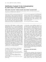
Fig. 1. Organization of the 3¢ region of human MUC4 gene. Boxes
indicate exons. They are numbered consecutively above the boxes
with 2 for the central exon (black box). Shaded grey box indicate
3¢-untranslated region. Horizontal lines indicate introns. They are
numbered below the lines. The length of the exons and introns are
showntoscale.
Ó FEBS 2002 Splice variants of MUC4 (Eur. J. Biochem. 269) 3639
secretion and corresponds to a high molecular mass
O-glycoprotein. It exhibits a VNTR polymorphism corre-
lated with the variation of one unit of repetition that
composes its central domain. In opposition with the strictly
secreted mucins, several transcripts of MUC4 were previ-
ously identified. This property, to be expressed under
numerous RNA forms for human mucins, appears to be
shared by the members of the membrane-bound mucin
subfamily. Indeed, four distinct transcripts were character-
ized for MUC1 as well as for MUC3 [26–30]. In both cases,
the perfect knowledge of the genomic organization allowed
the authors to assimilate transcripts with alternative splice
forms of the MUC1 and MUC3 genes.
Right now, MUC1 is the best known and characterized
mucin; therefore, some functions were identified and
associated with precise MUC1 splice form. For instance,
MUC1 is known to participate directly in the integrity of the
epithelial surface via its interaction with the b-catenin [31].
This interaction is regulated mainly by two mechanisms, via
the phosphorylation status of each partner of the b-catenin
pathwaycausedbytheGSK3b, or via the interaction of
MUC1 through two of its splice forms MUC1/SEC and
MUC1/Y [26,27]. MUC1/Y has also been referred to
enhance tumor initiation and progression in vivo [32].
MUC1/Y showed expression in various epithelial tumors,
such as breast and ovarian cancers but it is undetectable in
adjacent normal tissues.
For the MUC4 gene, 24 distinct transcripts have
already been isolated from various tissue samples as well
as cell lines [14,22,23]. They received the name of sv0- to
sv21-MUC4, MUC4/X and MUC4/Y. The sv0-MUC4
transcript is the main variant expressed by all the tissue
samples and cell lines studied right now [23]. It corres-
ponds to a 26.5-kb RNA encoding an apoprotein of
930 kDa organized in the following domains: mucin-type,
cysteine-rich, EGF-like, transmembrane, and cytoplasmic.
The sv0-MUC4 has a structural organization similar to its
rat homologue, SMC. SMC/rMuc4 is known to be a
heterodimeric complex composed of two subunits ASGP-1
and 2 [7–10]. The rMuc4 is believed to play an important
role in tumorigenesis and metastasis via anti-adhesive and
anti-immune recognition effects of its extracellular mucin
domain [33–35]. It is also shown to participate in the
ErbB2/neu signaling pathway [11], pointing out an
important role in cell proliferation and differentiation of
epithelial cells. A secreted form was identified for rMuc4;
however, it was shown not to be caused by alternative
splicing of its premRNA but by proteolytic cleavage of the
membrane-bound mucin [36]. Therefore at this point, only
one transcript was isolated for rMuc4,aswellasfor
mMuc1, mMuc3,andrMuc3.
In this study, we determined, for the first time, the
genomic organization of the MUC4 gene as well as the
mechanisms responsible for the diversity of MUC4 tran-
scripts. We have demonstrated that the events (deletions/
insertions) observed in the 24 MUC4 variants are generated
by different mechanisms of alternative splicing: alternative
use of exons, which is the mechanism most commonly used
to generate isoforms, and the use of cryptic donor/acceptor
splice sites. The identification of the same event in several
MUC4 variants, isolated from different tissues or cell lines,
and the molecular characterization of the splice events
strongly suggest that the diversity in the 3¢ region of MUC4
variants is not due to an error in the splicing process or an
artifact.
Altogether, it appears that the membrane-bound mucins
possess, at least from the transcriptional point of view, a
level of complexity in more than their animal homologues.
This complexity seems to be the highest for MUC4,asingle
gene code for at least 24 distinct transcripts. As no evidence
Table 2. Characteristics of the exon–intron junctions of the MUC4 gene.
3640 F. Escande et al. (Eur. J. Biochem. 269) Ó FEBS 2002
has been available to confirm the translation of the
transcripts, it is difficult to study their function. If translated,
the different transcripts will generate a complex family of
putative, membrane-bound, secreted and/or devoid of
functional domains (tandem repeat, cysteine-rich, EGF-like
domains) MUC4 isoforms (Table 4). Several splice variants
encode the same protein. The potential diversity of the
MUC4 isoforms in a single cell type may result in
modulation of the properties of the molecule. On the other
hand, the alternative RNA splice forms may only function
to reduce the level of expression of the main form sv0-
MUC4. Indeed, sv0-MUC4 (common in human and rat)
has been reported to play a key role for the epithelial
development and renewal as well as in tumorigenesis.
Therefore, its level of expression should be important in
maintaining the epithelial integrity. By analogy with
MUC1/Y splice variant, MUC4/X and MUC4/Y may have
important functions in tumorigenesis and activated
Table 3. Splice events detected in MUC4 cDNA variants. The constitutive exons are depicted as open boxes and alternative exons are shaded.
The solid lines show the splicing events in sv0-MUC4 and broken lines show the possible alternative splicing events in MUC4-variants. The
positions of the splice events were characterized according to the sequence AJ010901. The nomenclature used for the description of splicing
events corresponds to the consensus nomenclature [24].
Ó FEBS 2002 Splice variants of MUC4 (Eur. J. Biochem. 269) 3641
proliferative pathway. These hypotheses remain to be
verified. We are studying to find out if some MUC4
variants are very rare and occur only in specific tissues at
specific times during the development, and/or under certain
physiological conditions. Understanding how the complex
splicing of the transcripts encoded by this gene is regulated,
however, will help to elucidate how the specificity of their
expression is established as well as their putative functions.
Table 4. MUC4 splice variants and deduced peptides. NT1-NT3, N-terminal domains as described in [13]; TR, tandem repeat (central)
domain; CT1-CT12, C-terminal domains as described in [10].
3642 F. Escande et al. (Eur. J. Biochem. 269) Ó FEBS 2002
ACKNOWLEDGEMENTS
This work was supported by the Association de Recherche contre le
Cancer and a RO1 grant from the National Institutes of Health
(CA78590). We gratefully acknowledge D. Demeyer, C. Mouton,
M. Cre
´
pin for performing automatic sequences and A. Bernigaud,
D. Petitprez and V. Mortelec for the excellent technical assistance.
REFERENCES
1. Braga, V.M., Pemberton, L.F., Duhig, T. & Gendler, S.J. (1992)
Spatial and temporal expression of an epithelial mucin, Muc-1,
during mouse development. Development 115, 427–437.
2. Wesseling, J., Van der Valk, S.W., Vos, H.L., Sonnenberg, A. &
Hilkens, J. (1995) Episialin (MUC1) overexpression inhibits
integrin-mediated cell adhesion to extracellular matrix compo-
nents. J. Cell. Biol. 129, 255–265.
3. Buisine, M.P., Desreumaux, P., Leteurtre, E., Copin, M.C.,
Colombel, J.F., Porchet, N. & Aubert, J.P. (2001) Mucin gene
expression in intestinal epithelial cells in Crohn’s disease. Gut 49,
544–551.
4. Moniaux, N., Escande, F., Porchet, N., Aubert, J.P. & Batra. S.K.
(2001) Structural organization and classification of the human
mucin genes. Front. Biosci. 6, D1192–D1206.
5. Yin, B.W. & Lloyd, K.O. (2001) Molecular cloning of the CA125
ovarian cancer antigen: identification as a new mucin MUC16.
J. Biol. Chem. 276, 27371–27375.
6. Gum, J.R. Jr, Crawley, S.C., Hicks, J.W., Szymkowski, D.E. &
Kim, Y.S. (2002) MUC17, a novel membrane-tethered mucin.
Biochem. Biophys. Res. Commun. 291, 466–475.
7. Sherlom, A.P. & Carraway, K.L. (1980) A complex of two cell
surface glycoproteins from ascites mammary adenocarcinoma
cells. J. Biol. Chem. 255, 12051–12059.
8. Sheng, Z., Carraway, K.L. & Fregien, N. (1992) Molecular clon-
ing of the transmembrane component of the 13762 mammary
adenocarcinoma sialomucin complex. J. Biol. Chem. 267, 16341–
16346.
9. Wu, K., Fregien, N. & Carraway, K.L. (1994) Molecular
cloning and sequencing of the mucin subunit of a heterodimeric,
bifunctional cell surface glycoprotein complex of ascites
rat mammary adenocarcinoma cells. J. Biol. Chem. 269, 11950–
11955.
10. Moniaux, N., Nollet, S., Porchet, N., Degand, P., Laine, A. &
Aubert, J.P. (1999) Complete sequence of the human mucin
MUC4: a putative cell membrane-associated mucin. Biochem. J.
338, 325–333.
11. Carraway, K.L., Rossi, E.A., Komatsu, M., Price-Schiavi, S.A.,
Huang, D., Guy, P.M., Carvajal, M.E., Fregien, N. & Carraway,
C.A. (1999) An intramembrane modulator of the ErbB2 receptor
tyrosine kinase that potentiates neuregulin signaling. J. Biol.
Chem. 274, 5263–5266.
12. Porchet, N., Nguyen, V.G., Dufosse
´
,J.,Audie
´
, J.P., Guyonnet
Dupe
´
rat,V.,Gross,M.S.,Denis,C.,Degand,P.,Berheim,A.&
Aubert, J.P. (1991) Molecular cloning and chromosomal locali-
zation of a novel human tracheo-bronchial mucin cDNA con-
taining tandemly repeated sequences of 48 base pairs. Biochem.
Biophys. Res. Commun. 175, 414–422.
13. Nollet, S., Moniaux, N., Maury, J., Petitprez, D., Degand, P.,
Laine, A., Porchet, N. & Aubert, J.P. (1998) Human mucin gene
MUC4: organization of its 5¢ region and polymorphism of its
central tandem repeat array. Biochem. J. 332, 739–748.
14. Choudhury, A., Moniaux, N., Winpenny, J.P., Hollingsworth,
M.A.,Aubert,J.P.&Batra.S.K.(2000)HumanMUC4mucin
cDNA and its variants in pancreatic carcinoma. J. Biochem. 128,
233–243.
15. Audie
´
,J.P.,Janin,A.,Porchet,N.,Copin,M.C.,Gosselin,B.&
Aubert, J.P. (1993) Expression of human mucin genes in
respiratory, digestive, and reproductive tracts ascertained by in
situ hybridization. J. Histochem. Cytochem. 41, 1479–1485.
16. Audie
´
, J.P., Tetaert, D., Pigny, P., Buisine, M.P., Janin, A.,
Aubert, J.P., Porchet, N. & Boersma, A. (1995) Mucin
gene expression in the human endocervix. Hum. Reprod. 10,
98–102.
17. Balague, C., Audie, J.P., Porchet, N. & Real, F.X. (1995) In situ
hybridization shows distinct patterns of mucin gene expression in
normal, benign, and malignant pancreas tissues. Gastroenterology
109, 953–964.
18. Vandenhaute, B., Buisine, M.P., Debailleul, V., Cle
´
ment, B.,
Moniaux,N.,Dieu,M.C.,Degand,P.,Porchet,N.&Aubert,J.P.
(1997) Mucin gene expression in biliary epithelial cells. J. Hepatol.
27, 1057–1066.
19. Nguyen, P.L., Niehans, G.A., Cherwitz, D.L., Kim, Y.S. & Ho,
S.B. (1996) Membrane-bound (MUC1) and secretory (MUC2,
MUC3, and MUC4) mucin gene expression in human lung cancer.
Tumour Biol. 17, 176–192.
20. Copin, M.C., Buisine, M.P., Devisme, L., Leroy, X., Escande, F.,
Gosselin, B., Aubert, J.P. & Porchet, N. (2001) Normal
respiratory mucosa, precursor lesions and lung carcinomas:
differential expression of human mucin genes. Front. Biosci. 6,
1264–1275.
21. Balague, C., Gambus, G., Carrato, C., Porchet, N., Aubert, J.P.,
Kim, Y.S. & Real, F.X. (1994) Altered expression of MUC2,
MUC4, and MUC5 mucin genes in pancreas tissues and cancer
cell lines. Gastroenterology 106, 1054–1061.
22. Choudhury, A., Moniaux, N., Ringel, J., King, J., Moore, E.,
Aubert, J.P. & Batra. S.K. (2000) Alternative splicing at the 3¢-end
of the human pancreatic tumor-associated mucin MUC4 cDNA.
Teratog. Carcinog. Mutagen. 21, 83–96.
23. Moniaux, N., Escande, F., Batra, S.K., Porchet, N., Laine, A. &
Aubert, J.P. (2000) Alternative splicing generates a family of
putative secreted and membrane-associated MUC4 mucins. Eur.
J. Biochem. 267, 4536–4544.
24. den Dunnen, J.T. & Antonarakis, S.E. (2000) Mutation nomen-
clature extentions and suggestions to describe complex mutations:
a discussion. Hum. Mut. 15, 7–12.
25. Mount, S.M. (1982) A catalogue of splice junction sequences.
Nucleic Acids Res. 10, 459–472.
26. Wreschner, D.H., Hareuveni, M., Tsarfaty, I., Smorodinsky, N.,
Horev, J., Zaretsky, J., Kotkes, P., Weiss, M., Lathe, R. & Key-
dar, I. (1990) Human epithelial tumor antigen cDNA sequences:
Differential splicing may generate multiple protein forms. Eur. J.
Biochem. 189, 463–473.
27. Zrihan-Licht, S., Vos, H.L., Baruch, A., Elroy-Stein, O., Sagiv, D.,
Keydar, I., Hilkens, J. & Wreschner, D.H. (1994) Characterization
and molecular cloning of a novel MUC1 protein devoid of tandem
repeats, expressed in human breast cancer tissue. Eur. J. Biochem.
224, 787–795.
28. Ligtenberg, M.J., Vos, H.L., Gennissen, A.M. & Hilkens, J. (1990)
Episialin, a carcinoma-associated mucin, is generated by a poly-
morphic gene encoding splice variants with altenative amino ter-
mini. J. Biol. Chem. 265, 5573–5578.
29. Crawley, S.C., Gum, J.R., Hicks, J.W., Pratt, W.S., Aubert, J.P.,
Swallow, D.M. & Kim, Y.S. (1999) Genomic organization and
structure of the 3¢ region of human MUC3: alternative splicing
predicts membrane-bound and soluble forms of the mucin. Bio-
chem. Biophys. Res. Commun. 263, 728–736.
30. Williams, S.J., Munster, D.J., Quin, R.J., Gotley, D.C. &
McGuckin, M.A. (1999) The MUC3 gene encodes a transmem-
brane mucin and is alternatively spliced. Biochem. Biophy. Res.
Commun. 261, 83–89.
31. Yamamoto, M., Bharti, A., Li, Y. & Kufe, D. (1997)
Interaction of the DF3/MUC1 breast carcinoma-associated anti-
gen and beta-catenin in cell adhesion. J. Biol. Chem. 272, 12492–
12494.
Ó FEBS 2002 Splice variants of MUC4 (Eur. J. Biochem. 269) 3643
32. Baruch, A., Hartmann, M., Zrihan-Licht, S., Greenstein, S.,
Burstein, M., Keydar, I., Weiss, M., Smorodinsky, N. &
Wreschner, D.H. (1997) Preferential expression of novel MUC1
tumor antigen isoforms in human epithelial tumors and their
tumor-potentiating function. Int. J. Cancer 71, 741–749.
33. Komatsu, M., Yee, L. & Carraway, K.L. (1999) Overexpression of
sialomucin complex, a rat homologue of MUC4, inhibits tumor
killing by lymphokine-activated killer cells. Cancer Res. 59, 2229–
2236.
34. Carraway, K.L., Fregien, N., Carraway, K.L., 3rd & Carraway,
C.A. (1992) Tumor sialomucin complexes as tumor antigens and
modulators of cellular interactions and proliferation. J. Cell Sci.
103, 299–307.
35. Komatsu, M., Carraway, C.A., Fregien, N.L. & Carraway, K.L.
(1997) Reversible disruption of cell–matrix and cell–cell interac-
tions by overexpression of sialomucin complex. J. Biol. Chem. 272,
33245–33254.
36. Rossi, E.A., McNeer, R.R., Price-Schiavi, S.A., Van den Brande,
J.M., Komatsu, M., Thompson, J.F., Carraway, C.A., Fregien,
N.L. & Carraway, K.L. (1996) Expression as a soluble, secretable
form in lactating mammary gland and colon. J. Biol. Chem. 271,
33476–33485.
3644 F. Escande et al. (Eur. J. Biochem. 269) Ó FEBS 2002