Báo cáo Y học: Purification and characterization of VanXYC, a D,D-dipeptidase/D,D-carboxypeptidase in vancomycin-resistant Enterococcus gallinarum BM4174 docx
Bạn đang xem bản rút gọn của tài liệu. Xem và tải ngay bản đầy đủ của tài liệu tại đây (246.75 KB, 7 trang )
Purification and characterization of VanXY
C
,
a
D
,
D
-dipeptidase/
D
,
D
-carboxypeptidase in vancomycin-resistant
Enterococcus gallinarum
BM4174
Adrian H. B. Podmore and Peter E. Reynolds
Department of Biochemistry, University of Cambridge, UK
VanXY
C
, a bifunctional enzyme from VanC-phenotype
Enterococcus gallinarum BM4174 that catalyses
D
,
D
-pepti-
dase and
D
,
D
-carboxypeptidase activities, was purified as the
native protein, as a maltose-binding protein fusion and with
an N-terminal tag containing six histidine residues. The
kinetic parameters of His
6
–VanXY
C
were measured for a
variety of precursors of peptidoglycan synthesis involved in
resistance: for
D
-Ala-
D
-Ala, the K
m
was 3.6 m
M
and k
cat
,
2.5 s
)1
;forUDP-MurNAc-L-Ala-
D
-Glu-L-Lys-
D
-Ala-
D
-
Ala (UDP-MurNAc-pentapeptide[Ala]), K
m
was 18.8 m
M
and k
cat
6.2 s
)1
;for
D
-Ala-
D
-Ser, K
m
was 15.5 m
M
and k
cat
0.35 s
)1
.His
6
–VanXY
C
was inactive against the peptido-
glycan precursor UDP-MurNAc-
L
-Ala-
D
-Glu-L-Lys-
D
-
Ala-
D
-Ser (UDP-MurNAc-pentapeptide[Ser]). The rate of
hydrolysis of the terminal
D
-Ala of UDP-MurNAc-penta-
peptide[Ala] was inhibited 30% by 2 m
MD
-Ala-
D
-Ser or
UDP-MurNAc-pentapeptide[Ser]. Therefore preferential
hydrolysis of substrates terminating in
D
-Ala would occur
during peptidoglycan synthesis in E. gallinarum BM4174,
leaving precursors ending in
D
-Ser with a lower affinity for
glycopeptides to be incorporated into peptidoglycan. Muta-
tion of an aspartate residue (Asp59) of His-tagged VanXY
C
corresponding to Asp68 in VanX to Ser or Ala, resulted in a
50% increase and 73% decrease, respectively, of the specif-
icity constant (k
cat
/K
m
)for
D
-Ala-
D
-Ala. This situation is in
contrast to VanX in which mutation of Asp68fiAla pro-
duced a greater than 200 000-fold decrease in the substrate
specificity constant. This suggests that Asp59, unlike Asp68
in VanX, does not have a pivotal role in catalysis.
Keywords: vancomycin resistance;
D
,
D
-dipeptidase;
D
,
D
-
carboxypeptidase; Enterococcus gallinarum.
Glycopeptide antibiotics are effective Gram-positive anti-
bacterial agents that inhibit peptidoglycan synthesis by
binding to cell wall precursors terminating in
D
-Ala-
D
-Ala
[1]. VanA, VanB and VanD phenotypes have acquired
resistance to glycopeptides, and synthesize
D
-Ala-
D
-lactate
depsipeptides [2]. Three proteins are required for resistance:
VanH (VanH
B
,VanH
D
) reduces pyruvate to
D
-lactate [3,4];
VanA (VanB, VanD) ligases catalyse synthesis of
D
-Ala-
D
-
lactate [5] and VanX (VanX
B
)
D
,
D
-dipeptidase inhibits the
production of glycopeptide-susceptible precursors by
hydrolysing
D
-Ala-
D
-Ala [6]. A fourth enzyme, VanY, is a
D
,
D
-carboxypeptidase that hydrolyses terminal
D
-Ala from
UDP-MurNAc-pentapeptide [
D
-Ala] if
D
-Ala-
D
-Ala hydro-
lysis by VanX is incomplete [2] but is not necessary for
VanA-type resistance [2]. Regulation of expression of the
resistance genes is controlled by VanR (VanR
B
,VanR
D
)
andVanS(VanS
B
,VanS
D
), a two-component regulatory
system [7,8]. The VanA and VanB gene clusters are
contained on transposons that are integrated either into
self-transferable plasmids or the host chromosome [8,9].
VanC-type resistance is defined as intrinsic low-level
resistance to vancomycin (2–32 lgÆmL
)1
), but not to
teicoplanin [10]. It has been identified in Enterococcus
gallinarum, E. casseliflavus and E. flavescens [10,11]. Resist-
ance is based on the substitution of the terminal
D
-Ala in
peptidoglycan precursors by
D
-Ser [12,13]. VanC phenotype
glycopeptide resistance is mediated by VanC
D
-Ala:
D
-Ser
ligase [14], VanXY
C
D
,
D
-dipeptidase/
D
,
D
-carboxypeptidase
[15] and VanT
C
serine racemase [16]. These three proteins
eliminate
D
-Ala-terminating peptidoglycan precursors and
replace the terminal
D
-Ala-
D
-Ala with
D
-Ala-
D
-Ser. In
VanA and VanB phenotypes, the elimination of precursors
terminating in
D
-Ala requires two enzymes, VanX (VanX
B
),
astrict
D
,
D
-dipeptidase [6,17], and VanY (VanY
B
), a strict
membrane-bound
D
,
D
-carboxypeptidase [18]. Both of these
enzymes are also active with substrates terminating in
D
-Ser.
VanX is a metallo-protease: its catalytic, substrate-binding
and Zn
2+
-binding sites have been characterized by a
combination of kinetic [17], crystallographic [19] and site-
directed mutagenic studies [20,21]. In this study the
substrate specificity of purified VanXY
C
,acytoplasmic
bifunctional
D
,
D
-peptidase and
D
,
D
-carboxypeptidase, was
characterized kinetically and an investigation initiated of the
role of specific residues in determining the substrate
selectivity.
EXPERIMENTAL PROCEDURES
Strains, plasmids and growth conditions
Escherichia coli strains were grown in Luria–Bertani
medium, and maintained on Luria–Bertani agar (1.5%),
with the exception of E. coli JM83 [22] containing deriva-
Correspondence to A. H. B. Podmore, R & D Lab, Bioproducts
Laboratories (BPL), Dagger Lane, Elstree, WD6 3BX.
Tel.: + 44 208 2582200,
E-mail:
Abbreviations: MBP, maltose-binding protein.
(Received 28 January 2002, revised 11 April 2002,
accepted 19 April 2002)
Eur. J. Biochem. 269, 2740–2746 (2002) Ó FEBS 2002 doi:10.1046/j.1432-1033.2002.02946.x
tives of pMal-c2 (New England Biolabs), which was
grown in TYG medium [1% (w/v) tryptone, 0.5%
(w/v) yeast extract, 0.5% (w/v) NaCl, 0.2% (w/v) glucose,
pH 7.2] containing 100 lgÆmL
)1
ampicillin. Kanamycin
(25 lgÆmL
)1
) was added to the medium for E. coli M15
[pREP4] (Qiagen) and ampicillin (100 lgÆmL
)1
) was added
to the medium for E. coli M15[pREP4] containing deriva-
tives of pQE-30 (Qiagen) and E. coli JM83 containing
pAT704 [15].
DNA manipulations
Digestion with restriction endonucleases, cloning, isolation
of plasmid DNA, ligation and transformation were carried
out using standard protocols [23].
Plasmid construction
Plasmid pAT704 has been described previously [15]. Plas-
mid pAP1 (encoding a maltose binding protein-VanXY
C
fusion protein) was constructed as follows; Pfu polymerase
(Stratagene) was used to amplify vanXY
C
usingpAT704as
template with primers A (5¢-GCTA
GGTCTCAATGAAC
ACATTACAATT-3¢)andB(5¢-TATG
GAATTCTCATG
CGAACTGCCTCA-3¢) that included BsaIandEcoRI
restriction sites, respectively (underlined). The product was
purified, digested with BsaI, treated with DNA poly-
merase I large (Klenow) fragment, purified, digested with
EcoRI and cloned in pMal-c2 under the control of the tac
promoter. Plasmid pAP2 was constructed for production of
VanXY
C
with an N-terminal tag of six histidine residues.
Pfu polymerase was used to amplify vanXY
C
using pAT704
as template with primers C (5¢-CTCA
GGATCCAACACA
TTACAATTGATCAATA-3¢)andD(5¢-CACT
AAGCTT
TCATGCGAACTGCCTCAC-3¢) that included BamHI
and HindIII restriction sites, respectively (underlined).
The product was purified, digested with BamHI and
HindIII and cloned in pQE30 under the control of the T5
promoter.
Site-directed mutagenesis
Mutants D59S and D59A were constructed from a pAP2
template by PCR mutagenesis using the Expand Long
Template PCR System (Boehringer Mannheim) and sense/
antisense primer pairs D59S (CGTCTGGTA
TCTGGGT
AT/AATGTCCTTTTCTAGTCC), D59A (CGTCTG
GTA
GCTGGGTAT/AATGTCCTTTTCTAGTCC),P/W
(AGTTATGAA
TGGTGGCATTTTCG/GATACCGGT
GATCTCTTG) and Q/V (GGAAAAAGAA
GTGCGA
CG/GTACGATACCCATCTACC), respectively (mis-
match mutations are underlined). The purified PCR pro-
ducts were ligated and used to transform E. coli
M15[pREP4].
DNA sequence determination
DNA sequencing on both strands was carried out by the
dideoxynucleotide chain terminator method [24] using
fluorescent cycle sequencing with dye-labelled terminators
(ABI PrismTM Dye terminator Cycle Sequencing Ready
Reaction Kit, PerkinElmer) on a 373 A automated DNA
sequencer (PerkinElmer).
Protein quantitation
Protein concentration was determined by the method of
Bradford with bovine serum albumin as standard [25].
Purification of VanXY
C
A culture of E. coli JM83 containing pAT704(vanXY
C
)was
harvested after induction with 0.5 m
M
isopropyl thio-b-
D
-
galactoside for 3 h. The bacteria were then washed and
disrupted by sonication. After removal of cell debris,
purification was attempted using ammonium sulphate
fractionation (the majority of the enzyme was present in
the 45–60% fraction), followed by ion-exchange chroma-
tography on a MonoQ HR5/5 column (Pharmacia) and gel
exclusion chromatography on a FPLC Superdex 75 H
column. VanXY
C
eluted with an estimated mass of 42 kDa,
approximately twice that of the monomer, suggesting that
native VanXY
C
exists as a dimer. The activity of the
partially purified material (Fig. 1A) was only 3% of that
present in the original extract.
In order to improve recovery and purity, the enzyme was
produced as a fusion with the maltose binding protein. A
culture of E. coli JM83 containing pAP1(pMal-c2[vanXY
C
])
was incubated with 0.5 m
M
isopropyl thio-b-
D
-galactoside
for 30 min to induce the fusion protein. The bacteria were
harvested, washed, broken by sonication and the fusion
protein purified by affinity chromatography on an amylose
resin column. The yield, based on activity, was 50% and a
single band (60 000 kDa) was observed by SDS/PAGE
analysis (Fig. 1B). It proved impossible to release VanXY
C
from MBP-VanXY
C
by treatment with factor Xa.
A second fusion protein (His
6
–VanXY
C
)wasalso
purified by affinity chromatography after induction of
E. coli M15[pREP4] containing pAP2(pQE30[vanXY
C
])
for 30 min with 0.5 m
M
isopropyl thio-b-
D
-galactoside,
followed by sonication of the washed bacterial suspension.
The broken cell preparation was centrifuged at 43 000 g
for 20 min to pellet the cellular debris. The supernatant was
removed, and loaded onto a 4-mL Ni
2+
-nitrilotriacetic
acid/agarose column. The column was washed with 200 mL
50 m
M
1,3 bi[tris(hydroxymethyl)-methylamino] propane
(pH 7.5), 30 m
M
imidazole, 300 m
M
NaCl, and His
6
–
VanXY
C
was eluted with the same buffer in which the
imidazole concentration had been increased to 250 m
M
.
SDS/PAGE analysis showed a single band of 23 kDa
(Fig. 1C), whereas gel filtration on FPLC Superdex 75
column resulted in a peak with an apparent molecular mass
of 44 kDa. This method yielded 3–4 mg of His
6
–VanXY
C
from a culture volume of 1 L for enzyme characterization
studies and was also used for purification of His
6
–VanXY
C
in studies of site-directed mutagenesis.
Assay of
D
,
D
-dipeptidase and
D
,
D
-carboxypeptidase
activity
The method for assaying
D
-Ala-
D
-Ala dipeptidase activity
of VanXY
C
was based on that of Reynolds et al.[6].
Twenty microliters of 150 m
M
1,3 bi[tris(hydroxymethyl)-
methylamino] propane (pH 7.5) containing substrate
(
D
-Ala-
D
-Ala, unless stated otherwise) was mixed with
10 lL of enzyme preparation and incubated at 37 °C.
Samples were withdrawn at suitable time intervals and
Ó FEBS 2002 Purification and characterization of VanXYC (Eur. J. Biochem. 269) 2741
assayed using the
D
-amino acid oxidase assay [6], except for
kinetic studies when the modified cadmium-ninhydrin
method [17] was used, with
D
-alanine or
D
-serine as
standards. The sensitivity ranges of the
D
-amino acid
oxidase and cadmium ninhydrin assays were 2–20 nmol
D
-alanine or 4–40 nmol
D
-serine, and 10–80 nmol
D
-alanine
or
D
-serine, respectively.
Effect of divalent cations and EDTA on
D
,
D
-dipeptidase
and
D
,
D
-carboxypeptidase activity
The effect of divalent cations on the actual
D
-amino acid
oxidase assay was investigated as follows. Ten microliteres
of 10 m
M
Me
2+
(Fe
2+
,Cu
2+
,Mn
2+
,Co
2+
,Zn
2+
,Ni
2+
,
Mg
2+
as the metal chlorides) in 150 m
M
1,3 bi[tris
(hydroxymethyl)-methylamino] propane (pH 7.5) was
mixed with 10 lLofVanXY
C
, and incubated on ice for
15 min.
D
-Ala (20 nmol) was then added, to give a final
volume of 30 lL. These samples were assayed using the
D
-amino acid oxidase assay. Mn
2+
,Co
2+
and Cu
2+
caused a decrease in the absorbance at 460 nm (25% for
Mn
2+
and Cu
2+
37% for Co
2+
), but this would not have
masked a onefold or greater stimulation of
D
,
D
-dipeptidase
activity. To study the effect of metal ions on activity of
VanXY
C
,10lLof10m
M
Me
2+
in 150 m
M
1,3 bi[tris
(hydroxymethyl)-methylamino] propane was mixed with
10 lLVanXY
C
and incubated on ice for 15 min. Next,
10 lLof10m
MD
-Ala-
D
-Ala was added, incubated at
37 °C for 30 min, and assayed using the
D
-amino acid
oxidase assay. This experiment was repeated to determine
the effect of divalent cations on
D
,
D
-carboxypeptidase
activity, using Ni
2+
at concentrations of 1 and 5 m
M
,and
Zn
2+
at concentrations of 0.05 and 0.8 m
M
. The nucleo-
tide peptide substrate UDP-MurNAc-pentapeptide[Ala]
(3.3 m
M
)wasusedinsteadof
D
-Ala-
D
-Ala.
EDTA did not affect the
D
-amino acid oxidase assay at a
concentration of 0.05 m
M
and below. To test the effect of
EDTA on enzyme activity VanXY
C
was mixed with various
concentrations of EDTA (0.01, 0.05, 0.1, 1.0 and 5.0 m
M
)
and incubated on ice for 1 h. Samples were diluted with
50 m
M
1,3 bi[tris(hydroxymethyl)-methylamino] propane
(pH 7.5) to reduce the EDTA concentration to 0.05 m
M
.
10 lLEDTA-treatedVanXY
C
was mixed with 10 lLof
10 m
M
Me
2+
in 150 m
M
1,3 bi[tris(hydroxymethyl)-meth-
ylamino] propane and 10 lL10m
MD
-Ala-
D
-Ala, incuba-
ted at 37 °C for 30 min, and assayed for
D
,
D
-dipeptidase
activity using the
D
-amino acid oxidase assay.
Kinetic analysis of VanXY
C
His
6
–VanXY
C
(2.25 · 10
)7
M) was incubated at 37 °C
with various concentrations of
D
-Ala-
D
-Ala (2, 5, 10, 15, 20,
30, and 40 m
M
) in 100 m
M
1,3 bi[tris(hydroxymethyl)-
methylamino] propane (pH 7.5). Samples (30 lL) were
withdrawn at suitable time points and added to 750 lLof
cadmium-ninhydrin stock solution; 70 lL of distilled water
was added and incubated at 85 °C for 5 min. The absorb-
ance was measured at 505 nm and quantified with free
amino acid as standard. For determination of hydrolysis of
D
-Ala-
D
-Ser, His
6
–VanXY
C
was used at a concentration of
26 · 10
)7
M
. Rates of hydrolysis of 10, 15, 20, 25, and
30 m
MD
-Ala-
D
-Ser were determined using five time points.
The high A
505
at time 0 was attributable to
D
-Ala-
D
-Ser,
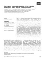
Fig. 1. SDS/PAGE analysis. (A) Purification of VanXY
C
.Lane1,
molecular mass standards (in kDa); Lane 2, partially pure VanXY
C
after gel filtration chromatography. (B) Purification of MBP-VanXY
C
.
Lane 1, molecular mass standards; lane 2, cytoplasm containing
MBP-VanXY
C
;Lane3,MBP–VanXY
C
after amylose resin chroma-
tography. (C) Purification of His
6
–VanXY
C
. Lane 1, molecular mass
standards; lane 2, cytoplasm containing His
6
–VanXY
C
;lane3His
6
–
VanXY
C
after Ni
2+
-nitrilotriacetic acid/agarose chromatography.
2742 A. H. B. Podmore and P. E. Reynolds (Eur. J. Biochem. 269) Ó FEBS 2002
determined using a
D
-Ala-
D
-Ser standard curve. For
determination of the rate of hydrolysis of UDP-MurNAc-
pentapeptide[Ala], His
6
–VanXY
C
was used at a concentra-
tion of 2.25 · 10
)7
M
. The rates of hydrolysis of 2, 5, 10, 15,
20 and 30 m
M
UDP-MurNAc-pentapeptide[Ala] were
determined using six time points. For studies of hydrolysis
of UDP-MurNAc-pentapeptide[Ser], His
6
–VanXY
C
was
used at a final concentration of 16 · 10
)7
M
.Therates
of hydrolysis of 2, 5, 10, 15, and 20 m
M
UDP-
MurNAc-pentapeptide[Ser] were determined using five time
points.
To confirm the degree of hydrolysis of the nucleotide-
peptide substances, measurements were also carried out
using HPLC by measuring the decrease in the amount of
substrate and increase in the amount of product (UDP-
MurNAc-tetrapeptide). His
6
–VanXY
C
(3.2 · 10
)7
M
)was
incubated with nucleotide-peptide substrates in 100 m
M
1,3 bi[tris(hydroxymethyl)-methylamino] propane (pH 7.5).
Samples were withdrawn at 0 and 30 min, heated at 90 °C
for 5 min, and analysed by HPLC following the method of
Reynolds et al. [13]. To determine whether
D
-Ala was
cleaved from UDP-MurNAc-tetrapeptide (2 m
M
), the
increase of UDP-MurNAc-L-Ala-
D
-Glu-
L
-Lys (UDP-Mur-
NAc-tripeptide) was also measured. No conversion of
UDP-MurNAc-tetrapeptide to UDP-MurNAc-tripeptide
was observed. The hydrolysis of 2 m
M
UDP-MurNAc-
pentapeptide[Ala] was also measured in the presence of
either UDP-MurNAc-pentapeptide[Ser] or
D
-Ala-
D
-Ser at
final concentrations of 2 m
M
.
RESULTS
Purification of VanXY
C
The vanXY
C
gene was expressed in E. coli JM83 (pAP1)
and conventional purification of native VanXY
C
attempted
as described in Materials and methods. The purification
procedure did not give good separation of VanXY
C
from
other proteins and the activity was spread over many
fractions during ion-exchange chromatography. This meth-
od yielded 0.2 mg of VanXY
C
together with contaminating
proteins from a culture volume of 1 L. Therefore, the
procedure was changed in order to synthesize and purify
VanXY
C
as a maltose-binding protein (MBP) fusion in
E. coli. The MBP–VanXY
C
fusion was purified to homo-
geneity using amylose affinity chromatography in yields up
to 3 mgÆL
)1
. MBP–VanXY
C
was kinetically characterized,
but it was not possible to remove MBP using factor Xa, and
compare the activity of the fusion protein with that of
VanXY
C
in the absence of MBP. This problem was caused
by the stringent steric requirements for factor Xa cleavage
that have been reported previously for this expression
system [27–29]. In order to investigate whether MBP might
be interfering with activity, an alternative purification
strategy was used. VanXY
C
was expressed with a smaller
N-terminal tag of six histidine residues (His
6
–VanXY
C
)in
E. coli M15 (pAP2) and purified to homogeneity using
nickel affinity chromatography in yields up to 4 mgÆL
)1
.
Kinetic analysis of His
6
–VanXY
C
revealed that its activity
was of the same order of magnitude, within the limits of the
assay, as that of MBP–VanXY
C
. Studies with VanX using
MBP attached or cut off did not affect the kinetic data
[20,21]. We therefore assumed (as the tag could not be
removed easily) that the smaller His tag was unlikely to
influence activity significantly. The predicted molecular
mass of His
6
–VanXY
C
and VanXY
C
, 23.6 kDa and
22.3 kDa, respectively, was consistent with the molecular
mass estimated by SDS/PAGE analysis. Gel permeation
chromatography of the native protein indicated a mobility
consistent with a mass of approximately 42–44 kDa. This
suggests that VanXY
C
exists as a dimer in its native form.
Effect of divalent cations and EDTA on
D
,
D
-dipeptidase
and
D
,
D
-carboxypeptidase activity
VanX copurified with near stoichiometric amounts of Zn
2+
[20]. The Zn
2+
binding residues of VanX were identified
using site-directed mutagenesis [20], and later confirmed by
analysis of the crystal structure [19]. Comparison of the
active site of VanXY
C
with VanX- and VanY-type enzymes
indicated that all of these enzymes contained the same Zn
2+
binding motif. VanX homologues present in the cyanobac-
terium Synechocystis strain PCC6803, and the glycopeptide
antibiotic producer Streptomyces toyocaensis had also been
purified as MBP-fusions and both of these enzymes
copurified with near stoichiometric quantities of Zn
2+
[21]. EDTA has been shown to abolish VanY activity [18],
but not VanX activity [17]. The addition of a low
concentration of Zn
2+
to EDTA-inactivated VanY resulted
in the recovery of activity. Replacement of Zn
2+
in VanX
by the direct addition of various divalent metal cations to
the purified protein affected the
D
-Ala-
D
-Ala dipeptidase
activity in some instances. When added to VanX at their
predetermined optimum stimulatory concentration, Zn
2+
,
Fe
2+
,Co
2+
and Ni
2+
increased the k
cat
by sixfold to 168-
fold [17,19,30]. All the divalent metals tested inhibited
VanXY
C
D
,
D
-dipeptidase activity. Mg
2+
inhibited activity
the least, followed by Ni
2+
,andthenZn
2+
(Table 1).
Co
2+
,Mn
2+
,Cu
2+
,andFe
2+
caused total inhibition of
D
,
D
-dipeptidase activity at 1.3 and 3.3 m
M
.Ni
2+
at
concentrations of 1 and 5 m
M
resulted in 10% and 75%
inhibition of
D
,
D
-carboxypeptidase activity. Zn
2+
at con-
centrations of 0.8 and 0.05 m
M
inhibited
D
,
D
-carboxypept-
idase activity by 50 and 20%, respectively. Therefore,
VanXY
C
, like VanY [18], was not stimulated by the direct
addition of metal ions. EDTA at concentrations between
Table 1. Effect of divalent cations on
D
,
D
-dipeptidase activity.
Cations
Concentration
of cation (m
M
)
Activity
(nmol
D
-Ala-
D
-Ala
hydrolysed per min)
a
– – 0.13
Mg
2+
1.3 0.11
Mg
2+
3.3 0.10
Mg
2+
(enzyme omitted) 3.3 0
Ni
2+
1.3 0.11
Ni
2+
3.3 0
Ni
2+
(enzyme omitted) 3.3 0
Zn
2+
1.3 0.06
Zn
2+
3.3 0
Zn
2+
(enzyme omitted) 3.3 0
a
Determined using the
D
,
D
-dipeptidase/
D
-amino acid oxidase
assay with VanXY
C
.
Ó FEBS 2002 Purification and characterization of VanXYC (Eur. J. Biochem. 269) 2743
0.01 and 5.0 m
M
did not affect the
D
,
D
-dipeptidase activity
of VanXY
C
, suggesting that the Zn
2+
molecule, if essential
for activity, is tightly bound in the active site.
Kinetic analysis of VanXY
C
The modified cadmium-ninhydrin method [17] alters sample
conditions such that ninhydrin preferentially binds to free
amino acids in the presence of peptides. The high A
505
values
at time zero were attributable to
D
-Ala-
D
-Ala,
D
-Ala-
D
-Ser
or UDP-MurNAc-pentapeptide[Ala] determined from the
relevant standard curves. The kinetic parameters for His
6
–
VanXY
C
acting as a
D
,
D
-peptidase and
D
,
D
-carboxypepti-
dase are given in Table 2 and the data from which these
were derived are plotted as Fig. 2A–C. K
m
and k
cat
were
determined by fitting the experimental data obtained to the
equation V
max
¼ v +[(v/s)ÆK
m
], using the direct linear plot
[31] and by plotting s/v against s using the equation
s/v ¼ K
m
/V +(1/V)Æs. This assay is crude, being two-part
rather than continuous, and repetitive measurement of the
K
m
value with the same enzyme preparation resulted in
values that were as much as twofold different.
Hydrolysis of terminal
D
-Ser from UDP-MurNAc-
L
-
Ala-
D
-Glu-
L
-Lys-
D
-Ala-
D
-Ser (UDP-MurNAc-pentapep-
tide[Ser]) was not detected.
The K
m
values for hydrolysis of
D
-Ala-
D
-Ser and UDP-
MurNAc-pentapeptide[Ala] were similar. Consequently the
extent of interference of precursors terminating in
D
-Ser on
the rate of hydrolysis of UDP-MurNAc-pentapeptide[Ala]
was measured using HPLC. The rate of removal of the
terminal
D
-Ala of 2 m
M
UDP-MurNAc-pentapeptide[Ala]
was shown by HPLC to be inhibited 30% by the presence of
either 2 m
MD
-Ala-
D
-Ser or 2 m
M
UDP-MurNAc-penta-
peptide[Ser].
Site-directed mutagenesis of vanXY
C
Mutated VanXY
C
fusion proteins containing an N-terminal
tag of six histidine residues were purified using the method
described for His
6
–VanXY
C
. The yields for mutants D59S
and D59A were similar to that obtained for His
6
–VanXY
C
.
No band corresponding to His
6
–VanXY
C
was identified in
the fractions eluted from the affinity column during
purification of His
6
–VanXY
C
harbouring P154W or
Q67V mutations, and no
D
,
D
-dipeptidase activity was
detected. The kinetic parameters for D59S and D59A
resultant proteins acting as a
D
,
D
-peptidase and
D
,
D
-
carboxypeptidase are given in Table 3. It was not possible
to determine the kinetic parameters of D59A for hydrolysis
of UDP-MurNAc-pentapeptide[Ala]. However, its activity
was comparable with that of D59S and His
6
–VanXY
C
.The
rates of hydrolysis of
D
-Ala-
D
-Ala and UDP-MurNAc-
pentapeptide[Ala] by His
6
–VanXY
C
are lower than those
estimated previously because the enzyme had lost activity
during storage probably due to aggregation of the enzyme.
DISCUSSION
The substrate specificity constant (k
cat
/K
m
) for hydrolysis of
D
-Ala-
D
-Ser was 24-fold lower than for
D
-Ala-
D
-Ala, the
result of a 3.8-fold increase in K
m
and a 6.3-fold decrease in
k
cat
.Wuet al. [17] determined that the substrate specificity
constant of VanX for
D
-Ala-
D
-Ser was only sevenfold lower
than for
D
-Ala-
D
-Ala, the result of a 2.8-fold increase in K
m
and a 2.6-fold decrease in k
cat
(the rate of hydrolysis of
D
-Ala-
D
-Ser was estimated using
DL
-Ala-
DL
-Ser with the
assumptions that a quarter of the racemic mixture is
D
-Ala-
D
-Ser and the other three isomers have no inhibition effect
Table 2. Kinetic parameters for His
6
–VanXY
C
. ND, hydrolysis not
detected.
Substrate
K
m
(m
M
)
k
cat
(s
)1
)
k
cat/
K
m
(m
M
)1
Æs
)1
)
D
-Ala-
D
-Ala 4.0 2.2 0.55
D
-Ala-
D
-Ser 15.5 0.35 0.02
UDP-MurNAc-pentapeptide[Ala] 17.0 5.9 0.35
UDP-MurNAc-pentapeptide[Ser] ND ND ND
Fig. 2. Initial velocity/substrate concentration vs. substrate plots of
His
6
–VanXY
C
for (A)
D
-Ala-
D
-Ala; (B)
D
-Ala-
D
-Ser; (C) UDP-Mur-
NAc-pentapeptide[Ala].
2744 A. H. B. Podmore and P. E. Reynolds (Eur. J. Biochem. 269) Ó FEBS 2002
on VanX) [17]. The likely effect on hydrolysis of terminal
D
-Ala from UDP-MurNAc-pentapeptide[Ala] in the pres-
ence of
D
-Ala-
D
-Ser was not clear from an examination of
their K
m
values, being 17 m
M
and 15 m
M
, respectively, as
determined using the cadmium-ninhydrin method. HPLC
analysis of the rate of UDP-MurNAc-pentapeptide[Ala]
hydrolysis in the presence of
D
-Ser-terminating precursors
was carried out. The presence of 2 m
MD
-Ala-
D
-Ser or 2 m
M
UDP-MurNAc-pentapeptide[Ser] caused a 30% reduction
intherateofhydrolysisof2m
M
UDP-MurNAc-penta-
peptide[Ala]. These data showed that His
6
–VanXY
C
selec-
tively hydrolysed
D
-Ala-terminating precursors in the
presence of
D
-Ser-terminating precursors. The role of VanX
in VanA-type resistance is to hydrolyse preferentially
D
-Ala-
D
-Ala but not
D
-Ala-
D
-Lactate: consequently it has
specificity for dipeptides. As a result, VanX hydrolyses
D
-Ala-
D
-Ser relatively rapidly in addition to
D
-Ala-
D
-Ala.
However, in the VanC phenotype, VanXY
C
must specific-
ally hydrolyse
D
-Ala-
D
-Ala with minimal activity against
D
-Ala-
D
-Ser, a very different type of specificity.
VanXY
C
, VanX-type and VanY-type enzymes contain
most of the active site residues identified in VanX. VanXY
C
has 39% identity and 74% similarity to VanY in an overlap
of 158 amino acids, and low amino-acid identity to VanX,
except for a stretch of 22 amino acids that constitute most of
theactivesite.However,VanXY
C
hydrolyses
D
,
D
-dipep-
tides such as
D
-Ala-
D
-Ala, whereas VanY is inactive against
this substrate. The small active site cavity of VanX, deduced
from crystallographic studies, only allows access of dipep-
tides [19]. Therefore, VanXY
C
and VanY-type enzymes
presumably have a less restrictive active site to accommo-
date larger substrates such as UDP-MurNAc-pentapep-
tide[Ala]. However VanY, unlike VanXY
C
, will not
hydrolyse
D
-Ala-
D
-Ala. His
6
–VanXY
C
has an almost
threefold higher k
cat
for UDP-MurNAc-pentapeptide[Ala]
than for
D
-Ala-
D
-Ala, which suggests that the active site can
accommodate the larger substrate more easily than dipep-
tides. Also, VanXY
C
and VanY showed activity against
UDP-MurNAc-pentadepsipeptide, albeit at a much
reduced level when compared to UDP-MurNAc-pentapep-
tide[Ala] [18]. VanX did not hydrolyse the depsipeptide
[6,17]. These data suggest that VanXY
C
has evolved from
an ancestor of the VanY-type enzymes to contain both
D
,
D
-
dipeptidase and
D
,
D
-carboxypeptidase activities, but it
remains unclear why VanXY
C
but not VanY can hydrolyse
D
-Ala-
D
-Ala [18]. To investigate the role of specific residues
in this selectivity some amino acids presumed to be involved
in binding or catalysis were targeted for site-directed
mutagenesis.
Asp68 is part of the Arg71-Asp68-Tyr35 hydrogen-
bonding triad that is believed to orientate Arg71 for
transition state stabilization in VanX [19]. This traid is
present in all VanX-type enzymes, but Asp68 and Tyr35
equivalents are absent in VanY-type enzymes. VanY-type
enzymes contain a conserved serine residue that has a
corresponding position to Asp68 in VanX and a glutamine
residue (Gln143) that is postulated to function as Asp68 in
VanX [32]. However, VanXY
C
contains both an Asp68
equivalent (Asp59) and a VanY-type Gln143 equivalent
(Gln67). Consequently, both of these residues were mutated
and the corresponding proteins purified as His-tagged
proteins to determine which of these residues may position
the arginine (Arg62) for transition-state stabilization in
VanXY
C
. Mutation of Asp59 to Ser or Ala in His
6
–
VanXY
C
resulted in a 50% increase and 73% decrease,
respectively, of the substrate specificity constant (k
cat
/K
m
)
for
D
-Ala-
D
-Ala. The mutation D59S caused a 1.3 fold
increase of the substrate specificity constant (k
cat
/K
m
)for
hydrolysis of UDP-MurNAc-pentapeptide[Ala]. The His-
tagged enzyme with the D59A mutation had similar activity
to the enzyme with the D59S mutation against UDP-
MurNAc-pentapeptide[Ala]. The effect of mutating this
aspartate residue is markedly different for VanX, where
mutation to Ala caused a 268-fold decrease in k
cat
and a
750-fold increase in K
m
. These results suggest that Asp59 in
VanXY
C
is unlikely to be involved in stabilizing Arg62.
Mutation of Gln67 to Val resulted in a complete
absence of His-tagged mutant protein. The same situation
resulted when a conserved Pro (corresponding to Trp182
in VanX) was mutated to Trp. This Pro residue is
conserved in all VanY-type enzymes (EPWH motif),
except the VanY homologue from Streptococcus mutans,
but Trp is present at this position in all VanX-type
enzymes (EWWH motif). The reason for the lack of
expression of these mutant His-tagged VanXY
C
enzymes
is unknown.
ACKNOWLEDGEMENTS
This work was carried out under the tenure of a BBSRC studentship to
A.H.B.P.WethankC.HillandJ.Lester,CambridgeCentrefor
Molecular Recognition for synthesis of oligonucleotides and automated
DNA sequencing, respectively.
REFERENCES
1. Reynolds, P.E. (1989) Structure, biochemistry and mechanism of
action of glycopeptide antibiotics. Eur. J. Clin. Microbiol. Infec.
Dis. 8, 943–950.
Table 3. Kinetic parameters for His
6
–VanXY
C
mutant products obtained by site-directed mutagenesis. ND, not determined.
Enzyme Substrate
K
m
(m
M
)
k
cat
(s
)1
)
k
cat/
K
m
(m
M
)1
Æs
)1
)
VanXY
C
a
D
-Ala-
D
-Ala 1.5 0.5 0.33
D59S
D
-Ala-
D
-Ala 2.5 1.4 0.56
D59A
D
-Ala-
D
-Ala 9.0 0.8 0.09
VanXY
C
a
UDP-MurNAc-pentapeptide[Ala] 9.5 0.8 0.08
D59S UDP-MurNAc-pentapeptide[Ala] 1.5 23 15
D59A UDP-MurNAc-pentapeptide[Ala] ND ND ND
a
His
6
–VanXY
C
.
Ó FEBS 2002 Purification and characterization of VanXYC (Eur. J. Biochem. 269) 2745
2. Arthur, M., Reynolds, P. & Courvalin, P. (1996) Glycopeptide
resistance in enterococci. Trends Microbiol. 4, 401–407.
3. Arthur, M., Molinas, C., Dutka-Malen, S. & Courvalin, P. (1991)
Structural relationship between the vancomycin-resistance protein
VanH and 2-hydroxycarboxylic acid dehydrogenases. Gene 103,
133–134.
4. Bugg, T.D., Wright, G.D., Dutka-Malen, S., Arthur, M.,
Courvalin, P. & Walsh, C.T. (1991) Molecular basis for vanco-
mycin resistance in Enterococcus faecium BM4147: biosynthesis of
a depsipeptide peptidoglycan precursor by vancomycin resistance
proteins VanH and VanA. Biochemistry 30, 10408–10415.
5. Bugg, T.D.H., Dutka-Malen, S., Arthur, M., Courvalin, P. &
Walsh, C.T. (1991a) Identification of vancomycin resistance pro-
tein VanA as a
D
-alanine:
D
-alanine ligase of altered substrate
specificity. Biochemistry 30, 2017–2021.
6. Reynolds, P.E., Depardieu, F., Dutka-Malen, S. & Courvalin, P.
(1994) Glycopeptide resistance mediated by enterococcal trans-
poson Tn1546 requires production of VanX for hydrolysis of
D
-alanyl-
D
-alanine. Mol. Microbiol. 13, 1065–1070.
7. Arthur, M., Molinas, C. & Courvalin, P. (1992) The VanS-VanR
two-component regulatory system controls synthesis of depsi-
peptide peptidoglycan precursors in Enterococcus faecalis
BM4174. J. Bacteriol. 174, 2582–2591.
8. Evers, S. & Courvalin, P. (1996) Regulation of vanB-type
vancomycin resistance gene-expression by the VanS (B)-VanR (B)
2-component regulatory system in Enterococcus faecalis V583.
J. Bacteriol. 178, 1302–1309.
9. Quintiliani, R. & Courvalin, P. (1994) Conjugal transfer of the
vancomycin resistance determinant VanB between enterococci
involves the movement of large genetic elements from chromo-
some to chromosome. FEMS Microbiol. Lett. 119, 359–363.
10. Dutka-Malen, S., Molinas, C., Arthur, M. & Courvalin, P. (1992)
Sequence of the vanC gene of Enterococcus gallinarum BM4174
encoding a
D
-alanine:
D
-alanine ligase-related protein necessary
for vancomycin resistance. Gene 112, 53–58.
11. Navarro, F. & Courvalin, P. (1994) Analysis of genes encoding
D
-alanine:
D
-alanine ligase-related enzymes in Enterococcus
casseliflavus and Enterococcus flavescens. Antimicrob.
Ag. Chemother. 38, 1788–1793.
12. Billot-Klein, D., Gutmann, L., Sable, S., Guittet, E. & Van
Heijenoort, J. (1994) Modification of peptidoglycan precursors is a
common feature of the low-level vancomycin-resistant VanB-type
Enterococcus D366 and of the naturally glycopeptide-resistant
species Lactobacillus casei, Pediococcus pentosaceus, Leuconostoc
mesenteroides,andEnterococcus gallinarum. J. Bacteriol. 176,
2398–2405.
13. Reynolds, P.E., Snaith, H.A., Maguire, A.J., Dutka-Malen, S. &
Courvalin, P. (1994) Analysis of peptidoglycan precursors in
vancomycin-resistant Enterococcus gallinarum BM4174. Biochem.
J. 301, 5–8.
14. Park, I.S., Chung-Hung, L. & Walsh, C.T. (1997) Bacterial re-
sistance to vancomycin: overproduction, purification and char-
acterization of VanC-2 from Enterococcus casseliflavus as a D-Ala:
D-Ser ligase. Proc. Natl Acad. Sci. USA 94, 10040–10044.
15. Reynolds, P.E., Arias, C.A. & Courvalin, C. (1999) Gene vanXY
C
encodes D,
D
-dipeptidase (VanX) and
D
,
D
-carboxypeptidase
(VanY) activities in vancomycin-resistant Enterococcus gallinarum
BM4174. Mol. Microbiol. 34, 341–349.
16. Arias, C.A., Martin-Martinez, M., Blundell, T.L., Arthur, M.,
Courvalin, P. & Reynolds, P.E. (1999) Characterization and
modelling of VanT: a novel, membrane-bound, serine racemase
from vancomycin-resistant Enterococcus gallinarum BM4174.
Mol. Microbiol. 31, 1653–1664.
17. Wu, Z., Wright, G.D. & Walsh, C.T. (1995) Overexpression,
purification, and characterization of VanX, a
D
,
D
-dipeptidase
which is essential for vancomycin resistance in Enterococcus
faecium BM4147. Biochemistry 34, 2455–2463.
18. Arthur, M., Depardieu, F., Cabanie, L., Reynolds, P. &
Courvalin, P. (1998) Requirement of the VanY and VanX
D
,
D
-peptidases for glycopeptide resistance in enterococci. Mol.
Microbiol. 30, 819–830.
19. Bussiere, D.E., Pratt, S.D., Katz, L., Severin, J.M., Holzman, T. &
Park, C.H. (1998) The structure of VanX reveals a novel amino-
dipeptidase involved in mediating transposon-based vancomycin
resistance. Mol. Cell 2, 75–84.
20. McCafferty, D.G., Lessard, I.A.D. & Walsh, C.T. (1997) Muta-
tional analysis of potential zinc-binding residues in the active site
of the enterococcal
D
-Ala-
D
-Ala dipeptidase VanX. Biochemistry
36, 10498–10505.
21. Lessard, I.A.D., Pratt, S.D., McCafferty, D.G., Bussiere, D.E.,
Hutchins,C.,Wanner,B.L.,Katz,L.&Walsh,C.T.(1998)
Homologs of the vancomycin resistance
D
-Ala-
D
-Ala dipeptidase
VanX in Streptomyces toyocaensis, Escherichia coli and Synecho-
cystis: attributes of catalytic efficiency, stereoselectivity and regu-
lation with implications for function. Chem. Biol. 5, 489–504.
22. Yanisch-Perron, C., Vieira, J. & Messing, J. (1985) Improved M13
phage cloning vectors and host strains: nucleotide sequences of the
M13mp18 and pUC18 vectors. Gene 33, 103–119.
23. Sambrook, J., Fritsch, E.F. & Maniatis, T. (1989) Molecular
Cloning: a Laboratory Manual, 2nd edn. Cold Spring. Harbour
Laboratory Press, Cold Spring Harbour, NY.
24. Sanger, F., Nicklen, S. & Coulson, A. (1977) DNA sequencing
with chain-terminating inhibitors. Proc. Natl Acad. Sci. USA 74,
5463–5467.
25. Bradford, M.M. (1976) A rapid and sensitive method for rapid
quantification of microgram quantities of protein utilising a
principle of protein-dye binding. Anal. Biochem. 72, 248–254.
26. Messer, J. & Reynolds, P.E. (1992) Modified peptidoglycan pre-
cursors produced by glycopeptide-resistant enterococci. FEMS
Microbiol. Lett. 94, 195–200.
27. Riggs, P.D. (1994) Expression and purification of maltose-binding
protein fusions. In Current protocols in Molecular Biology, John
Wiley & Sons, NY.
28. Liger, D., Masson, A., Blanot, D., Van Heijenoort, J. & Parquet,
C. (1995) Over-production, purification, and properties of the
uridine-diphosphate-N-acetylmuramyl:
L
-alanine ligase from
Escherichia coli. Eur. J. Biochem. 230, 80–87.
29. Pryor, K.D. & Leiting, B. (1997) High-level expression of soluble
protein in Escherichia coli using a His
6
-tag and maltose-binding
protein double-affinity fusion system. Prot. Expr. Purif. 10,
309–319.
30. Brandt, J.J., Chatwood, L.L. & Crowder, M.W. (1998) VanX, a
metalloenzyme conferring high-level vancomycin resistance.
J. Inorg. Biochem. 74,80.
31. Eisenthal, R. & Cornish-Bowden, A. (1974) A new graphical
procedure for estimating enzyme kinetic parameters. Biochem.
J. 139, 715–720.
32. Lessard, I.A.D. & Walsh, C.T. (1999) Mutational analysis of
active-site residues of the enterococcal
D
-Ala-
D
-Ala dipeptidase
VanX and comparison with Escherichia coli
D
-Ala-
D
-Ala ligase
and
D
-Ala-
D
-Ala carboxypeptidase VanY. Chem. Biol. 6,177–
187.
2746 A. H. B. Podmore and P. E. Reynolds (Eur. J. Biochem. 269) Ó FEBS 2002