- Trang chủ >>
- Khoa Học Tự Nhiên >>
- Vật lý
nanotechnology and nanomaterials promises for improved tissue regeneration
Bạn đang xem bản rút gọn của tài liệu. Xem và tải ngay bản đầy đủ của tài liệu tại đây (3.97 MB, 15 trang )
Nano Today (2009) 4, 66—80
available at www.sciencedirect.com
journal homepage: www.elsevier.com/locate/nanotoday
REVIEW
Nanotechnology and nanomaterials: Promises for
improved tissue regeneration
Lijie Zhang, Thomas J. Webster
∗
Divisions of Engineering and Orthopaedics, Brown University, 182 Hope Street, Providence, RI 02912, USA
Received 23 September 2008; received in revised form 14 October 2008; accepted 15 October 2008
KEYWORDS
Nanomaterials;
Tissue engineering;
Nanotechnology;
Scaffold;
Biomimetic;
Regenerative
medicine
Summary Tissue engineering and regenerative medicine aim to develop biological substitutes
that restore, maintain, or improve damaged tissue and organ functionality. While tissue engi-
neering and regenerative medicine have hinted at much promise in the last several decades,
significant research is still required to provide exciting alternative materials to finally solve the
numerous problems associated with traditional implants. Nanotechnology, or the use of nano-
materials (defined as those materials with constituent dimensions less than 100 nm), may have
the answers since only these materials can mimic surface properties (including topography,
energy, etc.) of natural tissues. For these reasons, over the last decade, nanomaterials have
been highlighted as promising candidates for improving traditional tissue engineering materials.
Importantly, these efforts have highlighted that nanomaterials exhibit superior cytocompatible,
mechanical, electrical, optical, catalytic and magnetic properties compared to conventional (or
micron structured) materials. These unique properties of nanomaterials have helped to improve
various tissue growth over what is achievable today. In this review paper, the promise of nano-
materials for bone, cartilage, vascular, neural and bladder tissue engineering applications will
be reviewed. Moreover, as an important future area of research, the potential risk and toxicity
of nanomaterial synthesis and use related to human health are emphasized.
© 2008 Elsevier Ltd. All rights reserved.
Nanotechnology and nanomaterials:
biomimetic tools for tissue regeneration
In 1959, Nobel award winner Richard Feynman first proposed
the seminal idea of nanotechnology by suggesting the devel-
∗
Corresponding author. Tel.: +1 401 863 2318;
fax: +1 401 863 9107.
E-mail address: Thomas
(T.J. Webster).
opment of molecular machines. Ever since, the scientific
community has investigated the role that nanotechnology
can play in every aspect of society. The intrigue of nanotech-
nology comes from the ability to control material properties
by assembling such materials at the nanoscale. The tunable
material properties that nanotechnology can provide were
stated in Norio Taniguchi’s paper in 1974 where the term
‘‘nanotechnology’’ was first used in a scientific publication
[1,2]. Nanotechnology has achieved tremendous progress in
the past several decades. Recently, nanomaterials, which
are materials with basic structural units, grains, particles,
1748-0132/$ — see front matter © 2008 Elsevier Ltd. All rights reserved.
doi:10.1016/j.nantod.2008.10.014
Nanotechnology and nanomaterials: Promises for improved tissue regeneration 67
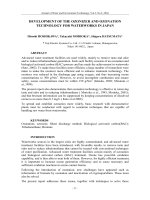
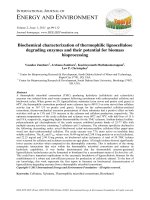
Figure 1 (A) Scanning electron microscopy (SEM) image of poly(L-lactic acid) (PLLA) nanofibrous scaffold with interconnected
spherical macropores created by a phase-separation technique [6]. (B) Electrospun polycaprolactone/hydroxyapatite/gelatin
(PCL/HA/gelatin, 1:1:2) nanofibers which significantly improved osteoblast functions for bone tissue engineering applications [7].
(C) Densely aligned single wall carbon nanotube (SWCNT) forest grown with novel water-assisted chemical vapor deposition in 10 min
[8]. (D) Transmission electron microscopy (TEM) image of monodispersed magnetic Fe
3
O
4
nanoparticles (6 nm) deposited from their
hexane dispersion and dried at room temperature [9].
fibers or other constituent components smaller than 100 nm
in at least one dimension [3], have evoked a great amount of
attention for improving disease prevention, diagnosis, and
treatment.
The intrigue in nanomaterial research for regenerative
medicine is easy to see and is wide spread. For example,
from a material property point-of-view, nanomaterials can
be made of metals, ceramics, polymers, organic materials
and composites thereof, just like conventional or micron
structured materials. Nanomaterials include nanoparti-
cles, nanoclusters, nanocrystals, nanotubes, nanofibers,
nanowires, nanorods, nanofilms, etc. To date, numerous top-
down and bottom-up nanofabrication technologies (such as
electrospinning, phase separation, self-assembly processes,
thin film deposition, chemical vapor deposition, chemical
etching, nano-imprinting, photolithography, and electron
beam or nanosphere lithographies [4]) are available to
synthesize nanomaterials with ordered or random nanoto-
pographies (Fig. 1, [6—9]). Nanomaterials can also be grown
or self-assembled into nanotubes/nanofibers which can even
more accurately simulate the dimensions of natural enti-
ties, such as collagen fibers. After decreasing material size
into the nanoscale, dramatically increased surface area, sur-
face roughness and surface area to volume ratios can be
created to lead to superior physiochemical properties (i.e.,
mechanical, electrical, optical, catalytic, magnetic proper-
ties, etc.) [5]. Therefore, nanomaterials with such excellent
properties have been extensively investigated in a wide
range of biomedical applications, in particular regenerative
medicine.
With the striking increase in the world’s population,
there are enormous demands each year for various biomed-
ical implants to repair diseased or lost tissues. However,
conventional tissue replacements (such as autografts and
allografts) have a variety of problems that cannot satisfy
high performance demands necessary for today’s patient.
Consequently, tissue engineering (or regenerative medicine)
emerged initially defined by Robert Langer and Joseph
Vacanti as ‘‘an interdisciplinary field that applies the
principles of engineering and life sciences toward the devel-
opment of biological substitutes that restore, maintain, or
improve tissue function’’ [10]. However, it is clear that
today, materials used in a wide range of tissue engineering
applications still require improvement. Since natural tissues
or organs are nanometer in dimension and cells directly
interact with (and create) nanostructured extra-cellular
matrices (ECM), the biomimetic features and excellent phys-
iochemical properties of nanomaterials play a key role in
stimulating cell growth as well as guide tissue regenera-
tion. Even though it was a field in its infancy a decade ago,
currently, numerous researchers fabricate cytocompatible
biomimetic nanomaterial scaffolds encapsulating cells (such
68 L. Zhang, T.J. Webster
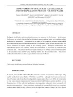
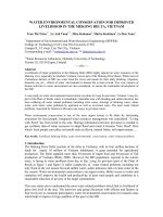
Figure 2 The biomimetic advantages of nanomaterials. (A) The nanostructured hierarchal self-assembly of bone. (B) Nanophase
titanium (top, the atomic force microscopy image) and nanocrystalline HA/HRN hydrogel scaffold (bottom, the SEM image). (C)
Schematic illustration of the mechanism by which nanomaterials may be superior to conventional materials for bone regeneration.
The bioactive surfaces of nanomaterials mimic those of natural bones to promote greater amounts of protein adsorption and
efficiently stimulate more new bone formation than conventional materials.
as stem cells, chondrocytes and osteoblasts, etc.) for tissue
engineering applications. In this review, we will focus on
the recent progress of the use of nanomaterials for bone,
cartilage, vascular, neural and bladder tissue engineering
applications in vitro and more importantly in vivo. As the
next frontier in nanotechnology research, toxicity concerns
of nanomaterials and nanoparticles during manufacturing
and/or implantation will be covered as well.
The promise of nanomaterials for bone and
cartilage tissue engineering applications
Today various bone fractures, osteoarthritis, osteoporosis
or bone cancers represent common and significant clin-
ical problems. The National Center for Health Statistics
(NCHS) reported that bone fractures for all sites num-
bered 1,039,000 in 2004 in the U.S. In addition, around
118,700 patients (home health care) had osteoarthritis and
associated disorders in 2000. The American Academy of
Orthopedic Surgeons also reported that in just a 4 year
period, there was an 83.72% increase in the number of hip
replacements performed from nearly 258,000 procedures in
2000 to 474,000 procedures in 2004 [11]. Such traumatic
bone and cartilage damage happens frequently each year. A
similar trend has been documented for other industrialized
countries as well. However, traditional implant materials
only last 10—15 years on average and implant failures orig-
inating from implant loosening, inflammation, infection,
osteolysis and wear debris frequently occur. It is clearly
urgent to develop a new generation of cytocompatible bone
and cartilage substitutes to regenerate bone/cartilage tis-
sue at defect sites that will last the lifetime of the patient.
Using nanotechnology for regenerative medicine
becomes obvious when examining nature. For example,
bone is a nanocomposite that consists of a protein based
soft hydrogel template (i.e., collagen, non-collagenous
proteins (laminin, fibronectin, vitronectin) and water)
and hard inorganic components (hydroxyapatite, HA,
Ca
10
(PO
4
)
6
(OH)
2
) [12,13] (Fig. 2A). Specifically, 70% of
the bone matrix is composed of nanocrystalline HA which
is typically 20—80 nm long and 2—5 nm thick [14]. Other
protein components in the bone ECM are also nanometer
in dimension. This self-assembled nanostructured ECM in
bone closely surrounds and affects mesenchymal stem cell,
osteoblast (bone-forming cell), osteoclast and fibroblast
adhesion, proliferation and differentiation. Moreover,
cartilage is a low regenerative tissue composed of a small
Nanotechnology and nanomaterials: Promises for improved tissue regeneration 69
percentage of chondrocytes but dense nanostructured ECM
rich in collagen fibers, proteoglycans and elastin fibers.
The limited regenerative properties of cartilage originates
from a lack of chondrocyte mobility in the dense ECM
as well as an absence of progenitor cells and vascular
networks necessary for efficient cartilage tissue repair [15].
Apparently, the design of novel nanomaterials which possess
not only excellent mechanical properties but that are also
biomimetic in terms of their nanostructure (Fig. 2B), has
become quite popular in order to improve bone cell and
chondrocyte functions.
In addition to the dimensional similarity to
bone/cartilage tissue, nanomaterials also exhibit unique
surface properties (such as surface topography, surface
chemistry, surface wettability and surface energy) due to
their significantly increased surface area and roughness
compared to conventional or micron structured materials.
As is known, material surface properties mediate specific
protein (such as fibronectin, vitronectin and laminin)
adsorption and bioactivity before cells adhere on implants,
further regulating cell behavior and dictating tissue regen-
eration [12]. Furthermore, an important criterion for
designing orthopedic implant materials is the formation of
sufficient osseointegration between synthetic materials and
bone tissue. Studies have demonstrated that nanostruc-
tured materials with cell favorable surface properties may
promote greater amounts of specific protein interactions to
more efficiently stimulate new bone growth compared to
conventional materials [16—18] (Fig. 2C). This may be one of
the underlying mechanisms why nanomaterials are superior
to conventional materials for tissue growth. Therefore, by
controlling surface properties, various nanophase ceramic,
polymer, metal and composite scaffolds have been designed
for bone/cartilage tissue engineering applications.
Nanophase ceramics, especially nano-hydroxyapatite
(HA, a native component of bone), are popular bone sub-
stitutes, coatings and other filler materials due to their
documented ability to promote mineralization. The nanome-
ter grain sizes and high surface fraction of grain boundaries
in nanoceramics increase osteoblast functions (such as adhe-
sion, proliferation and differentiation). For example, some
in vitro studies demonstrated that nanophase HA (67 nm
grain size) significantly enhanced osteoblast adhesion and
strikingly inhibited competitive fibroblast adhesion com-
pared to conventional, 179 nm grain size HA, after just 4 h of
culture [17]. Researchers believe they know why. They have
elucidated the highest adsorption of vitronectin (a protein
well known to promote osteoblast adhesion) on nanophase
ceramics, which may explain the subsequent enhanced
osteoblast adhesion on these materials [17]. In addition,
enhanced osteoclast-like cell functions (such as the synthe-
sis of tartrate-resistant acid phosphatase (TRAP) and the
formation of resorption pits) have also been observed on
nano-HA compared to conventional HA [19]. In a recent
study, Nukavarapu et al. fabricated a biodegradable nano-
hydroxyapatite/polyphosphazene microsphere 3-D scaffold
which had suitable mechanical properties (compressive
moduli of 46—81 MPa) and cytocompatibility properties for
bone tissue engineering applications [20]. It should not
be surprising that nanostructured composites have similar
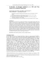
Figure 3 Histology of rat calvaria after 6 weeks of implantation of uncoated tantalum, conventional HA coated tantalum and
nanocrystalline HA coated tantalum. Greater amounts of new bone formation occur in the rat calvaria when implanting nanocrys-
talline HA coated tantalum than uncoated and conventional HA coated tantalum. Red represents new bone and blue represents
collagen. Images are adapted from [21].
70 L. Zhang, T.J. Webster
mechanical properties to bone since bone itself is a nanos-
tructured composite.
Importantly, such results have not been limited to in
vitro studies. In vivo (specifically, rat) studies also demon-
strated that nanocrystalline HA accelerated new bone
formation on tantalum scaffolds when used as an osteo-
conductive coating compared to uncoated or conventional
micron size HA coated tantalum [21]. Histological exami-
nation (Fig. 3) revealed that nanocrystalline HA coatings
promoted greater amounts of new bone growth in the rat
calvaria than uncoated or conventional HA coated tan-
talum after 6 weeks of implantation. Similar tendencies
have been reported for other nanoceramics including alu-
mina, zinc oxide and titania, thus, providing strong evidence
that, to some extent, it may not matter what implant
chemistry is fabricated to have nanometer surface features
to promote bone growth. For example, osteoblast adhe-
sion increased by 146% and 200% on nanophase zinc oxide
(23 nm) and titania (32 nm) compared to microphase zinc
oxide (4.9 m) and titania (4.1 m), respectively [22]. Fur-
thermore, nanophase zinc oxide, nanophase titania and
nanofiber alumina enhanced collagen synthesis, alkaline
phosphatase activity and calcium mineral deposition by
osteoblasts compared to conventional equivalents [22—23].
Because collagen in bone and cartilage is a triple helix
self-assembled into nanofibers 300 nm in length and 1.5 nm
in diameter, many recent efforts have been dedicated to
exploring the influence that novel biomimetic nanofibrous
or nanotubular scaffolds have on regenerative medicine
by following a bottom-up self-assembly process. Specifi-
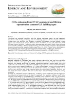
cally, Hartgerink et al. reported that a peptide-amphiphile
(PA) with the cell-adhesive ligand RGD (Arg-Gly-Asp) self-
assembled into supramolecular nanofibers (Fig. 4A and B)
[24]. By directly nucleating and aligning HA on the long axis
of a nanofiber, a new nanofiber composite was designed with
the same self-assembly pattern as collagen and HA crystals
in bone. Moreover, Hosseinkhani et al. investigated mes-
enchymal stem cell (MSC) behavior on self-assembled PA
nanofiber scaffolds [25]. Significantly enhanced osteogenic
differentiation of MSC occurred in the 3-D PA scaffold
compared to 2-D static tissue culture. RGD modified PA
nanofibers promoted the maximum amount of alkaline phos-
phatase activity and osteocalcin content by osteoblasts.
Promise has also been demonstrated for other novel
nanostructured self-assembled chemistries. For example,
osteogenic helical rosette nanotubes obtained through the
self-assembly of DNA base pairs (Guanine∧Cytosine) in aque-
ous solutions (Fig. 4C) have been reported for bone tissue
engineering applications. They have tailorable amino acid
and peptide side chains (such as lysine, RGD and KRSR (Lys-
Arg-Ser-Arg, which selectively promotes osteoblast adhesion
and inhibits fibroblast adhesion)) and are excellent mineral-
ization templates to assemble a biomimetic nanotube/HA
structure (Fig. 4D). Furthermore, significantly improved
osteoblast adhesion has been observed on helical rosette
nanotubes regardless of whether they are incorporated into
hydrogels or coated on titanium (compared to untreated
controls [26,27]). Cartilage tissue engineering has also
benefited from nanostructured self-assembled chemistries.
Kisiday et al. designed a self-assembling peptide (the pep-
tide KLD-12, Lys-Leu-Asp) hydrogel for cartilage repair [28].
The chondrocyte encapsulated scaffold supported chon-
drocyte differentiation and promoted the synthesis of a
cartilage-like ECM matrix (rich in proteoglycans and type
II collagen) in 3-D cell cultures after 4 weeks, thus, showing
promise for cartilage tissue engineering. In summary, by this
self-assembly process, one can create a biologically inspired
3-D scaffold with self-assembled biomimetic features more
suitable for reconstructing 3-D bone and cartilage.
In addition, due to their superior cytocompat-
ible, mechanical and electrical properties, carbon
nanotubes/nanofibers (CNTs/CNFs) are ideal scaffold
candidates for bone tissue engineering applications [29].
In a recent study by Price et al., 60 nm diameter CNFs sig-
nificantly increased osteoblast adhesion and concurrently
decreased competitive cell (fibroblast, smooth muscle cell,
etc.) adhesion in order to stimulate sufficient osseointegra-
tion [30]. Other research efforts have also demonstrated
that CNTs are suitable to promote osteoblast functions
[31]. Recently, Sitharaman and colleagues reported an
in vivo study of ultra-short SWCNT polymer nanocompos-
ites after implanting them into rabbit femoral condyles
and subcutaneous pockets for up to 12 weeks [32]. The
nanocomposites exhibited favorable hard and soft tissue
responses after 4 and 12 weeks. They induced a 300%
greater bone volume than all other experimental groups at
4 weeks and 200% greater bone growth at defect sites than
control polymers without CNTs after 12 weeks. CNT/CNF
reinforced polymer nanocomposites have also demonstrated
excellent electrical conductivity for tissue regeneration.
For instance, using biodegradable polylactic acid (PLA)/CNT
composites as an example, an 80%/20% (w/w) PLA/CNT
composite exhibited ideal electrical conductivity for bone
growth while PLA was an insulator and not appropriate
for electrically stimulating bone growth. Specifically, the
PLA/CNT composite promoted a 46% increase in osteoblast
proliferation and a 307% increase in calcium content after
electrical stimulation for 2 and 21 days compared to PLA
alone, respectively [33]. These studies indicated that the
CNTs/CNFs and their composites can serve as osteogenic
scaffolds with good cytocompatibility properties, reinforced
mechanical properties and improved electrical conductivity
to effectively enhance bone tissue growth.
As mentioned above, synthetic and natural polymers
(e.g., polyglycolic acid (PGA), poly(lactic-co-glycolic acid)
(PLGA), PLLA, PLA, gelatin, collagen, chitosan) are excellent
candidates for bone/cartilage tissue engineering applica-
tions due to their biodegradability and ease of fabrication.
Nanoporous or nanofibrous polymer matrices can be fab-
ricated via electrospinning, phase separation, particulate
leaching, chemical etching and 3-D printing techniques.
For cartilage applications, there has been great interest
in incorporating chondrocytes or progenitor cells (such as
stem cells) into the 3-D polymer or composite scaffolds
during electrospinning [34—36]. For example, Li et al. inves-
tigated in vitro chondrogenesis of MSCs in an electrospun
poly(-caprolactone) (PCL) nanofibrous scaffold [35]. The
differentiation of the stem cells into chondrocytes in the
nanofibrous scaffold was comparable to an established cell
pellet culture. However, the easily fabricated and modified
nanofibers possessed much better mechanical properties
to overcome the disadvantages of using cell pellets and,
thus, were presented as ideal candidates for stem cell
transplantation during clinical cartilage repair. Because the
Nanotechnology and nanomaterials: Promises for improved tissue regeneration 71
Figure 4 Self-assembled nanofibers and nanotubes for bone/cartilage tissue engineering applications. (A) Schematic illustration
of the self-assembly process of peptide-amphiphiles functionalized with RGD to form a nanofiber 7.6 ± 1nm in diameter. Images are
adapted from [24]. (B) TEM image of the above self-assembled nanofibers. (C) Schematic illustration of the self-assembly process
of the Guanine∧Cytosine DNA base pairs forming helical rosette nanotubes (HRNs). (D) SEM images of biomimetic nano-HA aligned
with HRNs on a porous carbon TEM grid.
Figure 5 (A) Schematic illustrating an efficient cell seeding method into a cell—nanofiber composite for cartilage tissue engi-
neering applications. (B) Image of a shiny cartilage-like tissue from the cell—nanofiber composite after 42 days of culture. (C)
Low-magnification histology showing well-dispersed chondrocyte distribution throughout the nanofiber scaffold after 1 day of cell
culture (the cross section). (D) High-magnification histology showing distinct cell populations among the nanofibers. Arrows point
to chondrocytes dispersed among nanofibers. Images are adapted from [36].
72 L. Zhang, T.J. Webster
infiltration of cells is usually inhibited by small pore sizes
of electrospun polymer nanofibers, leading to uneven cell
distributions in the scaffold, a recent study improved chon-
drocyte seeding technology and obtained a homogeneous
cell—PLLA nanofiber composite (Fig. 5) [36]. The results
showed that chondrocytes were uniformly present through-
out the entire cell—nanofiber composite, and the scaffold
developed into a smooth cartilage-like tissue with more
total collagen and improved mechanical properties in a
dynamic bioreactor relative to that obtained in static cul-
ture. Moreover, Park et al. reported significantly increased
chondrocyte functions (adhesion, proliferation and matrix
synthesis) on 3-D nanostructured PLGA created via chemical
etching [37].
For bone tissue engineering, there are a large number of
studies which report the promise of biomimetic 3-D nanos-
tructured polymer scaffolds which encapsulate stem cells
and/or osteoblasts. For instance, Venugopal and colleagues
electrospun a fibrous nanocomposite of PCL/HA/gelatin
at a ratio of 1:1:2 (Fig. 1B). The results demonstrated
that osteoblast proliferation, alkaline phosphatase activity
and mineralization were the highest on the highly flexi-
ble PCL/HA/gelatin nanocomposite when compared to other
PCL nanofibrous scaffolds [7]. Recently, Osathanon et al.
developed a novel polymer/calcium phosphate composite
for bone tissue engineering applications. These nanofibrous
fibrin-based composites promoted osteoblast alkaline phos-
phatase activity as well as osteoblast marker gene (mRNA)
expression to support bone maturation both in vitro and in
vivo in a mouse calvarial defect model [38].
Last but not the least, nanophase metals have been
extensively investigated for orthopedic applications due to
their higher surface roughness, energy, and presence of
more particle boundaries at the surface compared with
conventional micron metals. Webster et al. provided the
first evidence that nanophase Ti, Ti6Al4V and CoCrMo
significantly enhanced osteoblast adhesion compared to
respective conventional metals [39]. In addition, Puckett
et al. created linear patterns of nano-features of Ti via
electron beam evaporation. This study revealed that the
nanoregion of the patterned Ti induced greater osteoblast
adhesion than the micron-rough regions and also controlled
osteoblast morphology and alignment [40]. Moreover, an
electrochemical method known as anodization, a well-
established nanosurface modification technique, has been
used to fabricate highly porous TiO
2
nanotube layers on Ti.
Through the anodization of Ti in dilute hydrofluoric acid
(HF) electrolyte solutions, nanotubes with diameters around
100 nm and lengths around 500 nm can be implemented into
the TiO
2
layers of Ti. Yao et al. reported greatly improved
osteoblast functions on nanotubular anodized Ti compared
to unanodized Ti in vitro [41]. Moreover, increased chondro-
cyte adhesion was also observed on anodized nanotubular Ti
compared to unanodized Ti in a recent study, thus, suggest-
ing the possibility of promoting cartilage growth on anodized
Ti [42].
The promise of nanomaterials for vascular
tissue engineering applications
Due to the increasing prevalence of vascular diseases (such
as atherosclerosis), vascular grafts of greater efficacy to
replace damaged blood vessels are needed. For example,
the American Heart Association reported that coronary heart
disease mostly caused by atherosclerosis had led to 451,326
deaths in 2004 and is the single leading cause of death
in the U.S. today [94]. In addition, peripheral arterial dis-
ease related to blood vessels outside of the heart and brain
affects about 8 million Americans. Over 500,000 coronary
and periphery bypass surgeries were performed in the U.S.
in 2005. Since vascular tissue is a layered structure pos-
Figure 6 Fluorescent microscopy images of greatly increased endothelial cell proliferation on nanostructured Ti compared to
conventional Ti. Scale bar is 10 m. Images are adapted from [43].
Nanotechnology and nanomaterials: Promises for improved tissue regeneration 73
sessing numerous nanostructured features (i.e., due to the
presence of collagen and elastin in the vascular ECM), nano-
materials have shown much promise to improve vascular cell
(specifically, endothelial and smooth muscle cells) functions
to inhibit thrombosis and severe inflammation.
Choudhary et al. reported that vascular cell adhesion
and proliferation were greatly improved on nanostructured
Ti compared to conventional Ti (Fig. 6) [43]. Interestingly,
greater competitive endothelial cell adhesion, total elastin
and collagen synthesis were observed than respective vas-
cular smooth muscle cell functions on nanostructured Ti
after 5 days in culture. Since one of the current problems
with vascular stents is the overgrowth of smooth muscle
cells compared to endothelial cells, these results suggest
that endothelial cell functions were enhanced over that of
vascular smooth muscle cells, thus, increasing the proba-
bility of endothelialization on nanostructured stents. It was
speculated that the increased nano-roughness and particle
boundaries on nanostructured Ti contributed to the observed
favorable endothelial cell functions. In addition, Miller et al.
created biodegradable PLGA vascular grafts with nanome-
ter surface features through chemical etching in NaOH and
through a cast-mold technique [44—46]. Results demon-
strated that both those polymers created through chemical
etching and a polymer cast-mold technique possessed ran-
dom nanometer structures which promoted endothelial and
vascular smooth muscle cell proliferation compared to the
conventional PLGA [44]. A further study provided evidence
that nanostructured PLGA promoted more fibronectin and
vitronectin adsorption from serum than conventional PLGA,
thus, leading to the greater vascular cell responses on the
nanostructured PLGA [45]. In order to elucidate specific
nanometer surface features which promoted vascular cell
responses, 500, 200, and 100 nm polystyrene spheres were
used to cast PLGA [46]. Results demonstrated that the PLGA
with 200 nm structures promoted vascular cell responses and
greater fibronectin interconnectivity compared to smooth
PLGA and PLGA with 500 nm surface features (Fig. 7).
Such results have been translated into the design of
3-D polymer scaffolds as several random and aligned 3-
D nanofiber scaffolds have been fabricated for vascular
applications. For example, Lee and colleagues fabricated
and evaluated a variety of electrospun collagen, elastin
and synthetic polymer (such as PLLA, PLGA and PCL)
nanofiber scaffolds for vascular graft applications [47].
These scaffolds have tailorable mechanical properties
and exceptional cytocompatibility properties for vascular
applications. Specifically, extensive smooth muscle cell infil-
tration was observed in the collagen/elastin/PLLA scaffold
after 21 days of culture. By electrospinning on a rotat-
ing disk collector, Xu et al. fabricated an aligned PLLA-CL
(75:25) nanofibrous scaffold which mimicked the oriented
fibril structure in the medial layer of an artery [48]. Not only
did coronary artery smooth muscle cells favorably interact
with that scaffold, but cells also oriented along the fiber, fur-
ther emulating the natural environment. In addition to the
electrospinning method, self-assembled peptides have been
formulated into scaffolds to mimic the vascular basement
membrane showing excellent cytocompatibility properties
for vascular tissue repair. Genove et al. functionalized three
peptide sequences from two basement membrane proteins
(specifically, laminin and collagen IV) onto a self-assembled
peptide scaffold [49]. These tailorable self-assembled scaf-
folds enhanced endothelialization and improved nitric oxide
release and laminin as well as collagen IV deposition by
the endothelial cell monolayer. These results indicate the
promise of biomimetic nanoscaffolds for improving vascu-
lar tissue engineering applications and when coupled with
the aforementioned promise of nanomaterials for ortho-
pedic applications, suggests a possible wide spread use of
nanomaterials for numerous tissue engineering applications.
The promise of nanomaterials for neural
tissue engineering applications
In addition to aiding in orthopedic and vascular tissue regen-
eration, nanomaterials are also helping to heal damaged
nerves. In particular, nervous system injuries, diseases, and
disorders occur far too frequently. In the U.S., there are
about 250,000—400,000 patients suffering from a spinal cord
injury each year [50]. Although various cell therapies and
implants have been investigated, repairing damaged nerves
and achieving full functional recovery are still challeng-
Figure 7 Atomic force microscopy images of fibronectin (5 g/mL) coated PLGA cast nanosphere surfaces. (A) Phase images of
fibronectin adsorbed on PLGA with 500 nm surface features showed no interconnectivity between proteins. (B) Phase images of
fibronectin adsorbed on PLGA with 200 nm surface features showed significant interconnectivity between fibronectin. (C) PLGA with
200 nm surface features only. Images are adapted from [46].
74 L. Zhang, T.J. Webster
Figure 8 Schematic graphs of injured nerve regeneration in the central and peripheral nervous systems. (A) Central nervous
system recovery process with glial scar tissue formation and (B) peripheral nervous system recovery process involving the activity
of Schwann cells, macrophages, and monocytes. Images are redrawn and adapted from [52,29].
ing considering the complexity of the nervous system. For
example, nearly 50,000 patients die among the average 1.4
million Americans that sustain traumatic brain injuries each
year [51]. Generally, the nervous system can be divided
into two main parts: the central nervous system (CNS)
(including the brain and the spinal cord) and the peripheral
nervous system (PNS) (including the spinal and autonomic
nerves). These two systems have two different repair pro-
cedures after injury (Fig. 8) [52—54]. For the PNS, the
damaged axons usually regenerate and recover via prolifer-
ating Schwann cells, phagocytosing myelin by macrophages
or monocytes, forming bands of Bünger by the bundling of
Schwann cells and sprouting axons in the distal segment
[55]. However, it is difficult to re-extend and re-innervate
axons to recover functions in the CNS due to the absence
of Schwann cells. More importantly, due to the influence
of astrocytes, meningeal cells and oligodendrocytes, the
thick glial scar tissue typically formed around today’s neural
biomaterials will prevent proximal axon growth and inhibit
neuron regeneration [53]. For these reasons, CNS injuries
may cause severe functional damages and are much more
difficult to repair than PNS injuries.
The ideal materials for neural tissue engineering appli-
cations should have excellent cytocompatible, mechanical
and electrical properties. Without good cytocompatibility
properties, materials may fail to improve neuron growth and
at the same time may elicit severe inflammation or infec-
tion. Without sufficient mechanical properties, the scaffold
may not last long enough to physically support neural tis-
sue regeneration. In addition, superior electrical properties
of scaffolds are required to help stimulate and control
neuron behavior under electrical stimulation, thus, more
effectively guiding neural tissue repair. To date, various nat-
ural and synthetic materials have been adopted as nerve
grafts to repair severely damaged nerves by bridging nerve
gaps and guiding neuron outgrowth. However, there are still
many shortcomings for these neural biomaterials including:
for autografts, it is usually difficult to collect sufficient
donor nerves from patients and it is possible donor site
nerve functions may be impaired [56], and for allografts,
inflammation, rejection and transmission of diseases may
frequently occur leading to implant failures [57]. Other
traditional biomaterials (such as silicon probes used in neu-
roprosthetic devices and polymers used as nerve conduits)
Nanotechnology and nanomaterials: Promises for improved tissue regeneration 75
used for neural tissue repair have been limited by the exten-
sive formation of glial scar tissue around the material as
well as non-optimal mechanical and electrical properties for
nerve regrowth. Nanotechnology provides a wide platform to
develop novel and improved neural tissue engineering mate-
rials and therapy including designing nanofiber/nanotube
scaffolds with exceptional cytocompatibility and conduc-
tivity properties to boost neuron activities. Nanomaterials
have also been used to encapsulate various neural stem cells
and Schwann cells into biomimetic nanoscaffolds to enhance
nerve repair.
For example, work by Ramakrishna et al. has led to the
fabrication of various nanofibrous PLLA or PCL scaffolds
via electrospinning and phase separation; such scaffolds
have demonstrated excellent cytocompatibility properties
for neural tissue engineering applications [58—60]. Recently
this research group incorporated laminin (a neurite pro-
moting ECM protein) into electrospun PLLA nanofibers in
order to create a biomimetic scaffold for peripheral nerve
repair [58]. The results showed that neurite outgrowth
improved on laminin-PLLA scaffolds produced by facile
blended electrospinning. In another recent report, electro-
spun PCL/chitosan nanofiber scaffolds exhibited improved
mechanical properties compared to chitosan [59]. Schwann
cells also proliferated well on this PCL/chitosan nanofiber
scaffold. In addition, Zhang and colleagues also reported
favorable neural cell responses on the self-assembled
peptide nanofiber scaffold (called SAPNS). Holmes et al.
reported that the self-assembled peptide scaffold sup-
ported neuronal cell functions, neurite outgrowth and
functional synapse formation among neurons [61]. Further-
more, Ellis-Behnke et al. investigated SAPNS for in vivo axon
regeneration in the CNS [62]. The SAPNS aided in CNS regen-
eration to help axonal growth, even ‘‘knitting’’ the brain
tissue together and successfully improving functional recov-
ery.
Due to the fact that carbon nanotubes/fibers have excel-
lent electrical conductivity, strong mechanical properties,
and have similar nanoscale dimensions to neurites, they
have been used to guide axon regeneration and improve
neural activity as biomimetic scaffolds at neural tissue
injury sites. In particular, Mattson et al. found for the
first time that neurons grew on multiwalled carbon nan-
otubes (MWCNTs) [63]. They observed over a 200% increase
in total neurite length and nearly a 300% increase in
the number of branches and neurites on MWCNTs coated
with 4-hydroxynonenal compared to uncoated MWCNTs. Hu
et al. revealed that different surface charges of MWC-
NTs, obtained through chemical functionalization, resulted
in different neurite outgrowth patterns (such as neurite
length, branching and the number of growth cones) [64].
They demonstrated that positively charged MWCNTs signifi-
cantly increased the number of growth cones and neurite
branches compared to negatively charged MWCNTs, thus,
controlling neural growth. Lovat et al. demonstrated that
purified MWCNTs potentially boosted electrical signal trans-
fer of neuronal networks (Fig. 9A and B) [65]. Moreover,
highly ordered CNT/CNF matrices or free standing nanotube
films have been fabricated for neural tissue engineering
applications [66—67]. For instance, Gheith et al. inves-
tigated the biocompatibility of a freestanding positively
charged SWCNT/polymer thin-film membrane prepared by
layer-by-layer assembly [66]. They observed that 94—98%
of neurons were viable on the SWCNT/polymer films after
a 10 day incubation. The SWCNT/polymer films favor-
ably induced neuronal cell differentiation, guided neuron
extension and directed more elaborate branches than con-
trols.
In order to inhibit activated astrocyte functions which
result in the formation of glial scar tissue, McKenzie et
al. incorporated different weight ratios of high surface
energy CNFs into polymers and demonstrated for the first
time that astrocyte adhesion can be effectively inhib-
ited by using CNF/polymer composites [68]. In addition,
decreased astrocyte proliferation was observed on nanos-
tructured CNFs, thus, leading to decreased glial scar tissue
formation on such materials. On the other hand, Nguyen-
Vu et al. fabricated a vertically aligned CNF nanoelectrode
array by creating a thin conductive polymer film coating
(such as polypyrrole) for neural implants [69]. The verti-
cal CNF arrays had more open and mechanically robust 3-D
structures as well as better electrical conductivity which
contributed to forming an intimate neural-electrical inter-
face between cells and nanofibers (Fig. 9C—E). Gabay et
Figure 9 SEM images of neural cell adhesion on carbon nanotube/fiber substrates. (A) Neonatal hippocampal neurons adherent
on purified MWCNT glass substrates with extended neurites after 8 days; inset image (B) shows a single neurite in close contact to
CNTs. Images are adapted from [65]. (C), (D) and (E) PC12 neural cells grown freestanding on vertically aligned CNFs coated with
polypyrrole at different magnifications. Images are adapted from [69].
76 L. Zhang, T.J. Webster
al. developed a novel method to fabricate islands of CNT
on substrates. Neurons preferably attached on the CNT
islands and further extended their neurites to form intercon-
nected neural networks according to pre-designed patterns
[70]. In this manner, the CNTs/CNFs and their composites
are promising scaffold candidates for injured neural tissue
repair.
Studies have also provided evidence that individual
CNTs/CNFs may be useful in treating neurological dam-
age when combined with stem cells. Stem cells have the
potential to differentiate and self-renew into controllable,
desirable cell types: i.e., neural stem cells in the CNS can
differentiate into neurons and astrocytes [71]. Therefore,
many efforts have focused on impregnating multi-potential
stem cells into CNTs/CNFs and other nanoscaffolds, which
can be directly transplanted into injury sites and assist neu-
ral tissue recovery. However, a challenging problem has
been to determine how to effectively deliver and selec-
tively differentiate stem cells into favorable neuronal cell
types at injury sites in order to regenerate desirable tissue.
Although the underlying mechanisms triggering differen-
tiation of stem cells are not entirely clear, accumulated
evidence has indicated that novel biomimetic nanomate-
rials may contribute to selective stem cell differentiation
(without the use of growth factors) [72,73]. For exam-
ple, Lee et al. injected CNFs impregnated with stem cells
into stroke damaged neural tissue in rat brains and found
extensive neural stem cell differentiation with little glial
scar tissue formation in vivo [72]. After 1 and 3 weeks
of animal implantation, histological sections showed that
neural stem cells favorably differentiated into neurons
(Fig. 10A and B) and little to no glial scar tissue (Fig. 10C
and D) formed around CNFs compared to controls (only
implanting stem cells without CNFs or implanting CNFs
without cells). Furthermore, Jan et al. successfully dif-
ferentiated mouse embryonic neural stem cells including
neurospheres and single cells into neurons on layer-by-
layer assembled SWCNT/polyelectrolyte composites [73].
The layer-by-layer SWCNT composites promoted slightly
more neurons and fewer astrocytes on substrates during
a 7 day culture period than poly-
L-ornithine (a common
substrate for neural stem cell studies). Clearly, CNTs/CNFs
played an important role in effectively delivering stem
cells into injured sites and promoted stem cells to differ-
entiate into favorable neurons to repair damaged neural
tissues.
The promise of nanomaterials for bladder
tissue engineering applications
Nanomaterials have also been used in soft tissues, such as
the bladder. As the 6th most common cancer in the U.S.,
urinary bladder cancer affects over 53,200 Americans and
Figure 10 Histology of CNFs impregnated with stem cells into stroke damaged rat neural tissue after 3 weeks. (A) and (B), numerous
active neuroprogenitor cells and fully differentiated neurons (brown stained cells, marked by nestin and MAP2, respectively) were
found around CNFs. (C) and (D), few glial cells interacting with CNFs led to little or no glial scar tissue formation. GFAP is a marker
for astrocytes; CD11b is a marker for activated microglia cells. Black areas in the images are CNFs. Scale bar is 25 m. Images are
adapted from [72].
Nanotechnology and nanomaterials: Promises for improved tissue regeneration 77
Figure 11 Schematic illustration of the bilayer smooth muscle cell/urothelial cell (SMC-UC) encapsulation in a PA/PGA gel. Image
is adapted from [76].
leads to 12,200 deaths annually [74]. Although standard
treatments such as surgery to remove bladder tumors fol-
lowed by radiation, chemotherapy and immunotherapy have
improved, various complications (such as systemic infec-
tions, flu-like symptoms and cancer recurrence, etc.) with
these procedures are still too commonly reported. Some-
times, radical cystectomy by removing parts of and even the
entire bladder is needed. However, such a drastic approach
requires the implantation of a bladder tissue replacement
to quickly recover bladder functions. As emerging blad-
der tissue engineering materials, nanomaterials provide
a promising approach to more efficiently improve blad-
der tissue regeneration for the same reasons mentioned
earlier for other tissue systems (biologically inspired rough-
ness, increased surface energy, selective protein adsorption,
etc.). In particular, Harrington and colleagues have coated
a series of branched or linear self-assembling peptide-
amphiphile nanofibers containing cell-adhesive RGDS on
traditional PGA scaffolds [75]. Human bladder smooth mus-
cle cell densities on the branched PA/PGA nanocomposite
were greater than on the uncoated PGA after 17 days of cul-
ture. In a recent review, they encapsulated bladder smooth
muscle cells and urothelial cells into a PA/PGA nanofi-
brous gel containing specific growth factors (Fig. 11) [76].
Due to their ability to mimic the oriented nanostructured
bladder ECM, electrospun polymer nanofibers have been
used in bladder tissue engineering. Baker et al. showed
that bladder smooth muscle cells were aligned on oriented
electrospun polystyrene scaffolds similar to the native blad-
der tissue [77]. This study also demonstrated that argon
plasma treated electrospun polystyrene nanofibers signifi-
cantly improved smooth muscle cell attachment. Fibrinogen
has also been electrospun into a scaffold for urinary tract
tissue regeneration [78]. This study demonstrated that
human bladder smooth muscle cells rapidly migrated into,
proliferated onto and remodeled the 3-D fibrinogen scaf-
fold.
Other nanostructured polymers with superior biocom-
patibility properties have been widely investigated by
Haberstroh and colleagues for bladder tissue regener-
ation applications [79—81]. For instance, this research
group used nanotextured PLGA and poly(ether urethane)
(PU) films to successfully enhance bladder smooth muscle
cell functions [79]. Through chemical etching technolo-
gies, PLGA and PU were transformed from their native
nano-smooth surface features into those possessing a high
degree of nano-roughness. This study revealed that nano-
roughness played a critical role in promoting bladder
smooth muscle cell proliferation once the influence of
surface chemistry change was eliminated (through cast-
mold techniques using the chemical treated polymer as
the cast). Recently, Pattison et al. also demonstrated
that nanostructured PLGA and PU 3-D scaffolds prepared
by a solvent casting and salt leaching methods signifi-
cantly enhanced bladder smooth muscle cell functions and
ECM protein synthesis compared to conventional nano-
smooth polymers in vitro [80]. Furthermore, preliminary
in vivo studies have provided evidence that nanostruc-
tured polymer scaffolds form little to no calcium oxalate
stones (stone formation is a common problem during blad-
der replacements) in augmented rat bladders. Although
there are many unknowns for the use of nanomaterials
in bladder tissue engineering applications, utilizing these
biomimetic nanomaterials with progenitor cells is undoubt-
edly a promising future research direction to regenerate
bladder tissue in resected bladder cancerous tissue loca-
tions.
Potential risks of nanomaterials towards
human health
As described, nanotechnology has achieved tremendous
progress in a relatively short time period in medical applica-
tions. As a result, nanomaterials have begun to enter wide
spread industrial production. For instance, nanoceramics
are commercially available as new bone grafts or as implant
coating materials (i.e., nano-HA paste—–Ostim
®
from Obern-
burg, Germany; nano-beta-tricalcium phosphate-Vitoss from
Orthovita, USA) [82]. However, it is important to note that
the research on nanomaterials for tissue engineering appli-
cations is still at its infancy and, most importantly, the
78 L. Zhang, T.J. Webster
influence of nanomaterials on human health and the environ-
ment is not well understood. In particular, toxic responses to
nanoparticles generated from the degradation of implanted
nanomaterials, via wear debris from artificial joints with
nanofeatures, and heavy metals (iron, nickel and cobalt
catalysts) remaining in CNTs, have all been reported. Many
reports on the cellular uptake of nanoparticles in the lungs,
immune system, as well as other organs have been pub-
lished [83—85]. Nanoparticle uptake by endothelial cells,
alveolar macrophages, pulmonary or intestinal epithelium,
nerve cells etc. has been reviewed and, thus, may possess
a problem for this field if not thoroughly understood before
being applied widely [83]. Gutwein et al. investigated the
viability of osteoblasts in vitro when cultured in the pres-
ence of nanoalumina and titania particles for 6 h [84]. This
study demonstrated that ceramic nanoparticles were safer
to osteoblasts than conventional, micron-sized, ceramics
particles. In contrast, in an in vivo study, Lam et al. showed
that CNTs were more toxic than carbon black in the lungs,
which may be a serious occupational health hazard in chronic
inhalation exposures [85]. Sometimes nanoparticle interac-
tions with biomolecules in vivo or their aggregation states
may change their toxicity to humans. But the often con-
tradictory results of current studies are clearly not enough
to provide the final answer concerning nanomaterial tox-
icity. In depth investigations of nanomaterials on human
health and the environment are necessary to fully eluci-
date whether nanoparticles should be used in biomedical
applications.
Conclusions
To date, there has been an exponential increase in stud-
ies using nanotechnology for tissue engineering applications.
To be concise, this paper only covered the recent progress
using nanomaterials for bone, cartilage, vascular, neural
and bladder tissue regeneration. Other reviews of nanotech-
nology applications for the specific regeneration of tissues
can be found [15,76,86—92]. Nanotechnology approaches
for the regeneration of other types of tissues (such as
the muscle, skin, kidneys, liver, pancreas, and the immune
system) have also been reviewed [92,93]. Although many
challenges may lie ahead, synthetic nanomaterials can
mimic properties of the natural ECM and thus, show great
potential for numerous tissue engineering applications.
Particularly, due to their excellent cytocompatibility proper-
ties, research interest has been evoked to use nanomaterials
as the next generation of tissue repair materials. In the
future, the underlying mechanisms of the in vivo interac-
tions between nanomaterials and cells at the molecular
level will significantly advance the development of this
field.
References
[1] N. Taniguchi, Proc. of International Conference on Precision
Engineering (ICPE), Tokyo, Japan, 1974, pp. 18—23.
[2] T.J. Webster, Int. J. Nanomed. 2 (2007) 1.
[3] R.W. Siegel, G.E. Fougere, Nanostruct. Mater. 6 (1995) 205.
[4] J.W. Freeman, L.D. Wright, C.T. Laurencin, S. Bhattacharyya,
in: K.E. Gonsalves, C.R. Halberstadt, C.T. Laurencin, L.S. Nair
(Eds.), Biomedical Nanostructures, John Wiley & Sons, Inc.,
New Jersey, 2008, pp. 3—24.
[5] B.D. Fahlman, Materials Chemistry, Springer, Dordrecht,
Netherlands, 2007.
[6] V.J. Chen, P.X. Ma, Biomaterials 25 (2004) 2065.
[7] J.R. Venugopal, S. Low, A.T. Choon, A.B. Kumar, S. Ramakr-
ishna, Artif. Organs 32 (2008) 388.
[8] K. Hata, D.N. Futaba, K. Mizuno, T. Namai, M. Yumura, S. Iijima,
Science 306 (2004) 1362.
[9] S. Sun, H. Zeng, D.B. Robinson, S. Raoux, P.M. Rice, S.X. Wang,
et al., J. Am. Chem. Soc. 126 (2004) 273.
[10] R. Langer, J.P. Vacanti, Science 260 (1993) 920.
[11] American Academy of Orthopedic Surgeons (AAOS), online:
/>[12] T.J. Webster, in: J.Y. Ying (Ed.), Advances in Chemical Engi-
neering, Academic Press, New York, 2001, pp. 125—166.
[13] L. Zhang, S. Sirivisoot, G. Balasundaram, T.J. Webster, in: B.
Basu, D. Katti, A. Kumar (Eds.), Advanced Biomaterials: Funda-
mentals, Processing and Applications, John Wiley & Sons, Inc.,
New Jersey, in press.
[14] F.S. Kaplan, W.C. Hayes, T.M. Keaveny, A. Boskey, T.A. Einhorn,
J.P. Iannotti, in: S.R. Simon (Ed.), Orthopedic Basic Science,
American Academy of Orthopaedic Surgeons, Rosemont, 1994,
pp. 127—185.
[15] R. Vasita, D.S. Katti, Int. J. Nanomed. 1 (2006) 15.
[16] I. Degasne, M.F. Baslé, V. Demais, G. Huré, M. Lesourd, B.
Grolleau, et al., Calcified Tissue Int. 64 (1999) 499.
[17] T.J. Webster, C. Ergun, R.H. Doremus, R.W. Siegel, R. Bizios,
J. Biomed. Mater. Res. 51 (2000) 475.
[18] T.J. Webster, L.S. Schadler, R.W. Siegel, R. Bizios, Tissue Eng.
7 (2001) 291.
[19] T.J. Webster, C. Ergun, R.H. Doremus, R.W. Seigel, R. Bizios,
Biomaterials 22 (2001) 1327.
[20] S.P. Nukavarapu, S.G. Kumbar, J.L. Brown, N.R. Krogman, A.L.
Weikel, M.D. Hindenlang, et al., Biomacromolecules 9 (2008)
1818.
[21] M. Sato, Nanophase hydroxyapatite coatings for dental
and orthopedic applications, PhD Thesis, Purdue University,
2006.
[22] G. Colon, B.C. Ward, T.J. Webster, J. Biomed. Mater. Res. A 78
(2006) 595.
[23] T.J. Webster, E.L. Hellenmeyer, R.L. Price, Biomaterials 26
(2005) 953.
[24] J.D. Hartgerink, E. Beniash, S.I. Stupp, Science 294 (2001)
1684.
[25] H. Hosseinkhani, M. Hosseinkhani, F. Tian, H. Kobayashi, Y.
Tabata, Biomaterials 27 (2006) 4079.
[26] L. Zhang, S. Ramsaywack, H. Fenniri, T.J. Webster, Tissue Eng.
Part A 14 (2008) 1353.
[27] L. Zhang, Y. Chen, J. Rodriguez, H. Fenniri, T.J. Webster, Int.
J. Nanomed. 3 (2008) 323.
[28] J. Kisiday, M. Jin, B. Kurz, H. Hung, C. Semino, S. Zhang, et
al., Proc. Natl. Acad. Sci. 99 (2002) 9996.
[29] L. Zhang, B. Ercan, T.J. Webster, in: C. Liu (Ed.), The Area of
‘Carbon’, Research Signpost, Trivandrum, in press.
[30] R.L. Price, M.C. Waid, K.M. Haberstroh, T.J. Webster, Bioma-
terials 24 (2003) 1877.
[31] L.P. Zanello, B. Zhao, H. Hu, R.C. Haddon, Nano Lett. 6 (2006)
562.
[32] B. Sitharaman, X. Shi, X.F. Walboomers, H. Liao, V. Cuijpers,
L.J. Wilson, et al., Bone 43 (2008) 362.
[33] P.R. Supronowicz, P.M. Ajayan, K.R. Ullmann, B.P. Arulanan-
dam, D.W. Metzger, R. Bizios, J. Biomed. Mater. Res. 59 (2002)
499.
[34] C. Fecek, D. Yao, A. Kac¸orri, A. Vasquez, S. Iqbal, H. Sheikh,
et al., Tissue Eng. Part A 14 (2008) 1403.
[35] W.J. Li, R. Tuli, C. Okafor, A. Derfoul, K.G. Danielson, D.J. Hall,
et al., Biomaterials 26 (2005) 599.
Nanotechnology and nanomaterials: Promises for improved tissue regeneration 79
[36] W.J. Li, Y.J. Jiang, R.S. Tuan, Tissue Eng. Part A 14 (2008)
639.
[37] G.E. Park, M.A. Pattison, K. Park, T.J. Webster, Biomaterials 26
(2005) 3075.
[38] T. Osathanon, M.L. Linnes, R.M. Rajachar, B.D. Ratner, M.J.
Somerman, C.M. Giachelli, Biomaterials 29 (2008) 4091.
[39] T.J. Webster, J.U. Ejiofor, Biomaterials 25 (2004) 4731.
[40] S. Puckett, R. Pareta, T.J. Webster, Int. J. Nanomed. 3 (2008)
229.
[41] C. Yao, V. Perla, J.L. McKenzie, E.B. Slamovich, T.J. Webster,
J. Biomed. Nanotechnol. 1 (2005) 68.
[42] K. Burns, C. Yao, T.J. Webster, J. Biomed. Mater. Res. A,
doi:10.1002/jbm.a.31899, in press.
[43] S. Choudhary, K.M. Haberstroh, T.J. Webster, Tissue Eng. Part
A 13 (2007) 1421.
[44] D.C. Miller, A. Thapa, K.M. Haberstroh, T.J. Webster, Biomate-
rials 25 (2004) 53.
[45] D.C. Miller, K.M. Haberstroh, T.J. Webster, J. Biomed. Mater.
Res. 73A (2005) 476.
[46] D.C. Miller, K.M. Haberstroh, T.J. Webster, J. Biomed. Mater.
Res. 81A (2007) 678.
[47] S.J. Lee, J.J. Yoo, G.J. Lim, A. Atala, J. Stitzel, J. Biomed.
Mater. Res. 83A (2007) 999.
[48] C.Y. Xu, R. Inai, M. Kotaki, S. Ramakrishna, Biomaterials 25
(2004) 877.
[49] E. Genové, C. Shen, S. Zhang, C.E. Semino, Biomaterials 26
(2005) 3341.
[50] />stats.htm.
[51] J.A. Langlois, W. Rutland-Brown, K.E. Thomas, Traumatic Brain
Injury in The United States: Emergency Department Visits,
Hospitalizations, and Deaths, Centers for Disease Control and
Prevention, National Center for Injury Prevention and Control,
Atlanta, 2004.
[52] M. Bahr, F. Bonhoeffer, Trends Neurosci. 17 (1994) 473.
[53] N. Zhang, H. Yan, X. Wen, Brain Res. Rev. 49 (2005) 48.
[54] Y.C. Huang, Y.Y. Huang, Artif. Organs 30 (2006) 514.
[55] G.R.D. Evans, Anat. Rec. 263 (2001) 396.
[56] J.K. Terzis, D.D. Sun, P.K. Thanos, J. Reconstr. Microsurg. 13
(1997) 215.
[57] A.A. Zalewski, A.K. Gulati, Transplantation 31 (1981) 88.
[58] H.S. Koh, T. Yong, C.K. Chan, S. Ramakrishna, Biomaterials 29
(2008) 3574.
[59] M.P. Prabhakaran, J. Venugopal, T.T. Chyan, L.B. Hai, C.K.
Chan, A.L. Tang, et al., Tissue Eng. Part A 14 (2008) 1787.
[60] F. Yang, R. Murugan, S. Ramakrishna, X. Wang, Y.X. Ma, S. Wang,
Biomaterials 25 (2004) 1891.
[61] T.C. Holmes, S.D. Lacalle, X. Su, G. Liu, A. Rich, S. Zhang, PNAS
97 (2000) 6728.
[62] R.G. Ellis-Behnke, Y.X. Liang, S.W. You, D.K. Tay, S. Zhang, K.F.
So, et al., PNAS 103 (2006) 5054.
[63] M.P. Mattson, R.C. Haddon, A.M. Rao, J. Mol. Neurosci. 14
(2000) 175.
[64] H. Hu, Y. Ni, V. Montana, R.C. Haddon, V. Parpura, Nano Lett.
4 (2004) 507.
[65] V. Lovat, D. Pantarotto, L. Lagostena, B. Cacciari, M.
Grandolfo, M. Righi, et al., Nano Lett. 5 (2005) 1107.
[66] M.K. Gheith, V.A. Sinani, J.P. Wicksted, R.L. Matts, N.A. Kotov,
Adv. Mater. 17 (2005) 2663.
[67] I. Firkowska, M. Olek, N. Pazos-Peréz, J. Rojas-Chapana, M.
Giersig, Langmuir 22 (2006) 5427.
[68] J.L. McKenzie, M.C. Waid, R. Shi, T.J. Webster, Biomaterials 25
(2004) 1309.
[69] T.D.B. Nguyen-Vu, H. Chen, A.M. Cassell, R.J. Andrews, M.
Meyyappan, J. Li, IEEE Trans. Biomed. Eng. 54 (2007) 1121.
[70] T. Gabay, E. Jakobs, E. Ben-Jacob, Y. Hanein, Physica A 350
(2005) 611.
[71] B.A. Reynolds, S. Weiss, Science 255 (1992) 1707.
[72] J.E. Lee, J.H. Kim, J.Y. Kim, D. Kang, T.J. Webster, Int. J.
Nanomed., in press.
[73] E. Jan, N.A. Kotov, Nano Lett. 7 (2007) 1123.
[74] American Urological Association, logy-
health.org/adult/index.cfm?cat=03&topic=37.
[75] D.A. Harrington, E.Y. Cheng, M.O. Guler, L.K. Lee, J.L. Dono-
van, R.C. Claussen, et al., J. Biomed. Mater. Res. 78A (2006)
157.
[76] D.A. Harrington, A.K. Sharma, B.A. Erickson, E.Y. Cheng, World
J. Urol. 26 (2008) 315.
[77] S.C. Baker, N. Atkin, P.A. Gunning, N. Granville, K. Wilson, D.
Wilson, et al., Biomaterials 27 (2006) 3136.
[78] M. McManus, E. Boland, S. Sell, W. Bowen, H. Koo, D. Simpson,
et al., Biomed. Mater. 2 (2007) 257.
[79] A. Thapa, D.C. Miller, T.J. Webster, K.M. Haberstroh, Biomate-
rials 24 (2003) 2915.
[80] M. Pattison, T.J. Webster, J. Leslie, M. Kaefer, K.M. Haberstroh,
Macromol. Biosci. 7 (2007) 690.
[81] M.A. Pattison, S. Wurster, T.J. Webster, K.M. Haberstroh, Bio-
materials 26 (2005) 2491.
[82] V. Wagner, A. Dullaart, A.K. Bock, A. Zweck, Nat. Biotechnol.
24 (2006) 1211.
[83] P.H.M. Hoet, I. Bruske-Hohlfeld, O.V. Salata, J. Nanobiotech-
nol. 2 (2004) 2.
[84] L.G. Gutwein, T.J. Webster, Biomaterials 25 (2004) 4175.
[85] C.W. Lam, J.T. James, M. Richard, R.L. Hunter, Toxicol. Sci. 77
(2004) 126.
[86] E.M. Christenson, K.S. Anseth, J.J. van den Beucken, C.K.
Chan, B. Ercan, J.A. Jansen, et al., J. Orthop. Res. 25 (2007)
11.
[87] T.J. Webster, E.S. Ahn, Adv. Biochem. Eng. Biotechnol. 103
(2007) 275.
[88] L. Zhang, S. Sirivisoot, G. Balasundaram, T.J. Webster, in: A.
Khademhosseini, J. Borenstein, M. Toner, S. Takayama (Eds.),
Micro and Nanoengineering of the Cell Microenvironment: Tech-
nologies and Applications, Artech House, Norwood, 2008, pp.
431—460.
[89] D.C. Miller, T.J. Webster, K.M. Haberstroh, Expert Rev. Med.
Devices 1 (2004) 259.
[90] S.K. Seidlits, J.Y. Lee, C.E. Schmidt, Nanomedicine 3 (2008)
183.
[91] P. Liu-Snyder, T.J. Webster, Expert Rev. Med. Devices 3 (2006)
683.
[92] S.P. Nukavarapu, S.G. Kumbar, L.S. Nair, C.T. Laurencin, in: K.E.
Gonsalves, C.R. Halberstadt, C.T. Laurencin, L.S. Nair (Eds.),
Biomedical Nanostructures, John Wiley & Sons, Inc., New Jer-
sey, 2008, pp. 377—407.
[93] A. Khademhosseini, J. Borenstein, M. Toner, S. Takayama,
Micro and Nanoengineering of the Cell Microenvironment:
Technologies and Applications, Artech house, Norwood,
2008.
[94] American Heart Association, />Lijie Zhang received a BS degree in chemical
engineering from Tianjin University, China,
in 2001 and a MS degree in chemical engi-
neering from Brown University in 2007. She is
currently pursuing her PhD in biomedical engi-
neering at Brown University. She has interests
in developing novel nanobiomaterials for
bone tissue engineering, orthopedic implant
and vascular stent applications. She has pub-
lished over fourteen journal papers as well as
conference proceedings, three book chapters
and has presented her work at over eighteen conferences. She
was also the recipient of the Society for Biomaterials STAR Award
(2007).
80 L. Zhang, T.J. Webster
Thomas J. Webster is an associate profes-
sor of Engineering and Orthopedics at Brown
University. His degrees are in chemical engi-
neering from the University of Pittsburgh
(BS, 1995) and in biomedical engineering
from Rensselaer (MS, 1997; PhD, 2000). His
research addresses the design, synthesis, and
evaluation of nanophase materials for various
regenerative medicine applications. His lab
group has generated 4 books, 33 book chap-
ters, 85 invited presentations, 215 literature
articles, and 245 conference presentations. He is the found-
ing editor-in-chief of the International Journal of Nanomedicine
and is on the editorial board for 10 other journals. Among
other awards, Dr. Webster received the 2002 Biomedical Engi-
neering Society Rita Schaffer Young Investigator Award, 2004
Outstanding Young Investigator Award from Purdue University, 2005
Wallace Coulter Foundation Early Career Award, and in 2007 was
elected as a Fellow of the American Academy of Nanomedicine.
His research has led to the formation of two nanotech
companies.