- Trang chủ >>
- Khoa Học Tự Nhiên >>
- Vật lý
stability of anodic aluminum oxide membranes with nanopores
Bạn đang xem bản rút gọn của tài liệu. Xem và tải ngay bản đầy đủ của tài liệu tại đây (321.28 KB, 5 trang )
Physics Letters A 318 (2003) 440–444
www.elsevier.com/locate/pla
Stability of anodic aluminum oxide membranes with nanopores
S.G. Yang
a,∗
,T.Li
a
,L.S.Huang
a
,T.Tang
a
, J.R. Zhang
a
,B.X.Gu
a
,Y.W.Du
a
,
S.Z. Shi
b
,Y.N.Lu
b
a
National Laboratory of Solid State Microstructures, Nanjing University, Nanjing, China
b
College of Materials Science and Engineering, Nanjing University of Technology, Nanjing 210009, China
Received 17 August 2003; received in revised form 22 September 2003; accepted 22 September 2003
Communicated by R. Wu
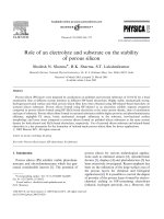
Abstract
The anodic aluminum oxide membranes (AAOMs) with nanopores, produced by electrochemical etching method in
oxalic/sulphuric/phosphoric acid, were transformed from amorphous phase to crystalline phase by annealing. Differential
thermal analysis and X-ray diffraction were performed for the phase transformation studies. Scanning electron microscopy
studies show that the crystalline AAOMs keep the nanopore morphology and become very stable in the violent acid and violent
alkali solutions.
2003 Elsevier B.V. All rights reserved.
PACS: 81.40.Ef; 81.70.Pg; 81.05.Rm; 82.33 z
Keywords: Anodic aluminum oxide membrane; Nanopore; Stability
Nanoscale materials have being widely studied
because of their peculiar properties and potential
applications. One-dimensional nanoscale materials,
nanowires and nanotubes, have attracted much atten-
tion in recent years [1–3]. One of the most impor-
tant method forthe preparation of the one-dimensional
nanoscale materials is the template method, which use
the membranes with nanopore channels as the tem-
plate. In the template method, anodic aluminum oxide
membranes (AAOMs), prepared by electrochemical
etching aluminum foil in oxalic/sulphuric/phosphoric
acid solution are the most popular membranes used
*
Corresponding author.
E-mail address: (S.G. Yang).
in the experiments. For example, the AAOMs have
been used in the fabrication of ordered carbon nano-
tube arrays [4], wide gap semiconductor nanowires
[5–8] and superconductor nanowire arrays [9]. The
magnetic nanowire arrays prepared by electrodeposit-
ing the corresponding materials into the nanopores
of the AAOMs are regarded as a potential media in
high density magnetic storage [10–12]. In all these
experiments, the synthesis environmental condition is
vapor or nearly neutral solutions. Many experiments
have shown that the AAOMs are unstable and can be
dissolved in the violent acid or alkali solutions. This
property confines the application areas ofthe AAOMs.
In the previous studies, thermal treatment and solu-
bility of the AAOMs have been performed carefully
in the view point of chemistry [17]. In this Letter, in
0375-9601/$ – see front matter 2003 Elsevier B.V. All rights reserved.
doi:10.1016/j.physleta.2003.09.051
S.G. Yang et al. / Physics Letters A 318 (2003) 440–444 441
the view point of one-dimensional nanoscale materi-
als synthesis, we studied the stability difference of the
AAOMs before and after calcinations.
The AAOMs can be formed in oxalic acid, sul-
phuric acid or phosphoricacid solutions [13]. By using
high purity (99.999%) aluminum foil as the starting
material, three groups of AAOMs were prepared in ox-
alic acid/sulphuric acid/phosphoricacid solution sepa-
rately with all the acid concentration of 0.4 mol/l. The
electrochemical etching time was 20 hours for all the
samples. The environmental condition was ice/water
which kept the acid solution temperature with 0
◦
C
in the whole process. The anodic voltages used in
the experiments were 60, 28 and 140 V for oxalic
acid, sulphuric acid and phosphoric acid, respectively.
The distance between the neighboring pore centers
was fixed after the electrochemical etching, they were
about 150, 65 and 400 nm, respectively. The diameter
of the nanopores can be adjusted by phosphoric acid
solution. After removal of the remaining aluminum,
the AAOMs were put into a 30
◦
C phosphoric acid
with 10 wt% concentration for 40 minutes to adjust the
nanopore diameters. The diameters of the nanopores
were about 70, 35 and 120 nm for the three groups of
prepared AAOMs. These results will be shown in the
following scanning electron microscopy (SEM) stud-
ies. These AAOMs were used as the prepared mate-
rials for further studies in this Letter. For simplicity,
the AAOMs prepared in the oxalic acid were used as
an example. The properties of the AAOMs prepared
in sulphuric acid and phosphoric acid are similar with
that of the AAOMs prepared in the oxalic acid.
In the stability studies of the AAOMs, a sulphuric
acid with 1 mol/l concentration and a sodium hydrox-
ide solution with 1 mol/l were used as the testing solu-
tion. The temperature of the testing solution was fixed
at 50
◦
C with water bath. These two solutions are typi-
cal for violent acid and violent alkali. Generally speak-
ing, if the AAOMs are stable in these testing solutions,
they will be stable in other violent acid or violent al-
kali solutions.
Two pieces of prepared AAOMs were put into
the testing solutions separately. The AAOMs will be
decomposedwithin 3 minutes in the sodium hydroxide
testing solution and 2 hours in the sulphuric acid
testing solution. This shows the instability of the
AAOMs when used in the violent acid and alkali
environments. In order to show the etching effect of
Fig. 1. The unstability of the as-prepared AAOMs in testing
solutions. (A) as-prepared AAOM, (B) after 100 seconds etched in
alkali solution, (C) after 20 minutes etched in acid solution.
the testing solutions, two pieces of AAOMs were
used in the SEM studies. One AAOM was draw out
from the sodium hydroxide solution after 100 seconds
immersing,and another AAOM was draw out from the
sulphuric acid solution after 20 minutes immersing.
Both of the two pieces of AAOMs were washed many
times immediately by distilled water after they were
draw out from the testing solution.
The morphology of the AAOMs was performed by
a scanning electron microscopy (SEM, JEOL, JSM-
5900). Fig. 1 shows the top view of the AAOMs as-
442 S.G. Yang et al. / Physics Letters A 318 (2003) 440–444
Fig. 2. DTA curve of the AAOM prepared in oxalic acid solution.
prepared (A) and etched for 100 seconds in the alkali
(B) and for 20 minutes in the sulphuric acid (C). The
diameters of the nanopores were 70, 90 and 120 nm,
respectively. These results reveal that the AAOMs will
be dissolved in the sodium hydroxide and sulphuric
acid solution. When the time is short for immersing in
the testing solutions, the nanopores will be widened,
and the diameter will increase. When the time is long
enough for the membrane immersing in the testing
solutions, the membrane will be decomposed totally.
As shown in the following,the as-prepared AAOMs
are in amorphous phase. Some researches showed
that there are many voids in the membrane, which is
formed in the preparation of the AAOMs [14]. The
voids distributed all over the AAOMs make it easy to
occur for the chemical reaction between the AAOM
body and the testing solutions. This property has been
used in the preparation of alumina nanotubes [15,16].
Perhaps it is the reason why the AAOMs can be dis-
solved in the testing solutions. This reveals us a way,
crystallization, may be used to get more stable mem-
brane. Annealing is the general method for the crys-
tallization of amorphous materials. In order to find the
crystallization temperature, differential thermal analy-
sis (DTA) was employed in the study of the ther-
mal property of the AAOMs. Fig. 2 shows a typical
DTA curve of the oxalic prepared membrane. From
the DTA results, two peaks can be observed at 903
◦
C
and 1219
◦
C, which correspond to the crystallization
temperature of amorphous phase to γ -Al
2
O
3
phase
Fig. 3. The XRD patterns of the AAOMs.
and the transition temperature from γ -Al
2
O
3
phase to
α-Al
2
O
3
, respectively.
In order to get crystalline phase of the AAOMs,
the AAOMs were annealed at different temperatures
for about 10 hours in the atmosphere ambient. The
crystalline structure was performed by X-ray diffrac-
tion (XRD). Fig. 3 shows the XRD patterns of the
AAOMs. An amorphous curve is observed for the as-
prepared AAOM. The AAOMs after 500
◦
C annealing
also shows amorphous outline in the XRD pattern.The
AAOM annealed at 910
◦
C shows crystalline peaks
in the XRD pattern. The XRD peaks belong to the
γ -Al
2
O
3
. All the peaks in XRD pattern of the 1220
◦
C
annealed AAOM belong to α-Al
2
O
3
.Thisrevealsthat
after 910
◦
C and 1220
◦
C annealing, the AAOMs pre-
pared in oxalic acid crystallize to crystalline phases
γ -Al
2
O
3
and α-Al
2
O
3
, respectively. The similar re-
sults have been observed in the AAOMs prepared in
sulphuric acid and phosphoric acid.
There is a possibility that the nanopore microstruc-
ture of the AAOMs can be destroyed when annealing.
For illustrating the stability of the annealed AAOMs,
SEM studies were performed for the annealed sam-
ples. Fig. 4 is the top view of the AAOMs annealed at
910
◦
C and 1220
◦
C. Compared with Fig. 1(A), no ap-
parent changes of the nanostructure can be observed.
In order to study the stability of the annealed
AAOMs in alkali solution, the annealed AAOMs were
put into the sodium hydroxide testing solution for 120
minutes. The experimental time is 150 minutes for
the stability study in sulphuric acid testing solution.
After washed in distilled water for several times,
S.G. Yang et al. / Physics Letters A 318 (2003) 440–444 443
Fig. 4. Top view of the AAOMs annealed at (A) 910
◦
C,
(B) 1220
◦
C.
the morphologies of the AAOMs were performed by
SEM. Fig. 5 is the top view of the AAOMs annealed at
910
◦
C and etched in testing solutions. In these figures
no apparent changes of the nanostructure is observed
after etching in the testing solutions. The diameters
of all these membranes are almost the same with
the as-prepared AAOMs. The same results have been
observed for the samples annealed at 1220
◦
C. This
result reveals that the crystallized AAOMs are stable
in sulphuric acid and sodium hydroxide solutions.
Comparing the SEM images in Figs. 1, 4 and 5,
for the AAOMs prepared in oxalic acid solution, the
diameter of the AAOMs is almost the same before
and after annealing. This shows that the annealing
treatment will not change the nanopore structure of
the membranes below 1220
◦
C. The diameter of the
AAOMs annealed above 910
◦
C cannot be widened
in the sulphuric acid or sodium hydroxide testing
solution. In other words, the annealed AAOMs are
stable in alkali and acid solutions.
The AAOMs prepared by sulphuric acid and phos-
phoric acid have the similar properties. The as-pre-
pared AAOMs will decompose in the testing solution.
Otherwise, they will become very stable after crystal-
Fig. 5. SEM images of the AAOMs annealed at 910
◦
Cetched(A)
in sodium hydroxide testing solution, (B) in sulphuric acid testing
solution.
lization by annealing. SEM studies have illustrated the
stability of these AAOMs.
In summary, the as-prepared AAOMs are amor-
phous, which can be decomposed in violent acid
and alkali solutions. This amorphous aluminum ox-
ide can be transformed to crystalline phase when an-
nealing above 910
◦
C. The annealed AAOMs keep the
nanopore microstructures as they were prepared. The
crystalline phase of the AAOMs is very stable in ei-
ther sulphuric acid solution or sodium hydroxidesolu-
tion. This result reveals that the annealed AAOMs can
be used in a more widely condition, including violent
acid solutions and violent alkali solutions.
Acknowledgements
This work is supported by Natural Science Foun-
dation of Jiangsu Province (BK2001404) and The Key
Project of Fundamental Research in China (G199906-
4508).
444 S.G. Yang et al. / Physics Letters A 318 (2003) 440–444
References
[1] Y.N. Xia, P.D. Yang, Adv. Mater. 15 (5) (2003) 351.
[2] A. Chatterjee, B.L. Deopura, Fiber Polym. 3 (4) (2002) 134.
[3] S. Liu, J. Zhu, Appl. Phys. A 70 (2002) 673.
[4] S.H. Jeong, H.Y. Hwang, K.H. Lee, et al., Appl. Phys.
Lett. 78 (14) (2001) 2052.
[5] G.S. Cheng, L.D. Zhang, Y. Zhu, et al., Appl. Phys.
Lett. 75 (16) (1999) 2455.
[6] Z. Wang, H.L. Li, Appl. Phys. A 74 (2002) 201.
[7] Y. Li, G.W. Meng, L.D. Zhang, et al., Appl. Phys. Lett. 76 (15)
(2000) 2011.
[8] X.S. Peng, Y.W. Wang, J. Zhang, et al., Appl. Phys. A 74
(2002) 437.
[9] G. Yi, W. Schwarzacher, Appl. Phys. Lett. 74 (12) (1999) 1746.
[10] S.G. Yang, H. Zhu, D.L. Yu, et al., J. Magn. Magn.
Mater. 222 (1–2) (2000) 97.
[11] D.H. Qin, C.W. Wang, Q.Y. Sun, H.L. Li, Appl. Phys. A 74
(2002) 761.
[12] Y.W. Wang, G.Z. Wang, S.X. Wang, et al., Appl. Phys. A 74
(2002) 577.
[13] G.E. Thompson, Thin Solid Films 297 (1997) 192.
[14] Y.F. Mei, X.L. Wu, X.F. Shao, et al., Phys. Lett. A 309 (2003)
109.
[15] L. Pu, X.M. Bao, J.P. Zou, et al., Angew Chem. Int. Ed. 40 (8)
(2001) 1490.
[16] Z.L. Xiao, C.Y. Han, U. Welp, et al., Nano Lett. 2 (11) (2002)
1293.
[17] P.P. Mardilovich, A.N. Govyadinov, N.I. Mukhurov, A.M.
Rzhcvskii, R. Paterson, J. Membr. Sci. 98 (1995) 131.