- Trang chủ >>
- Khoa Học Tự Nhiên >>
- Vật lý
synthesis, structure and magnetic properties of iron-doped tungsten oxide nanorods
Bạn đang xem bản rút gọn của tài liệu. Xem và tải ngay bản đầy đủ của tài liệu tại đây (638.14 KB, 5 trang )
Physica B 392 (2007) 154–158
Synthesis, structure and magnetic properties of
iron-doped tungsten oxide nanorods
P.Z. Si
a,b,c,Ã
, C.J. Choi
b
, E. Bru
¨
ck
c
, J.C.P. Klaasse
c
, D.Y. Geng
a
, Z.D. Zhang
a
a
Shenyang National Laboratory for Materials Science and International Centre for Materials Physics, Institute of Metal Research,
Chinese Academy of Sciences, Shenyang 110016, China
b
Korea Institute of Machinery and Materials, 66 Sangnam-dong, Changwon 641-010, South Korea
c
Van der Waals Zeeman Institute, University of Amsterdam, Valckenierstr 65, NL-1018 XE Amsterdam, The Netherlands
Received 19 June 2006; received in revised form 6 November 2006; accepted 9 November 2006
Abstract
Iron-doped tungsten oxide nanorods of 20–30 nm in diameter and 60–2000 nm in length have been prepared by an arc discharge route
using W as cathode and a mixture of Fe and NiO as anode, in which NiO serves as oxygen source. The characteristics of the nanorods
were investigated systematically by using X-ray diffraction, transmission electron microscopy, energy dispersive spectra, X-ray
photoelectron spectroscopy, and superconducting quantum interference device magnetometer. The nanorods were mainly composed of
tungsten, iron and their oxides. The iron-rich phase in the nanorods exhibits soft ferromagnetic behaviors with zero coercivity and zero
remanence and a decreased Curie temperature of 1000 K. Heat-treatment of the sample in air induces oxidation of elemental Fe, resulting
in the reduction of the magnetization.
r 2006 Elsevier B.V. All rights reserved.
PACS: 75.75.þa; 81.07.Wx
Keywords: Iron; Nanorods; Magnetic properties; Tungsten oxide
1. Introduction
Nanomaterials have been the subject of intense research
in recent years because of their unique properties in
comparison with the bulk counterparts an d their existing
and/or potential applications in a wide variety of areas
such as information storage, electronics, sensors, structural
components, catalysis, etc. Two-dimensional WO
3
films
have been widely studied for their use in gas sensors [1].
One-dimensional WO
3
nanorods, which can be prepared
by using a few different approaches, as partially described
below, are attracting increasingly attention recently.
Nanorods of the mixtures of WO
2
and WO
3
were obtaine d
via amorphous tungsten oxide nanoparticles [2]. Electro-
chemical etching followed by heating yielded WO
3
nanorods on W substrates [3]. Through the controlled
removal of surfactant from the pre-synthesized mesola-
mellar at elevated temperature, WO
3
nanowires were
obtained [4].WO
3
nanorods have also been generated by
heating the tungsten filament using SiO
2
[5],B
2
O
3
[6], air
[7], and H
2
O as oxygen sources [8]. In this work, we report
on the formation of Fe-doped tungsten oxide nanorods by
arc discharge method, using NiO as oxygen sources.
The magnetic behaviors of atomic and bulk transition
metals are i ntrinsically d ifferent. Consequently, t he magnetic
properties of nanoparticles as a bridge in the atomic and bulk
materials are very sensitive to size, composition, and local
atomic environment, thus showing a wide variety of intriguing
phenomena [9] . In this work, the magnetic properties o f t he
WO
3
/Fe nanorods were investigated systematically.
2. Experimenta l
The WO
3
/Fe nanorods were prepared by using the
traditional arc discharge method, which had been widely
ARTICLE IN PRESS
www.elsevier.com/locate/physb
0921-4526/$ - see front matter r 2006 Elsevier B.V. All rights reserved.
doi:10.1016/j.physb.2006.11.011
Ã
Corresponding author. Department of Physics, China Jiliang Uni-
versity, 310018, Hangzhou, China. Tel.:+86 571 81302373.
E-mail address: (P.Z. Si).
employed to synthesize magnetic nanocapsules in our
previous work [10–12]. The compacted mixture of 120 g
Fe and 14 g NiO powders was used as anode, while a W
needle was used as cathode. The chamber was vacuumized
to be below 1 Pa and further filled with Ar to 14 000 Pa. An
arc with a current of 200 A was struck between the anode
and the cathode. Part of the as-prepared products was
annealed in air at 573 K for 10 h.
Powder X-ray diffraction (XRD) was performed with Cu
K
a
radiation (l ¼ 1:54178
˚
A) at room temperature to
identify the crystal structure of the products. The products
were then dispersed in ethanol and deposited on copper
grids for transmission electron microscope (TEM) imaging
and energy dispersive X-ray spectroscopy (EDX). Addi-
tionally, the chemical bonding structure of the as-prepared
products was determined by X-ray photoelectron spectro-
scopy (XPS) employing a 1486.8 eV source. Spectra of the
original sample surface and surface after argon–ion
bombardment for 150 s were recorded by XPS on a
compacted plate with diameter of 10 mm and thickness of
1 mm. Magnetic hysteresis were measured by using a
superconducting quantum interference device magnet-
ometer (SQUID) in fields up to 5 T. The hysteresis loops
were measured at selected temperatures. Curie points were
determined by using a Faraday magnetometer from 330 to
1150 K in a magnetic field of 0.05 T. The as-prepared
products for Faraday magnetometer measurements were
first loaded into a quartz tube and then sealed in 0.14 bar
argon atmosphere.
3. Results and discussion
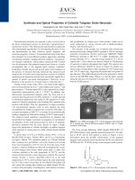
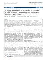
Fig. 1 shows the XRD patterns of the as-prepared and
the annealed samples. In order to show up the weaker
peaks, the data in Fig. 1 were plotted on a logarithmic
intensity scale. Both spectra show WO
3
and W diffraction
peaks. The additional weaker WO
2
lines could also be
indexed in the spectra for the as-prepared sample. The
results indicate that most of the WO
2
and part of the W
could be oxidized to WO
3
after air annealing at 573 K. Iron
and its oxide could be indexed in the XRD patterns of both
samples. However, the weak diff raction peaks for iron and
its oxide were broadened significantly, indicating very tiny
crystallites in size or a very small weight percentage. The
high saturation magnetization of the as-prepared sample
and EDX analysis of the air-oxidized sample as discussed
below indicate a considerable weight percentage of iron
and its compounds in the samples.
The morphologies of the as-prepared products are
demonstrated in Figs. 2a and b. The products show
obvious rod-like shape up to 2000 nm in length and
20–30 nm in diameter. Shown in Fig. 2b is a typical TEM
image, in which the nanorods were covered by a thin film of
approximately 3–4 nm in thickness, estimated by the
contrast. Nanoclusters adhering to the nanorods or
protuberances could also be observed over the surface of
the nanorods. At higher magnification, as shown in Figs. 2c
and d, the nanorods exhibit clear fringes parallel to their
long axis. The lattice spacing of two parallel planes was
0.39 nm, which could be indexed best as (0 0 1) of WO
3
,
according to JCPDS card No. 20-1324. Figs. 2c and d also
show that the thin layer covering the nanorods is in
amorphous. The thickness of the amorphous coatings and
the size of the nanoclusters adhering to the nanorods are
much smaller, compared with the diameter and size of the
well-crystallized WO
3
matrix. Therefore, there should be
more elemental W than Fe in the sample.
The XPS technique probes mostly the surface atoms of
the sample. Fig. 3 represents the XPS spectra of the W 4f,
Fe 2p
3/2
,Fe2p
1/2
, and Ni 2p photoelectrons in the as-
prepared nanorods for original surface and surface after
argon-ion etching for 150 s, respectively. The original
surface consists of WO
3
and non-stoichiometric tungsten
oxide (WO
3
/W) [13]. Additional W peaks could be detected
in the spectra for the etched surface of the sample. The XPS
results are in good agreement with that of the XRD
analysis, which could be index ed to WO
3
,WO
2
and W. In
fact, a number of tungsten oxides, including WO
3
,WO
2
,
W
3
O
8
,W
5
O
14
,W
17
O
47
,W
18
O
49
,W
20
O
58
,andW
40
O
118
etc.,
could be formed, which could coexist or progressively
change one into the other with changing temperature and
oxygen partial pressure. Usually, oxidation to metal is
controlled by oxygen atomic diffusion process. Therefore,
the non-stoichiometric tungsten oxide could be formed in
the nanoscale sample consisting of WO
3
and W. There are
two characteristic binding energies (707.4 and 711.4 eV) for
the photoelectron line of Fe 2p
3/2
, as illustrated in Fig. 3.
The binding energies of 707.4 and 711.4 eV are in good
agreement with that of Fe and FeO
x
(Fe
3
O
4
or Fe
2
O
3
) [14],
respectively. However, the binding energy (724 eV) for the
Fe 2p
1/2
peak agrees well with that of Fe
2
O
3
instead of
Fe
3
O
4
[15]. Therefore, we can conclude that Fe is present in
the forms of elemental Fe an d Fe
2
O
3
in the as-prepared
ARTICLE IN PRESS
Fig. 1. X-ray diffraction patterns of (a) the as-prepared powders and (b)
that after annealing at 573 K in air for 10 h.
P.Z. Si et al. / Physica B 392 (2007) 154–158 155
sample. As shown in Fig. 3, no Ni 2p peak was observed in
both the original and the etched surfaces of the sample.
Since XPS technique is very sensitive to elements, the
absence of Ni 2p peak indica tes the absence of elemental Ni
or its compounds in the products, in good agreement with
the XRD results above and the EDX results to be discussed
below. Note that the photoelectron lines for elemental W
are much stronger than those for elemental Fe, indicating a
larger W content than Fe content in the sample.
The electron-induced X-ray fluorescence (EDX) analysis
was employed to determine the composition of the air-
oxidized WO
3
/Fe nanorods. Since air oxidation could not
change the elemental ratio except ratio to oxygen, the EDX
results for the air-oxidized sample can to some extent
represent that of the as-prepared sample. Fig. 4 shows the
TEM images and the EDX spectra for the air-annealed
products. It is obvious that the rod-like shape and
morphology of the products were maintained after air-
oxidation. Only W, Fe, O, and Cu elements were detected,
as shown in Fig. 4. The presence of a Cu signal arises from
the sample holder, thus the nanorods were composed of W,
Fe, and O. The peak intensity for W is much stronger than
that for Fe, indicating that the nanorods, at least within the
selected area for recording EDX spectra, contain more W
than Fe. It should be noted that the Ni atoms expected
from the anode were not detected by EDX.
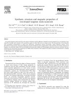
In Fig. 5 we show the results of magnetic measur ements
for the as-prepared and the heat-treated samples. Even
though the XPS spectra proved the presence of Fe
2
O
3
in
the as-prepared sample, we cannot exclude the presence of
ARTICLE IN PRESS
Fig. 2. TEM images of the as-synthesized Fe-doped tungsten oxide
nanorods: (a) low magnification image shows the morphology of
the nanorods, (b) high magnification image shows a thin film covering
the nanorods and several nanoparticles adhering to the surface of the
nanorods, ðc; dÞ high resolution TEM images show amorphous film over
the surface of a nanorod and well-crystallized nanorods with 0.39 nm
interplanar distance corresponding to the (0 0 1) interplanar spacing
of WO
3
.
Fig. 3. XPS spectra of the W 4f, Fe 2p
3/2
,Fe2p
1/2
and Ni 2p
photoelectrons in the as-prepared nanorods.
P.Z. Si et al. / Physica B 392 (2007) 154–158156
other iron oxides because of the limited difference between
Fe 2p binding energies in different iron oxides. In order to
determine the magnetization contribution of different
magnetic phases, a basic knowledge for the magnetic
properties of bulk Fe and its oxides is crucial. It is well
known that bulk iron (or Fe
3
O
4
) is a ferromagnet (or
ferrimagnet) with T
C
¼ 1043 K (or 850 K) and M
s
¼
222 Am
2
=kg (or 84 Am
2
=kg), while FeO is antiferromag-
netic with T
N
¼ 198 K. The Fe
2
O
3
exists in amorphous
form or other four polymorphs (alpha, beta, gamma, and
epsilon) [16]. Amorphous Fe
2
O
3
is paramagnetic at
temperatures above T
N
¼ 80 K with a magnetic moment
of 2:5m
B
per atom of iron [17]. The a-Fe
2
O
3
phase is
antiferromagnetic (paramagnetic) at temperatures
To260 K (T4T
N
950 K) while a destabilization of their
perfect antiparallel arrangement and development of weak
ferromagnetism occurs between 260 and 950 K [16]. b-
Fe
2
O
3
exhibits paramagnetic behavior at temperatures
above 119 K [16]. The thermal instability of ferrimagnetic
g-Fe
2
O
3
disables direct determination of its T
C
. For well
developed g-Fe
2
O
3
crystals (M
s
¼ 74 Am
2
=kg) a direct
g-Fe
2
O
3
! a-Fe
2
O
3
transformation occurs at approxi-
mately 673 K [16]. For very small g -Fe
2
O
3
particles, a
notably higher transformation temperature was observed
with e-Fe
2
O
3
being an intermediate of the g-Fe
2
O
3
!
a-Fe
2
O
3
structural transformation [18]. The e-Fe
2
O
3
is a
non-collinear ferr imagnet with T
C
near 470 K [16,18] .
In the plot of M vs H, both the samples reach saturation
in fields above 0.8 T. The magnetization at 5 K of the as-
prepared sample in an applied field of 5 T is as large as
56 Am
2
=kg, arising from the magnetization of metallic Fe
and its oxides. Assuming a magnetic moment of 2 :2m
B
per
iron atom in the sample, we find the most conservative
estimation of Fe content in the sample is 25.5 wt%.
Considering the effects that could reduce the total
magnetization, including the formation of iron oxides
and atomi c disorder in small particles, the actual elemental
Fe content in the sample should be much higher than
25.5 wt%. However, most analytical results mentioned
above, including XRD, XPS, EDX, and TEM observa-
tions, support a lower content of elemental Fe than that of
W in the sample. We speculate that the large magnetization
of the sample might partially be due to a possible enhanced
magnetic moment of Fe atom in the Fe nanoclusters. In
fact, an enhanced magnetic moment, 3m
B
per Fe atom at
120 K for clusters containing 25–130 Fe atoms, has been
observed in small iron clusters [19]. In comparison with
iron, iron oxides have a much lower saturation magnetiza-
tion, being just slightly larger than the saturation
magnetization of the WO
3
/Fe nanorods. Both the as-
prepared and the heat-treated samples exhibit soft ferro-
magnetic behavior as shown by hysteresis loops in Fig. 5.
Even at 5 K, both the samples exhibit zero coercivity (less
than 5 Oe) and zero remanence, which are quite different
from those of well-crystallized Fe nanoparticles, in which
enhanced coercivity and enhanced remanence magnetiza-
tion in comparison with that of bulk Fe were observed [10].
Shown in the left inset of Fig. 5 is the plot of the
magnetization at an applied field of 5 T vs T
3=2
. The
magnetization for the heat-treated sample is approximately
48% of that of the as-prepared sample, owing mainly to the
oxidation of the Fe clusters. The as-prepared and heat-
treated samples show a linear T
3=2
dependence of M at
temperatures above 50 and 90 K, respectively, following
the well-known Bloch’s law [20,21]. How ever, the curves at
temperatures below these temperatures deviate significantly
from Bloch’s law. Magnetization deviation from Bloch ’s
law towards lower magnetization has been observed in
amorphous iron at temperatures below 50 K [22]. However,
our samples exhibit magnetization deviations to larger
magnetization as shown in the left inset of Fig. 5. The
sharp magnetization increase in the low temperature region
ARTICLE IN PRESS
Fig. 4. EDX spectrum shows the composition profiles of the Fe-doped
tungsten oxide nanorods after annealing in air. The inset shows the
morphology of the annealed sample and the area for recording EDX
spectrum.
Fig. 5. M vs H plot for the as-prepared (black) and heat-treated (gray)
samples at several temperatures. The left inset shows the magnetization vs
the
3
2
power of temperature for both the samples. The line is a fit to Bloch’s
law. Shown in right inset is the Curie point determination curve for the
as-prepared sample obtained on a Faraday balance.
P.Z. Si et al. / Physica B 392 (2007) 154–158 157
might indicate the presence of paramagnetic phases in the
sample, which usually has little (large) contrib ution to
magnetization in high (low) temperature regions. The air-
oxidized sample and the as-prepared samples show
deviation at temperatures below 90 and 50 K, respectively.
This indicates that the presence of iron oxides should be
one reason for these deviations because iron oxides usually
do not follow Bloch’s law.
In the right inset of Fig. 5 we show the magnetization
measurement for the as-prepared nanorods in an applied
field of 0.05 T. The magnetization starts to decrease at
800 K with increasing temperature and vanishes at 1000 K.
Among all iron oxides, Fe
3
O
4
and g-Fe
2
O
3
exhibit the
largest saturation magnetization of 84 and 74 Am
2
/kg,
respectively. The slightly decreasing feature between 800
and 900 K is very likely due to the Curie point of Fe
3
O
4
and the structural transformation of g -Fe
2
O
3
4a-Fe
2
O
3
with e-Fe
2
O
3
being an intermediate [18]. A notably higher
transformation temperature for g-Fe
2
O
3
4a-Fe
2
O
3
than
673 K of bulk g-Fe
2
O
3
has been observed in nanoscale g-
Fe
2
O
3
[18]. The sharp magnetization decrease feature at
temperatures between 940 and 1000 K, which is slightly
lower than the T
C
(1043 K) of bulk Fe, is ascribed to the
Curie point for tiny Fe clusters. Usually, amorphous iron
or very small iron clusters exhibit decreased Curie
temperature [19,22].
4. Conclusi ons
In summary, iron-doped tun gsten oxide nanorods with
diameters ranging from 20 to 30 nm and lengths up to
60–2000 nm have been synthesized by an arc discharge
route using W as cathode and a mixture of Fe and NiO as
anode, in which NiO serves as an oxygen source. The
nanorods were composed of W, Fe, and their oxides. Most
of the nanorods were covered by an amorphous film with
3–4 nm in thickness, in which nanoclusters adhering to the
surface of the nanorods were frequently observed. XPS
shows that the surface layers were mainl y composed of
tungsten oxide, iron and its oxide. Faraday balance
measurements show that the magnetization of the sample
vanishes at temperatures above 1000 K, indicating a
decreased Curie temperature for tiny Fe clusters comparing
with that of bulk Fe. Heat-treatment of the sample in air
induces oxidation of eleme ntal Fe, resulting in the
reduction of the magnetization. Both the as-prepared and
the heat-treated samples show zero coercivity and zero
remanence.
Acknowledgments
The work was supported by the Center for Nanostruc-
tured Materials Techn ology under ‘21st Century Frontier
R&D Programs’ (Grant no. 05K1501-00310), the National
Natural Science Foundation of China (Gr ants nos.
59725103, 50332020, and 50171070), and the scientific
exchange program between China and The Netherlands.
References
[1] A. Hoel, L.F. Reyes, P. Heszler, V. Lantto, C.G. Granqvist, Curr.
Appl. Phys. 4 (2004) 547.
[2] Y. Koltypin, S.I. Nikitenko, A. Gedanken, J. Mater. Chem. 12 (2002)
1107.
[3] G. Gu, B. Zheng, W.Q. Han, S. Roth, J. Liu, Nano Lett. 2 (2002)
849.
[4] X.L. Li, J.F. Liu, Y.D. Li, Inorg. Chem. 42 (2003) 921.
[5] Y.Q. Zhu, W.B. Hu, W.K. Hsu, M. Terrones, N. Grobert, J.P. Hare,
H.W. Kroto, D.R.M. Walton, H. Terrones, Chem. Phys. Lett. 309
(1999) 327.
[6] Z.W. Liu, Y.S. Bando, C.C. Tang, Chem. Phys. Lett. 372 (2003) 179.
[7] Y.B. Li, Y. Bando, D. Golberg, K. Kurashima, Chem. Phys. Lett.
367 (2003) 214.
[8] A. Rothschild, J. Sloan, R. Tenne, J. Am. Chem. Soc. 122 (2000)
5169.
[9] J. Bansmann, S.H. Baker, C. Binns, J.A. Blackman, J.P. Bucher, J.D.
Da
´
vila, V. Dupuis, L. Favre, D. Kechrakos, A. Kleibert,
K.H.M. Broer, G.M. Pastor, A. Perez, O. Toulemonde, K.N.
Trohidou, J. Tuaillon, Y. Xie, Sur. Sci. Rep. 56 (2005) 189.
[10] P.Z. Si, Z.D. Zhang, D.Y. Geng, C.Y. You, X.G. Zhao, W.S. Zhang,
Carbon 41 (2003) 247.
[11] X.L. Dong, Z.D. Zhang, Q.F. Xiao, X.G. Zhao, Y.C. Chuang, S.R.
Jin, W.M. Sun, Z.J. Li, Z.X. Zheng, H. Yang, J. Mater. Sci. 33 (1998)
1915.
[12] Z.D. Zhang, J.G. Zheng, I. S
ˇ
korva
´
nek, G.H. Wen, J. Kova
´
e
`
, F.W.
Wang, J.L. Yu, Z.J. Li, X.L. Dong, S.R. Jin, W. Liu, X.X. Zhang,
J. Phys.: Condens. Matter 13 (2001) 1921.
[13] I. Kojima, M. Kurahashi, J. Electron Spectrosc. Relat. Phenom. 42
(1987) 177.
[14] G.C. Allen, M.T. Curtis, A.J. Hooper, P.M. Tucker, J. Chem. Soc.
Dalton Trans. 14 (1974) 1525.
[15] B.J. Tan, K.J. Klabunde, P.M.A. Sherwood, Chem. Mater. 2 (1990)
186.
[16] R. Zboril, M. Mashlan, D. Petridis, Chem. Mater. 14 (2002) 969.
[17] N. Heiman, N.S. Kazama, J. Appl. Phys. 50 (1979) 7633.
[18] E. Tronc, C. Chane
´
ac, J.P. Jolivet, J. Solid State Chem. 139 (1998) 93.
[19] I.M.L. Billas, J.A. Becker, A. Chaˆ telain, W.A. de Heer, Phys. Rev.
Lett. 71 (1993) 4067.
[20] F. Bloch, Z. Physik 61 (1930) 206.
[21] U. Ko
¨
bler, J. Phys.: Condens. Matter 14 (2002) 8861.
[22] M.W. Grinstaff, M.B. Salamon, K.S. Suslick, Phys. Rev. B 48
(1993) 269.
ARTICLE IN PRESS
P.Z. Si et al. / Physica B 392 (2007) 154–158158