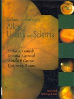
Sankara Nethralaya''s Atlas of Uveitis and Scleritis pptx
Bạn đang xem bản rút gọn của tài liệu. Xem và tải ngay bản đầy đủ của tài liệu tại đây (20.97 MB, 179 trang )
••
Sankara
Nethralaya's
Atlas
of
U
Itis
and
Scleritis
Su()ba
I(
Gal1esb
Mamta Agarwa[
Ama[a E
George
'
J~otirmo~
Biswas
Sankara
Nethralaya's
Atlas
of
Uveitis
and
Scleritis
Sudha
K
Ganesh
Mamta Agarwal
Amala E George
Jyotirmoy
Biswas
Department of Uvea
Sankara Nethralaya, 18 College Road, Chennai
t
JAYPEE
BROTHERS
MEDICAL PUBLISHERS (
P)
L
TO
New Delhi
•
Published
by
Jitendar P Vij
Jaypee
Brothers
Medical
Publishers
(P) Ltd
EMCA House,
23
/
23B
Ansari Road, Daryaganj
New
Delhi
1
10
002, India
Phones: +91-11-23272143, +91-11-23272703,
+91
·11·23282021 , +91·1 1·23245672
Fax: +91-11-23276490, +91-11-23245683 e-mail: jpmedpub@
deI2.vsnl.netin
Visit our website: ww
w.
jaypeebrothers.com
Branches
• 202 Batavia Chambers, 8 Kumara Krupa Road, Kumara Park East,
Bangalore
560001,
Phones: +91-80-22285971, +91-80-22382956, +9
1-
80-30614073
Tele Fax: +91·80·22281761 e·mail:
in
• 282 IIIrd Floor, Khaleal Shirazi Estate, Fountain Plaza
Pantheon Road , Chennal 600
008, Phones: +91-44-28262665, +91-44-28269897
Fax: +91-44-28262331 e-mail:
· 4-2-1067/1-3,
1st
Floor, Balaji Buildi
ng
, Ramkole
Cross Road, Hyderabad 500
095, Phones: +91·40-55610020, +91-40-24758498
Fax:
+91
-40-24758499 a-mail:
• 1 A Indian Mirror Street, Wellington Square
Kolkala 700 013,
Phone: +91-33-22451926 Fax: +91-33-22456075
e-mail:
• 106 Ami! Industrial Estate, 61 Dr S8
Rao
Road, Near
MGM
Hospital
Parel, Mumbai
400 012, Phones: +91-22-24124863, +91·22·24104532, +91-22·30926896
Fax: +9 1-22-24160828 e-mail:
Ssnksrs Nethralaya's Atlas
of
Uveitis
and
Scleritis
C 2005, Authors
All rights reserved.
No
part of this publication should
be
reproduced, stored
in
a retrieval system , or transmitted
In
any form or
by
any means: electronic, mechanical, photocopying, recording, or otherwise, without
the
prior
written permission
of
the authors
and
t
he
publisher
This book has been published
in
good faith that the material provided
by
authors is original. Every effort
is
made
to
ensure accuracy of material, but the publisher, printer
and
authors will not
be
he
ld responsible for any
inadvertent
errort
s)
. In case of anv
diSDute
al1leoal matters to be settled under Delhi iurisdiction onlv.
First Edition: 2005
ISBN
81
-8061-466-2
Typeset at JPBMP typesetti
ng
unit
Printed
at
Paras Offset Pvt Ltd
.,
C 176 Naraina Industrial Area, Phase
1,
New Delhi 110028
FOREWORD
Several uveitic entities
are
recognized
based
on
their clinical features
and!
or
pathogenesis; such entities include Toxoplasma retinochoroiditis, Pars
pianitis, Serpiginous choroiditis and several others.
In
many instances
however, determining exactly what initiated the process of uveitis has
been
a challenge, particularly when the trigger
is
an
infectious agent. The
detection process
is
complicated by the diverse clinical manifestations of
uveitis that are induced
by
the infectious agent, by the unavailability of
infected tissue
for
examination and by the lack of specific and sensitive
diagnostic tests.
For
example, Mycobacterium tuberculosis can present
with an anterior
or
posterior uveitis, with or without granulomas. Moreover
this
agent
can
induce a hypersensitivity reaction with clinical features of
retinal vasculitis. iridocyclitis
or
multifocal choroiditis
or
wi
th features
suggestive of Serpiginous choroiditis.
In
such a clinical spectrum. Mycobacterium tuberculosis can
be
isolated from the granulomatous lesions, but such samples rarely become available.
In
hypersensitivity
reactions such
as
retinal vasculitis. the infectious organisms
appear
to
be
absent
from the retina, yet
some
patients
respond
positively to antituberculous agents suggesting that mycobacterium
does
indeed
have
a role in this entity.
Sputum
analysis
and
chest X-ray findings may not help pinpoint the diagnosis
in these patients.
In
recent years, molecular diagnostic procedures have detected infectious agents in
some
cases
wherein organisms could not
be
cultured
or
detected
by other microbiological procedures.
Among
the
various molecular procedures used, the analysis of intraocular fluid by polymerase chain reaction
(PCR)
in
uveitis
has
shown great promise
in
detecting traces of
an
infectious
agent
in
the form of a microbial
specific DNA sequence. This procedure has
been
successfully used by several laboratories including
laboratory of
Sankara
Nethralaya to detect bacteria, viruses
and
protozoa in intraocular inflammation.
Based
on
well
documented
clinical findings. laboratory
in
vestigations including PCR analysis. the
authors
of this well illustrated atlas provide succinctly main clinical diagnostic features
and
management
of
various uveitic entities.
The
authors profusely illustrate clinical examples of
both
anterior
and
posterior
uveitis
and
scleral inflammation
seen
in
their practice over a
decade.
This atlas should
be
valuable to
ophthalmologists
in
clinical practice
and
to postgraduate students
who
are
in
the process of acquiring
knowledge in the field
of
uveitis, a leading
cause
of blindness
in
the developing world.
Narsing A Rao MD
Los Angeles. California
USA
PREFACE
Uveitis
is
an
emerging subspeciality of ophthalmology that presents with a constellation of clinical
findings. Accurate diagnosis of uveitis
and
successful treatment of these patients remain a challenge.
The field of uvei
tis
has been revolutionized over the past
50
years.
OUf
understanding of ocular
inflammatory diseases has progressed rapidly
and
we can accurately diagnose
and
treat these diseases.
Basic science
and
research
in
immunology has led to new therapeutic approaches to the patient with
uveitis
and
scleritis. This atlas gives the reader a concise overview of the clinical manifestations,
investigations, diagnosis and management
of
uveitis and scleritis.
We
emphasize on careful clinical
assessment
and
accurate diagnosis. This book
is
not intended to be a textbook but a practical guide to
the diagnosis,
in
obtaining appropriate investigations
and
management. The uvea department has a
team of four consultants
and
we examine about a thousand new cases of uveitis every year. The
emphasis
on
maintaining good clinical record with digital archiving was
an
excellent source
for
selecting
pictures
for
this atlas.
All
cases included were patients seen at Sankara Nethralaya, Chennai.
Authors
'0
ACKNOWLEDGEMENTS
-
'
Dr SS Badrinath
has
been
the driving force behind this atlas. Without his encouragement, this
book
would
not
have come into existence. We are deeply indebted to him for his active support.
We are very grateful to
Mr
SP
Govindarajan,
Mr
5
Jayaraman.
Mr
Anand
and
Ms
Lakshmi (Nursing
Staff)
in
photography department at Sankara Nethralaya. Special thanks to
Dr
Rajesh
FogJa
and
Dr
Mani Baskaran for their help
in
taking digital
slit
lamp photographs of the anterior segment.
We would like to give
our
special thanks to
Mr
V MuraU,
Mr
Mohan
and
Mr
Elango
in
our
multimedia
department
who
helped us
in
scanning the images.
We
also extend
our
sincere thanks to
our
corresponding
secretaries. We also acknowledge the efforts
of
Jaypee
Brothers Medical Publishers
(P)
Ltd for their
technical assistance, printing
and
publishing this atlas
on
time.
[n
particular, we like to thank Shri J P
Vij,
Chairman
and
Managing Director of
Jaypee
Brothers
and
Mr
R
Jayanandan,
author
Co-ordinator of
Chennai
branch.
Finally, we thank
aU
our patients for trusting us
and
allowing us to
par
ticipate
in
the
management
of
their ocular problems.
•
CONTENTS
Section
1:
Anterior
Uveitis
1.
Idiop
a
th
ic
Ant
erio
r Uve
it
is
3
2.
Granul
o
matous
Ant
e
rior
Uv
eiti
s
.
9
3.
Fuch
s'
Heterochromic
Iridoc
yc
litis
13
4.
Juvenil
e
Idiopathic
Arthritis
(
JIA
)
Associat
ed
Uveitis
17
5.
Lens
Indu
c
ed
Uveitis
.
2 1
6.
Intraocular
Lens
Induced
Uve
itis
25
Section
2:
Intermediate
Uveitis
7.
Intermediat
e Uve
itis
.
31
Section
3:
Posterior
Uveitis
8.
Serpiginous
Choroidit
is
37
9.
Mult
i
ple
Evanescent
Whit
e
Dot
Syndrome
(MEWDS)
49
10
.
Acute
Posterior
Multifocal
Pla
c
oid
Pigment
Epitheliopath
y (
APMPPE
)
.
53
11.
Birdshot
Retinochoroidopathy
57
12
.
Punctate
Inner
Choroidopathy
(PIC)
6 1
13.
Choroiditis
65
14
.
Ocular
Toxoplasmosis
71
15.
Parasitic
Uveitis
.
.
79
16
. Viral Re
tinitis
.
89
17
.
Retinal
Vasculitis
97
Section
4:
Panuveitis
18.
Sarcoidosis
1
07
19. Vogt-K
oy
anagi
-Ha
rada
(VKH)
Syndr
ome .
113
2
0.
Symp
a
the
tic
Ophthalmi
a
.
121
2 1. 8 e
hc
et's
Dis
ea
se
12
7
22 . Tuber
cu
los
is
131
Section
5:
Infectious
Uveitis
23.
End
o
genous
End
o
phth
a
lm
itis
143
xii
Atlas
of
Uveitis and Scleritis
Section
6 :
Systemic
Diseases
associated
with
Uveitis
24
. Sy
stemic
Di
se
a
ses
ass
ociat
ed
with
Uve
itis
149
Section
7:
Complications
of
Uveitis
25
. C
omplicati
ons of
Uv
e
itis
159
Section
8 :
Investigations
in
Uveiti
s
26
. Inve
stigations
in Uve
itis
.
167
Section
9:
Masquerade
Syndrome
27
.
Masqu
erade
Syndrome
179
Section
10
:
Acquired
Immunodeficiency
Syndrome
(AIDS)
28
. Ac
quir
ed
Immun
ode
fi
ci
en
cy
Syn
drome
(
AIDS
)
185
Section
11:
Scleritis
29
. Scle
riti
s
.
197
Index
. 203
CHAPTER
ONE
Idio thic Anterior Uveitis
•
4
I.:;,
Atlas
of
Uveitis
a~ld
Scleritis
Inflammation
of
the
iris
and
ciliary
body
is
called anterior
uveitis. It can
be
nongranulomatous or granulomatous.
Idiopathic nongranulomatous anterior uveitis
is
the
most common
type
of
anterior uveitis which
is
known to
be
associat
ed
with the
human
leukocyte antigen
(HLA)
827
in
50-60 percent of patients. It
is
characterized by
male
predominance
and
may
be
associated
with
seronegative arthritic syndromes such as ankylosing
spondylitis, Reiter syndrome, psoriatic arthritis, and
inflammatory bowel disease.
Ocular Features
Symptoms
Pain, redness, photophobia, decreased vision.
Signs
Acute
• Circumcorneal congestion
• Fine keratic precipitates
•
Aqueous
fl
are
and
cells (
graded
according to severity)
• H
ypopyon
• Posterior synechiae
•
Iris
bombe
Chronic
• Old pigmented keratic precipitates
• Broken posterior synechiae
• Seclusio and occlusio pupillae
• Posterior subcapsular cataract
Investigations
• Complete blood count
• Rheumatoid factor, antinuclear antibody
• Human leucocyte antigen (HLA-B27)
Treatment
• Topical steroids: Prednisolone acetate, Betame
th
a-
sone, Dexamethasone.
In acute cases, topical steroids
can
be
used every hourly and gradually tapered.
•
Cycloplegic
agents:
Atropine,
Homatropine
,
Cyclopentolate
•
In
severe cases, periocularl systemic steroids can be
used.
REFERENCES
1. Biswas
J.
Narain S. Das
D.
Ganesh
SK
. Pattern of uveitis
in
a
referral uveitis
clinic
in
India.
Int
Ophtha
lmol.
1996-97;20:223-8.
2.
Martin
TM.
Smith
JR.
Rosenbaum
JT.
Anterior uveitis: Current
concepts
of
pathogenesis
and
interactions
with the
spondyloarthropathies.
Curr Opin
Rh
euma
tol.
2002: 14:337·4
1.
Clinical Features
of
Nongranulomatous Anterior Uveitis
Fig
. 1.1: Silt lamp photograph showing circumcorneal
congestion
in
a case of acute anterior uveitis
Fig. 1.3: Hypopyon
in
an
HlA
B27 positive patient with
ankylosing spondylitis
Fig. 1
.2:
Anterior chamber sl
it
beam photograph showing
intense
flare due
to
breakdown of blood aqueous barrier
Fig.
1.
4: Slit beam photograph showing small to medium sized
keratic precipitates
Fig. 1
.5:
Slit lamp examination
of
anterior chamber showing
circumcorneal congesti
on,
fine keratic precipitates, aqueous
flare,
cells and seve
re
fibrinous reaction
in
anterior chamber
•
••
Fig. 1.6: Posterior synechiae following attacks of recurrent
anterior uveitis
Fig. 1.7: Slit lamp photograph showing circumcorneal congestion,
iris nodules and posterior synechiae (arrow)
•
••
Systemic
Associations
of
Idiopathic
Anterior
Uveitis
Fig
. 1.9: Active chondritis involving the
external ear in a patient wi
th
relapsing
polychondrilis who also developed
recurrent attacks of anterior uveitis
Figs
1.BA
to
C
A. A 40-year-old man suffering from ankylosing spondylitis
B. X-ray hip showed sclerosis of sacroiliac joints (arrows)
C. X-ray spine showed complete spinal fusion, i.e. bamboo spine
Fig.
1.10: External photograph of a patient with psoriasis showing
exfoliative skin lesions with silvery scales, onycholysis, pitting and
nail discoloration along with
swelling of distal interphalangeal joints
(sausage digits).
•
CHAPTER
TWO
Granulomatous
Anterior
Uveitis
Granulomatous anterior uvei
tis
is
a chronic subclinical Investigations
inflammation of the
iris
and
ciliary body. It may
be
due
• Complete blood count
to
an
idiopathic reaction or from the host's immune
response to a systemic infectious process such as syphilis,
tuberculosis, Hansen's disease or local reactivation of
herpetic
viral
infection or sarcoidosis.
It
is
usually bilateral,
involve
all
parts of the eye
and
can
be
part of a systemic
disease process or
an
ocular syndrome.
Ocular Features
Symptoms
Decreased vision, dull aching eye, pain, photophobia.
Signs
External
• Herpetic
uveitis-C
utaneous vesicles
in
the dismbution
of the ophthalmic branch of the
fifth
cranial nelVe
•
Hansen's
disease-Madarosis,
leonine facies
Ocular
• No circumcorneal congestion
• Large (mutton-fat) keratic precipitates
• Aqueous flare
and
cells
• Koeppe nodules, Busacca's nodules
• Sectoral
iris
atrophy
in
herpetic uveitis
• Generalized iris atrophy
in
Hansen's disease
• Raised intraocular pressure especially
in
herpetic
uveitis
• Inflammatory nodules
in
the angle
• Erythrocyte sedimentation rate
• Mantoux test
• Serum angiotensin converting enzyme
• Fluorescent treponemal
antibody
absorption test
(ITA
-ABS)
• Venereal Disease Research Laboratory Test
(VORL)
• Rapid Plasma Reagin (RPR)
• Chest
X-ray
Treatment
• Topical steroids: Prednisolone acetate, Betametha-
sone, Dexamethasone
•
Cycloplegic
agents:
Atropine,
Homatropine
,
Cyclopentolate
• Treatment of specific diseases (Tuberculosis, syphilis,
sarcoidosis, Hansen's disease)
•
In
case of noninfectious posterior segment involve-
ment, systemic steroids / immunosuppressive agents
are required.
REFERENCES
1. Kristeva
M.
Biswas J, Pararajasegaram G, 5evanian A
and
Rao
NA:
'Histochemical analysis of experimental granulomatous uveitis'.
Ophthalmic
Research. 1991; 23:73-78.
2.
Nussenblatt
RB,
Whitcup SM, Palestine
AG:
Uveitis: Fundamentals
and
Clinical Practice. 2nd oon. Mosby-Year Book; 1996.
3.
Rito
NA.
Cousins S, Forster
D:
Intraocular lnnammation
and
Uveitis:
Basic
and
Clinical Science Course 2002.
•
"-
~
-
,
~~
,"
.
- ~
••
Fig. 2.1: Multiple mutton-fat keratic precipitates
in
a patient
with sarcoidosis
Fig. 2.3: Koeppes
nodules at the pupillary border
in
a case of
sarcoidosis
Fig
. 2.2: Slit lamp photograph shows large, round, translucent
keratic precipitates
in
a patient
wi
th Hansen's disease
Fig. 2
.4:
Slit lamp photograph showing large iris nodules
(Busacca nodules) and keratic precipitates in a patient with
pulmonary tuberculosis
"'II
12 , "_
Atlas
of
Uveitis and Scleritis
Fig
. 2.5: Slit lamp photograph showing sectoral iris atrophy in
a patient with herpet
ic
a
nt
erior uveitis
Fig
. 2.7: Herpet
ic
keralouveitis
with
disciform stromal oedema
Fig
. 2.6: Dendritic ulcer stained with fluorescein
with few keratic precipitates
Fig. 2.8: Herpes
zosterophlha
lmicus
with anterior uveitis
•
CHAPTER THREE
_ _
I
Fuchs' Heterochromic Iridocyclitis
Fuchs' heterochromic
ir
id
ocyclitis
is
a chronic, unilateral
nongranulomatous anterior uveitis characterized by a
white eye,
ste
ll
ate keratic precipitates,
iris
atrophy with
or
without
iris
heterochromia, abnormal angle vessels
and absence of posterior synechiae.
It
is
bilateral
in
approximately 10 percent of patients. The etiology of
the disease
is
still
un
kn
own. Several theories including
infection
from Toxoplasma
gondii
,
an
immune
dysfunction, infiltration of sensitized lymphocytes, and
chronic herpetic infection have been proposed.
Ocular Features
Symptoms
Decreased vision due to cataract, floaters due to vitreous
opacification.
Signs
• Small, nonpigmented, translucent stellate
ke
ratic
precipitates with
fine
filament projections described
as
ste
ll
ate scattered over the entire poster
io
r corneal
surface.
• Minimal anterior chamber cells and flare.
• I
ris
heterochromia
is
present
in
most patients and
usually seen as flatt
en
ing of the anterior i
ris
features
resulting from loss of the anterior i
ris
border layer
and
iris
stroma, especially
in
the peripupillary area.
• White
iris
nodules may occur along the border.
• Posterior synechiae are absent. Radial
iris
vessels are
seen on the
ir
is surface because of
iris
atrophy.
• Fine blood vessels on the trabecu
la
r meshwork may
bleed when the
in
traocular pressure suddenly drops
during surgery or paracentesi
s.
(Amsler sign).
• Posterior subcapsular cataract
• Vitreous opacification
• Secondary glaucoma
is
seen
in
about 60 percent cases
Investigations
Diagnosis
is
always clinical. No laboratory investigations
are required.
Treatment
Topical corticosteroids
for
the anterior segment inflam-
mation. Cataract
and
glaucoma are
managed
with
surgery with steroid prophylaxis.
REFERENCES
L Jones
NP:
Fuchs'
heterochromic uveitis:
An
updaie.
Su
IV
Ophthalmol
1993: 37:
253-72
2.
La
Hey
E.
de Jong
PT
.
Kij
ls
lTa
A. Fuchs' heterochromic cyclitis:
Review of the
li
tera
tu
re on the pathogenetic me
ch
anisms.
ST
J
Oph
thillmoL
1994 :78:307-12.
3.
V
el
ill
a S. Dios
E.
Herreras
JM
.
(alonge
M. Fuchs' heterochromic
iridocyc
lit
is:
a review of 26 cases. Ocul Immunol lnflamm. 2001; 9:
169-75.
•
••
Figs 3.1A and B: Clinical photograph of the right (Fig.
A)
and lett eye (Fig. 8) of a 30-year-old
man
who complained of
decreased vision in his left eye for the last 6
months.
Slillamp
examination showed diffuse stellate keratic precipitates, pupillary
sphincter atrophy, moth-ealen appearance of the iris and posterior subcapsular cataract in left eye. The patient was diagnosed
to have Fuchs' heterochromic iridocyclitis
Fig. 3.2: Slit beam photograph under high magnification
showing
small stellate keratic precipitates with fibrillary
extensions
Fig. 3.3: Multiple keratic precipitates spread over entire corneal
endothelium in an eye with Fuchs' heterochromic iridocyclitis
CHAPTER FOUR
Juvenile Idiopathic Arthritis (JIA)
associated Uveitis
•
18
J
Atlas
of
Uveitis alld Scleritis
Juvenile idiopathic arthritis (JI
A)
associated with uveitis
is
the
most frequent
cause
of chronic intraocular
inflammation
among
children. Chronic iridocyclitis occurs
in 10-20 percent of all patients with JIA. Severe loss of
vision
may
occur
in
30
·
40
percent of patients with JIA-
associated uvei
tis.
JIA
is
subd
ivided into three types.
• Systemic
onset
JIA (S
till
's
disease)
• Polyarticular
onset
JIA
• Paudarticular
onset
J IA
Ocular Features
Symptoms
Decreased vision.
Signs
• Bilateral nongranul
omatous
anterior uveitis (70-80
%)
•
Band
keratopathy, small-medium sized keratic
precipitates
• Anterior
chambe
r flare
• Posterior synechiae; pupillary
membrane
• Posterior subcapsular cataracts
• Vitritis, CME, hypotony macu]opathy
• Secondary glaucoma or hypotony late
in
the disease.
Investigations
• Rheumatoid factor
• Antinuclear antibody
(AN
A)
Treatment
• Topical steroids: Prednisolone acetate, Betametha-
sone,
Dexamethasone
•
Cycloplegic
agents:
Atropine
,
Homatropine
,
Cyclopentolate.
• Periocular/ systemic steroids
and
immunosuppressive
agents
are
frequently required in J IA-related anterior
uveitis
in
refractory cases or/
and
as
a steroid-sparing
agent.
REFERENCES
1. Kadayofcilar S. Eidem
B.
Tumer
B.
Uveitis
in
childhood. J Pediatr
Ophthalmol Strabismus.
2003:40:33540.
2.
Foster
CS.
Diagnosis
and
treatment of juvenile idiopathic arthritis-
associated
uveitis.
CUT!
Opin Ophthalmol. 2003:14:395-98.
3.
Kotaniemi K,Sayolain
en
A, Karma
A,
Aho
K.
Recent advances
in
uveitis of juvenile idiopathic arthritis.
SUN
Ophthalmol.
2003
;48:
489-
502. Review
•
••
Fig. 4.1: Slit lamp examination of a 10-year
-o
ld patient
of
JI
A
showing band shaped keratopathy, aqueous celis a
nd
flare
and complicated cataract
Fig. 4.
2:
Band keratopathy
in
juvenile chronic a
rt
hritis
-"
./
-
/
Figs 4.
3A
and
B: Polyarti
cu
lar onset JIA Involving small joints
of hand as
well as knee, ankle and wrist
•
CHAPTER FIVE
Lens Induced Uveitis
•
I
L-~
~
==~====~
________
~
Atlas
0 Uveitis alld Scleritis
Phacogenic
or
lens induced uveitis occurs
as
a result of
leakage of lens proteins
in
hypermature cataract or
traumatic cataract
or
residual cortex after cataract surgery
or loss of cortical matter into the vitreous following a
posterior capsular rupture during cataract surgery. It
is
an immunologic response to lens proteins that become
autoantigenic
upon
release
into
the aqueous humor. Lens
induced uveitis may
be
of
wo
types :
1. Phacoanaphylactic uveitis
2. Phacolytic glaucoma
Ocular Features
Phacoanaphylactic uveitis
is
a granulomatous inflam-
mation of abrupt onset, presenting with large mutton
fat keratic precipitates, thick flare
and
abundant
anterior
chamber cells and hypopyon. Lens
is
opaque with a
wrinkled anterior capsule. It occurs
days
to
weeks after
surgical
or
non-surgical disruption of the lens capsule.
Phacolytic gl
aucoma
is
an
acute glaucoma that occurs
in
hypennature
cataract where lens protein leaks through
the capsule
and
is
engulfed by macrophages that obstruct
the trabecular meshwork
and
cause increased intraocular
pressure.
Investigations
Aqueous
aspirate which shows
macrophages
engulfed
with l
ens
material.
Treatment
Surgical removal of the cataractous lens
and
the residual
lens material.
Topical corticosteroids
and
cycloplegic agents
REFERENCES
1. Smith
RE.
Nozik RAUveitis . A Clinical Approach
to
Diagnosis
and
Management.
ed
2,Baltimore:Williams
and
Wilkins. 1989, 198-203.
2. Marak
G:
Phacoanaphylaclic endophthalmi!is.
SUlV
Ophthalmol
1992;36:325·339
3.
FilipeJC.PaImaresJ. Delgado
L.
LopesJM. BorgesJ. Castro-Correja
J.
Phacolytic glaucoma
and
lens-induced uveitis,
In!
Ophthalmol
1993; 17:289-93 .