frog tongue acts as muscle powered adhesive tape
Bạn đang xem bản rút gọn của tài liệu. Xem và tải ngay bản đầy đủ của tài liệu tại đây (1.07 MB, 9 trang )
Downloaded from on February 10, 2017
rsos.royalsocietypublishing.org
Research
Cite this article: Kleinteich T, Gorb SN. 2015
Frog tongue acts as muscle-powered adhesive
tape. R. Soc. open sci. 2: 150333.
/>
Received: 10 July 2015
Accepted: 4 September 2015
Subject Category:
Biology (whole organism)
Subject Areas:
biomechanics/biomaterials/materials science
Keywords:
adhesion, biomaterials, feeding,
amphibians, biomechanics
Author for correspondence:
Thomas Kleinteich
e-mail:
Electronic supplementary material is available
at or via
.
Frog tongue acts as
muscle-powered
adhesive tape
Thomas Kleinteich and Stanislav N. Gorb
Zoological Institute: Functional Morphology and Biomechanics, Kiel University,
Am Botanischen Garten 9, 24118 Kiel, Germany
Frogs are well known to capture fast-moving prey by flicking
their sticky tongues out of the mouth. This tongue projection
behaviour happens extremely fast which makes frog tongues
a biological high-speed adhesive system. The processes at the
interface between tongue and prey, and thus the mechanism
of adhesion, however, are completely unknown. Here, we
captured the contact mechanics of frog tongues by filming
tongue adhesion at 2000 frames per second through an
illuminated glass. We found that the tongue rolls over the
target during attachment. However, during the pulling phase,
the tongue retractor muscle acts perpendicular to the target
surface and thus prevents peeling during tongue retraction.
When the tongue detaches, mucus fibrils form between the
tongue and the target. Fibrils commonly occur in pressuresensitive adhesives, and thus frog tongues might be a biological
analogue to these engineered materials. The fibrils in frog
tongues are related to the presence of microscopic papillae
on the surface. Together with a layer of nanoscale fibres
underneath the tongue epithelium, these surface papillae will
make the tongue adaptable to asperities. For the first time, to
the best of our knowledge, we are able to integrate anatomy
and function to explain the processes during adhesion in
frog tongues.
1. Introduction
The evolution of a muscular tongue has been a major innovation of
terrestrial vertebrates [1,2] and is directly related to the emergence
of a wide variety of feeding modes on land [3]. Especially,
the projectile tongues of chameleons [4–9], several salamander
species [10–15] and many species of frogs [16–21] have received
considerable attention because of their ability to reach distant
prey items at high velocities. Tongue adhesion generally is a
highly dynamic process, and the time spent for contact formation
between the tongue and the target is in the range of only a
few milliseconds [7,11,17,22]. For frogs, the forces acting on the
tongues during impact and retraction can even be beyond the
body weight of the animals [22].
2015 The Authors. Published by the Royal Society under the terms of the Creative Commons
Attribution License which permits unrestricted
use, provided the original author and source are credited.
Downloaded from on February 10, 2017
2.1. Specimens and housing
Experiments to film the contact dynamics were performed with three adult specimens of horned frogs
(Anura: Ceratophryidae: Ceratophrys sp.). One frog represented the species Ceratophrys cranwelli, two
individuals belonged to the so-called fantasy colour morph, which does not occur in the wild and is
achieved by crossing C. cranwelli with Ceratophrys cornuta. All specimens were captive bred individuals
and purchased from the pet trade. These frogs are fossorial sit-and-wait predators native to Argentina,
Bolivia, Brazil and Paraguay [26]. The animals were individually housed at temperatures of 26–29◦ C
during the day and 24–26◦ C at night. Each terrarium was filled with loose substrate to a depth of
approximately 5 cm in which the frogs could bury themselves. Usually, the frogs were found to be half
buried and only occasionally, they moved to a different location. We moistened the terrariums daily to
sustain a relative humidity of approximately 70–80%. Before the onset of the experiment, the animals
were fed twice a week an alternating diet of crickets (Gryllus bimaculatus), grasshoppers (Schistocerca
gregaria), wax-worms (Zophobas morio), earthworms (Lumbricus terrestris) and rodents (Mus musculus).
For the description of the tongue anatomy, two preserved adult specimens of Ceratophrys ornata were
made available by the Zoological Museum Hamburg (ZMH A11916 and ZMH A11917).
2.2. Contact dynamics experiment
We filmed the tongue contact dynamics during the normal feeding routine of the frogs. During each
experimental session, a maximum of four trials per individual was recorded. Overall, we filmed 25
tongue contacts.
For visualization of tongue contact formation and release, we created a frame that fitted a regular glass
object slide by using the three-dimensional design software SKETCHUP MAKE v. 14.1 (Trimble Navigation
Limited, available at www.sketchup.com). This frame was open to one side to easily change the glass
slides after each experimental trial. The other side of the frame was designed as a cylinder that had the
same diameter as the gooseneck of a cold light source (Zeiss CL 1500 Eco, Carl Zeiss AG, Germany). With
the light source attached, light was directed into the glass slide (figure 1a). Further, a mount for a force
transducer (World Precision Instruments, Sarasota, FL, USA) was modelled onto the frame, but owing to
the rigidity of the gooseneck, simultaneous force measurements could not be performed during contact
area filming, and this mount was only used to fixate the frame during the experiment. We created real
frames by rapid prototyping the three-dimensional designed model with ABS plastic on a CubeX Duo
three-dimensional printer (3D Systems, Rock Hill, SC, USA).
For experimental trials, the frame was placed into the terrariums of the frogs in a vertical position
approximately 2 cm in front of the animals (figure 1c). Generally, the frogs did not move and remained
half buried during the set-up procedure of the experiment. We then placed a cricket immediately behind
the glass slide and held it in place with tweezers. As the frogs tried to capture the cricket, their tongues
attached to the glass slides (figure 1d). Owing to the effect of frustrated total internal reflection, light was
transmitted from the illuminated glass slides only at regions where the tongues actually were in contact
................................................
2. Methods
2
rsos.royalsocietypublishing.org R. Soc. open sci. 2: 150333
Despite the remarkable performances of projectile tongues in vertebrates as biological high-speed
adhesive systems, little is known on the actual cause of their adhesive properties. In chameleons, it was
demonstrated that a negative pressure can be generated inside the specialized tip of the tongue that
forms a pouch [4]; in frogs and salamanders, the mechanism behind tongue adhesion is unknown. Based
on the force over time relationship during detachment of the tongue of the horned frog Ceratophrys sp.
from a glass slide, we hypothesized that frog tongues may act as pressure-sensitive adhesives (PSAs)
[22], which is a class of materials comprising commonly used adhesives, such as sticky tape [23–25].
While we determined the forces acting during tongue adhesion in frogs in a previous study [22], here
we went into detail and focused on the adhesive mechanism of a tongue against a smooth surface. We
managed for the first time to actually capture the processes at the interface between a projected frog
tongue and a target in vivo. The aim of this study is to explain the contact mechanical behaviour of
adhesive projectile tongues in frogs by integrating in vivo observations of the processes during tongue
adhesion with a detailed anatomical description at multiple organizational levels ranging from the
tongue itself to nanoscale tongue elements.
Downloaded from on February 10, 2017
(a)
(c)
3
1 cm
(d )
0 ms
1 ms
2 ms
3 ms
4 ms
5 ms
155 ms
5 ms
355 ms
555 ms
755 ms
855 ms
1 cm
(e)
(g)
(i)
400
400
0.5 cm
300
area (mm2)
area (mm2)
300
200
(f)
200
(h)
100
100
0
0
0
5
time (ms)
10
0
400
time (ms)
800
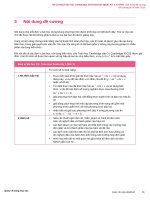
Figure 1. Contact dynamics of frog tongues. We used the effect of frustrated total internal reflection to visualize frog tongues during
contact with a glass slide. For this, we created a frame that fitted a glass slide and that allowed light from a gooseneck light source to be
guided into the glass. Light was totally reflected within the glass (a) and was only transmitted at contact regions (b; here demonstrated
with a fingerprint). We then mounted these frames in the terrariums with the frogs (Ceratophryidae: Ceratophrys sp.) (c) and presented a
cricket to the frogs behind the glass slide. If the frogs tried to catch the cricket, their tongues adhered to the glass and the illumination of
the glass slide at the contact area was captured with a high-speed video camera at 2000 frames per second (d). (e,g) The development of
the tongue contact area over time for contact formation (e) and release (g) for the exemplary experimental trial shown in (d); arrowheads
correspond to individual frames in (d). Tongue contact formation happens at least one order of magnitude faster than detachment.
(f ,h) Tongue shape changes during contact; for this, we traced the outlines of the tongue for selected frames in the high-speed video
recordings. Different colours for the outlines relate to different frames and correspond to the coloured arrowheads in (e,g). Contact
formation progresses from proximal regions of the tongue distally (f ); contact release happens from the outside of the tongue contact
area to the inside (h). During detachment, mucus fibrils are clearly visible (i).
with the glass. We filmed this light transmission at the tongue contact interface with a Photron Fastcam
SA1.1 high-speed-video camera (Photron Europe Limited, Bucks, UK) at 2000 frames per second. To
avoid a negative learning effect for the frogs, we fed them the crickets after each experimental trial.
We filmed the tongue contact at an angle of approximately 20–45◦ relative to the axis perpendicular to
the glass slide to avoid having the contact area of the tongue obscured by the presence of the cricket on the
side of the glass slide facing the high-speed video camera. This resulted in a slight perspective distortion
of the glass surface in the recorded videos and in a gradual decrease of brightness of the illuminated
................................................
(b)
rsos.royalsocietypublishing.org R. Soc. open sci. 2: 150333
1 cm
Downloaded from on February 10, 2017
We used a multi-scale approach for microcomputed tomography (micro-CT) imaging: (i) we micro-CT
scanned entire frogs, (ii) we micro-CT scanned isolated tongues which we dissected from the specimens,
and (iii) we prepared small pieces of tongue tissue that we scanned separately at highest resolution.
To visualize soft tissues with micro-CT imaging, we used 4% LUGOL’s iodine and potassium iodine
solution as the staining agent following the protocol by Metscher [28], but we increased the staining time
to two weeks to stain entire frogs. All micro-CT scans were performed in distilled water. For this, frog
specimens were placed in plastic containers, tongues were mounted inside Falcon tubes, and pieces of
tongue surface tissue were squeezed into the tips of water-filled pipettes and wrapped with laboratory
film (Parafilm M , Bemis Company Inc., Oshkosh, WI, USA).
For micro-CT scanning, we used a Skyscan 1172 desktop micro-CT scanner (Bruker micro-CT,
Kontich, Belgium). The frog specimens were scanned at a source voltage of 100 kV and a source current
of 100 µA; isolated tongue specimens were scanned with 70 kV at 140 µA; small pieces of tongue
surface tissue were scanned at 40 kV and 250 µA. Micro-CT scanning resulted in image stacks of X-ray
projections; between each projection, the specimen was rotated by 0.3◦ , respectively 0.25◦ for highresolution scans of tongue pieces. From these X-ray projections, stacks of virtual cross sections through
the specimens were reconstructed with the software NRECON (Bruker micro-CT, Kontich, Belgium). The
reconstructed cross-sectional images had pixel resolutions of 26.7 µm for entire animal scans, 22.1 µm
for tongue scans and 0.9 µm for scans of tongue tissue pieces. The reconstructed micro-CT datasets for
each scan are available online at The stacks of cross-sectional
images were imported as volumetric data into the three-dimensional visualization software package
AMIRA v. 5.4.2 (FEI) for three-dimensional rendering.
2.4. Scanning electron microscopy
After micro-CT imaging, we prepared pieces from the isolated frog tongues for scanning electron
microscopy. For this, we first dehydrated the specimens by a stepwise transfer to 100% ethanol (with
steps of 30%, 50%, 70%, 90%; each step was maintained for 24 h). During this process, the iodine
potassium iodide stain was washed out from the specimens. After dehydration, the tongue specimens
were critical point dried with a Quorum E3000 critical point drying system (Lewes, UK). In a first step,
the specimen chamber was cooled down to 5◦ C, and the ethanol was substituted by liquid CO2 . The
specimen chamber was then heated to 40◦ C, which corresponded to a pressure of 95 bar and is beyond
the critical point of CO2 (35◦ C, 80 bar). The pressure was then slowly released, whereas the temperature
was kept constant at around 40◦ C, causing the CO2 to evaporate. After drying, we fractured one tongue
fragment with a sharp razor blade. Prior to scanning electron microscopy, we coated the specimens with
a 10 nm gold–palladium layer by using a Leica SCD05 Sputter Coater. For scanning electron microscopy,
we used a Hitachi S-4800 scanning electron microscope at an accelerating voltage of 3 kV.
................................................
2.3. Microcomputed tomography imaging
4
rsos.royalsocietypublishing.org R. Soc. open sci. 2: 150333
contact area from the side facing the camera to the far side. We corrected for the distortion by using the
Landmark Correspondence plugin [27] of the FIJI distribution of the open source image analysis software
IMAGEJ (available at www.fiji.sc). As reference shape for distortion correction, we used a rectangle that
had the exact height/width ratio as the glass slides. After distortion correction, we defined regions of
interest in the datasets to exclude structures that were visible in the high-speed video recordings but that
were not in contact with the glass surface (e.g. the frog moving behind the glass slide or the illuminated
ABS plastic frames). Within these regions of interest, we used the Analyse Particles function of IMAGEJ to
automatically measure the size of the contact area for each recorded frame.
Statistical analysis of the resulting data for contact area over time was performed with the open source
statistical computing package R v. 3.1.1 (available at www.r-project.org). For each experimental trial, we
plotted the development of the contact area over time. We extracted the following variables: (i) maximum
contact area, (ii) duration of contact formation; i.e. the time from the first contact to the time at which
the maximum contact area was measured, and (iii) duration of detachment; i.e. the timeframe from
maximum contact to the final release of the tongue from the glass slide. From visual inspection of the
area over time relationships, it appeared that for substantial amounts of time the decrease of contact area
over time is almost perfectly linear (figure 1g). We visually defined the onset and end of this linear phase
and calculated the relative portion of the duration of contact breakage in which this linear phase was
observed. Further, we fitted a linear regression line of contact area over time to this particular phase of
contact release. The slope of this regression line is a measure on how fast the contact breaks.
Downloaded from on February 10, 2017
3. Results
3.2. Morphology
The tongue in C. ornata is built from two muscles: (i) the tongue-protracting musculus genioglossus
(m. genioglossus) and (ii) the tongue-retracting musculus hyoglossus (m. hyoglossus). In C. ornata, fibres
of the m. genioglossus originate from the lingual face of the rostral parts of the lower jaw and enter
the tongue immediately dorsocaudad to the jaw symphysis (figure 2b). The m. genioglossus is oriented
parallel to the tongue surface, and bundles of muscle fibres are branched from this muscle as it runs from
proximal-to-distal parts of the tongue (figure 2e). The m. hyoglossus enters the tongue at its centre in
the shape of a thick bundle of muscle fibres from which subsequently smaller subunits of muscle fibres
are branched off to run towards the periphery of the tongue (figure 2c). The fibres of the m. hyoglossus
run perpendicular to the tongue surface and thus perpendicular to the fibres of the m. genioglossus with
which they are interwoven (figure 2). The dentrite-like pattern of the m. hyoglossus results in an even
distribution of m. hyoglossus fibres over the tongue surface (figure 2e).
The dorsal tongue surface in C. ornata is covered by two different kinds of papillae (figure 2f ) that
are referred to as fungiform and filiform. Scanning electron microscopy shows that wide parts of the
filiform papillae are covered by mucus (figure 2h). By fracturing critical point dried C. ornata tongue
pieces, we found approximately 30 nm thin fibres that are mostly oriented perpendicular to the tongue
surface underneath the dorsal tongue epithelium (figure 2h–j).
4. Discussion
For the first time, to the best of our knowledge, we are able to integrate macroscale anatomy with the
surface features to understand the contact mechanical behaviour of adhesive projectile tongues in frogs.
While the contact is quickly established by a progressive increase of the contact area from the proximal to
the distal regions of the tongue, the contact release happens much slower and progresses from the outside
to the inside of the tongue contact area. During detachment, we observed fibrillation—a process that is
typical for PSAs [24,25]. Our data thus show that frog tongues, indeed, act as PSAs as we previously
suggested [22].
In frogs, tongue projection is caused by a rapid opening of the lower jaw and action of the tongueprotracting muscle m. genioglossus, which will result in a rotation of the tongue around the jaw
symphysis that acts as a pivotal point [19,21,29–31] (electronic supplementary material, video S3). During
tongue projection, the distal parts of the tongue travel the longest distance and will hit the surface
with some delay relative to the proximal regions. This time-lag between proximal and distal tongue
................................................
To visualize the processes at the interface between a frog tongue (Ceratophrys sp.) and a target, we applied
the principle of frustrated total internal reflection by which light is transmitted out of an illuminated glass
slide only at regions of contact with the tongue (figure 1a,b). This light transmission was then captured
with a high-speed video camera at 2000 frames per second (figure 1d; electronic supplementary material,
videos S1 and S2). Tongue impact happens rapidly, on average it takes only 19.2 ± 16.1 ms (n = 24) from
the first contact to reach a peak contact area which is on average 331.8 ± 151.7 mm2 . The leading edge of
contact formation always progresses from the proximal parts of the tongue, i.e. close to the connection
with the tip of the lower jaw, distally towards the terminal tongue lobes (figure 1d,f ).
Detachment is two orders of magnitude slower than contact formation and takes on average
1177.3 ± 952.3 ms (n = 24). During detachment, the tongue surface area shrinks from the outside towards
the centre of the tongue (figure 1h and electronic supplementary material, video S2). We found that
detachment starts with a sudden decrease of the tongue contact area that is then followed by a phase in
which the contact area shrinks almost constantly over time (figure 1g). During this phase of detachment,
fibrils are visible that maintain contact with the glass surface in regions where the main body of the
tongue is already detached (figure 1i and electronic supplementary material, video S2). The phase of
linear decrease of tongue surface area over time lasts then for on average 59.7 ± 14.9% (n = 24) of the total
duration of the detachment phase. In all experimental trials herein, the relationship of contact area over
time can well be represented by a regression line (adjusted r2 > 0.91 except for one trial with r2 = 0.69;
p < 2 × 10⁻¹⁶ ; electronic supplementary material, table S1). During the period of linear decrease, the
attached tongue surface shrinks on average by 2.59 ± 1.38 cm2 s−1 (n = 25).
rsos.royalsocietypublishing.org R. Soc. open sci. 2: 150333
3.1. Tongue contact dynamics
5
Downloaded from on February 10, 2017
(a)
(c)
6
................................................
rsos.royalsocietypublishing.org R. Soc. open sci. 2: 150333
m. genioglossus
m. hyoglossus
(b)
10 mm
( f ) filiform
papillae
(h)
fungiform
papillae
(d )
0.2 mm
(i)
10 µm
(e)
(g)
(j)
0.5 µm
0.2 mm
5 mm
Figure 2. Tongue anatomy in Ceratophrys ornata. (a–g) high-resolution micro-computed tomography of frog tongues at the level of the
organism (a, lateral view; b, ventral view), the organ (c, ventral view; d,e, dorsal views) and the surface (f ,g, dorsal views). The body
of the tongue is composed by two muscles, the m. genioglossus (brown arrowheads) and the m. hyoglossus (orange arrowheads); the
orientation of the arrowheads depicts the fibre orientation of the muscles. The m. genioglossus is oriented parallel to the tongue surface,
the m. hyoglossus inserts perpendicular to the tongue epithelium and the m. genioglossus. Both muscles show a branching pattern from
proximal to distal and the fibres of the two muscles are tightly interwoven (e,g). The tongue surface is covered by two types of papillae: the
larger and spherical fungiform papillae are surrounded by smaller and more hair-like filiform papillae. (h–j) Scanning electron microscopy
of critical point dried and fractured pieces of frog tongue. Most of the filiform papillae are covered in mucus but some could clearly be
identified along the fractured edge (h). Underneath the papillae, we found a network of thin fibres that were oriented in a perpendicular
direction towards the tongue surface (i–j).
areas explains the progression of the contact formation from proximal to distal which we observed
herein (figure 3). The time to establish a full contact is approximately twice as fast as the time it
takes to reach the maximum tongue impact force (39.1 ± 19.6 ms, n = 80), which we measured for the
same frogs in a previous experiment [22]. This delay between maximum contact area and maximum
impact force suggests that the tongue continues to push against the surface while it is already in
full contact.
During retraction, the even distribution of m. hyoglossus muscle fibres inside the body of the tongue
causes the tongue to be pulled perpendicular to the interface with the target (figure 3). Thus other than
peeling, where one would expect the contact to break from one side to the other, in Ceratophrys, the
tongue is pulled over its entire surface area simultaneously and the contact area shrinks from the outside
to the inside (figure 1h). For peeling, the force to break a contact only depends on the width of the leading
edge and the peeling angle [32]. In Ceratophrys, however, the tongue-pulling force depends on the size of
the contact area [22]. Prevention of peeling by pulling the entire tongue perpendicular to the contact area
is suggested to be a mechanism that will allow for high tongue-pulling forces before the contact breaks.
Downloaded from on February 10, 2017
tongue projection
tongue retraction
7
filiform papillae
fibrillation
mucus
prey surface
impact
retraction
detachment
Figure 3. Schematic of the events on the macroscopic and microscopic level during frog tongue feeding. During projection, the tongue
performs a rotational movement that is caused by the momentum of the lower jaw during depression and action of the m. genioglossus
(brown arrowheads). Tongue retraction is caused by action of the m. hyoglossus (orange arrwoheads). The fibres of the m. hyoglossus
are spread evenly over the tongue and insert perpendicular to the tongue surface. This arrangement of muscle fibres causes an equal
force distribution over the contact area and thus allows for high pulling forces. The presence of filiform papillae and a layer of fibrous
material underneath the tongue epithelium allows for a good adaptability of the tongue to variable surfaces. The entire system is
submerged in mucus. If detachment occurs, fibrils of mucus still maintain contact with the prey surface and thus allow for continued
pulling forces. The effect of fibrillation is predicted to benefit from the presence of filiform papillae, which will aid in anchoring the
mucus fibrils.
During natural feeding, this will allow these frogs to lift heavy prey, such as rodents [33] or other frogs
[34] off the ground.
Further, simultaneous pulling over the entire contact area in combination with detachment of the
tongue from the outside to the inside of the contact area suggests that frog tongues detach by mode I,
according to the classification of contact release modes by Carbone et al. [35]. In this mode of detachment,
the flat punch contact between an adhesive material and a target breaks by crack propagation from
the outside to the inside while the pulling force is evenly distributed [35]. The force needed to break a
contact in this contact release mode is predicted to be independent from the presence of imperfections
in the contact interface, such as dirt particles [35], which frogs will encounter during regular
feeding events.
The first rapid decrease of tongue contact during retraction probably corresponds to a phase in which
peak pulling forces act while the following phase of linear reduction of the contact area over time
suggests constant forces. During natural feeding, the latter phase will play a minor role because for
a feeding strike, the tongue will move quickly back into the mouth of the frog. Once in the mouth,
other detachment mechanisms (e.g. scraping the tongue against the palate or reduction of the tongue
stickiness in a moist environment) might help to release the tongue from the target. However, provoking
the frogs to detach their tongues from a glass surface allows us to reveal some basic mechanisms behind
the adhesiveness of the tongue in frogs. The observed pattern of a linear reduction of the contact area
over time matches our previous force over time measurements in Ceratophrys [22] and is also known
from manufactured PSAs [23,24]. In PSAs, almost constant pulling forces during detachment are caused
by the process of fibrillation, i.e. the PSA is not separated instantaneously from the opposing surface but
instead the contact area breaks slowly by cavities that emerge between fibrils of PSA material [23,24].
Our observation of the formation of mucus fibrils during frog tongue adhesion in vivo (figure 1i and
electronic supplementary material, video S2) thus provides further evidence that frog tongues are PSAs.
................................................
musculature
m. genioglossus
m. hyoglossus
fibre layer
rsos.royalsocietypublishing.org R. Soc. open sci. 2: 150333
m. hyoglossus
m. genioglossus
Downloaded from on February 10, 2017
Universität Kiel and the Ministry of agriculture, the environment, and rural areas of the federal state SchleswigHolstein, Germany (reference no.: V 242-7224.121-29 (63-6/14)).
Data accessibility. Electronic supplementary material, table S1: data from the contact dynamics experiment. Electronic
supplementary material, video S1: high-speed video recording of tongue impact in Ceratophrys sp. against a glass
surface; filmed at 2000 frames per second and replayed at 12 frames per second (i.e. slowed down by a factor of
approximately 167). Electronic supplementary material, video S2: high-speed video recording of tongue impact and
retraction in Ceratophrys sp. against a glass surface; filmed at 2000 frames per second and replay speed corresponds
to 90 frames per second (i.e. slowed down by a factor of 22). Electronic supplementary material, video S3: highspeed video recording of a Ceratophrys sp. catching a cricket; filmed at 1000 frames per second and replayed at
24 frames per second, i.e. 42 times slowed down. Micro-CT datasets: micro-CT datasets of the head, tongue and
tongue tissue of C. ornata are available at the Dryad repository under the following link: ( />dryad.066mr).
Authors’ contributions. T.K. and S.G. designed the study, analysed the data, wrote the article and gave final approval for
publication; T.K. conducted the experiment.
Competing interests. The authors declare no competing financial interests.
Funding. T.K. is supported by the German Research Foundation (DFG grant no. KL2707/2-1).
Acknowledgements. We are grateful for the support by the members of the functional morphology and biomechanics
group at Kiel University, who contributed comments and discussions to this study. We thank Alexander Haas and
Jakob Hallermann from the Zoological Museum at the University of Hamburg for providing access to the C. ornata
specimens. The help by Nienke Bijma in housing the animals is appreciated here.
................................................
Ethics. The in vivo experiment herein was approved by the animal welfare representative of the Christian-Albrechts-
8
rsos.royalsocietypublishing.org R. Soc. open sci. 2: 150333
However, it shall be noted that for PSAs, it was predicted that cavities first emerge from the centre of
the contact area where stresses are highest [36]. In Ceratophrys, we observed fibrils around the periphery
of the tongue contact with the glass. This pattern might be related to the flexibility of frog tongues while
PSAs are usually tested on rigid probes [37,38]. This flexibility will cause the tongue to deform under
tension in a way that the tongue is stretched perpendicular to the target surface, whereas its cross section
and thus the contact area becomes narrower.
Fibrillation during detachment might also be related to the surface profile of the tongue in Ceratophrys,
which is covered by numerous filiform papillae and several fungiform papillae. The later were previously
described as chemoreceptors [39–42], whereas the filiform papillae are known to be the regions of mucus
production in frogs [43]. Although the mechanical and chemical properties of the mucus remain to be
resolved, it is likely that the interaction between the mucus and the filiform papillae is important for the
adhesive mechanism of frog tongues. The presence of numerous hair-like filiform papillae on the tongue
surface might increase the frictional forces acting on the leading edge of contact release during tongue
retraction. A similar mechanism of higher shearing resistance by hair-like structures was previously
suggested for other biological wet adhesive systems, such as the suctorial disc of the northern clingfish
Gobiesox maeandricus [44] and octopus suction cups [45,46]. Further, comparable to a brush applying
paint to a surface, the filiform papillae will interact with the mucus and fibrils are likely to form inbetween neighbouring filiform papillae (figure 3). The filiform papillae are also suggested to increase the
adaptability of the tongue to asperities on the opposing target surface. A high adaptability of the tongue
is crucial for contact formation at any surface profile, especially given the extremely short time intervals
for attachment reported here.
The biological tissue inside the tongue is supposed to contain large amounts of liquids in vivo (inside
individual cells as well as an extracellular matrix), which will cause the tongue to have an almost constant
volume under compression (i.e. during impact) and tension (i.e. during retraction). However, in the
experiment described herein, the tongue appeared very flexible and deforms notably during impact and
retraction, thus it might be best compared with a water-filled balloon. The presence of nanoscale-thin
fibres underneath the tongue surface will further increase the adaptability of the tongue in compression
by lowering its deformation resistance (figure 3). However, under tension, these fibres will add strength
to the body of the tongue, which is needed to withstand the high pulling forces during retraction
(figure 3).
Here, we show that the tongue of frogs is highly specialized to its extremely fast and reliable
adhesive performance on multiple levels of its organization comprising the distribution of muscle
fibres, the presence of microscale surface structures that interact with viscous mucus, and the internal
organization of underlying material layers. Experimental data show that frog tongues act as PSAs and
may provide a new model system in the development of improved biomimetic and environment-friendly
adhesive materials.
Downloaded from on February 10, 2017
References
33. Duellman WE, Lizana M. 1994 Biology of a
sit-and-wait predator, the leptodactylid frog
Ceratophrys cornuta. Herpetologica 50,
51–64.
34. Kleinteich T. 2015 To have a frog in the throat:
micro-CT imaging of anuran prey in Ceratophrys
ornata (Anura: Ceratophryidae). Salamandra 51,
209–211.
35. Carbone G, Pierro E, Gorb SN. 2011 Origin of the
superior adhesive performance of
mushroom-shaped microstructured surfaces. Soft
Matter 7, 5545–5552. (doi:10.1039/c0sm01482f)
36. Yamaguchi T, Morita H, Doi M. 2006 Modeling on
debonding dynamics of pressure-sensitive
adhesives. Eur. Phys. J. E 20, 7–17. (doi:10.1140/
epje/i2005-10078-6)
37. Creton C, Lakrout H. 2000 Micromechanics of
flat-probe adhesion tests of soft viscoelastic
polymer films. J. Polym. Sci. B, Polym. Phys. 38,
965–979. (doi:10.1002/(SICI)1099-0488(200004
01)38:7<965::AID-POLB7>3.0.CO;2-8)
38. Glassmaker NJ, Hui CY, Yamaguchi T, Creton C. 2008
Detachment of stretched viscoelastic fibrils. Eur.
Phys. J. E 25, 253–266. (doi:10.1140/epje/i200710287-y)
39. Stensaas LJ. 1971 The fine structure of fungiform
papillae and epithelium of the tongue of a South
American toad, Calyptocephalella gayi. Am. J. Anat.
131, 443–461. (doi:10.1002/aja.1001310405)
40. DeHan R, Graziadei PPC. 1971 Functional anatomy of
frog’s taste organs. Experientia 27, 823–826.
(doi:10.1007/BF02136888)
41. Iwasaki S-I, Wanichanon C. 1993 An ultrastructural
study of the dorsal lingual epithelium of the
crab-eating frog, Rana cancrivora. J. Morphol. 215,
89–100. (doi:10.1002/jmor.1052150106)
42. Jaeger CB, Hillman DE. 1976 Morphology of
gustatory organs. In Frog neurobiology (eds R Llinás,
W Precht), pp. 588–606. Berlin, Germany: Springer.
43. Iwasaki S, Iwabuchi Y, Okumura Y. 1998 Histological
and ultrastructural studies of the effects of
tachykinins on protein secretion from the lingual
epithelium and the lingual gland of the Tokyo
daruma pond frog (Rana porosa porosa). Arch. Oral.
Biol. 43, 463–471. (doi:10.1016/S0003-9969(98)
00023-5)
44. Wainwright DK, Kleinteich T, Kleinteich A, Gorb SN,
Summers AP. 2013 Stick tight: suction adhesion on
irregular surfaces in the northern clingfish. Biol.
Lett. 9, 20130234. (doi:/10.1098/rsbl.2013.
0234)
45. Tramacere F, Appel E, Mazzolai B, Gorb SN. 2014
Hairy suckers: the surface microstructure and its
possible functional significance in the Octopus
vulgaris sucker. Beilstein J. Nanotechnol. 5, 561–565.
(doi:10.3762/bjnano.5.66)
46. Tramacere F, Kleinteich T, Tramacere F, Kovalev A,
Kovalev A, Gorb SN, Gorb SN, Mazzolai B, Mazzolai
B. 2014 Structure and mechanical properties of
Octopus vulgaris suckers. J. R. Soc. Interface 11,
20130816. (doi:10.1098/rsif.2013.0816)
................................................
17. Nishikawa KC, Gans C. 1996 Mechanisms of tongue
protraction and narial closure in the marine toad
Bufo marinus. J. Exp. Biol. 199, 2511–2529.
18. Gans C, Gorniak GC. 1982 How does the toad
flip its tongue? Test of two hypotheses. Science
216, 1335–1337. (doi:10.1126/science.216.4552.
1335)
19. Gans C, Gorniak GC. 1982 Functional morphology of
lingual protrusion in marine toads (Bufo marinus).
Am. J. Anat. 163, 195–222. (doi:10.1002/aja.10016
30302)
20. Regal PJ, Gans C. 1976 Functional aspects of the
evolution of frog tongues. Evolution 30, 718–734.
(doi:10.2307/2407812)
21. Lappin AK, Monroy JA, Pilarski JQ, Zepnewski ED,
Pierotti DJ, Nishikawa KC. 2006 Storage and
recovery of elastic potential energy powers ballistic
prey capture in toads. J. Exp. Biol. 209, 2535–2553.
(doi:10.1242/jeb.02276)
22. Kleinteich T, Gorb SN. 2014 Tongue adhesion in the
horned frog Ceratophrys sp. Sci. Rep. 4, 5225.
(doi:10.1038/srep05225)
23. Zosel A. 1989 Adhesive failure and deformation
behaviour of polymers. J. Adhes. 30, 135–149.
(doi:10.1080/00218468908048202)
24. Creton C. 2003 Pressure-sensitive adhesives: an
introductory course. MRS Bull. 28, 434–439.
(doi:10.1557/mrs2003.124)
25. Creton C, Lakrout H, Sergot P. 1999 Direct
observation of cavitation and fibrillation
in a probe tack experiment on model acrylic
pressure-sensitive-adhesives. J. Adhes.
69, 307–359. (doi:10.1080/002184699080
17233)
26. Amphibia Web. 2015 Information on amphibian
biology and conservation. Berkeley, CA:
AmphibiaWeb. See: />27. Saalfeld S, Tomancák P. 2008 Automatic landmark
correspondence detection for ImageJ. Proc. ImageJ
User Dev. Conf. 2008, 128–133.
28. Metscher BD. 2009 MicroCT for comparative
morphology: simple staining methods allow
high-contrast 3D imaging of diverse
non-mineralized animal tissues. BMC Physiol. 9, 11.
(doi:10.1186/1472-6793-9-11)
29. Deban SM, Nishikawa KC. 1992 The kinematics of
prey capture and the mechanism of tongue
protraction in the green tree frog Hyla cinerea.
J. Exp. Biol. 170, 235–256.
30. Nishikawa KC. 2000 Feeding in frogs. In Feeding:
form, function, and evolution in tetrapod vertebrates
(ed. K Schwenk), pp. 117–147. San Diego, CA:
Academic Press.
31. Mallett ES, Yamaguchi GT, Birch JM, Nishikawa KC.
2001 Feeding motor patterns in anurans: insights
from biomechanical modeling. Am. Zool. 41,
1364–1374. (doi:10.1668/0003-1569(2001)041
[1364:FMPIAI]2.0.CO;2)
32. Kendall K. 1975 Thin-film peeling: the elastic term.
J. Phys. D, Appl. Phys. 8, 1449–1452.
(doi:10.1088/0022-3727/8/13/005)
rsos.royalsocietypublishing.org R. Soc. open sci. 2: 150333
1. Iwasaki S-I. 2002 Evolution of the structure and
function of the vertebrate tongue. J. Anat. 201,
1–13. (doi:10.1046/j.1469-7580.2002.00073.x)
2. Helff OM. 1929 Studies on amphibian
metamorphosis. IV. Growth and differentiation of
anuran tongue during metamorphosis. Physiol.
Zool. 2, 334–341.
3. Nishikawa KC, Schwenk K. 2001 Ingestion in reptiles
and amphibians. In Encyclopedia of life sciences,
pp. 1–7. Chichester, UK: John Wiley and Sons.
(doi:10.1038/npg.els.0001835)
4. Herrel A, Meyers JJ, Aerts P, Nishikawa KC. 2000 The
mechanics of prey prehension in chameleons. J. Exp.
Biol. 203, 3255–3263.
5. Wainwright PC, Bennett AF. 1992 The mechanism of
tongue projection in chameleons: I.
Electromyographic tests of functional hypotheses. J.
Exp. Biol. 168, 1–21.
6. Wainwright PC, Bennett AF. 1992 The mechanism of
tongue projection in chameleons: II. Role of shape
change in a muscular hydrostat. J. Exp. Biol. 168,
23–40.
7. Wainwright PC, Kraklau DM, Bennett AF. 1991
Kinematics of tongue projection in Chamaeleo
oustaleti. J. Exp. Biol. 159, 109–133.
8. de Groot JH, van Leeuwen JL. 2004 Evidence for an
elastic projection mechanism in the chameleon
tongue. Proc. R. Soc. Lond. B 271, 761–770.
(doi:10.1098/rspb.2003.2637)
9. Anderson CV, Deban SM. 2010 Ballistic tongue
projection in chameleons maintains high
performance at low temperature. Proc. Natl Acad.
Sci. USA 107, 5495–5499. (doi:10.1073/
pnas.0910778107)
10. Deban SM, Richardson JC. 2011 Cold-blooded
snipers: thermal independence of ballistic tongue
projection in the salamander Hydromantes
platycephalus. J. Exp. Zool. 315A, 618–630.
(doi:10.1002/jez.708)
11. Deban SM, O’Reilly JC, Dicke U, van Leeuwen JL.
2007 Extremely high-power tongue projection in
plethodontid salamanders. J. Exp. Biol. 210,
655–667. (doi:10.1242/jeb.02664)
12. Deban SM, Dicke U. 2004 Launching a ballistic
tongue. J. Exp. Biol. 207, 218, (doi:10.1242/jeb.01037)
13. Deban SM, Wake DB, Roth G. 1997 Salamander with
a ballistic tongue. Nature 389, 27–28. (doi:10.1038/
37898)
14. Wake DB, Roth G, Wake MH. 1983 Tongue evolution
in lungless salamanders, family Plethodontidae. III.
Patterns of peripheral innervation. J. Morphol. 178,
207–224. (doi:10.1002/jmor.1051780302)
15. Lombard RE, Wake DB. 1976 Tongue evolution in the
lungless salamanders, family Plethodontidae. I.
Introduction, theory and a general model of
dynamics. J. Morphol. 148, 265–286.
(doi:10.1002/jmor.1051480302)
16. Nishikawa KC, Cannatella DC. 1991 Kinematics of
prey capture in the tailed frog Ascaphus truei
(Anura: Ascaphidae). Zool. J. Linn. Soc. 103,
289–307. (doi:10.1111/j.1096-3642.1991.tb00906.x)
9