ionizing radiation modulates human macrophages towards a pro inflammatory phenotype preserving their pro invasive and pro angiogenic capacities
Bạn đang xem bản rút gọn của tài liệu. Xem và tải ngay bản đầy đủ của tài liệu tại đây (1.65 MB, 15 trang )
www.nature.com/scientificreports
OPEN
received: 24 June 2015
accepted: 25 November 2015
Published: 06 January 2016
Ionizing radiation modulates
human macrophages towards
a pro-inflammatory phenotype
preserving their pro-invasive and
pro-angiogenic capacities
Ana Teresa Pinto1,2,3, Marta Laranjeiro Pinto1,2,4, Ana Patrícia Cardoso1,2,3, Cátia Monteiro1,2,
Marta Teixeira Pinto1,5, André Filipe Maia1,6, Patrícia Castro1,5, Rita Figueira7,
Armanda Monteiro7, Margarida Marques7, Marc Mareel8, Susana Gomes dos Santos1,2,4,
Raquel Seruca1,5,9, Mário Adolfo Barbosa1,2,4, Sónia Rocha10 & Maria José Oliveira1,2,9
In order to improve the efficacy of conventional radiotherapy, attention has been paid to immune
cells, which not only modulate cancer cell response to therapy but are also highly recruited to tumours
after irradiation. Particularly, the effect of ionizing radiation on macrophages, using therapeutically
relevant doses, is not well understood. To evaluate how radiotherapy affects macrophage behaviour
and macrophage-mediated cancer cell activity, human monocyte derived-macrophages were subjected,
for a week, to cumulative ionizing radiation doses, as used during cancer treatment (2 Gy/fraction/day).
Irradiated macrophages remained viable and metabolically active, despite DNA damage. NF-kappaB
transcription activation and increased Bcl-xL expression evidenced the promotion of pro-survival
activity. A significant increase of pro-inflammatory macrophage markers CD80, CD86 and HLA-DR, but
not CCR7, TNF and IL1B was observed after 10 Gy cumulative doses, while anti-inflammatory markers
CD163, MRC1, VCAN and IL-10 expression decreased, suggesting the modulation towards a more proinflammatory phenotype. Moreover, ionizing radiation induced macrophage morphological alterations
and increased their phagocytic rate, without affecting matrix metalloproteases (MMP)2 and MMP9
activity. Importantly, irradiated macrophages promoted cancer cell-invasion and cancer cell-induced
angiogenesis. Our work highlights macrophage ability to sustain cancer cell activities as a major concern
that needs to be addressed to improve radiotherapy efficacy.
Radiation therapy is a widely used and highly cost effective cancer treatment modality1,2. The biological principle
of its application relies mainly on the direct effect on cancer cells, as they usually exhibit higher proliferative rates
and impairment in DNA repair mechanisms, when compared to host cells3. Although physics and technological
evolution in the field of radiotherapy, involving new imaging modalities and more advanced software/equipment,
have largely contributed to improve local control, it is necessary to improve radiotherapy targeting, control disease
progression and predict treatment outcome4,5. To achieve the desired effectiveness, new therapeutic strategies,
1
I3S-Instituto de Investigaỗóo e Inovaỗóo em Saỳde, Universidade do Porto, Porto, 4200-135, Portugal. 2INEBInstitute of Biomedical Engineering, University of Porto, Porto, 4200-465, Portugal. 3FEUP-Faculty of Engineering,
University of Porto, Porto, 4200-465, Portugal. 4ICBAS-Institute of Biomedical Sciences Abel Salazar, University
of Porto, Porto, 4050-313, Portugal. 5IPATIMUP-Institute of Molecular Pathology and Immunology, University of
Porto, Porto, 4200-465, Portugal. 6IBMC-Institute for Molecular and Cell Biology, University of Porto, Porto, 4200465, Portugal. 7Radiotherapy Service, Centro Hospitalar S. João, EPE, Porto, 4200–319, Portugal. 8Department
of Radiation Oncology and Experimental Cancer Research, Ghent University Hospital, Ghent, B-9000, Belgium.
9
Department of Pathology and Oncology, Faculty of Medicine, University of Porto, Porto, 4200–319, Portugal.
10
Centre for Gene Regulation and Expression, College of Life Sciences, University of Dundee, Dundee, DD1 5EH, UK.
Correspondence and requests for materials should be addressed to M.J.O. (email: )
Scientific Reports | 6:18765 | DOI: 10.1038/srep18765
1
www.nature.com/scientificreports/
administered concomitantly, before or after radiotherapy, are required6. The key may rely on a better understanding
of the effect of ionizing radiation on tumour stromal cells, as they are crucial for disease progression and treatment
outcome, and are also irradiated7–10. Ionizing radiation-induced cancer cell death releases death-signals, which
lead to the recruitment of more immune cells, including monocytes which differentiate into macrophages at the
injured region11. Within the different immune cells present at the tumour microenvironment, macrophages are
particularly relevant, as they constitute, in many tumours, the major inflammatory stromal component and are
also known as obligate partners for cancer cell migration, invasion and metastasis12,13. Due to their sophisticated
phagocytic ability, macrophages perform a crucial role in clearing dying cells, contributing to the induction of
tolerance, or stimulation of adaptive antitumour immunity11,14.
Nevertheless, it is not well understood how ionizing radiation, namely clinically relevant doses, directly affects
macrophage behaviour as well as macrophage regulation of cancer progression. Till now a clear limitation on
determining the clinical effect of ionizing radiation on elements of the tumour microenvironment are the model
systems analysed and the dose of radiation used. Ionizing radiation for cancer treatment is usually delivered in a
multi-fractionated regimen, with daily doses of typically 2 Gy (5× /week), often delivered during several weeks15.
However, the majority of the studies aiming to reveal the effect of ionizing radiation on macrophages are performed
in mouse models and make use of single, low (< 0.1 Gy) or moderate (0.1 Gy–1 Gy) doses16–20, thus not relevant
in a human clinical context.
The present work aims to reveal the effect of fractionated ionizing radiation on human macrophages, mimicking a week of cancer patients’ treatment. In order to achieve this, human monocyte-derived macrophages were
differentiated in the presence of M-CSF, a factor involved in the recruitment of monocytes to the tissues, and
exposed to cumulative ionizing radiation doses of 2 Gy each, up to 10 Gy. A plethora of functions in macrophages
was then characterized. We demonstrate that irradiated macrophages are viable and metabolically active, activate
NF-κ B, exhibit a reduced anti-inflammatory profile, increased phagocytosis and unaltered MMP-2 and 9-mediated
proteolysis. We also evidenced that irradiated macrophages are still able to promote tumour cell invasion and angiogenesis. Overall, our work adds novelty to the current literature and reinforces the idea of targeting macrophage
differentiation and/or their molecular targets as a complementary strategy to improve radiotherapy efficacy.
Results
Irradiated macrophages are viable and metabolically active, despite DNA damage. To confirm
DNA damage induced by the fractionated irradiation protocol, 10 Gy irradiated macrophages were fixed and
immunostained for phosphorylated H2AX (Ser139) (γ H2AX), a sensitive marker of DNA double-strand breaks
(DSBs)21. Immunofluorescence images and respective quantification indicated that a significantly (P < 0.01)
higher percentage (87.17 ± 7.99%) of irradiated macrophages presented γ H2AX foci, in comparison to non-irradiated ones (6.85 ± 1.03%) (Fig. 1A). Upon ionizing radiation exposure, DNA damage signals are generally
sensed and propagated through a kinase cascade, including Ataxia Telangiectasia Mutated Kinase (ATM) and also
Checkpoint kinase 2 (Chk2)22. Therefore, Chk2 expression and phosphorylation status were evaluated by western
blot analysis. Chk2 activation was consistently observed in all donors at 1, 6 and 24 h after cumulative ionizing radiation doses (2, 6 and 10 Gy) (Fig. 1B). This data confirms that the irradiation protocol applied induced
macrophage DNA damage, from single to several cumulative doses, and that the triggered signalling response
involves Chk2 activation.
Depending on the level of radiation-induced DNA damage, cell death may or not occur22. Apoptosis is known
as the major cell death modality induced by ionizing radiation in cells from the myeloid lineage, which is composed by progenitor and mature effector myeloid cells, such as macrophages, dendritic cells, erythrocytes and
platelets23–25. In the last years, a heterogeneous population of immature myeloid cells with immunosuppressive
properties, termed myeloid-derived suppressor cells (MDSCs), has been defined26. Together with macrophages,
this population is frequently recruited into tumours after ionizing radiation exposure and less radiosensitive than
other lymphocyte subsets27–30.
Complementary approaches to detect cell apoptosis may include evaluation of several cellular aspects, such
as caspase activation, cell morphology and mitochondrial status31. If apoptosis was occurring in irradiated macrophages, effector caspases, those responsible for the apoptosis execution phase, such as caspase-7, should be
functionally activated, this is proteolytically cleaved31. Therefore, caspase-7 expression was evaluated by western blot
analysis, using an antibody which detects both the full length caspase-7 (35 kDa) and the large fragment of cleaved
caspase-7 (20 kDa) (Fig. 1C). We demonstrated that there was no caspase-7 cleavage in irradiated macrophages.
Additionally and focusing on the strongest cumulative dose (10 Gy) (6 h time point) (Supplementary Fig. S1), we
confirmed, by using a positive control, that caspase-7 was not being cleaved in irradiated macrophages. At the same
time, we also evaluated the cleavage of caspase-3, which is is a critical executioner of apoptosis31, and the cleavage
of PARP, a substrate of several cell death proteases32, including caspase-3 (Supplementary Fig. S1). The antibody
which detects the full length caspase-3 (35 kDa) also recognizes the large fragment of cleaved caspase-3 (17 kDa).
Positive controls for caspase-3 and -7 activation as well as for PARP cleavage are presented in this western blot
panel. As demonstrated by membrane overexposure, no cleavage of caspase-3/– 7 nor even of PARP were observed
in irradiated macrophages, suggesting that apoptosis was not occurring.
In order to identify apoptosis-related morphological features in macrophages after exposure to cumulative ionizing radiation doses, cells were observed and photographed after each irradiation dose. No apoptotic signs, such
as cell shrinkage, pyknosis or loss of membrane integrity31, were observed in 10 Gy irradiated and non-irradiated
macrophages, suggesting that both populations were similarly viable (Fig. 1D).
To complement previous observations, macrophage mitochondrial function was evaluated 20 h after exposure
to 2, 6 and 10 Gy cumulative doses, using the resazurin reduction assay (Fig. 1E). Results evidenced that, independently of the dose, ionizing radiation did not affect macrophage metabolic activity.
Scientific Reports | 6:18765 | DOI: 10.1038/srep18765
2
www.nature.com/scientificreports/
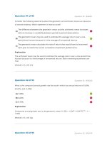
Figure 1. Irradiated human monocyte-derived macrophages are viable and metabolically active, despite
DNA damage. (A) Radiation-induced macrophage DNA damage is demonstrated by immunocytochemistry
for Ser139-phosphorylated H2AX (γ H2AX) (red), while nuclei were counterstained with DAPI (blue). Scale
bar represents 20 μ m. Graph indicates the percentage of macrophages (n = 3 and 400 cells/donor counted)
exhibiting γ H2AX foci. Data was analysed with paired t-test. **P < 0.01. (B) Western blot analysis of total and
phosphorylated Chk2 (Thr68) expression on non-irradiated (− ) or irradiated (2, 6 and 10 Gy) (+ ) macrophages
(n = 3), upon 1, 6 and 24 h. (C) Western blot analysis of caspase-7 expression on non-irradiated (− ) or
irradiated (2, 6 and 10 Gy) (+ ) macrophages (n = 3), upon 1, 6 and 24 h. In all Western blots β -actin was used
as loading control. (D) Brightfield microscopic images of non-irradiated (0 Gy) and irradiated macrophages
(10 Gy). Scale bar represents 100 μ m. (E) Quantification of the metabolic activity of irradiated macrophages (2,
6 or 10 Gy) (n = 8), normalized to the activity of non-irradiated ones, and expressed as percentage. One-sample
t-test was performed.
Scientific Reports | 6:18765 | DOI: 10.1038/srep18765
3
www.nature.com/scientificreports/
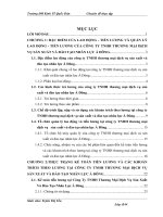
Figure 2. Ionizing radiation induces macrophage NF-κB activation and increases Bcl-xL expression.
(A) Evaluation of RelA phosphorylation (Ser536) and RelB, cRel, p100/p52 and p105/p50 subunit expression,
1 and 6 h after irradiation (2, 6 and 10 Gy). (B) RelB nuclear translocation 6 h after macrophage irradiation
(10 Gy). Histone deacetylase 1 (HDAC1) and β -actin were used as loading controls for nuclear and cytoplasmic
fractions, respectively. (C) Evaluation of Bcl2 and Bcl-xL expression after macrophage irradiation. Western
blot images are representative of protein expression/phosphorylation status in distinct donors (at least n = 4),
evaluated in two independent experiments.
Overall, we concluded that irradiated macrophages are viable and metabolically active, and do not activate
apoptosis, despite radiation-induced DNA damage.
Ionizing radiation activates macrophage pro-survival and NF-κB signalling pathway. Considering macrophage survival following ionizing radiation exposure, a typical radiation-induced
survival pathway mediated by NF-κ B33, was next investigated. Western blot analysis for the five NF-κ B family
subunits, RelA, RelB, cRel, p52/p100 and p50/p105 (Fig. 2A) as well as for Iκ Bα (Supplementary Fig. S2), a
NF-κ B inhibitor, were performed. Densitometry analysis (Supplementary Fig. S3) confirmed that ionizing radiation increased the expression of RelB at every time points in every donors and tend to slightly increase cRel
expression (Supplementary Fig. S3A). Additionally, ionizing radiation also tend to slightly increase p100 processing, as suggested by a reduced p52/p100 ratio at 2 and 6 Gy. No major alterations were found in phRel/RelA and
p105/p50 levels. Moreover, alterations in RelB suggested that ionizing radiation upregulates macrophage NF-κ B,
mainly through the non-canonical pathway. As RelB was the most consistently upregulated subunit in response to
radiation, we next confirmed RelB nuclear translocation, by evaluating its expression, in nuclear and cytoplasmic
extracts, 6 h after 10 Gy cumulative dose (Fig. 2B). In fact, ionizing radiation increased RelB nuclear expression
(Supplementary Fig. S3B), suggesting its nuclear translocation and subsequent activation. As NF-κ B can induce
the expression of anti-apoptotic proteins, such as Bcl2 and Bcl-xL34, we evaluated the expression of these targets
after macrophage irradiation (2, 6 and 10 Gy) (Fig. 2C). The most relevant alteration was the Bcl-xL increased
expression, particularly at 10 Gy (Supplementary Fig. S3C). The increase was visible at 1 h and sustained after
6 h, suggesting induction of macrophage pro-survival activity. Since Bcl-xL maximum expression tends to occur
after 10 Gy, all subsequent experiments were performed with macrophages submitted to this cumulative ionizing
radiation dose.
Ionizing radiation induces a reduction in anti-inflammatory macrophage phenotype. To evaluate the effect of ionizing radiation on macrophage polarization profile, we characterized the pattern of expression
of pro- or anti-inflammatory cytokines/chemokines and cell surface receptors, by a combination of quantitative
Scientific Reports | 6:18765 | DOI: 10.1038/srep18765
4
www.nature.com/scientificreports/
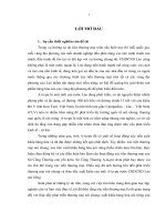
Figure 3. Irradiated macrophages present a reduced anti-inflammatory phenotype. (A) mRNA expression
profile of pro-(CXCL8, CD80, IL1B, TNF, CCR7, CCL2, IL6) and anti-inflammatory (CD163, MRC1, VCAN)
macrophage markers, by real-time PCR, 20 h after 10 Gy. Graphs represent mRNA expression of irradiated
macrophages compared to non-irradiated ones (dotted line) (at least n = 7 per marker). β -actin was used
as housekeeping gene. Wilcoxon signed rank test was used to compare the median of each dataset against a
hypothetical median value of 1. (B) Expression of a monocyte/macrophage lineage (CD14), pro-(HLA-DR and
CD86) and anti-inflammatory (CD163) macrophage markers was determined, by flow cytometry, 20 h after
irradiation (at least n = 6 per marker). Paired t-test was used for statistical analysis. (C) Levels of pro-(IL-6,
IL-12/IL-23(p40)) and anti-inflammatory (TGF-β 1 and IL-10) cytokines were determined in macrophage CM
(n = 9) by ELISA, 20 h after irradiation. Data was normalized to protein concentration. Wilcoxon matched pair
test was used for statistical analysis. *P < 0.05, **P < 0.01. Median is represented by the horizontal line inside
the box plots, while the average is indicated by a “+ ”. In IL-6 and IL-10 graphics outliers are also indicated.
real-time PCR, flow cytometry and ELISA. Ionizing radiation induced a significant increase (P < 0.05) of CD80
expression, but not of other pro-inflammatory gene markers. In fact, IL6 and CCL2 were significantly downregulated (P < 0.01) after macrophage irradiation (Fig. 3A). Furthermore, all anti-inflammatory gene markers tested,
CD163, MRC1 and VCAN were significantly downregulated (P < 0.05 or P < 0.01) upon irradiation (Fig. 3A). To
investigate how these changes could translate into cell surface receptor expression, flow cytometry was then performed. Results evidenced that ionizing radiation significantly increased HLA-DR (P < 0.01) and CD86 (P < 0.05)
(macrophage pro-inflammatory markers), and tend to decrease CD163 (macrophage anti-inflammatory marker),
but did not alter the expression of the monocyte/macrophage lineage marker CD14 (Fig. 3B). To further complete
the polarization profile analysis, levels of pro-(IL-6 and IL-12/IL-23(p40)) and anti-inflammatory (TGF-β 1 and
IL-10) cytokines were evaluated by ELISA, using conditioned medium (CM) from non-irradiated (0 Gy) and irradiated (10 Gy) macrophages, normalized to protein concentration (Fig. 3C). Interestingly, only IL-10 levels were
significantly downregulated (P < 0.01) by ionizing radiation. IFN-γ and TNF-α levels were also investigated, but
Scientific Reports | 6:18765 | DOI: 10.1038/srep18765
5
www.nature.com/scientificreports/
Figure 4. Upon exogenous stimulation, irradiated macrophages polarize towards a pro- or an antiinflammatory phenotype. Non-irradiated and 10 Gy irradiated macrophages (n = 6) were stimulated, for
20 h, with LPS (100 ng/ml) and IFN-γ (20 ng/ml) towards a pro-inflammatory, or with M-CSF (10 ng/ml) and
IL-10 (20 ng/ml) towards an anti-inflammatory phenotype. (A) Expression of monocyte/macrophage lineage
(CD14), pro-(HLA-DR and CD86) and anti-inflammatory (CD163) macrophage markers was determined by
flow cytometry. (B) Macrophage CM levels of pro-(TNF-α , IL-6, IL-12/IL-23(p40)) and anti-inflammatory
(TGF-β 1 and IL-10) cytokines were determined by ELISA. Data was normalized to protein concentration.
Paired t-test and one-way ANOVA were used for statistical analysis. *P < 0.05, **P < 0.01, ***P < 0.001. Median
is represented by the horizontal line inside the box plots, while the average is indicated by a “+ ”.
were undetectable for both macrophage populations. Altogether, this data suggests that ionizing radiation directs
macrophages towards a reduced anti-inflammatory phenotype, without achieving however a complete classical
pro-inflammatory profile.
Upon exogenous stimulation, irradiated macrophages polarize towards pro- or
anti-inflammatory phenotypes. Unstimulated M-CSF differentiated macrophages typically exhibit a
more anti-inflammatory-like phenotype, but they are able to polarize towards a pro- or an anti-inflammatory
phenotype, after proper exogenous stimulation35,36. The functional response to external stimuli, termed plasticity, is one of the major hallmarks of the mononuclear phagocyte system37. To assess how ionizing radiation
affects macrophage plasticity, we evaluated whether irradiated macrophages were still able to polarize towards a
pro- or an anti-inflammatory phenotype, upon proper exogenous stimulation. Therefore, upon 10 Gy cumulative
dose exposure, macrophages were stimulated for 20 h with LPS (100 ng/mL) and IFN-γ (20 ng/mL), towards a
pro-inflammatory phenotype, or with M-CSF (10 ng/mL) and IL-10 (20 ng/mL), towards an anti-inflammatory
one. The expression of pro- and anti-inflammatory cell surface markers and cytokines/chemokines was evaluated
by flow cytometry (Fig. 4A) and ELISA (Fig. 4B), respectively. Comparison between exogenously stimulated
(LPS/IFN-γ or M-CSF/IL-10) non-irradiated (0 Gy) and irradiated (10 Gy) macrophages indicated that ionizing
radiation did not affect macrophage ability to polarize towards a pro- or an anti-inflammatory phenotype, upon
proper exogenous stimuli. LPS/IFN-γ -stimulated macrophages (both non-irradiated and irradiated) present
higher levels of pro-inflammatory markers such as HLA-DR, CD86, TNF-α , IL-6 and IL-12/IL-23 (p40), being
the last four significantly different (from P < 0.05 to P < 0.001), when compared to M-CSF/IL-10-stimulated macrophages (Fig. 4A,B). On the other side, LPS/IFN-γ -stimulated macrophages tend to present lower levels of the
anti-inflammatory marker CD163, comparing to M-CSF/IL-10-stimulated macrophages (Fig. 4A). Our results
evidenced that, contrary to expectations, the pro-inflammatory stimuli (LPS/IFN-γ ) also induced higher levels of
immunosuppressive IL-10 cytokine than the anti-inflammatory ones (M-CSF/IL-10). However, this phenomenon
was also previously reported in M-CSF-conditioned dendritic cell precursors, which exhibit a rapid IL-10 release
upon LPS stimulation and may therefore participate in the modulation of inflammation and immune response38.
Scientific Reports | 6:18765 | DOI: 10.1038/srep18765
6
www.nature.com/scientificreports/
Figure 5. Ionizing radiation increases macrophage area, aspect ratio and phagocytic rate, but does not
alter MMP-2 and -9 activities. (A) Actin (green) and tubulin (red) stainings of non-irradiated and 10 Gy
irradiated macrophages (n = 4). Quantification of cell area and aspect ratio was performed using Fiji software.
Scale bar indicates 20 μ m. (B) Phagocytic ability of non-irradiated and 10 Gy-irradiated macrophages (n = 5)
was determined after 1 h incubation with FITC-labelled (green) Staphylococcus aureus particles. F-actin was
stained with rhodamine phalloidin (red). The percentage of macrophages able to phagocyte S.aureus particles
was quantified using Fiji software. Scale bar indicates 20 μ m. (C) MMP-2 and -9 activity was evaluated by
gelatin zymography, using 1 and 15 μ g of protein from CM of non-irradiated and 10 Gy-irradiated macrophages
(n = 10). White bands of proteolytic activity were revealed on a Coomassie Blue-stained gelatin gel. All data was
analysed with paired t-test. **P < 0.01, ***P < 0.001. Median is represented by the horizontal line inside the box
plots, while the average is indicated by a “+ ”.
Interestingly, irradiated macrophages stimulated with LPS/IFN-γ presented significantly (P < 0.05) higher
levels of HLA-DR, when compared to their non-irradiated counterparts (Fig. 4A). On the other hand, under
stimulation with M-CSF/IL-10, irradiated macrophages tend to exhibit lower CD163 and higher HLA-DR levels,
when compared to non-irradiated macrophages (Fig. 4A,B). Also, irradiated macrophages polarized towards a
pro- or an anti-inflammatory phenotype presented a significant decrease (P < 0.05) in IL-10 levels, when compared
to non-irradiated and also exogenous stimulated ones (Fig. 4B). Altogether, these results demonstrate that, upon
exogenous stimulation, irradiated macrophages are still able to polarize towards a pro- or an anti-inflammatory
phenotype. However, ionizing radiation by itself tends to promote the pro-inflammatory phenotype.
Ionizing radiation induces macrophage morphological alterations and increases their phagocytic rate, without altering MMP-2 and MMP-9 activities. In order to characterize morphological
changes, macrophage area and aspect ratio were determined 1 h after exposure to 10 Gy (Fig. 5A). Results indicated that irradiated macrophages presented a significantly higher (P < 0.05) cell area (2,471 ± 513.3 μ m2) and
tend to increase cell aspect ratio (quotient between major and minor cell axes) (2.38 ± 0.63), when compared to
non-irradiated ones (area 2,047 ± 616.3 μ m2 and aspect ratio 1.72 ± 0.24).
Besides functional and morphological plasticity, other features like phagocytic ability and proteolysis are
also characteristic of macrophages39. To evaluate the effect of ionizing radiation on macrophage phagocytic
ability, non-irradiated and 10 Gy irradiated macrophages were incubated, for 1 h, with FITC-labelled and killed
Staphylococcus (S.) aureus particles. Results revealed that ionizing radiation significantly increased (P < 0.01) the
percentage of macrophages able to phagocyte S. aureus particles (Fig. 5B).
The impact of ionizing radiation on the activity of MMP-2 and MMP-9, two matrix metalloproteases involved in
the promotion of cancer cell invasion and angiogenesis40, was determined by gelatin zymography. Using 1 μ g of protein from macrophage CM, no differences in pro-MMP-9 proteolytic activity were found, between non-irradiated
and 10 Gy irradiated macrophages (Fig. 5C). The same conclusion was extended to MMP-9 and pro-MMP-2 proteolytic bands, when 15 μ g of macrophage CM protein were loaded. Our results evidence that ionizing radiation
induces macrophage morphologic alterations and increases their phagocytic rate, without affecting macrophage
MMP-2 and MMP-9 proteolytic activities.
Irradiated macrophages promote cancer cell invasion and angiogenesis. To investigate the effect
of ionizing radiation on macrophage pro-invasive cancer cell activity, non-irradiated or 10 Gy irradiated macrophages and irradiated colorectal cancer cells were confronted on Matrigel invasion assays (Fig. 6A). Irradiated
as non-irradiated macrophages significantly promoted (P < 0.001 or P < 0.01) the invasion of irradiated RKO
Scientific Reports | 6:18765 | DOI: 10.1038/srep18765
7
www.nature.com/scientificreports/
Figure 6. Ionizing radiation does not affect macrophage ability to promote cancer cell invasion and
cancer cell-mediated angiogenesis. (A) Matrigel invasion assays were established confronting RKO cells
(upper compartment) and macrophages (lower compartment) after being separately exposed or not to 10 Gy
cumulative ionizing radiation dose. The six possible combinations are represented in the scheme. Invasive
cells were counterstained with DAPI and counted on the microscope. (B) RKO cells were inoculated, for 72 h,
with CM from non-irradiated or 10 Gy irradiated macrophages, in rings (inoculation areas), on top of CAM.
The comparison of RKO+ CM Mac 0 Gy versus RKO+ CM Mac 10 Gy was evaluated in two rings within the
same fertilized egg (n = 18), while the condition RKO+ RPMI was performed in a single distinct egg (n = 16).
Analysis of RKO-induced angiogenesis was performed through quantification of the number of new vessels in
control and experimental conditions. ANOVA analysis demonstrated a significant difference between groups.
*P < 0.05, **P < 0.01, ***P < 0.001. The median is represented by the horizontal line inside the box plots, while
the average is indicated by a “+ ”.
cells. To clarify if macrophage pro-invasive ability was dependent on the presence of irradiated colorectal cancer
cells, we used non-irradiated RKO cells. Herein, we verified the same effect previously observed indicating that
irradiated macrophages do not lack the ability to promote cancer cell invasion. Moreover, we also observed that
irradiation of RKO cells alone significantly decrease (P < 0.05) its invasive potential. We could indeed predict
that RKO cell invasive potential was going to be highly affected by radiation exposure due to the RKO intrinsic
radiosensitivity, previously reported in the literature. An accumulation of about 70% of RKO cells was described
in G2/M-phase 16 ± 24 h after 12 Gy irradiation in pH 7.5 medium41. RKO cells were also included in a group
of radiosensitive cells, upon evaluation of the clonogenic survival of 27 human tumour cell lines after ionizing
radiation exposure42.
In order to investigate the effect of irradiated macrophages on cancer cell-induced angiogenesis, a chick embryo
chorioallantoic membrane (CAM) assay was performed. CM from both non-irradiated and 10 Gy irradiated macrophages significantly promoted (P < 0.05) RKO-induced angiogenesis (Fig. 6B). No differences were observed
between RKO cells treated with CM derived from non-irradiated and irradiated macrophages. Altogether, Matrigel
invasion and CAM-based assays demonstrated that, in the present experimental context, ionizing radiation per se
was not able to restrain macrophages’ endogenous ability to promote cancer cell invasion and cancer cell–induced
angiogenesis, which constitute two main hallmarks of cancer3.
Discussion
The present work aims to understand the effect of ionizing radiation on human macrophages, as they are important
components of the tumour microenvironment, and also highly recruited into tumours during radiation therapy43.
The recurrent use of mouse models and lack of clinically relevant doses in other studies have not allowed a full
understanding of this effect16–20. In the present study, we characterized, for the first time, the response of human
primary macrophages to cumulative ionizing radiation doses, using the same fractionated scheme as used during
Scientific Reports | 6:18765 | DOI: 10.1038/srep18765
8
www.nature.com/scientificreports/
cancer patients’ treatment (2 Gy/fraction/day), up to 10 Gy cumulative dose. As a model, we used M-CSF-cultured
macrophages, differentiated from peripheral-blood monocytes, as it is considered the predominant in vitro system to study human tissue macrophages44. M-CSF is a growth factor involved in the recruitment of monocytes/
macrophages to tissues and also in the regulation of macrophage function within tumours45. Taking into account
that the cellular radiation response is a complex process46, several features like DNA damage, NF-κ B signalling
pathway, polarization profile, plasticity, phagocytosis, proteolysis and the ability to promote cancer cell activities,
were evaluated in irradiated human macrophages.
Macrophage resistance to ionizing radiation was first reported in mouse models some decades ago47,48.
Nowadays, it is recognized that human macrophages, similarly to regulatory T cells (Tregs), dendritic cells, Natural
Killer (NK) cells and thrombocytes, as well as MDSCs, display a more radiation resistant phenotype than other
immune cell populations, such as monocytes27,29,30,46. However, most of the studies were performed in mouse macrophages irradiated with X-ray doses, which barely mimic the fractionated scheme used in cancer patients’ treatment. Our results demonstrated that irradiated macrophages exhibited higher DNA damage, confirmed through
increased H2AX phosphorylation, than non-irradiated ones. DNA damage induced by ionizing radiation is known
to lead to Chk2-specific phosphorylation (Thr68) at sites of DSBs49. In agreement, we demonstrated that Chk2
phosphorylation increased along time and according to the exposure doses. Despite DNA damage, irradiated macrophages remain viable and metabolically active, leading to understand which survival pathways were activated.
We focused on NF-κ B signalling, which is known to induce a pro-survival response in cells exposed to single 2 Gy
doses33,50. Our results demonstrated that RelB expression was consistently increased after macrophage exposure
to 2, 6 and 10 Gy cumulative doses. RelB nuclear translocation suggested the involvement of the non-canonical
NF-κ B pathway in radiation-induced macrophage response. Our data also demonstrated an increased expression
of the anti-apoptotic Bcl-xL protein, which also contributes to the promotion of pro-survival activity. Interestingly,
a recent study demonstrated that ionizing radiation induces RelB to activate Bcl-xL in cancer cells51.
NF-κ B transcription factors are not only important for cell survival upon irradiation, but are also considered
major regulators of inflammation processes and, particularly in macrophages, their activation is required for
the anti- to pro-inflammatory phenotype transition52–54. Radiation-induced NF-κ B alterations in macrophages
led to the characterization of macrophage inflammatory status upon irradiation, which data was summarized
in the scheme of Fig. 7. In the present study, 10 Gy cumulative ionizing radiation dose significantly decreased
both anti-inflammatory gene markers (CD163, MRC1, VCAN) and the immunosuppressive cytokine IL-10, and
increased HLA-DR and CD86 expression of M-CSF differentiated macrophages (Fig. 7). Additionally, we also
demonstrated that bacterial phagocytosis, a classical feature of pro-inflammatory macrophages, was found to be
significantly increased in irradiated macrophages. In fact, mouse macrophages subjected to 8 Gy single dose were
also described to slightly enhance phagocytosis of inert latex beads55. Together, our data supports the hypothesis that ionizing radiation may drive macrophages towards a pro-inflammatory phenotype. Although irradiated
macrophages exhibited increased CD80 expression, other classical pro-inflammatory markers, like IL1B, TNF
and IL6 were unaltered, downregulated, or undetectable at the cytokine level. This suggests that irradiated macrophages did not reach a classical pro-inflammatory phenotype, despite the observed reduction of their original
anti-inflammatory-like phenotype. The mechanism behind this shift in macrophage phenotype may indeed rely
on radiation-induced NF-κ B alterations, particularly in RelB subunit as its expression is increased in irradiated
macrophages. In fact, very recently, a switch of anti-inflammatory to pro-inflammatory macrophages was found to
be directly mediated by RelB induction in M-CSF and TNF-stimulated osteoclast precursors56. However, further
experiments are still required to confirm RelB involvement in macrophage anti-inflammatory phenotype reduction,
suggested to occur after macrophage ionizing radiation exposure.
To simplify the understanding of our hypothesis, we represented the typical pro- and anti-inflammatory macrophage profiles as two extremes of a continuous polarization spectrum (generally represented as a line). However,
macrophage polarization has been revealed as a very complex and dynamic system and new representations are
emerging as long as new knowledge in this area is coming out. Recently, Ruffell and Coussens summarized the
macrophage polarization system as a circle, which does not consider pro- and anti-inflammatory macrophage
profiles, but rather emphasize that macrophage functional roles (angiogenesis, cytotoxicity, stimulation, suppression or chemotaxis) (included in an inner circle) are dictated by the integration of multiple stimuli (represented
in an outer circle)57. Therefore, we may speculate that ionizing radiation exposure could also constitute one of
the distinct stimuli capable of polarizing macrophages into a different phenotype, which does not completely
corresponds to a pro- or an anti-inflammatory one, but could rather include features from both phenotypes, as we
demonstrated in this work, or even from other phenotypes still to be defined. Overall, our study defines, for the
first time, a molecular profile for human macrophages subjected to cumulative ionizing radiation doses, emphasizing the important role of fractionated ionizing radiation doses, as used in radiotherapy, to direct macrophages
towards a pro-inflammatory phenotype, which is recognized to be tumour cytotoxic37. According to the literature,
the relation between ionizing radiation exposure and inflammatory response seems to be dependent not only on
the cell type analysed and radiation quality, but mainly on the delivered dose58. Low doses (maximum of 12 Gy at
≤ 1.0 Gy/fraction), usually applied in non-malignant disorders or received by normal tissues outside the tumour
target volume, induce an anti-inflammatory phenotype, while higher doses (single doses ≥ 2 Gy, total doses ≥ 40 Gy)
are reported to have a pro-inflammatory effect59,60. Most of the studies, aiming to reveal the role of irradiation on
macrophage inflammatory status, are performed using in vitro or ex vivo mouse macrophages. Although mouse
models have widely contributed to our understanding of ionizing radiation-induced effect on macrophages, it is
also well recognized that mouse and human macrophages present many distinct features61. Particularly, mouse
macrophages are high producers of nitric oxide (NO) and L-citrulline from L-arginine, via inducible nitric oxide
synthase (iNOS) activation, while NOS and arginase activities in human macrophages are quite debatable62,63. These
considerations should be taken into account when extrapolating data from one species to another.
Scientific Reports | 6:18765 | DOI: 10.1038/srep18765
9
www.nature.com/scientificreports/
Figure 7. Schematic representation of the effect of ionizing radiation on human blood monocyte-derived
macrophages. Two main macrophage functional polarization status are recognized: a pro-inflammatory,
responsible for killing intracellular pathogens and antitumour activity, and an anti-inflammatory one, which
induces tissue repair, angiogenesis and promotes tumour activity. Pro-inflammatory macrophages produce high
levels of TNF-α , IL-6 and IL-1β cytokines and exhibit CD80, CD86, HLA-DR, CCR7 and NF-κ B increased
expression, while anti-inflammatory ones express CD163, MRC1 and produce high levels of TGF-β 1 and
IL-10 cytokines. In the present study, we demonstrated that irradiated macrophages exhibit a decrease of antiinflammatory (CD163, MRC1 and IL-10) and an increase of other pro-inflammatory (CD80, CD86, HLA-DR)
markers. Although irradiated macrophages are more effective than non-irradiated ones at phagocytosis, a
typical feature of pro-inflammatory macrophages, they fail to reach a classical pro-inflammatory phenotype,
as they do not produce high levels of TNF-α , IL-6, IL-1β and CCR7. On the other hand, and similarly to
their counterparts, irradiated macrophages are able to promote cancer cell invasion and cancer cell-induced
angiogenesis. Our data suggests that M-CSF differentiated macrophages, exposed to cumulative ionizing
radiation doses up to 10 Gy, exhibit a reduced anti-inflammatory-like phenotype, compared to non-irradiated
ones, probably moving towards a pro-inflammatory phenotype. However, although irradiated macrophages
exhibit characteristics from both pro- and anti-inflammatory phenotypes, they do not perfectly match to any of
these typical profiles, appearing to acquire intermediate characteristics.
We have also demonstrated that irradiated macrophages are as able as their non-irradiated counterparts to
promote RKO cancer cell invasion and cancer cell-induced angiogenesis. This is the first report demonstrating
that, at least during the first week (5 days) of radiotherapy, human macrophages sustain its ability to promote
cancer cell invasion and cancer cell-induced angiogenesis, which is a matter of concern. Nevertheless, we need to
be cautious when extrapolating this data to the clinic, as during further neoadjuvant treatment the situation may
change. Not only because cell response along treatment time may be different but also because in vivo other host
cells and several environmental factors may contribute to tumour response to radiotherapy. As our team previously
demonstrated, MMP activity is known to be an important factor for macrophage-mediated cancer cell invasion64.
Therefore, the sustained promotion of cancer cell invasion by irradiated macrophages may be associated with the
fact that MMP-2 and MMP-9 activity is not being affected by radiation exposure. Furthermore, we also showed
that irradiated macrophages are still able to promote cancer cell-induced angiogenesis.
In summary, our work adds valuable data on characterization of a plethora of functions in human macrophages,
subjected to cumulative ionizing radiation doses, as used during cancer patients’ treatment, which were not
addressed before. We have characterized important aspects like plasticity, proteolysis, phagocytosis and cancer cell
activity promotion. We demonstrated that human macrophages subjected to cumulative doses of ionizing radiation
are viable, metabolically active and exhibit increased survival signalling, through NF-κ B activation and increased
Scientific Reports | 6:18765 | DOI: 10.1038/srep18765
10
www.nature.com/scientificreports/
Bcl-xL expression, despite DNA damage. Irradiated macrophages also present a reduced anti-inflammatory profile,
increased phagocytosis and unaltered MMP-2 and -9-mediated proteolysis. Pro-inflammatory-like macrophages
are known to be cytotoxic and exhibit antitumoural activities and may therefore contribute to the efficacy of local
radiotherapy37. Our data also demonstrates that irradiation maintains macrophage ability to promote cancer cell
invasion and cancer cell-induced angiogenesis. Overall, although radiotherapy mainly induces cancer cell death,
other components of the microenvironment, particularly macrophages, are also irradiated and could persist still
sustaining the activity of residual radioresistant cancer cells. Furthermore, this knowledge opens new perspectives
for macrophage clinical targeting, prior, after or concomitantly to ionizing radiation, as a strategy to improve
radiotherapy efficacy.
Material and methods
Ethics statement. In the present study, human monocytes were obtained from buffy coats, which are a
highly leukocyte-enriched waste-product that results from a whole blood donation, from healthy blood donors.
A collaboration protocol between our Institution and Centro Hospitalar São João (CHSJ), where blood donations
of Portugal North region are performed, allows the use of these products for investigation purposes. All studies
using this human material were approved by CHSJ Ethics Committee for Health (References 259 and 260/11),
in agreement with the Helsinki declaration. Informed consent was obtained from all subjects before each blood
donation.
Human monocyte isolation and macrophage differentiation. Human monocytes were isolated as previously described64. Following this procedure, over 80% of isolated monocytes were found to be
CD14-positive64. For monocyte-macrophage differentiation, 1.2 × 10⁶ cells/9.6 cm2 (6-well plate) were cultured in complete RPMI1640 medium with GlutaMax (Invitrogen) in the presence of 50 ng/mL of macrophage
colony-stimulating factor (M-CSF) (ImmunoTools). Culture medium (1.5 mL/well) was replaced after one week
and macrophage differentiation was completed 13 days after monocyte isolation, as at this stage macrophages
were shown to provide a higher stimulus for cancer-cell invasion, than with shorter differentiation times64.
Cell culture. RKO (CRL-2577) cells, derived from a human colon carcinoma, were purchased from the
American Type Culture Collection (ATCC). Cells were maintained at 37 °C, 5% CO2 humidified-atmosphere,
in RPMI1640 (L-Glutamine) (Invitrogen) supplemented with 10% FBS (Lonza, Basel, Switzerland), 100 U/mL
penicillin and 100 μ g/mL streptomycin (Invitrogen).
Ionizing radiation exposure. Prior irradiation, a dosimetry plan was established (ELEKTA CMS
XiO v.4.7.0). Culture plates were submitted to a Computerized Tomography (CT) scan and the volume occupied by two entire plates was defined as the target volume. Two beam fields, one anterior-posterior and other
posterior-anterior, were arranged to deliver 2 Gy per fraction to this target volume. Inside the defined volume, the
total dose varied from 198 cGy to 202 cGy. As the 4 cGy difference was not significant, the same dose was considered homogenously distributed through plates. To guarantee this uniform dose and to avoid the build-up region
of the 18 MV photon beam, 5 water plates were added above, and 5 below the culture plates during irradiation.
Medium was renewed before the first irradiation. Both macrophages and RKO cells were then exposed to 1–5
cumulative ionizing radiation doses (2 Gy/fraction/day), for a week (Monday to Friday). Therefore, the maximum
cumulative irradiation dose, equivalent to 5 fractions, totalized 10 Gy (Supplementary Fig. S4). Photon beam was
produced by a PRIMUS (Siemens, Malvern, PA, USA) linear particle accelerator, used for human radiotherapy
sessions, operated at 18 MV at the Radiotherapy Service of CHSJ. To avoid differences between non-irradiated
and irradiated cells, caused by medium agitation during transport to/from the Radiotherapy Service, control cells
were also transported, but were not radiation-exposed.
Cell viability. For a proper follow-up of macrophage during irradiation week, cells were carefully observed
under a light microscope (Olympus) and daily pictures were taken. To complement this qualitative data, macrophage metabolic activity was determined through resazurin reduction assay, which was considered a sensitive,
reproducible and non-destructive assay to measure cell response to irradiation65. Briefly, 20 h after irradiation
(2, 6 or 10 Gy), macrophages were incubated with resazurin redox dye (0.01 mg/mL) (Sigma-Aldrich) for 3 h
at 37 °C and 5% CO2. Fluorescence intensity was measured (530 nm Ex/590 nm Em), using the multi-mode
microplate reader Synergy MX (BioTek) and values were normalized to protein concentration in the CM, measured with detergent-compatible (DC) protein assay (BioRad). Data from irradiated macrophages was then compared with the respective controls and expressed as percentage.
Protein extraction and Western Blot. Whole cell protein-extracts were performed 1, 6 and 24 h after
irradiation (2, 6 and 10 Gy) (n = 4), using lysis buffer supplemented with a cocktail of proteases/phosphatases
inhibitors, as previously described64. Nuclear/cytoplasmic extracts were performed 6 h after 10 Gy (n = 5), using
appropriate lysis buffer [10 mM Hepes pH 7.9, 1.5 mM MgCl2, 10 mM KCL, 1 mM DTT, 0.1% Igepal, protease/
phosphatase inhibitors cocktail]. Cytoplasmic extracts were obtained after centrifugation at 14 000 rpm for
10 min, at 4 °C. For nuclear extracts, pellets were resuspended in another lysis buffer [20 mM Hepes pH 7.9 ,
420 mM KCl , 1.5 mM MgCl2, 1 mM DTT, 25% Glycerol, protease/phosphatase inhibitors cocktail], rocked for
15 min at 4 °C, centrifuged at 14 000 rpm for 15 min, and sonicated. Following SDS-PAGE, gels were transferred
onto polyinylidine difluoride (PVDF) membrane, which were then incubated, for 1 h, with primary antibodies against the following proteins: p105/p50, p100/p52 (Millipore), Bcl-xL (BD Biosciences), phospho-Iκ Bα
(Ser32/36) (clone 5A5), Iκ Bα (clone 44D4), phospho-RelA (Ser536) (clone 93H1), Bcl-2 (clone 50E3),
phospho-Chk2 (Thr68), Chk2, caspase-3, caspase-7, cleaved PARP (clone D64E10) (Cell Signalling), RelA, RelB,
Scientific Reports | 6:18765 | DOI: 10.1038/srep18765
11
www.nature.com/scientificreports/
cRel (Santa Cruz Biotechnology). Positive controls for caspase-3 and -7 activation as well as for PARP cleavage
were used. Antibody against β -actin (clone 8H10D10) (Cell Signalling) was used to normalize protein expression. Goat anti-rabbit or horse anti-mouse-Horseradish peroxidase (HRP)-conjugated secondary antibodies (Cell
Signalling) were used for 1 h, followed by ECL-Detection (Thermo Fisher Scientific). Densitometry analysis of
western blot images from Fig. 2 was performed with Quantity One software (BioRad).
RNA extraction, cDNA preparation and quantitative PCR analysis. Total RNA, from
non-irradiated or 10 Gy irradiated macrophages, was extracted using TriPure Isolation Reagent (Roche),
according to manufacturer’s instructions. RNA was converted to cDNA using 150 U of SuperScript II
Reverse Transcriptase, 1× first strand buffer, 10 mM DTT 0.1 M (Invitrogen), 0.5 mM dNTPs 10 mM (Bioron),
8U of rRNasin (Promega) and RNase/DNase free water (Gibco). To evaluate mRNA expression levels of proand anti-inflammatory gene markers, quantitative PCR using Brilliant II Sybr green kit (Stratagene/Agilent
Technologies) and specific MX3005P 96-well semi-skirted plates, were performed. Samples were analysed on
the MX3005P qPCR platform (Stratagene/Agilent). The following primers, from Invitrogen, were used for
RT-qPCR: CXCL8, F: 5′ -CCAGGAAGAAACCACCGGA-3′ , R: 5′ -GAAATCAGGAAGGCTGCCAAG-3′ ;
IL1B, F: 5′-GGCAGGGAACCAGCATC-3′ , R: 5′-CCGACCACCACTACAGCAA-3′ ; TNF F: 5′GGCTGGAGCTGAGAGATA-3′ , R: 5′ -CAGCCTTGGCCCTTGAAGA-3′ . Primer sets for ACTB (used as a
normalizing gene), CXCL12 and CCL2 were obtained from Qiagen, while probes for CD80, CCR7, IL6, CD163,
MRC1 and VCAN were from Applied Biosystems.
™
Macrophage polarization. After 10 Gy cumulative ionizing radiation exposure, macrophages were
stimulated, during 20 h, with 100 ng/mL LPS (Sigma-Aldrich) plus 20 ng/mL IFN-γ (Immunotools) towards a
pro-inflammatory phenotype (M1-like), or with 10 ng/mL M-CSF plus 20 ng/mL IL-10 (Immunotools) towards
an anti-inflammatory (M2-like) one44.
Flow cytometry. For cell surface receptor expression analysis, non-irradiated and 10 Gy irradiated macrophages, subjected or not to further cytokine-induced polarization, as above detailed, were kept on ice, washed
with PBS, gently detached by scraping and resuspended in FACs buffer [PBS, 2% FBS (Lonza), 0.01% sodium
azide]. Stainings with anti-human CD14-APC (clone MEM-18), HLA-DR-PE (MEM-12), CD86-FITC (clone
BU63) (Immunotools) and CD163-PE (clone GHI/61) (R&D Systems) antibodies were performed in the dark
for 30 min. After additional washing steps, macrophages were fixed for 15 min in 4% paraformaldehyde (PFA).
Isotype-matched antibodies were used as negative controls, to define background staining. Cells were acquired
on a FACS Calibur Flow Cytometer (BD Biosciences), using Cell Quest Software (collecting 1 × 10⁴ cells).
Analysis was performed with FlowJo software (v7.6.5). Mean fluorescent intensity was calculated by subtracting
the respective isotype control intensity.
™
Enzyme-linked immunosorbent assay (ELISA). IL-6, IL-12/IL-23(p40), TNF-α , TGF-β 1 free active
and IL-10 cytokine levels were determined, according to manufacturer’s instructions (BioLegend), in CM from
non-irradiated and 10 Gy irradiated macrophages, subjected or not to further cytokine-induced polarization as
above detailed. Briefly, 50 μ L of cell culture supernatant were added to a 96-well plate pre-coated with the capture antibody of interest. The soluble proteins bound to the capture antibody were detected using a biotinylated
antibody, followed by an avidin-HRP conjugated solution. Finally, the addition of TMB substrate, resulted in a
colour change, which intensity was proportional to the amount of antigen captured. Absorbance was then read at
450 and 570 nm. Cytokine levels were determined by plotting values on a standard curve and normalizing them
to CM protein concentration.
Immunocytochemistry. DNA damage and morphology were evaluated, by immunocytochemistry, in
non-irradiated and 10 Gy irradiated macrophages (4 × 10⁴). After 1 h, macrophages were fixed with 4% PFA
for 20 min and immunocytochemistry procedure was then performed as previously described64. Macrophages
were incubated with monoclonal antibodies for phosphorylated histone-H2AX (Ser139) (γ H2AX) (clone
JBW301) (Millipore) or α - tubulin (Sigma-Aldrich), for 1 h, followed by goat-anti mouse AlexaFluor594-conjugated-secondary antibody (Invitrogen) incubation, for 45 min in the dark. F-actin was stained for
15 min with 0.5 μ M Phalloidin-FITC (Sigma-Aldrich), 0.1 M EGTA and 1 M MgSO4. Finally, nucleus was stained
with 10 μ g/mL 4′ , 6-diamidino-2-phenylindole (DAPI) solution. Multiwell plate-based screening was performed
with a Leica DMI6000 B inverted motorized fluorescence microscope (Leica Microsystems). Microscopic images
are represented at 300× magnification.
Phagocytosis. Ready-made pHrodo green Staphylococcus aureus BioParticles Conjugate (1 μ m diame-
ter) (Invitrogen) were resuspended in PBS up to 1 mg/mL and gently vortexed and sonicated for homogenous
dispersion. Non-irradiated and 10 Gy irradiated macrophages (4 × 10⁴) were then incubated with 1.6 × 10⁶ S.
aureus particles at 37 °C and 5% CO2, to evaluate phagocytic activity. After 1 h, macrophages were washed in
PBS and fixed with 4% PFA for 20 min. For cell identification, F-actin was stained with rhodamine-labeled phalloidin (1:100 dilution) (Invitrogen) for 30 min, after previous permeabilization with 0.2% Triton X-100 and 5%
bovine serum albumin (BSA) blocking. Finally, for nuclei visualization, macrophages were incubated with 10 μ g/
mL DAPI solution for 5 min. Multiwell plate-based screening was performed with IN Cell Analyzer 2000 (GE
Healthcare). Microscopic images are represented at 150× magnification. The number of cells able to phagocyte S.
aureus particles was then determined, using Fiji software66.
Scientific Reports | 6:18765 | DOI: 10.1038/srep18765
12
www.nature.com/scientificreports/
Gelatin zymography. Macrophage CM (1 and 15 μ g of protein), collected 24 h after 10 Gy cumulative ion-
izing radiation exposure, were used to evaluate MMP-2 and MMP-9 activities, through gelatin-zymography, as
previously described64.
Matrigel invasion assays. To evaluate macrophage-mediated RKO cell invasion, non-irradiated or 10 Gy
irradiated RKO cells (5 × 10⁴) were seeded on the upper compartment of Matrigel-coated inserts of 8-μ m pore
size (BD Biosciences), while non-irradiated or 10 Gy irradiated macrophages (2 × 10⁵), were added on the bottom, for 24 h, as previously described64.
Chick embryo in vivo angiogenesis assay. The chick embryo CAM model was used to evaluate
RKO-induced angiogenic response in the presence of macrophage CM. Therefore, commercially available fertilized chick (Gallus gallus) eggs were horizontally incubated at 37.5 °C, in a humidified atmosphere. On embryonic
development day (EDD)3, a square window was opened in the shell after removal of 1.5–2 mL of albumen, to
allow detachment of the developing CAM. The window was sealed with a transparent adhesive tape and eggs
re-incubated. On EDD10, RKO cells (1 × 10⁶) resuspended in CM from non-irradiated or 10 Gy irradiated macrophages were placed on top of the same CAM, into two independent 3 mm silicone rings, under sterile conditions. For control, RKO cells resuspended in RPMI medium were inoculated in a different egg. Eggs were
re-sealed and returned to the incubator for additional 72 h. On EDD13, rings were removed, the CAM was
excised from embryos and photographed ex-ovo under a stereoscope, using a 20× magnification (Olympus,
SZX16 coupled with a DP71 camera). The number of new vessels (< 20 μ m diameter) growing radially towards
the inoculation area was counted in a blind fashion.
Statistical analysis. All graphs and statistical analysis were performed using GraphPad Prism Software v5
(GraphPad-trial version). Data was analysed for Gaussian distribution using the D’Agostino and Pearson normality test, when n ≥ 8. To test the hypothesis that irradiated macrophages are different from non-irradiated ones,
Wilcoxon matched pairs test was used for non-parametric samples, while t-test (either paired t-test or one sample
t-test) was used for parametric data or when n < 8. For other comparisons, one-way ANOVA test was performed.
Statistical significance was achieved when P < 0.05.
References
1. Delaney, G., Jacob, S., Featherstone, C. & Barton, M. The role of radiotherapy in cancer treatment: estimating optimal utilization
from a review of evidence-based clinical guidelines. Cancer 104, 1129–1137 (2005).
2. Ringborg, U. et al. The Swedish Council on Technology Assessment in Health Care (SBU) systematic overview of radiotherapy for
cancer including a prospective survey of radiotherapy practice in Sweden 2001–summary and conclusions. Acta Oncol 42, 357–365
(2003).
3. Hanahan, D. & Weinberg, R. A. Hallmarks of cancer: the next generation. Cell 144, 646–674 (2011).
4. Bernier, J., Hall, E. J. & Giaccia, A. Radiation oncology: a century of achievements. Nat Rev Cancer 4, 737–747 (2004).
5. Allen, B. J., Bezak, E. & Marcu, L. G. Quo vadis radiotherapy? Technological advances and the rising problems in cancer management.
Biomed Res Int 2013, 749203 (2013).
6. Begg, A. C., Stewart, F. A. & Vens, C. Strategies to improve radiotherapy with targeted drugs. Nat Rev Cancer 11, 239–253 (2011).
7. Madani, I., De Neve, W. & Mareel, M. Does ionizing radiation stimulate cancer invasion and metastasis? Bull Cancer 95, 292–300
(2008).
8. Hofmeister, V., Schrama, D. & Becker, J. C. Anti-cancer therapies targeting the tumor stroma. Cancer Immunol Immunother 57, 1–17
(2008).
9. Di Caro, G., Marchesi, F., Laghi, L. & Grizzi, F. Immune cells: plastic players along colorectal cancer progression. J Cell Mol Med 17,
1088–1095 (2013).
10. Jain, R. K. Normalizing tumor microenvironment to treat cancer: bench to bedside to biomarkers. J Clin Oncol 31, 2205–2218 (2013).
11. Lauber, K., Ernst, A., Orth, M., Herrmann, M. & Belka, C. Dying cell clearance and its impact on the outcome of tumor radiotherapy.
Front Oncol 2, 116 (2012).
12. Condeelis, J. & Pollard, J. W. Macrophages: obligate partners for tumor cell migration, invasion, and metastasis. Cell 124, 263–266
(2006).
13. Joyce, J. A. & Pollard, J. W. Microenvironmental regulation of metastasis. Nat Rev Cancer 9, 239–252 (2009).
14. Vacchelli, E. et al. Trial Watch: Anticancer radioimmunotherapy. Oncoimmunology 2, e25595 (2013).
15. Hellevik, T. & Martinez-Zubiaurre, I. Radiotherapy and the Tumor Stroma: The Importance of Dose and Fractionation. Front Oncol
4, 1 (2014).
16. UNSCEAR. Biological mechanisms of radiation actions at low doses. A white paper to guide the Scientific Committee’s future
programme of work. (United Nations 2012).
17. College, O. in College Physics Vol. 3 (ed OpenStax College) Ch. 32 (2012).
18. Frischholz, B. et al. Reduced secretion of the inflammatory cytokine IL-1beta by stimulated peritoneal macrophages of radiosensitive
Balb/c mice after exposure to 0.5 or 0.7 Gy of ionizing radiation. Autoimmunity 46, 323–328 (2013).
19. Tsukimoto, M., Homma, T., Mutou, Y. & Kojima, S. 0.5 Gy gamma radiation suppresses production of TNF-alpha through upregulation of MKP-1 in mouse macrophage RAW264.7 cells. Radiat Res 171, 219–224 (2009).
20. Wunderlich, R. et al. Low and moderate doses of ionizing radiation up to 2 Gy modulate transmigration and chemotaxis of activated
macrophages, provoke an anti-inflammatory cytokine milieu, but do not impact upon viability and phagocytic function. Clin Exp
Immunol 179, 50–61 (2015).
21. Mah, L. J., El-Osta, A. & Karagiannis, T. C. gammaH2AX: a sensitive molecular marker of DNA damage and repair. Leukemia 24,
679–686 (2010).
22. Sulli, G., Di Micco, R. & d’Adda di Fagagna, F. Crosstalk between chromatin state and DNA damage response in cellular senescence
and cancer. Nat Rev Cancer 12, 709–720 (2012).
23. Radford, I. R., Murphy, T. K., Radley, J. M. & Ellis, S. L. Radiation response of mouse lymphoid and myeloid cell lines. Part II. Apoptotic
death is shown by all lines examined. Int J Radiat Biol 65, 217–227 (1994).
24. Eriksson, D. & Stigbrand, T. Radiation-induced cell death mechanisms. Tumour Biol 31, 363–372 (2010).
25. Seita, J. & Weissman, I. L. Hematopoietic stem cell: self-renewal versus differentiation. Wiley Interdiscip Rev Syst Biol Med 2, 640–653
(2010).
Scientific Reports | 6:18765 | DOI: 10.1038/srep18765
13
www.nature.com/scientificreports/
26. Youn, J. I. & Gabrilovich, D. I. The biology of myeloid-derived suppressor cells: the blessing and the curse of morphological and
functional heterogeneity. Eur J Immunol 40, 2969–2975 (2010).
27. Kozin, S. V. et al. Recruitment of myeloid but not endothelial precursor cells facilitates tumor regrowth after local irradiation. Cancer
Res 70, 5679–5685 (2010).
28. Xu, J. et al. CSF1R signaling blockade stanches tumor-infiltrating myeloid cells and improves the efficacy of radiotherapy in prostate
cancer. Cancer Res 73, 2782–2794 (2013).
29. Vatner, R. E. & Formenti, S. C. Myeloid-derived cells in tumors: effects of radiation. Semin Radiat Oncol 25, 18–27 (2015).
30. Barker, H. E., Paget, J. T., Khan, A. A. & Harrington, K. J. The tumour microenvironment after radiotherapy: mechanisms of resistance
and recurrence. Nat Rev Cancer 15, 409–425 (2015).
31. Elmore, S. Apoptosis: a review of programmed cell death. Toxicol Pathol 35, 495–516 (2007).
32. Chaitanya, G. V., Steven, A. J. & Babu, P. P. PARP-1 cleavage fragments: signatures of cell-death proteases in neurodegeneration. Cell
Commun Signal 8, 31 (2010).
33. Rashi-Elkeles, S. et al. Parallel induction of ATM-dependent pro- and antiapoptotic signals in response to ionizing radiation in murine
lymphoid tissue. Oncogene 25, 1584–1592 (2006).
34. Tamatani, M. et al. Tumor necrosis factor induces Bcl-2 and Bcl-x expression through NFkappaB activation in primary hippocampal
neurons. J Biol Chem 274, 8531–8538 (1999).
35. Jaguin, M., Houlbert, N., Fardel, O. & Lecureur, V. Polarization profiles of human M-CSF-generated macrophages and comparison
of M1-markers in classically activated macrophages from GM-CSF and M-CSF origin. Cell Immunol 281, 51–61 (2013).
36. Martinez, F. O., Gordon, S., Locati, M. & Mantovani, A. Transcriptional profiling of the human monocyte-to-macrophage
differentiation and polarization: new molecules and patterns of gene expression. J Immunol 177, 7303–7311 (2006).
37. Mantovani, A. et al. The chemokine system in diverse forms of macrophage activation and polarization. Trends Immunol 25, 677–686
(2004).
38. Kwan, W. H., Boix, C., Gougelet, N., Fridman, W. H. & Mueller, C. G. LPS induces rapid IL-10 release by M-CSF-conditioned
tolerogenic dendritic cell precursors. J Leukoc Biol 82, 133–141 (2007).
39. Burke, B. & Lewis, C. E. The Macrophage. 2nd edn (Oxford University Press, 2002).
40. Deryugina, E. I. & Quigley, J. P. Matrix metalloproteinases and tumor metastasis. Cancer Metastasis Rev 25, 9–34 (2006).
41. Park, H. J., Lyons, J. C., Ohtsubo, T. & Song, C. W. Cell cycle progression and apoptosis after irradiation in an acidic environment.
Cell Death Differ 7, 729–738 (2000).
42. Williams, J. R. et al. Overview of radiosensitivity of human tumor cells to low-dose-rate irradiation. Int J Radiat Oncol Biol Phys 72,
909–917 (2008).
43. Shiao, S. L. & Coussens, L. M. The tumor-immune microenvironment and response to radiation therapy. J Mammary Gland Biol
Neoplasia 15, 411–421 (2010).
44. Murray, P. J. et al. Macrophage activation and polarization: nomenclature and experimental guidelines. Immunity 41, 14–20 (2014).
45. Lin, E. Y., Nguyen, A. V., Russell, R. G. & Pollard, J. W. Colony-stimulating factor 1 promotes progression of mammary tumors to
malignancy. J Exp Med 193, 727–740 (2001).
46. Heylmann, D., Rodel, F., Kindler, T. & Kaina, B. Radiation sensitivity of human and murine peripheral blood lymphocytes, stem and
progenitor cells. Biochim Biophys Acta 1846, 121–129 (2014).
47. McLennan, G., Oberley, L. W. & Autor, A. P. The role of oxygen-derived free radicals in radiation-induced damage and death of
nondividing eucaryotic cells. Radiat Res 84, 122–132 (1980).
48. Perkins, E. H., Nettesheim, P. & Morita, T. Radioresistance of the engulfing and degradative capacities of peritoneal phagocytes to
kiloroentgen x-ray doses. J Reticuloendothel Soc 3, 71–82 (1966).
49. Ward, I. M., Wu, X. & Chen, J. Threonine 68 of Chk2 is phosphorylated at sites of DNA strand breaks. J Biol Chem 276, 47755–47758
(2001).
50. Brach, M. A. et al. Ionizing radiation induces expression and binding activity of the nuclear factor kappa B. J Clin Invest 88, 691–695
(1991).
51. Zhu, L. et al. RelB regulates Bcl-xl expression and the irradiation-induced apoptosis of murine prostate cancer cells. Biomed Rep 2,
354–358 (2014).
52. Hagemann, T., Biswas, S. K., Lawrence, T., Sica, A. & Lewis, C. E. Regulation of macrophage function in tumors: the multifaceted
role of NF-kappaB. Blood 113, 3139–3146 (2009).
53. Biswas, S. K. et al. A distinct and unique transcriptional program expressed by tumor-associated macrophages (defective NF-kappaB
and enhanced IRF-3/STAT1 activation). Blood 107, 2112–2122 (2006).
54. Lawrence, T. & Natoli, G. Transcriptional regulation of macrophage polarization: enabling diversity with identity. Nat Rev Immunol
11, 750–761 (2011).
55. Conrad, S., Ritter, S., Fournier, C. & Nixdorff, K. Differential effects of irradiation with carbon ions and x-rays on macrophage function.
J Radiat Res 50, 223–231 (2009).
56. Zhao, Z. et al. TNF Induction of NF-kappaB RelB Enhances RANKL-Induced Osteoclastogenesis by Promoting Inflammatory
Macrophage Differentiation but also Limits It through Suppression of NFATc1 Expression. PLoS One 10, e0135728 (2015).
57. Ruffell, B. & Coussens, L. M. Macrophages and therapeutic resistance in cancer. Cancer Cell 27, 462–472 (2015).
58. Rodel, F. et al. Immunomodulatory properties and molecular effects in inflammatory diseases of low-dose x-irradiation. Front Oncol
2, 120 (2012).
59. Seegenschmiedt, M. H., Makoski, H. B., Trott, K. R. & Brady, L. W. E. Radiotherapy for Non-Malignant Disorders. (Springer Verlag
2008).
60. Rodel, F., Frey, B., Multhoff, G. & Gaipl, U. Contribution of the immune system to bystander and non-targeted effects of ionizing
radiation. Cancer Lett 356, 105–113 (2015).
61. Mestas, J. & Hughes, C. C. Of mice and not men: differences between mouse and human immunology. J Immunol 172, 2731–2738
(2004).
62. Schneemann, M. et al. Nitric oxide synthase is not a constituent of the antimicrobial armature of human mononuclear phagocytes.
J Infect Dis 167, 1358–1363 (1993).
63. Thomas, A. C. & Mattila, J. T. “Of mice and men”: arginine metabolism in macrophages. Front Immunol 5, 479 (2014).
64. Cardoso, A. P. et al. Macrophages stimulate gastric and colorectal cancer invasion through EGFR Y(1086), c-Src, Erk1/2 and Akt
phosphorylation and smallGTPase activity. Oncogene 33, 2123–2133 (2014).
65. Anoopkumar-Dukie, S. et al. Resazurin assay of radiation response in cultured cells. Br J Radiol 78, 945–947 (2005).
66. Schindelin, J. et al. Fiji: an open-source platform for biological-image analysis. Nat Methods 9, 676–682 (2012).
Acknowledgements
This work was financially supported by the Portuguese Science and Technology Foundation FCT/MEC (PTDCSAU-ONC/112511/2009 and UID/BIM/04293/2013), through National Funds and, when applicable, co-financed
by the FEDER via the PT2020 Partnership Agreement under the 4293 Unit I&D. We also acknowledge the
Program COMPETE FCOMP-01-0124-FEDER-010915and the Prize L’Óreal for Women in Science (Foundation
Scientific Reports | 6:18765 | DOI: 10.1038/srep18765
14
www.nature.com/scientificreports/
L’Óreal/FCT/UNESCO). Authors also thank the International Iberian Nanotechnology Laboratory (INL),
FCT (PhD fellowships: SFRH/BD/74144/2010 and SFRH/BD/81103/2011; FCT-Program Ciência2008 and
FCT2012-Investigator Program), EMBO and ESTRO travel Fellowships, North Region Operational Program
(ON.2) (NORTE-07-0124-FEDER-000005-QREN), Cancer Research UK (C99667/A12918) and Wellcome Trust
(097945/B/11/Z) for their grant support. Finally, we would like to perform a special acknowledgement to all
members of Radiotherapy Service (CHSJ), especially to radiotherapy technicians, for the welcome, commitment,
availability and support provided to this project.
Author Contributions
A.T.P. designed the study, performed the experiments and drafted the manuscript. M.L.P. and A.P.C. contributed to
experimental planning, protocol optimization and in vitro experiments. C.M. helped with macrophage polarization
experiments. M.T.P. carried out in vivo C.A.M. assay experiments. A.F.M. and P.C. provided technical support on
image acquisition and analysis. R.F., A.M. and M.M. elaborated the dosimetric planning, managed the irradiation
schedule and provided clinical input. S.G.S. helped in the acquisition and interpretation of macrophage polarization
data. M.M., R.S., M.A.B. and S.R. provided scientific input and reviewed the manuscript. M.J.O. supervised the
study data and drafted the manuscript. All authors read and approved the final manuscript.
Additional Information
Supplementary information accompanies this paper at />Competing financial interests: The authors declare no competing financial interests.
How to cite this article: Teresa Pinto, A. et al. Ionizing radiation modulates human macrophages towards a proinflammatory phenotype preserving their pro-invasive and pro-angiogenic capacities. Sci. Rep. 6, 18765; doi:
10.1038/srep18765 (2016).
This work is licensed under a Creative Commons Attribution 4.0 International License. The images
or other third party material in this article are included in the article’s Creative Commons license,
unless indicated otherwise in the credit line; if the material is not included under the Creative Commons license,
users will need to obtain permission from the license holder to reproduce the material. To view a copy of this
license, visit />
Scientific Reports | 6:18765 | DOI: 10.1038/srep18765
15