Sparing the larynx during gynecological laparoscopy: a randomized trial comparing the LMA Supremet and the ETT pptx
Bạn đang xem bản rút gọn của tài liệu. Xem và tải ngay bản đầy đủ của tài liệu tại đây (232.96 KB, 6 trang )
Sparing the larynx during gynecological laparoscopy:
a randomized trial comparing the LMA Supremet
and the ETT
W. ABDI
1
,R.AMATHIEU
1
,A.ADHOUM
1
,C.PONCELET
2
,V.SLAVOV
1
,W.KAMOUN
1
,X.COMBES
3
and G. DHONNEUR
1
Departments of
1
Anesthesia and Intensive Care Medicine,
2
Department of Gynecology, Obstetric and Fertility, Jean Verdier University Hospital
of Paris, Bondy, France and
3
Department of Anesthesia, Intensive Care Medicine, and Prehospital Emergency Medicine, Henri Mondor
University Hospital of Paris, Paris, France
Background: We designed a prospective randomized sin-
gle-blind study to compare efficiency and post-operative
upper airway morbidity when the laryngeal mask airway
(LMA) Supremet is used as an alternative to the endo-
tracheal tube (ETT) .
Methods: One hundred and thirty-eight elective pelvic
laparoscopic ASA I–II femalepatientswereassignedto
receive either the LMA Supreme
s
or the ETT for airway
management. Balanced anesthesia and ventilation techni-
ques were standardized to control end-tidal CO
2
and BIS
value in the range 4.5 –5 kPa and 40–50, respectively, and to
maintain adequate hemodynamic stability. A single sur-
geon blinded to the airway management technique per-
formed all surgical procedures. The ventilation efficiency
of each airway was evaluated. Anesthesia- and surgery-
related times were calculated and anesthesia details were
recorded. Post-operative pain and pharyngolaryngeal
morbidity were measured in a blind fashion using a
numerical ra ting scale (N RS) (0 –100).
Results: Surgery duration was similar in both groups.
Airway management duration was shorter with the
LMA Supreme
s
. Post-operative pharyngolaryngeal mor-
bidity incidence and all symptoms’ intensity were signifi-
cantly increased after ETT as compared with LMA
Supreme
s
anesthesia. At the end o f the PACU stage, the
incidence and mean NRS of post-operative hoarseness
were reduce d when L MA Suprem e
s
was used as an al ter-
native to the ETT (16% vs. 47%; Po0. 01 and 9 vs. 19,
Po0.01 , respectively).
Conclusion: We demonstrated that choosing an LMA
Supreme
s
was an efficient pharyngola ryngeal morbidity-
sparing strategy. Moreover, we showed that t he LMA
Supreme
s
and the ETT were equally effective airways for
a routine gynecological laparoscopy procedure.
Accepted for publication 9 June 2009
r 2009 The Authors
Journal compilation r 2009 The Acta Anaesthesiologica Scandinavica Foundation
T
HE safety and efficacy of laryngeal mask airway
(LMA) during general anesthesia is now estab-
lished.
1–3
However, few randomized studies have
prospectively compared the laryngeal mask and
endotracheal tube (ETT) anesthesia outcome in
routine clinical practice mainly because the poten-
tial differences could only be established through
studies in which the LMAs were used by specia-
lists.
4
Because skill acquisition with LMA use was
quite a long process, most clinical studies per-
formed by non-expert users precluded meaningful
comparisons of the airway devices. Two years ago,
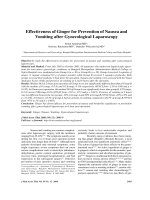
a new gastric access LMA (Fig. 1), LMA Supremet,
became available. Increasingly, it appeared that the
LMA Supremet (SUP) with the possibility to suc-
tion the stomach could be an interesting alternative
to the ETT during laparoscopic procedures, mainly
because of its overall simplicity of use for non-
expert laryngeal mask users and ventilation per-
formance. We hypothesized that choosing the SUP
during gynecologic pelvic laparoscopy would in-
fluence upper airway management outcome. We
designed a study to compare post-operative phar-
yngolaryngeal discomfort and ventilation effi-
ciency when the SUP was used as an alternative
to the ETT.
Methods
After obtaining Local Research Ethics Committee
approval and written informed consent, 138 ASA
status I–II adult female patients scheduled for
elective pelvic laparoscopy were enrolled in this
randomized single-blind study. Patients with
1
Acta Anaesthesiol Scand 2009; ]]: 1–6
Printed in Singapore. All rights reserved
r 2009 The Authors
Journal compilation r 2009 The Acta Anaesthesiologica Scandinavica Foundation
ACTA ANAESTHESIOLOGICA SCANDINAVICA
doi: 10.1111/j.1399-6576.2009.02095.x
known or predicted difficult airway, increased risks
of regurgitation, ongoing upper respiratory tract
and those unable to understand the definitions of
the post-operative upper airway symptoms (hoar-
seness of voice, sore throat and dysphagia) or the
use of a visual analogue scale (VAS) or requiring
operative use of a nasogastric tube were not in-
cluded in the trial.
Anesthetic and airway management procedures
Premedication consisted of oral hydroxizine 1 mg/
kg given 1 h before the planned surgery time. Upon
arrival in the operating room, the patients were
randomly assigned (sealed envelops) to receive
either the ETT or the SUP for airway management.
Standard monitoring systems including non-inva-
sive arterial blood pressure, heart rate, hemoglobin
arterial oxygen saturation, end-tidal carbon diox-
ide (EtCO
2
) and sevoflurane (EtSevo), BIS and
adductor pollicis neuromuscular function were
attached.
Anesthetists involved in the present trial were
requested to maintain optimal anesthesia depth as
indicated by hemodynamic stability (within 20%
pre-induction values) and BIS values ranging be-
tween 40 and 50. Airway management was per-
formed by senior anesthesiologists skilled with the
LMA Supremet (SEBAC, Pantin, France). After
3 min of pre-oxygenation, anesthesia was induced
with sufentanil (0.15–0.25 mcg/kg), followed 45 s
later by propofol (2–2.5 mg/kg) and atracurium
(0.5 mg/kg).
Managing the airway with a tracheal tube
In the ETT group, tracheal intubation maneuvers
were initiated with adductor pollicis muscle re-
sponse 5 0 to train-of-four stimulation of the ulnar
nerve. Facemask ventilation was discontinued and
conventional laryngoscopy was performed with a
plastic single-use size 3 or 4 blade. The trachea
was intubated using a single-size 7.0 mm internal
diameter high-volume, low-pressure tracheal tube.
The cuff was inflated and maintained at 25 cmH
2
O
(Pressure controller, Endotest Rusch, 67660 Betsch-
dorf, France). In the case of difficult laryngoscopy,
an Eschman bougie was recommended to facilitate
tracheal access.
Managing the airway with the LMA Supremet
In the SUP group, with optimal jaw relaxation, a
humidified size 3, 4 and 5 laryngeal mask [depend-
ing on patient height (o155, 4155 and 4170 cm,
respectively] was inserted into the oral cavity with
the head of the patient slightly placed in the
sniffing position as soon as an optimal was ob-
tained. Once orally placed, the laryngeal mask was
distally blocked in the pharynx while the head
returned to the neutral position. Then the cuff of
the laryngeal mask was inflated to 50 cmH
2
O pres-
sure and ventilation was attempted. In case of
failure to ventilate after initial placement, the cuff
was deflated to zero cuff pressure before the
optimal position (up–down maneuver) was chal-
lenged using a small-volume bag ventilation-in-
duced end-tidal CO
2
curve appearing on the
monitor. Then the cuff of the laryngeal mask was
inflated and maintained at 50 cmH
2
O of controlled
pressure and the airway was fixed with an adhe-
sive tape. In case of impossible ventilation with the
SUP after two insertion attempts we planned to
place an ETT. When ventilation was confirmed
with the SUP, a 14-Gauge probe was placed in the
stomach to suction its content, which was recorded,
and then removed from the laryngeal mask.
Standardized procedures for both groups
When an adequate level of anesthesia was ob-
tained, the leak pressure was measured with both
airways using the ventilator (Julian, Dra
¨
ger Medi-
cal, Antony, France). Three cycles of pressure-con-
trolled ventilation at three levels of 20, 25 and
30 cmH
2
O were applied while antero-lateral neck
auscultation was performed. Then the three in-
spiratory pressure-induced tidal volumes were
used during volume-controlled ventilation to mea-
sure cycle-to-cycle inspiratory–expiratory leak vo-
lume. The sealing pressure was defined as the
highest inspiratory pressure free from audible
and measured gas leak.
Ventilation port
Ventilation face
Drainage tube to
suction the stomach
Cuff
Gastric access
port
Fig. 1. The LMA Supremet. LMA, laryngeal mask airway.
W. Abdi et al.
2
After airway managements, the patients of both
groups received tidal volume (8 ml/kg) controlled
mechanical ventilation with a respiratory rate of
12 breaths/min adapted to maintain end-tidal
CO
2
in the range 4.5–5 kPa with a fresh gas flow
of 1.5 l/min.
Forced-air warming of the upper body and limbs
was systematically applied to maintain tympanic
temperature above 36.5 1C. Anesthesia was main-
tained with sevoflurane in combination with ni-
trous oxide (50%) in oxygen. The concentration of
sevoflurane and re-injections of sufentanil and
atracurium were adjusted to maintain adequate
anesthesia depth and neuromuscular blockade as
confirmed by o2/4 responses of the corrugator
supercilii muscle to TOF stimulation of the
ophthalmic branch of the facial nerve.
During the surgery, all patients received an
intravenous Ringer lactate solution administered
at the rate of 15 ml/kg/h, propacetamol (1 g),
ibuprofen (100 mg) and tramadol (20 mg). Laparo-
scopic technique pneumoperitoneum was main-
tained with carbon dioxide and adjusted to a
pressure of 15–20 mmHg. At the end of the proce-
dure, the surgeon systematically administered
0.75% ropivacaine (10 ml) in the peritoneum
through the main umbilical trocar, and residual
neuromuscular blockade was reversed with neos-
tigmine 1.5–2.0 mg and atropine 1.0–1.5 mg de-
pending upon adductor pollicis muscle number
of response (! 2/4) to train-of-ratio stimulation of
the ulnar nerve at the wrist. Sevoflurane was then
discontinued, the ventilatory circuit was opened
and the patient was allowed to awakening. With
return to spontaneous ventilation, the cuff of the
airway was deflated until its removal when the
patient responded to simple commands.
Operating room recordings and measurements
A single surgeon blinded to the airway manage-
ment technique performed all the laparoscopic
procedures. An unblinded assessor not attending
to the patient recorded airway management details
and timed all anesthesia and surgical sequences.
Airways placement and weaning duration were
defined as the time elapsing between injection of
atracurium to definitive ventilation through the
airway (chest/neck auscultation and EtCO
2
normal
qualitative and quantitative aspects) and the last
trocar to airways removal, respectively. Surgery
time lasted from the first peritoneal puncture to
the last suture. Airway management quality was
assessed using a difficulty VAS (0 5 no difficulty,
100 impossible airway management) rated by the
senior anesthetist caring for the patient just after
removal of the airways.
Post-operative recordings and measurements
A second assessor blinded to the allocation group
recorded on a separate data sheet post-operative
parameters such as the occurrence of nausea and/
or vomiting, and the need for any therapeutic
interventions including the analgesic requirements,
and questioned all the patients just before leaving
the PACU about pharyngolaryngeal discomfort.
The patients were asked whether they experienced
any abdominal pain or pharyngolaryngeal discom-
fort. Hoarseness of the voice was defined as being
either a change in the voice tone or a painful
phonation. Sore throat was defined as a permanent
soreness of the throat. Dysphagia was defined as a
pain triggered by on-command saliva swallows.
The questions were: ‘do you have abdominal pain,’
‘do you have hoarseness of voice or sore throat’ ‘do
you feel any discomfort or pain when you swallow
your saliva.’ If the answer was yes to any of these
questions, the intensity of the complaint or pain
was assessed using a 101-point numerical rating
scale (NRS 0 5 no discomfort or no pain to
100 5 extreme discomfort or maximal imaginable
pain). Symptoms’ intensity was also rated just
before leaving the hospital. Similar questions con-
cerning post-operative upper-airway discomfort
were asked by the surgeon at the day 5–7 post-
operative visit.
Sample size calculation and statistics
We measured in 35 post-operative patients leaving
the PACU, whose trachea was intubated for gyne-
cologic pelvic laparoscopy, a mean Æ SD NRS for
hoarseness of voice of 21 Æ 14. We hypothesized
that SUP, compared with ETT anesthesia, would
reduce by at least 50% the intensity of NRS for
hoarseness, considered as the primary objective
variable. A total of 138 patients included in two
equal groups were requested to declare this hy-
pothesis (a 5 0.05 and power 5 1 À b 5 0.9) using a
two-sided test. Values are mean Æ SD or median
(range). Parametric variables were compared using
Student’s t-test, and non-parametric variables were
compared using the Mann–Whitney U-test or the
w
2
test with Yates’ continuity correction, as appro-
LMA or ETT anesthesia for pelvic laparoscopy?
3
priate. A P value of o0.05 was considered statisti-
cally significant.
Results
The demographic characteristics of the patients
were comparable between the two groups (Table
1). No ventilation failure occurred with the two
airways. The average time to airways placement
and weaning was of a shorter duration in the SUP
group as compared with the ETT group. Three
patients of the ETT group required an Eschman
bougie assistance to facilitate tracheal access, and
11 patients of the SUP group required manipula-
tions to promote a seal airway. Median airway
management difficulty VAS was 5, similar in both
groups. In the SUP group, a mean (extreme) of 4 (0–
21) ml of gastric secretion was suctioned. Operating
room recordings and measurements are presented
in Table 2. Surgery duration was similar in the two
groups. Post-operative evaluations showed that the
incidence of nausea and vomiting and the require-
ment for analgesic treatment were similar in both
groups. Post-operative pain and pharyngolaryn-
geal discomfort evaluation is presented in Table 3.
Post-operative abdominal pain incidence and NRS
intensity were similar in the two groups. Post-
operative pharyngolaryngeal discomfort incidence
and NRS intensity were significantly increased
after ETT as compared with SUP anesthesia. Two
patients of the ETT group as compared with none
of the SUP group complained of hoarseness of
voice still persisting at the Day 5–7 evaluation visit.
In both cases, symptoms resolved within the 15
post-operative days.
Discussion
We demonstrated that using the SUP as an alter-
native to the ETT was an efficient pharyngolaryn-
geal morbidity-sparing strategy for the patients
included in the present trial. We showed that the
SUP and the ETT were equally effective ventilatory
devices for routine gynecological laparoscopic pro-
cedures.
We calculated post-operative pharyngolaryngeal
morbidity incidence and measured all symptoms’
intensity during the first post-operative week.
Although we standardized the anesthesia techni-
que, allowing similar anesthesia depth and recov-
ery room requirement, and surgical technique
promoting similar post-operative pain, we ob-
served that all upper airway-related symptoms
were reduced in incidence and in intensity if the
SUP was used as an alternative to the ETT. Our
primary objective variable, the hoarseness of voice,
which best characterizes laryngeal dysfunction re-
Table 1
Demographic characteristics of the patients.
LMA Supremet
group (n 5 69)
ETT group
(n 5 69)
Age (years) 33 (9) 33(8)
Weight (kg) 66 (14) 66 (17)
Height (m) 1.65 (8) 1.64 (6)
ASA I/II (number of patients) 45/16 39/12
Smoking (410 cigarettes/day,
number of patients)
54
Values are mean (SD) or numbers.
ETT, endotracheal tube; LMA, laryngeal mask airway; SD,
standard deviation.
Table 2
Operating room recordings and measurements.
LMA
Supremet
group
(n 5 69)
ETT
group
(n 5 69)
Anesthetic agents
Induction
Propofol (mg) 185 (35) 196 (37)
Sufentanil (mcg) 12 (3) 14 (4)
Atracurium (mg) 32 (7) 31 (7)
During maintenance
Sufentanil (mcg) 6 (6) 10 (5)
Atracurium (mg) 3 (6) 4 (6)
Airway characteristics
Sealing pressure
430 cmH
2
O (%)
95 100
Sealing pressure
425 cmH
2
O (%)
5–
Maximum peak airway pressure
during anesthesia (cmH
2
O)
25 (6) 23 (5)
Duration of timed sequences
Airway placement (min) 2.2 (0.6) 3.8 (0.7)
Airway weaning (min) 4.5 (3.0) 7.5 (2.5)
Surgery (min) 54 (21) 57 (23)
Airway management difficulty VAS
(0–100) [mean/median (range)]
5/5 (0–25) 5/5 (0–40)
Visual analogue scale of airway management difficulty (0 5 no
difficulty and 100 impossible airway management).
The sealing pressure was defined as the highest peak inspira-
tory pressure (20–25–30 cmH
2
O), free from audible and mea-
sured gas leak. Airway placement and weaning time duration
were defined as the time elapsing between injection of atracur-
ium to definitive ventilation through the airway (chest/neck
auscultation and EtCO
2
normal qualitative and quantitative
aspect) and last trocar to airway removal, respectively. Surgery
time lasted from the first peritoneal puncture to the last suture.
ETT, endotracheal tube; LMA, laryngeal mask airway; VAS,
visual analogue scale.
W. Abdi et al.
4
sulting from airway management, was much more
intense after ETT than SUP anesthesia. Although
upper airway symptoms rapidly decreased in in-
cidence and intensity, two patients of the ETT group
vs. none of the SUP group suffered from persistent
dysphonia reported to the surgeon at the follow-up
visit. Although all patients were placed under
best-relaxed conditions
5
at the time tracheal intuba-
tion maneuvers were undertaken, some minor
trauma of the vocal cords during tracheal intuba-
tion
6,7
or tracheal tube maintenance
8
possibly oc-
curred, resulting in prolonged post-operative voice
dysfunction. Interestingly, some patients of the
SUP group suffered from hoarseness of the voice
of minor intensity. Two reasons may explain why
the SUP may affect post-operative laryngeal func-
tion. First, 11 patients (16%) required manipula-
tions and repositioning of the laryngeal mask to
improve ventilation and seal. During the up–down
maneuver, we speculated that in case this specific
feature occurred, minor laryngeal trauma might
have occurred. The second reason might be linked
to the structure and shape of the SUP. Indeed, when
correctly positioned, the distal cuff of the laryngeal
mask is inflated at the level of or just below the
cricopharyngeal upper-esophageal sphincter mus-
cle. Although we maintained the pressure in the
cuff of the laryngeal mask at or below 50 cmH
2
O,
some changes in arytenoids shape or position
might have resulted in transient vocal cord palsy
and hoarseness of the voice. Finally, we evidenced
in one patient of the SUP group a surprising
audible stridor resulting from expiration through
the SUP just after insertion. Reducing cuff pressure
of the laryngeal mask to 45 cmH
2
O relieved this
abnormal expiratory sound. Endoscopic studies are
requested to evaluate the influence of SUP cuff
pressure on the glottis shape and post-operative
laryngeal function.
Large-scale studies have already reported high
success and low complication rates when a LMA
was used during general anesthesia.
1–3
Similarly,
some trials have shown that the LMA ProSealt
was an efficient alternative to the ETT for laparo-
scopic cholecystectomy
9
and gynecological sur-
gery.
10
However, routine ‘LMA ProSeal’ use for
laparoscopic procedure remained relatively infre-
quent due to the lack of reliability of this airway if
placed by a non-expert anesthesiologist. In con-
trast, we demonstrated that the SUP resulted in
very low airway management difficulty VAS, with
most anesthesiologists rating both ETT and SUP
airway management techniques as very simple.
Interestingly, the SUP was found to be an efficient
ventilation device compared with the ETT. We
confirmed that the SUP promoted high sealing
pressure exceeding 30 cm in most (95%) cases.
Moreover, four patients of the SUP group required
a transient increase to more than 20 mmHg of the
carboperitoneum pressure, resulting in a peak in-
spiratory pressure exceeding 35 cmH
2
O. For these
patients we could not evidence any gas leak,
suggesting that the sealing pressure of SUP could
exceed 35 cmH
2
O in some patients.
6
Of interest, we
Table 3
Post-operative pain and pharyngolaryngeal discomfort evalua-
tion.
LMA Supremet
group (n 5 69)
ETT group
(n 5 69)
Post-anesthesia care unit evaluation
Abdominal pain
Incidence (%) 81 79
Intensity NRS [mean/
median (range)]
28/30 (0–45) 27/30 (0–40)
Hoarseness of voice
Incidence (%) 16 47*
NRS [mean/median
(range)]
3/0 (0–40) 19* /0 (0–65)
Dysphagia
Incidence % 16 26*
NRS, mean/median
(range)
7/0 (0–50) 15* /0 (0–60)
Sore throat
Incidence (%) 19 32*
NRS, mean/median
(range)
7/0 (0–50) 10/0 (0–60)
Ward evaluation just before leaving the hospital
Abdominal pain
Incidence (%) 54 50
NRS [mean/median
(range)]
18/15 (0–40) 20/20 (0–40)
Hoarseness of voice
Incidence, % 9 37*
NRS, mean/median
(range)
1/0 (0–25) 10* /0 (0–70)
Dysphagia
Incidence (%) 9 19*
NRS [mean/median
(range)]
2/0 (0–35) 5/0 (0–30)
Sore throat
Incidence (%) 5 15*
NRS [mean/median
(range)]
3/0 (0–25) 3/0 (0–45)
In order to evaluate post-operative pain and pharyngolaryngeal
discomfort, the patients were asked whether they experienced any
abdominal pain or pharyngolaryngeal discomfort. The questions
asked were: ‘do you have abdominal pain,’ ‘do you have hoarse-
ness of voice or sore throat’ ‘do you feel any discomfort or pain
when you swallow your saliva.’ If the answer was yes to any of these
questions, the intensity of the complaint or pain was measured using
a 101-point numerical rating scale (NRS: 0 5 no discomfort or no
pain, to 100 5 extreme discomfort or maximal imaginable pain).
ETT, endotracheal tube; LMA, laryngeal mask airway.
*Po0.05 vs. LMA Supreme group.
LMA or ETT anesthesia for pelvic laparoscopy?
5
were able to drain the gastric content of all the
patients of the SUP group. We believe that this easy
gastric access associated with the use of the SUP is
an additional safety argument favoring the use of
this laryngeal mask in this type of surgery. Similar
gastric drainage performed in the ETT group may
have resulted in a difficulty in placing the probe in
the stomach of some patients and would have
certainly led to a higher pharyngolaryngeal mor-
bidity rate.
Our study has limitations. Firstly, we could not
blind the operating room observer who timed all
the events during the procedure. A strict double-
blind design for such a study would have been
difficult under our working conditions. Secondly,
our airway management techniques were com-
pared in a single surgical indication. Thus, our
results may not be generalizable to other abdom-
inal surgery types because restriction of the use of
SUP may be of concern until its safety is demon-
strated. Thirdly, we studied only female patients,
and our trial cannot be considered as a gender-
related study. However, we have used a relatively
small inner diameter ETT in order to limit the risk
of abnormally high hoarseness of the voice in both
the incidence and the intensity in the ETT group.
Finally, in all patients of the SUP group the airway
was placed after a muscle relaxant injection, which
may not be considered as a usual practice. The SUP
was inserted with optimal jaw relaxation, which
was obtained in all cases during the 60 s following
propofol injection, but before a peripheral maximal
neuromuscular block was installed. Because pa-
ralysis was required for surgical purposes and the
mean duration of the procedure was short (o1 h),
we decided to insert the SUP a few seconds after
the muscle relaxant injection. We are quite confi-
dent that this strategy did not affect our results.
In conclusion, we demonstrated that using the
SUP instead of an ETT allowed reducing the post-
operative pharyngolaryngeal morbidity resulting
from airway management. We also showed that
the SUP was an efficient alternative to the ETT for
airway management during infertility pelvic la-
paroscopy procedures.
Acknowledgements
Received from the Anesthesia and Intensive Care Department
of Jean Verdier University Hospital of Paris (APHP), 93143
Bondy, France.
This trial was funded by departmental sources.
References
1. Verghese C, Brimacombe JR. Survey of laryngeal mask
airway usage in 11,910 patients: safety and efficacy for
conventional and nonconventional usage. Anesth Analg
1996; 82: 129–33.
2. Brimacombe J. Analysis of 1500 laryngeal mask airway uses
by one anaesthetist in adults undergoing anaesthesia.
Anaesthesia 1996; 51: 76–80.
3. Oczenski W, Krenn H, Dahaba AA, Binder M, El-Schahawi-
Kienzl I, Kohout S, Schwarz S, Fitzgerald RD. Complica-
tions following the use of the Combitube, tracheal tube and
laryngeal mask airway. Anaesthesia 1999; 54: 1161–5.
4. Brimacombe J. The advantages of the LMA over the
tracheal tube or face mask: a meta-analysis. Can J Anaesth
1995; 42: 1017–23.
5. Mencke T, Echternach M, Kleinschmidt S, Lux P, Barth V,
Plinkert PK, Fuchs-Buder T. Laryngeal morbidity and
quality of tracheal intubation: a randomized controlled
trial. Anesthesiology 2000; 98: 1049–56.
6. van Zundert A, Brimacome J. The LMA Supreme – a pilot
study. Anaesthesia 2008; 63: 209–10.
7. Joshi GP, Inagaki Y, White PF, Taylor-Kennedy L, Wat LI,
Gevirtz C, McCraney JM, McCulloch DA. Use of the
laryngeal mask airway as an alternative to the tracheal
tube during ambulatory anesthesia. Anesth Analg 1997; 85:
573–7.
8. Tanaka A, Isono S, Ishikawa T, Sato J, Nishino T. Laryngeal
resistance before and after minor surgery: endotracheal
tube versus laryngeal mask airway. Anesthesiology 2003;
99: 252–8.
9. Maltby JR, Beriault MT, Watson NC, Liepert DJ, Fick GH.
LMA-Classic and LMA-ProSeal are effective alternatives to
endotracheal intubation for gynecologic laparoscopy. Can J
Anaesth 2003; 50: 71–7.
10. Swann DG, Spens H, Edwards SA, Chestnut RJ. Anaesthe-
sia for gynaecological laparoscopy: a comparison between
the laryngeal mask airway and tracheal intubation. Anaes-
thesia 1993; 48: 43–4.
Address:
Gilles Dhonneur
De
´
partement d’Anesthe
´
sie et Re
´
animation CHU (APHP)
Jean Verdier
Av du 14 Juillet
93143 Bondy
France
e-mail:
W. Abdi et al.
6