role for rip1 in mediating necroptosis in experimental intracerebral hemorrhage model both in vivo and in vitro
Bạn đang xem bản rút gọn của tài liệu. Xem và tải ngay bản đầy đủ của tài liệu tại đây (5.34 MB, 14 trang )
OPEN
Citation: Cell Death and Disease (2017) 8, e2641; doi:10.1038/cddis.2017.58
Official journal of the Cell Death Differentiation Association
www.nature.com/cddis
Role for RIP1 in mediating necroptosis in experimental
intracerebral hemorrhage model both in vivo and
in vitro
Haitao Shen1,2, Chenglin Liu1,2, Dongping Zhang1,2, Xiyang Yao1, Kai Zhang1, Haiying Li*,1 and Gang Chen*,1
Cell death is a hallmark of second brain injury after intracerebral hemorrhage (ICH); however, the mechanism still has not been fully
illustrated. In this study, we explored whether necroptosis, a type of regulated necrosis, has an essential role in brain injury after
ICH. We found that inhibiting receptor-interacting protein 1 (RIP1) – a core element of the necroptotic pathway – by a specific
chemical inhibitor or genetic knockdown attenuated brain injury in a rat model of ICH. Furthermore, necroptosis of cultured
neurons could be induced by conditioned medium from microglia stimulated with oxygen hemoglobin, and this effect could be
inhibited by TNF-α inhibitor, indicating that TNF-α secreted from activated microglia is an important factor in inducing necroptosis
of neurons. Undoubtedly, overexpression of RIP1 increased conditioned medium-induced necroptosis in vitro, but this effect was
partially diminished in mutation of serine kinase phosphorylation site of RIP1, showing that phosphorylation of RIP1 is the
essential molecular mechanism of necroptosis, which was activated in the in vitro model of ICH. Collectively, our investigation
identified that necroptosis is an important mechanism of cell death in brain injury after ICH, and inhibition of necroptosis may be a
potential therapeutic intervention of ICH.
Cell Death and Disease (2017) 8, e2641; doi:10.1038/cddis.2017.58; published online 2 March 2017
Intracerebral hemorrhage (ICH) is the second largest type of
stroke, accounting for ~ 15% of all patients with stroke.1,2 ICH
is associated with fast progression, high mortality and high
morbidity. It has been reported that, with mortality rates close
to 40% in 1 month, patients have legacy paralysis, aphasia
and other severe disabilities after ICH. The incidence of ICH
also increased significantly with the increase of population
age.3 Primary brain injury after ICH is due to hematoma mass
effect and mechanical damage to adjacent brain tissues,4 and
the secondary brain injury is a key reason to cause nerve
function damage in patients with ICH.5,6 In secondary brain
injury after ICH, pathological changes, including cell death,
cerebral edema and blood–brain barrier (BBB) damage, occur
in brain tissues surrounding hematoma.7 Mechanism of
secondary brain injury after ICH includes excitatory aminoacid toxicity, inflammatory response, expression of proteolytic
enzyme and the toxic effects of hematoma release product.5,7
Cell death is an important factor in secondary brain injury
after ICH.
In recent years, necrosis as another form of cell death
causes more attention. The definition of necrosis is based on
its pathological morphological characteristics; it is a passive
cell death mostly caused by overwhelming stress such as
dramatic changes in temperature or pH. Necrotic cells rapidly
lose cell membrane integrity, and cell membrane swelling and
mitochondrial dysfunction occur. The membrane rupture
caused a large number of cytoplasmic components to
leak from the cell and then induced inflammation in the
surrounding tissues.8 Previous studies suggested that necrosis is an occasional and irregular event and is difficult to study.9
However, Yuan and co-workers10 reported, for the first time,
necroptosis as a form of necrosis that can be regulated.10
Necroptosis has similar morphological characteristics (including early membrane integrity, cell and intracellular organelle
swelling) with necrosis, but it is caspase-independent
programmed cell death.11
Recent studies have shown that death receptors on the
cell membrane mediated different pathways of cell death
depending on the state of cell or local physiological or
pathophysiological microenvironment; for example, in the
event of TNF-α-induced cell death, TNF-α and its receptors
firstly formed a trimer, and then recruited proteins containing
the death domain (DD) formed a complex, which activated the
downstream apoptotic pathway. However, if caspase
activity was inhibited, the cells would go to the necroptotic
pathway. RIP1 was recruited to TNFR1 through its DD, which
bound directly to the DD of TNFR1, and it was activated by
multiple forms of ubiquitination. The activation of RIP1 led to
the recruitment of RIP3, MLKL and caspase-8, forming a
complex called 'necrosome' involved in necroptotic pathway.
Thus, RIP1 played the role of adaptor in mediating necroptosis. Activation of RIP3 activated three key enzymes of cell
metabolic pathways – glycogen phosphorylase, glutamine
synthetase, and glutamate dehydrogenase – which caused
overproduction of reactive oxygen species (ROS). ROS
caused DNA damage, damage to mitochondrial membrane
1
Department of Neurosurgery and Brain and Nerve Research Laboratory, The First Affiliated Hospital of Soochow University, Suzhou, Jiangsu Province, China
*Corresponding author: G Chen or H Li, Department of Neurosurgery and Brain and Nerve Research Laboratory, The First Affiliated Hospital of Soochow University,
188 Shizi Street, Suzhou 215006, China. Tel: +86 13771908806; Fax: +86 051267780165; E-mail: or Tel:+86 15716201037; Fax:+86 0512
67787180; E-mail:
2
These authors contributed equally to this work.
Received 27.10.16; revised 28.12.16; accepted 03.1.17; Edited by Y Shi
RIP1-mediated necroptosis in ICH
H Shen et al
2
permeability and lysosome damage, eventually leading to cell
death.11,12
Previous studies have confirmed that microglia was
quickly activated in brain tissues after ICH, and it secreted a
large number of inflammatory factors, including the TNF-α,
which can induce necroptosis.13 Under the stimulation of
Cell Death and Disease
TNF-α, a part of the brain cells undergo apoptosis and a part
may undergo necroptosis in brain tissues after ICH. Necrotic
cell death is common in a wide variety of pathological
conditions, including stroke.14 Compared with numerous
investigations on the mechanisms of apoptotic cell death,
fewer studies have explored necrotic cell death in ICH. Here
RIP1-mediated necroptosis in ICH
H Shen et al
3
we studied the role of necroptosis, a type of regulated
necrosis, in ICH.
Results
Necroptosis was activated in brain tissues after ICH. To
explore the involvement of necroptosis in brain tissues after
ICH, we detected propidium iodide-positive (PI+) cells in
frozen brain sections at 24 h after ICH. Notably, compared
with that in the Sham group, the PI+ cells were remarkably
increased in brain tissues surrounding hematomas after ICH
(Po0.0001, n = 6; Figures 1a and b). We also determined the
expression of RIP1, which is a major regulator for necroptosis
in brain tissues; the results of western blot suggested that the
expression levels of RIP1 were progressively upregulated
and peaked at 24 h after ICH (P = 0.0003 versus Sham group,
n = 3; Figures 1c and d). The results of immunofluorescent
staining also showed that the expression level of RIP1 in
neurons was remarkably increased at 24 h after ICH
(Figure 1e).
We further evaluated the formation of necrosome, which is
an important hallmark of activation of necroptosis in brain
tissues after ICH; anti-RIP1 antibody was used by immunoprecipitation (IP), and RIP3, MLKL and caspase-8 were
detected by immunoblotting. Increased interactions of RIP1
and RIP3, RIP1 and MLKL, and RIP1 and caspase-8 were
observed in brain tissues after ICH (all Po0.01 versus Sham
group, n = 3, Figures 1f and g). It suggested that the formation
of necrosome was significantly increased in brain tissues after
ICH than that in the Sham group. The results of immunofluorescence also showed that expressions of these four
proteins were significantly upregulated after ICH than that in
the Sham group (all Po0.01 versus Sham group, n = 6,
Figures 1h and i). These results indicated that necroptosis was
activated in brain tissues after ICH.
Inhibitor of necroptosis and apoptosis differentially
attenuates brain injury after ICH. To determine whether
necroptosis contributes to brain injury after ICH, the specific
inhibitor of RIP1, necrostatin-1 (Nec-1), was used, and
z-VAD, a caspase inhibitor, which can inhibit apoptosis, was
also used as a positive control in a rat model of ICH. Nec-1
reduced the PI+ cells but had no significant effect in TUNEL+
cells, whereas z-VAD only downregulated the TUNEL+ cells,
and did not affect the PI+ cells in brain tissues after ICH. The
combination of z-VAD and Nec-1 both obviously reduced the
PI+ cells and TUNEL+ cells (all Po0.01, n = 6; Figures 2a
and b). The results of IP also demonstrated that interactions
of RIP1 and RIP3, RIP1 and MLKL, and RIP1 and caspase-8
were significantly inhibited by Nec-1 but not by z-VAD
(all Po0.05, n = 3; Figures 2c and d). These results indicated
that necroptosis and apoptosis occurred in different cells at
same times and were independent of each other in brain
tissues after ICH.
BBB permeability was assessed using albumin extravasation, and the western blot was used to test the albumin level in
brain tissues; the results also suggested that treatment with
Nec-1 in ICH rats can improve albumin extravasation and BBB
injury (P = 0.0461, n = 3; Figures 2e and f). Brain water content
was calculated by the wet and dry weight method; the results
showed that, compared with ICH group, the brain water
content was significantly reduced in the ICH+Nec-1 group
(P = 0.0109 in Ipsi-CX and P = 0.0077 in Ipsi-BG, n = 6;
Figure 2g). The neurological score of the ICH+Nec-1 group
was significantly lower than that in the ICH group (P = 0.0094,
n = 6; Table 1). The levels of TNF-α in the cerebrospinal fluid
(CSF) were measured by ELISA; the results confirmed that the
level of TNF-α was obviously reduced by Nec-1 treatment,
indicating that Nec-1 inhibits inflammation in ICH rats
(Po0.0001, n = 6; Figure 2h). These results suggested that
necroptosis contributes to brain injury after ICH, including
neuronal dysfunction, brain water content, BBB permeability
and inflammation.
Blockade necroptosis by knockdown of RIP1 improves
brain injury after ICH. To further define the role of RIP1mediated necroptosis in neuronal dysfunction after ICH, we
used knockdown by small interfering RNA (siRNA) interference and overexpression by recombinant adenoviruses
transfection of RIP1 as treatment in rat model of ICH.
Knockdown of RIP1 reduced the PI+ cells, but overexpression of RIP1 increased the PI+ cells in brain tissues after ICH
(both Po0.0001, n = 6; Figures 3a and b). The results of IP
also demonstrated that interactions of RIP1 and RIP3, RIP1
and MLKL, and RIP1 and caspase-8 were significantly
inhibited by knockdown of RIP1, but they were increased
by overexpression of RIP1 (all Po0.05, n = 3; Figures 3c and
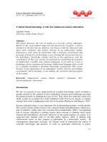
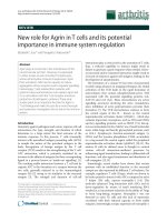
Figure 1 Indicators of necroptosis were discovered in brain tissues after ICH. (a) The necroptosis of cells in brain tissues was detected by PI labeling. As shown, compared
with the Sham group, considerable PI+ cells were detected in frozen sections of brain tissues in rats at 24 h after ICH. Arrows point to PI+ cells. Scale bar = 50 μm. (b) Related
with (a), it revealed relative levels of PI+ cells, **Po0.0001 versus Sham group, unpaired t-test, n = 6. (c) The results of western blot suggested that the expression levels of RIP1,
a major regulator for necroptosis, were obviously upregulated, and it reached peak at 24 h in brain tissues after ICH. (d) Related with (c), quantitative analysis of expression levels
of RIP1 in brain tissues within 1 week after ICH. **P = 0.0003 versus Sham group, unpaired t-test, n = 3. (e) Double immunofluorescence (IF) analysis was performed with
antibodies for RIP1 (green) and NeuN (red). Nuclei were fluorescently labeled with DAPI (4',6-diamidino-2-phenylindole) (blue). Representative images of the Sham group and the
ICH (24 h) group were shown. Scale bar = 10 μm. (f) The formation of necrosome was detected by immunoprecipitation (IP) by using anti-RIP1 antibody (rabbit immunoglobulin
G (IgG) was also used as a negative control), and RIP3, MLKL and caspase-8 were detected by immunoblotting. The results suggested that increased interactions of RIP1 and
RIP3, RIP1 and MLKL, and RIP1 and caspase-8 were observed in brain tissues at 24 h after ICH. Input, 5% of extract before IP. (g) Quantitative analysis of IP. **P = 0.0011 versus
Sham group; &&P = 0.0011 versus Sham group; #P = 0.0152 versus Sham group; all were unpaired t-test, n = 3. Besides, results of IF (h) showed that expressions of these four
proteins, which constituted necrosome, were increased significantly in brain tissues at 24 h after ICH than those in the Sham group. Scale bar = 100 μm. Related statistic of data
was revealed in (i). **Po0.0001 versus Sham group; &&Po0.0001 versus Sham group; ##Po0.0001 versus Sham group; $$Po0.0001 versus Sham group; all were unpaired ttest, n = 6. All data are expressed as means ± S.E.M., mean value for the Sham group was normalized to 1.0
Cell Death and Disease
RIP1-mediated necroptosis in ICH
H Shen et al
4
d). These results indicated that knockdown of RIP1 can
effectively block necroptotic pathway.
The results of BBB permeability also suggested that
treatment with RIP1 siRNA in ICH rats can inhibit albumin
extravasation and BBB permeability (P = 0.0493, n = 3;
Figures 3e and f). The results of brain water content declared
that, compared with the ICH group, the brain water content
Cell Death and Disease
was significantly reduced in the RIP1 siRNA group (Po0.0001
in Ipsi-CX and P = 0.0209 in Ipsi-BG, n = 6; Figure 3g). The
neurological score of the ICH+Si-RIP1 group was significantly
lower than that of the ICH group (P = 0.0185, n = 6; Table 1).
The levels of TNF-α in the CSF were detected by ELISA; the
results confirmed that the level of TNF-α was reduced by RIP1
siRNA treatment, indicating that downregulation of RIP1 could
RIP1-mediated necroptosis in ICH
H Shen et al
5
inhibit inflammation in brain tissues after ICH (P = 0.0002,
n = 6; Figure 3h). These results further proved that necroptosis
played an important role in brain injury after ICH.
Conditioned medium from activated microglia can
induce necroptosis of cultured neuron in vitro. To further
explore the mechanism of necroptosis after ICH, we used two
in vitro models of ICH: one is oxygen hemoglobin (OxyHb) to
deal directly with the neurons, and the other is to prestimulate
microglia with OxyHb, collect the supernatant as conditioned
medium and then treat neurons with the conditioned medium.
After these treatments, neurons were digested by trypsin into
cell suspension, stained with Annexin V and PI and then
detected by flow cytometry. The results of flow cytometry
Table 1 Clinical behavior scores in each group (n = 6)
Group
Normal
Sham
ICH
ICH+vehicle
ICH+Nec-1
ICH+z-VAD
ICH+Nec-1 and z-VAD
ICH+Si-NC
ICH+Si-RIP1
ICH+Ad-GFP
ICH+Ad-RIP1
Score (mean ± S.E.M.)
0.17 ± 0.17
0.33 ± 0.21
2.67 ± 0.33a
3.17 ± 0.31b
1.67 ± 0.21c
1.83 ± 0.17d
1.17 ± 0.17e
2.67 ± 0.21f
1.67 ± 0.21g
3.00 ± 0.37h
4.33 ± 0.21i
Abbreviations: Ad-GFP, adenovirus with GFP; Ad-RIP1, adenovirus with RIP1;
ICH, intracerebral hemorrhage; Nec-1, necrostatin-1; RIP1, receptor-interacting
protein 1; Si-NC, Si-negative control.
a
P = 0.0043 versus Sham group (Mann–Whitney test).
b
P = 0.2959 versus ICH group (unpaired t-test).
c
P = 0.0094 versus ICH+vehicle group (Mann–Whitney test).
d
P = 0.0096 versus ICH+vehicle group (Mann–Whitney test).
e
P = 0.0341 versus ICH+z-VAD group (Mann–Whitney test).
f
P = 0.9290 versus ICH group (Mann–Whitney test).
g
P = 0.0185 versus ICH+Si-NC group (Mann–Whitney test).
h
P = 0.5155 versus ICH group (unpaired t-test).
i
P = 0.0205 versus ICH+Ad-GFP group (Mann–Whitney test).
showed that, compared with control group, there was a higher
apoptotic ratio (~27.8%) in OxyHb directly stimulated neuron
group, whereas there was a higher necroptotic ratio in
conditioned medium treatment group (~31.5%). In addition,
Nec-1 treatment, but not z-VAD, significantly reduced the
conditioned medium-induced neuron necroptosis (data not
shown). However, the TNF-α inhibitor pretreatment significantly reduced the percentage of necroptotic neurons
(~13.5%; all Po0.01, n = 3; Figures 4a and b).
The results of IP also demonstrated that, compared with the
control, interactions of RIP1 and RIP3, RIP1 and MLKL, and
RIP1 and caspase-8 were significantly increased by treatment
with conditioned medium. However, interactions of RIP1 and
RIP3, RIP1 and MLKL, and RIP1 and caspase-8 were
remarkably inhibited by TNF-α inhibitor pretreatment than that
in the conditioned medium group (all Po0.01, n = 3;
Figures 4c and d). To further clarify the interaction among
RIP1-RIP3-MLKL-caspase-8, immunofluorescence staining
of RIP1/MLKL/caspase-8 and RIP3/MLKL/caspase-8 was
also performed on primary neurons (Figure 4e). Consistent
with the results of IP, there were increased colocalizations of
RIP1-MLKL-caspase-8 and RIP3-MLKL-caspase in conditioned medium-treated neurons, which were significantly
inhibited by TNF-α inhibitor. PI and Hoechst staining also
showed that treatment with conditioned medium increased the
ratio of necroptosis in neurons, but this can be inhibited by
TNF-α inhibitor pretreatment (both Po0.0001, n = 6;
Figures 4f and g). These results suggested that TNF-α in
conditioned medium may be an important factor of inducing
necroptosis in neurons.
Phosphorylation of RIP1 has an essential role in activation of necroptosis in neuron in vitro. Phosphorylation of
RIP1 is an essential element of activation of necroptosis.15
We detected the phosphorylation of RIP1 by IP using antiRIP1 antibody followed by immunoblotting for antiphosphorylation serine (p-Ser) antibody. The results of IP
suggested that the phosphorylation (serine site) of RIP1 was
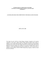
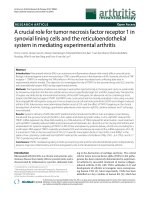
Figure 2 Different effects of Nec-1 and z-VAD on brain injury after ICH. Nec-1 (the specific inhibitor of necroptosis) and z-VAD (a caspase inhibitor) were used to explore
whether necroptosis contributes to brain injury after ICH. (a) The necroptosis in cells were detected by PI staining and apoptosis by terminal deoxynucleotidyl transferase dUTP
nick-end labeling (TUNEL) staining. Nec-1 reduced the PI+ cells, whereas it had no significant effects on TUNEL+ cells (apoptosis), and z-VAD only downregulated the TUNEL+
cells. Additionally, combination of Nec-1 and z-VAD obviously both reduced the PI+ cells and TUNEL+ cells. Scale bar = 100 μm. (b) Corresponding bar graph revealed relative
levels of PI/TUNEL+ cells. **Po0.0001 (both in PI and TUNEL) versus Sham group; NS, not significant difference (P = 0.0909 in PI and P = 0.0857 in TUNEL) versus ICH group;
&&
Po0.0001 in PI and NS (P = 0.4233) in TUNEL versus vehicle group; NS (P = 0.4233) in PI and ##Po0.0001 in TUNEL versus vehicle group; $$Po0.0001 in PI and NS
(P = 0.0663) in TUNEL versus z-VAD group; all were unpaired t-test, n = 6. (c) IP showed that Nec-1 reduced interactions of RIP1 and RIP3, RIP1 and MLKL, and RIP1 and
caspase-8, indicating that the formation of necrosome was inhibited. Input, 5% of extract before IP. (d) Corresponding bar graph revealed relative levels of association.
**P = 0.0097 in RIP1, **P = 0.0099 in RIP3, **P = 0.0010 in MLKL and **P = 0.0022 in caspase-8 versus Sham group; NS, not significant difference (P = 0.3077 in RIP1,
P = 0.7942 in RIP3, P = 0.9047 in MLKL and P = 0.4930 in caspase-8) versus ICH group; &P = 0.0102 in RIP1, &P = 0.0493 in RIP3, &&P = 0.0074 in MLKL and NS
(P = 0.9349) in caspase-8 versus vehicle group; NS (P = 0.8658) in RIP1, NS (P = 0.6857) in RIP3, #P = 0.0205 in MLKL and #P = 0.0281 in caspase-8 versus vehicle group; $
$
P = 0.0065 in RIP1, $P = 0.0499 in RIP3, $P = 0.0403 in MLKL and NS (P = 0.6746) in caspase-8 versus z-VAD group; all were unpaired t-test, n = 3. (e) Expression of albumin,
which is regarded as the index of BBB injury, was increased after ICH, whereas it could be significantly decreased when treated with Nec-1 and/or z-VAD. (f) Corresponding bar
graph revealed relative levels of albumin. *P = 0.0200 versus Sham group; NS, not significant difference (P = 0.9779) versus ICH group; &P = 0.0461 versus vehicle group;
#
P = 0.0496 versus vehicle group; $P = 0.0126 versus z-VAD group; all were unpaired t-test, n = 6. (g) Compared with ICH group, the brain water content was partially attenuated
in Nec-1 and/or z-VAD group, **Po0.0001 (both in Ipsi-CX and Ipsi-BG) versus Sham group; NS, not significant difference (P = 0.9022 in Ipsi-CX and P = 0.9660 in Ipsi-BG)
versus ICH group; &P = 0.0109 in Ipsi-CX and &&P = 0.0077 in Ipsi-BG versus vehicle group; #P = 0.0162 in Ipsi-CX and NS (P = 0.0727) in Ipsi-BG versus vehicle group; $
$
P = 0.0015 in Ipsi-CX and $$P = 0.0039 in Ipsi-BG versus z-VAD group; all were unpaired t-test, n = 6. (h) The levels of TNF-α in the CSF were measured by ELISA and reduced
by Nec-1 treatment. **Po0.0001 versus Sham group, NS, not significant difference (P = 0.7897) versus ICH group; &&Po0.0001 versus vehicle group, ##P = 0.0008 versus
vehicle group, $$Po0.0001 versus z-VAD group; all were unpaired t-test, n = 6. All data are expressed as means ± S.E.M. and mean value for Sham group was normalized
to 1.0.
Cell Death and Disease
RIP1-mediated necroptosis in ICH
H Shen et al
6
obviously increased in the conditioned medium treatment
group and could be inhibited by TNF-α inhibitor pretreatment
(both Po0.01, n = 3; Figures 4c and d). To further explore the
molecular mechanism of RIP1 in necroptosis after ICH, we
Cell Death and Disease
used overexpression with a mutation of phosphorylation site
(S166A) of RIP1. After transfection and being cultured for
another 24 h, neurons were digested by trypsin into cell
suspension, stained with Annexin V and PI and then detected
RIP1-mediated necroptosis in ICH
H Shen et al
7
by flow cytometry. The results of flow cytometry revealed that
upregulating the expression of RIP1 could increase the
percentage of necroptosis (~66.7%) in neurons, but overexpression of mutation of phosphorylation site (S166A) of
RIP1 has no obvious effect in inducing necroptosis of
neurons (~34.2%), and Nec-1 treatment can also inhibit the
necroptosis induced by RIP1 overexpression (~21.7%, all
Po0.01, n = 3; Figures 5a and b).
The results of IP also demonstrated that, compared with the
control, interactions of RIP1 and RIP3, RIP1 and MLKL, and
RIP1 and caspase-8 were significantly increased by overexpression of RIP1. However, interactions of RIP1 and RIP3,
RIP1 and MLKL, and RIP1 and caspase-8 were remarkably
inhibited by mutation of phosphorylation site (S166A) of RIP1
than those in the overexpression of wild type of the RIP1 group
(all Po0.01, n = 3; Figures 5c and d). PI and Hoechst
staining also showed that overexpression of wild-type RIP1
increases the ratio of necroptosis in neurons, but this effect
can be inhibited by mutation of the phosphorylation site
(S166A) of RIP1 (all Po0.0001, n = 6; Figures 5e and f).
These results all suggested that phosphorylation in the 166th
site of RIP1 may be an important element of inducing
necroptosis in neurons.
Discussion
Cell death is the important reason leading to brain injury after
ICH. Almost previous studies believed that the form of cell
death is apoptosis in brain tissues after ICH; few researches
put attention to the role of necrosis. Initiation factors of
apoptosis include downregulation of blood flow and energy
metabolism around the hematoma; a variety of enzymes that
are activated in the blood after ICH activate the apoptosis
signal, and mechanical damage of hematoma directly causes
apoptosis.5,16,17 Apoptosis theory can partly explain the
mechanism of cell death in brain tissues after ICH. However,
recent study found that the dead brain cells release a series of
proteins from the cytoplasm after ICH; these proteins were
known as danger-associated molecular patterns. The most
typical representative is the high-mobility group protein 1,
which can stimulate the inflammatory response that aggravates secondary brain injury after ICH.18 These results pose
challenge to apoptosis theory in ICH; scholars widely
recognized that cell apoptotic process and released apoptotic
bodies do not cause inflammation.15 Thus, presumably,
another form of cell death in addition to apoptosis might exist
in brain tissues and can stimulate inflammatory response after
ICH. On the other hand, microglia were rapidly activated after
ICH and released substantial inflammatory factors (such as
TNF-α); previous studies have confirmed that TNF-α can not
only induce apoptosis but also necroptosis; so whether
necroptosis exists in brain tissues after ICH becomes a
question.
Our present study showed that conditioned medium,
especially the key composition TNF-α, from activated microglia induced necrosis of neurons after ICH for the first time. We
found that necroptosis existed in brain tissues after ICH, and it
had an important role in neuronal dysfunction, brain edema
and BBB permeability after ICH. RIP1 inhibitor Nec-1 can
remarkably attenuate neurological dysfunction, brain edema
and BBB injury, and combinational use with apoptosis inhibitor
z-VAD has a better effect, suggesting that necroptotic pathway
and RIP1 may become targets for the treatment of brain injury
after ICH. We also observed that treatment of ICH rats with
Nec-1 does not affect the apoptosis of brain cells, and the use
of z-VAD treatment of ICH rats also does not affect the
necroptosis in brain tissues; these results indicated that
apoptosis and necroptosis were coexisting and were relatively
independent after ICH. Nec-1 and z-VAD could inhibit
apoptosis and necroptosis, respectively, whereas combinational treatment of Nec-1 and z-VAD was more effective in
treating ICH than each inhibitor alone. In addition, there may
be a crosstalk between apoptosis and necroptosis in the
progression of ICH, which needs further investigation. In
addition, while our research was in progress, Su et al.19
reported that Nec-1 ameliorated brain injury after ICH in a
collagenase-induced ICH model in mice.19 A recent report
also showed that Nec-1 reduced neurovascular injury after
ICH in a collagenase-induced ICH model in mice.20 As our
present study showed that, TNF-α, an important inflammatory
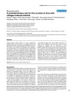
Figure 3 Knockdown of RIP1 reduced necroptosis in brain tissues after ICH. Knockdown and overexpression of RIP1 were used to study the role of RIP1-mediated
necroptosis after ICH in rats. (a) PI+ cells decreased in the Si-RIP1 group (knockdown), whereas they increased in the Ad-RIP1 group (overexpression). Arrows point to PI+ cells.
Scale bar = 200 μm. (b) Related with (a), it revealed relative levels of PI+ cells. **Po0.0001 versus Sham group; NS, not significant difference (P = 0.8453) versus ICH group;
&&
Po0.0001 versus Si-NC group; NS, not significant difference (P = 0.9211) versus ICH group; ##Po0.0001 versus Ad-GFP group; all were unpaired t-test, n = 6. (c) IP
demonstrated that interactions of RIP1 and RIP3, RIP1 and MLKL, and RIP1 and caspase-8 were significantly inhibited in the RIP1-knockdown group, whereas they increased in
the overexpression group. Input, 5% of extract before IP. Quantitative analysis of IP was shown in (d), *P = 0.0177 in RIP1, *P = 0.0261 in RIP3, **P = 0.0072 in MLKL and
*P = 0.0209 in caspase-8 versus Sham group; NS, not significant difference (P = 0.7939 in RIP1, P = 0.6515 in RIP3, P = 0.4640 in MLKL and P = 0.8812 in caspase-8) versus
ICH group; &P = 0.0254 in RIP1, &P = 0.0184 in RIP3, &&P = 0.0019 in MLKL and &&P = 0.0010 in caspase-8 versus Si-NC group; NS (P = 0.7254) in RIP1, NS (P = 0.5590) in
RIP3, NS (P = 0.3154) in MLKL and NS (P = 0.6284) in caspase-8 versus ICH group; #P = 0.0164 in RIP1, #P = 0.0138 in RIP3, #P = 0.0367 in MLKL and ##P = 0.0083 in
caspase-8 versus Ad-GFP group; all were unpaired t-test, n = 3. (e) Expression of albumin was elevated in the Ad-RIP1 group, whereas it was opposite in the Si-RIP1 group. (f)
Bar graph related to (e). **P = 0.0007 versus Sham group; NS, not significant difference (P = 0.8780) versus ICH group; &P = 0.0493 versus Si-NC group; NS, not significant
difference (P = 0.9361) versus ICH group; #P = 0.0470 versus Ad-GFP group; all were unpaired t-test, n = 3. (g) Brain water content decreased in the Si-RIP1 group, whereas it
was opposite in the Ad-RIP1 group,**Po0.0001 (both in Ipsi-CX and Ipsi-BG) versus Sham group; NS, not significant difference (P = 0.4041 in Ipsi-CX and P = 0.5003 in IpsiBG) versus ICH group; &&Po0.0001 in Ipsi-CX and &P = 0.0209 in Ipsi-BG versus Si-NC group; NS, not significant difference (P = 0.5276 in Ipsi-CX and P = 0.6015 in Ipsi-BG)
versus ICH group; ##Po0.0001 (both in Ipsi-CX and Ipsi-BG) versus Ad-GFP group; all were unpaired t-test, n = 6. (h) The levels of TNF-α in the CSF were reduced in RIP1knockdown group, whereas the effect was diametric in the RIP1 overexpression group. **Po0.0001 versus Sham group; NS, not significant difference (P = 0.5570) versus
ICH group; &&P = 0.0002 versus Si-NC group; NS, not significant difference (P = 0.9151) versus ICH group; ##Po0.0001 versus Ad-GFP group; all were unpaired t-test, n = 6.
All data are expressed as means ± S.E.M., and mean values for Sham group were normalized to 1.0. Ad-GFP, adenovirus with GFP; Ad- RIP1, adenovirus with RIP1; Si-NC,
Si-negative control.
Cell Death and Disease
RIP1-mediated necroptosis in ICH
H Shen et al
8
cytokine, may be a key factor in neurons' necrosis after ICH,
the side effects of collagen on inflammatory response should
not be ignored in necrosis-related studies. In this study, we
researched neurons' necrosis in autologous blood injection
ICH model for the first time.
Cell Death and Disease
In many previous studies, stimulation of neurons with
OxyHb was an in vitro model of ICH.21 However, the
inflammation is an important event in second brain injury after
ICH; activated microglia released an amount of inflammatory
cytokines such as TNF-α and IL-1β, which promote brain injury
RIP1-mediated necroptosis in ICH
H Shen et al
9
after ICH.22 Thus, in this study, we used OxyHb-stimulated
microglia and collected the supernatant as conditioned
medium, and then used conditioned medium to treat the
neurons, as an ICH model in vitro. This model highlights the
stimulation of inflammatory factor on neurons, and results of
ELISA also suggested that the level of TNF-α was significantly
increased in conditioned medium compared with the medium
of nontreated microglia (data not shown). We found that
necroptotic rate of neurons in this model was significantly
higher than that in the OxyHb-treated neurons, suggesting that
inflammatory factors released by activated microglia are a key
factor leading to cell necroptosis in brain tissues. A previous
study reported that, in a murine model of ICH, postinjury
treatment with the TNF-α antibody resulted in less neuroinflammation and reduction in functional deficit;23 combining
with our results, we can conclude that ameliorating brain
injury after ICH by blocking TNF-α is partially because of
reducing the necroptosis in brain tissues. Our results also
suggested that multiple stimuli factors coexisted in brain
tissues after ICH, and led to cell death by different pathways; the dominant stimulus in local microenvironment might
be the main reason leading to brain cell death. At the same
time, our results demonstrated that use of multiple in vitro
models may be more appropriate in the study of brain injury
following ICH.
Unlike apoptosis, the release of cell contents will cause
inflammation after the cell necroptosis. Interestingly, an
important initial factor of necroptosis is stimulation of
inflammatory factor (such as TNF-α), and necroptosis also
can further promote the inflammation, these suggested that
possibly have a positive feedback relationship between
necroptosis and inflammation in brain injury after ICH. The
effect of this positive feedback relationship in the pathophysiological processes after ICH needs to be further confirmed.
There are also some deficiencies in this study; although the
role of necroptosis in the brain injury following ICH was
explored, the molecular mechanism is still uncertain. On the
other hand, there are many kinds of inflammatory cytokines
released by activated microglia, such as IL-1β, IL-6 and IL-18;
whether these inflammatory factors also can cause cell
necroptosis in brain tissues after ICH is not clear.
In summary, our study confirmed that the RIP1-mediated
necroptosis exists in brain tissues, and it has an important role
in brain injury following ICH; on the other hand, we used the
in vitro model of ICH suggesting that the release of TNF-α from
activated microglia might be an important factor inducing
necroptosis in ICH (Figure 6). These findings further revealed
the causes of cell death and the relationship between cell
death and inflammation in brain injury after ICH and provide a
potential therapeutic target for secondary brain injury
after ICH.
Materials and Methods
Animals. Adult male Sprague–Dawley (SD) rats, weighing ~ 300 g, were
provided by the Animal Center of Chinese Academy of Sciences (Shanghai,
China). Experimental protocols were approved by the Animal Care and Use
Committee of Soochow University, and were implemented with reference to the
National Institutes of Health guidelines. The animals were freely fed and housed in a
quiet environment (indoor temperature of ~ 18–22 °C). Additionally, we strived as
much as possible to minimize the number of animals that were used and reduce
their suffering. Besides, primary neuronal and microglial cultures (in vitro) were
prepared using 16–18-day-old pregnant SD rats.
Experimental design. In experiment 1, 54 rats (70 rats were used, 54 rats
survived after the surgery) were randomly assigned to nine groups of six rats each;
the normal group, the Sham group and seven experimental groups were arranged
by time – 3, 6, 12, 24, 48, 72 h and 7 days after ICH. Arriving at separate time
points after SAH, all rats were killed and cerebral tissue samples were collected for
analysis (Figure 7a). In experiment 2, 60 rats (75 rats were used, 60 rats survived
after the surgery) were randomly divided into 10 groups – the Sham group, the ICH
group, the ICH+vehicle group, the ICH+Nec-1 group, the ICH+z-VAD group, the ICH
+Nec-1+z-VAD group, the ICH+Si-negative control group, the ICH+SiRNA-RIP1
group, the ICH+Ad-GFP group and the ICH+Ad-RIP1 group. At 24 h after ICH, all
rats were examined for behavioral impairment, and then brain samples were
collected (Figure 7b). In experiment 3, primary cultured neurons were used and
partitioned into six groups – the control group, the OxyHb group, the conditioned
medium group, the conditioned medium+TNF-α inhibitor group, the conditioned
medium+Ad-RIP1 group and the conditioned medium+Ad-RIP1-S166A group
(Figure 7c). Detailed information about each group was shown in special
procedures as below.
Establishment of ICH model. ICH model in vivo was established by
injection of autologous blood.24 After anesthesia with intraperitoneal injection of 4%
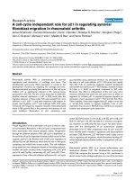
Figure 4 Conditioned medium-induced necroptosis in neurons in vitro. We used two in vitro ICH models to further explore the role of inflammatory factors, such as TNF-α, in
inducing necroptosis: one is neurons treated with OxyHb directly, and the other is using supernatant of culture of microglia (stimulated with OxyHb in advance) as neurons’
conditioned medium, and treated with or without inhibitor of TNF-α. (a) The necroptosis and apoptosis of neurons in vitro were detected by PI and Annexin V double staining and
flow cytometry analysis, respectively. PI − /Annexin V − represented survival neurons, PI+/Annexin V − represented necroptotic neurons, PI − /Annexin V+ represented
apoptotic neurons, and PI+/Annexin V+ represented a mixed damage of neurons. The results of flow cytometry indicated a higher ratio (~27.8%) of apoptosis and a lower ratio
(~11.4%) of necroptosis when neurons were stimulated with OxyHb. However, in conditioned medium treatment group, it was a higher percentage of necroptosis (~31.5%),
whereas the ratio could be significantly reduced when treated with TNF-α inhibitor (~13.5%). (b) Related bar graph showed four different conditions of neurons in various groups;
NS, not significant difference (P = 0.4642) in PI+/Annexin V − cells and **P = 0.0007 in PI − /Annexin V+ cells versus Control group; &&P = 0.0001 in PI+/Annexin V − cells and
NS, not significant difference (P = 0.1498) in PI − /Annexin V+ cells versus Control group; ##P = 0.0004 in PI+/Annexin V − cells and NS, not significant difference (P = 0.3401)
in PI − /Annexin V+ cells versus Conditioned medium group; all were unpaired t-test, n = 3. (c) IP revealed that when treated with conditioned medium, interactions of RIP1 and
RIP3, RIP1 and MLKL, and RIP1 and caspase-8 were remarkably increased. And these results were attenuated when pretreated with TNF-α inhibitor. (d) Consistent data
analysis of IP. **P = 0.0047 in p-Ser, **Po0.0001 in RIP3, **Po0.0001 in MLKL and **P = 0.0002 in caspase-8 versus Control group; &&Po0.0001 in p-Ser, &&P = 0.0006 in
RIP3, &&Po0.0001 in MLKL and &&Po0.0001 in caspase-8 versus Control group; ##P = 0.0003 in p-Ser, ##P = 0.0018 in RIP3, ##P = 0.0012 in MLKL and ##P = 0.0002 in
caspase-8 versus conditioned medium group. The mean values for the control group were normalized to 1.0, all were unpaired t-test, n = 3. (e) Immunofluorescence analysis was
performed with antibody for RIP1 /RIP3 (green), MLKL (red) and caspase-8 (purple) in cultured primary neurons under indicated treatment. Nuclei were fluorescently labeled with
DAPI (blue). Representative images were shown. Arrows indicated the colocalization of RIP1-MLKL-caspase-8 and RIP3-MLKL-caspase. Scale bar = 20 μm. (f) PI and Hoechst
double staining was also used in detection of necroptosis. The results showed that neurons in conditioned medium had higher ratio of necroptosis (as arrows point to, PI+/Hoechst
+ cells), which could be inhibited by TNF-α inhibitor. Scale bar = 50 μm. (g) Numbers of PI+/Hoechst+cells. **Po0.0001 versus control group, &&Po0.0001 versus control
group, ##Po0.0001 versus conditioned medium group; all were unpaired t-test, n = 6. All data are expressed as means ± S.E.M.
Cell Death and Disease
RIP1-mediated necroptosis in ICH
H Shen et al
10
chloral hydrate at a dosage of 1 ml/100 g, 100 μl of autologous blood was collected
from the heart, and then the rats were fixed in the stereotaxic frame (Zhenghua
Biological Equipment Co. Ltd, Anhui, China). The scalp was exposed and made
drilling a hole corresponding to right basal ganglia (0.2 mm anterior to the
intersection between the coronal suture and sagittal midline and 3.5 mm to the right
Cell Death and Disease
of the sagittal suture). A microsyringe was affixed to the stereotactic frame and a
needle was slowly inserted (5.5 mm in depth), and then 100 μl of autologous blood
was slowly injected (20 μl/min). Before slowly withdrawing the needle, it was kept in
place for another 5 min. Rats in the Sham group were intracerebrally injected with
100 μl physiological saline solution. The bone hole was sealed with bone wax, and
RIP1-mediated necroptosis in ICH
H Shen et al
11
skin incision was disinfected and sutured. During the establishment of the model,
rats’ vital signs were monitored and maintained in normal level.
Primary neuron- and microglia-enriched cultures. Neuron-enriched
cultures were prepared from brains of fetal rats (from 16–18-day-old pregnant SD
rats).25 The meninges and blood vessels were removed from the brain and then
brain tissues were digested with 0.25% trypsin (with EDTA) for 5 min at 37 °C. The
tissues were washed three times with PBS to terminate trypsin digestion. Then,
brain tissue suspensions were centrifuged at 1500 r.p.m. for 5 min, and the cells
were suspended in a Neurobasal-A medium containing 2% B27, 2 mM L-glutamine,
50 U/ml penicillin and 50 U/ml streptomycin (all from Gibco, Carlsbad, CA, USA).
Finally, cells were plated in 6- or 12-well plates in a fresh medium and later half the
medium was changed with fresh medium every 2 days.
For primary microglial cultures, the whole brains of 1- day-old rats were used.26
The digestion was similar to neurons, but the pellets after centrifugation were
suspended in DMEM/F12 containing 10% fetal bovine serum, 1 mM sodium pyruvate,
2 mM L-glutamine, 100 mM nonessential amino acids, 50 U/ml penicillin and 50 U/ml
streptomycin (all from Gibco). Then, cells were seeded into 150 cm2 culture flask in a
fresh medium and half the medium was changed with fresh medium every 2 days.
Two weeks after initial seeding, a confluent polylayer of glial cells could be observed.
Figure 6 Hypothesized model for molecular mechanism involved in necroptosis in brain tissues after ICH. After ICH occurs, microglia were rapidly activated and released an
amount of inflammatory cytokines such as TNF-α. TNFR1 can spontaneously trimerize at the plasma membrane. When TNF-α binds to TNFR1, the conformation of these
receptor trimers would be changed, allowing their cytosolic tails recruit multiple proteins, and then develop a complex (complex I) including TRADD (TNF-α receptor-associated
death domain), RIP1, TRAF2 (TNFR associated factor 2), cIAP1 and cIAP2 (cellular inhibitor of apoptosis 1/2). Deubiquitylation of RIP1 mediates its transition from complex I to
complex II, and then cooperates with RIP3 for recruitment of MLKL (mixed lineage kinase domain-like protein), FADD (FAS-associated protein with a death domain) and caspase8. In this complex IIb, RIP1 and RIP3 are inhibited by caspase-8. When caspase-8 is inactive, complex IIb would carry out the TNF-α-mediated necroptotic pathway. Further,
complex of MLKL and RIP3 transfers to the cell membrane and forms a channel, and then the internal flow of Ca2+ or Na+ is caused. Finally, the cell is dead by the necroptotic
pathway. As necroptosis can further promote inflammation, it suggests that it can possibly have a positive feedback relationship between necroptosis and inflammation in brain
injury after ICH.
Figure 5 Phosphorylation of RIP1 switched necroptosis in cultured neurons in model of ICH in vitro. As phosphorylation of RIP1 has an essential role of necroptosis, we used
overexpression and with a mutation of phosphorylation site (S166A) of RIP1 to investigate its role in necroptosis in ICH. (a) Flow cytometry indicated that, compared with the
conditioned medium group, it turned up a higher ratio of necroptosis (~66.7%) and a lower ratio of apoptosis (~0.9%) in neurons during overexpression of RIP1 by Ad-RIP1
transfection. Besides, this phenomenon could be significantly reduced in the S166A group (necroptosis ~ 34.2%) and in the Nec-1 treatment group (necroptosis ~ 21.7%). (b)
Related bar graph showed four different conditions of neurons that had been stated above. **P = 0.0001 in PI+/Annexin V − cells versus the conditioned medium group;
&&
P = 0.0027 in PI+/Annexin V − cells versus the Ad-RIP1 group; $$Po0.0001 in PI+/Annexin V − cells and $$Po0.0001 in PI − /Annexin V+ cells versus the Ad-RIP1 group;
all were unpaired t-test, n = 3. (c) IP demonstrated that interactions of RIP1 and RIP3, RIP1 and MLKL, and RIP1 and caspase-8 were increased in the Ad-RIP1 group. These
increased interactions could be attenuated remarkably in the S166A group. (d) Consistent data analysis of IP. **P = 0.0015 in p-Ser, **P = 0.0004 in RIP3, **P = 0.0013 in MLKL
and **P = 0.0003 in caspase-8 versus the conditioned medium group; &P = 0.0493 in p-Ser, &P = 0.0126 in RIP3, &&P = 0.0036 in MLKL and &&P = 0.0018 in caspase-8 versus
the Ad-RIP1 group. The mean values for the control group were normalized to 1.0; all were unpaired t-test, n = 3. (e) PI and Hoechst staining indicated that the ratio of necroptosis
upregulated (as arrows point to, PI+/Hoechst+ cells) in the overexpression group, whereas mutation of the phosphorylation site inhibited this effect. Scale bar = 50 μm. (f)
Numbers of PI+/Hoechst+ cells, **Po0.0001 versus the conditioned medium group; &&Po0.0001 versus Ad-RIP1 group; all were unpaired t-test, n = 6. All data are expressed
as means ± S.E.M.
Cell Death and Disease
RIP1-mediated necroptosis in ICH
H Shen et al
12
Figure 7 Experimental designs. (a) Experiment 1 was designed to show the time course of RIP1 expression after ICH and to determine a time point for the next experiment.
(b) Experiment 2 was designed to explore the roles of RIP1 and necroptosis in brain injury after ICH in vivo. (d) Experiment 3 was designed to study the potential mechanism of
RIP1 and necroptosis in brain injury after ICH in vitro.
Table 2 Neurobehavioral evaluation
Category Behavior
Score
Appetite
Finished meal
Left meal unfinished
Scarcely ate
0
1
2
Activity
Walk and reach at least three corners of the cage
Walk with some stimulations
Almost always lying down
0
1
2
Deficits
No deficits
Unstable walk
Impossible to walk
0
1
2
The microglia was separated from astrocytes by shaking the flask at 150 r.p.m. for
4 h, and then it was collected by centrifugation and reseeded in 12-well plates with
fresh medium.
Drug administration. Nec-1 was prepared in DMSO at a concentration of
1 μg/3 μl,27 and z-VAD (all from Santa Cruz Biotechnology, Santa Cruz, CA, USA)
was prepared in DMSO at a concentration of 100 μM.10 At 1–2 h before ICH, both
the inhibitors were injected into the lateral cerebral ventricle at a volume of 3 μl.
Equal volumes of DMSO were used as vehicle. For in vitro experiments, the TNF-α
inhibitor (Santa Cruz Biotechnology) was also dissolved in DMSO at a final
concentration of 50 μM in a neuronal medium. The final concentration of Nec-1 and
z-VAD was 30 and 100 μM, respectively, in a neuronal medium.
Cell Death and Disease
ICH models in vitro. An ICH model in vitro was established by neuronal
stimulation using OxyHb.21 Neurons were treated with OxyHb (10 μM) for 6 h at
37 °C in 5% CO2, and then cell medium was removed, washed three times with
PBS and followed by other experiments.
The other in vitro ICH model used conditioned medium to treat neurons. First,
microglia were treated with OxyHb (10 μM) and then incubated for 24 h at 37 °C with
5% CO2. The supernatant collected was centrifuged at 10 000 r.p.m. for 5 min and
then was transferred to a conditioned medium. Half of the neuronal medium was
changed with conditioned medium and incubated for another 6 h at 37 °C with 5%
CO2. The cell medium was removed, washed three times with PBS and followed by
other experiments.
Transfection of siRNA and adenoviruses in vivo and in vitro. The
following three kinds of recombinant adenoviruses were used: (1) adenoviruses
containing rat RIP1 (Ad-RIP1; Genbank ID: 157824040) that was used to overexpress
RIP1 protein; (2) adenovirus with rat RIP1 with mutation of S166A (Ser166 of RIP1
was mutated to alanine, Ad-RIP1-S166A); and (3) adenovirus with human GFP (AdGFP) as a control to Ad-RIP1. Ad-RIP1 and Ad-RIP1-S166A (both 6 × 10⁹ PFU/ml)
and Ad-GFP (2 × 10⁹ PFU/ml) were produced by Genescript (Nanjing, China). All of
them were stored at − 80 °C and diluted to 1 × 10⁹ PFU/ml in an enhanced
transfection solution (Genescript) before intracerebroventricular injection in vivo and
diluted to 1 × 10⁸ PFU/ml before being transfected to the cultured neuron.
The following two kinds of siRNAs were used: (1) disorganizing rat RIP1 mRNA
(Si-RIP1) to silence its transcription and (2) scramble siRNA (Si-negative control)
(both from Genescript). According to the manufacturer’s instructions for Entransterin vivo RNA transfection reagent (Engreen, Shanghai, China), 500 pmol RIP1 siRNA
and 500 pmol scramble siRNA were dissolved in 5 μl RNase-free water. Then, 10 μl
Entranster-in vivo RNA transfection reagent was added to 5 μl siRNA or 5 μl
scramble siRNA. After mixing for another 15 min, Entranster-in vivo-siRNA mixture
was injected intracerebroventricularly 24 h before ICH. RIP1 siRNA sequences were
RIP1-mediated necroptosis in ICH
H Shen et al
13
as follows: sense, 5′-GGAACAACGGAGUAUAUAAdTdT-3′ and Antisense:
3′-dTdTCCUUGUUGCCUCAUAUAUU-5′.
and were monitored for appetite, activity and neurological defects, as reported
previously30 (for more details see Table 2).
Western blot analysis. Western blot analysis was performed as indicated
previously.25 Briefly, the brain samples or extracted cells were mechanically lysed in
RIPA lysis buffer (Beyotime, Shanghai, China). Then we used enhanced BCA
Protein Assay Kit (Beyotime) to measure protein concentrations by the bicinchoninic
acid method. The protein samples (50 μg per lane) were then loaded onto a 10%
SDS-polyacrylamide gel, separated and electrophoretically transferred to a
polyvinylidene difluoride membrane (Millipore Corporation, Billerica, MA, USA),
which was then blocked with 5% bovine serum albumin (BioSharp, Anhui, China)
(1 h at room temperature). Then, the membrane was incubated for 12 h at 4 °C with
primary antibodies. The primary antibodies used were RIP1, RIP3, MLKL, caspase8, p-Ser (all from Santa Cruz Biotechnology) and albumin (Abcam, Cambridge, UK;
ab106582). Besides, the β-tubulin was also detected and served as a loading
control. Later, the membrane was incubated with related HRP-conjugated
secondary antibody (Santa Cruz Biotechnology) for 2 h at room temperature. We
revealed the band signals via the Enhanced Chemiluminescence (ECL) Kit
(Beyotime), and the relative quantity of proteins was analyzed via the Image J
Software (NIH, Bethesda, MD, USA) and normalized to that of the loading control as
discussed previously. In addition, the levels of phosphorylation were evaluated as
the ratio of phosphoprotein to total protein.
Assay of inflammatory cytokines (TNF-α). We used specific Rat Tumor
necrosis factor α, TNF-α ELISA KIT (Bio-Swamp; Hubei, China) to quantify the
levels of TNF-α in the CSF according to the manufacturer's instructions.
Immunoprecipitation. Briefly, according to our previous report,25 the brain
samples were lysed in ice-cold RIPA lysis buffer (Beyotime). For IP, the lysate was
incubated with specific antibodies against RIP1 or rabbit IgG (negative control)
overnight at 4 °C with agitation. Protein A+G Sepharose beads were then added to
each immune complex, and the lysate–bead mixture was incubated for 4 h at 4 °C
under rotary agitation. SDS-PAGE and immunoblotting were then performed for
further protein separation and detection.
Immunofluorescent staining. The brain samples were fixed in 4%
paraformaldehyde, embedded in paraffin and then cut into 4 μm sections. The
cultured neurons were fixed in 4% paraformaldehyde. Then, the sections and
neurons were incubated with primary antibody against RIP1, RIP3, MLKL and
caspase-8 and secondary antibodies. Normal rabbit IgG, normal mouse IgG
and normal goat IgG were used as negative controls for the immunofluorescence
assay (data not shown). Nuclei were stained with DAPI mounting medium. Finally,
the sections and neurons were observed in a fluorescence microscope (OLYMPUS
BX50/BX-FLA/DP70; Olympus Co., Tokyo, Japan). The relative fluorescence
intensity was analyzed with the Image J program. The quantitative analysis was
performed by an observer who was blinded to the experimental group.
PI and TUNEL staining. For all experiments, PI (Sigma-Aldrich, St. Louis, MO,
USA) was administered intraperitoneally (1 μg/g) while the ICH model was
established.28 According to the manufacturer’s protocol (Roche, Mannheim,
Germany), we detected cell apoptosis via TUNEL staining. The brain was removed
and frozen in liquid nitrogen and then stored at − 80 °C. Then, brain sections (12 μm)
were cut on a cryostat 150 to 200 μm apart along the anterior–posterior lesion and
were placed on poly-L-lysine-coated glass slides (−80 °C). Next, the sections were
incubated with TUNEL staining (37 °C for 1 h). After washing three times with PBST,
sections were visualized by a fluorescence microscope (OLYMPUS BX50/BX-FLA/
DP70; Olympus Co.). In conclusion, PI/TUNEL+ cells were counted by observers
blinded to the groups of experiment. To evaluate necropoptosis/apoptosis of cells, we
examined and photographed six microscopic fields per sample and the index was
defined as the average number in each section. As reported previously, PI+/TUNEL −
is defined as the pure necropoptosis, PI − /TUNEL+ is defined as pure apoptosis, and
PI+/TUNEL+ is defined as the mixed cell death.29
Brain edema and BBB injury. As described previously,21 the wet–dry
method was adopted to evaluate the index of brain edema and BBB injury. Briefly,
after brain tissues were removed and collected, the samples were weighed
immediately (wet weight), dried (100 °C for 72 h) and then reweighed (dry weight).
Thus, brain edema was calculated as ((wet weight − dry weight)/wet weight) ×
100%. Meanwhile, the level of albumin in brain tissues was regarded as the index of
BBB injury. The western blot was used to test the level of albumin.
Neurobehavioral evaluation. At 24 h after ICH, all the rats in the
experiments were examined for behavioral impairment using a scoring system
Annexin V and PI staining in vitro. After various treatments, neurons were
trypsinized by 0.25% trypsin (without EDTA) and centrifuged at 1500 r.p.m. for
5 min, and the resulting cell pellet was resuspended in 500 μl binding buffer. Later,
5 μl Annexin V and 5 μl PI (Beyotime, Shanghai, China) were added to the cell
suspension. After 20 min of incubation at 37 °C in the dark, the cells were analyzed
by flow cytometry (FACS Cabibur; BD, San Diego, CA, USA) and at least 20 000
events per sample were recorded.
PI and Hoechst staining in vitro. After various treatments, add 5 μl
Hoechst and 5 μl PI (Beyotime, Shanghai, China) to the cell medium. After 20 min
of incubation at 4 °C in the dark, the cells were analyzed by a fluorescence
microscope (OLYMPUS BX50/BX-FLA/DP70; Olympus Co., Tokyo, Japan.)
Statistical analysis. All data are presented as mean ± S.E.M. GraphPad
Prism 5.0 software (GraphPad, San Diego, CA, USA) was used for statistical
analysis. Data sets were tested for normality of distribution with Kolmogorov–
Smirnov test. Data groups (two groups) with normal distribution were compared
using two-sided unpaired Student’s t-test, and the Mann–Whitney U-test was used
for nonparametric data. Po0.05 indicates a statistically significant difference.
Conflict of Interest
The authors declare no conflict of interest.
Acknowledgements. This work was supported by Suzhou Key Medical Center
(Szzx201501), grants from the National Natural Science Foundation of China (No.
81601007), Scientific Department of Jiangsu Province (No. BL2014045), Education
Department of Jiangsu Province (No. 16KJB320008), Suzhou Government (No.
SZS201413 and SYS201608) and a project funded by the Priority Academic Program
Development of Jiangsu Higher Education Institutions.
1. Joseph MJ, Caliaperumal J, Schlichter LC. after intracerebral hemorrhage, oligodendrocyte
precursors proliferate and differentiate inside white-matter tracts in the rat striatum. Transl
Stroke Res 2016; 7: 192–208.
2. Behrouz R. Re-exploring tumor necrosis factor alpha as a target for therapy in intracerebral
hemorrhage. Transl Stroke Res 2016; 7: 93–96.
3. Jiang B, Li L, Chen Q, Tao Y, Yang L, Zhang B et al. Role of glibenclamide in brain injury after
intracerebral hemorrhage. Transl Stroke Res 2016; 10.1007/s12975-016-0506-2.
4. Dang G, Yang Y, Wu G, Hua Y, Keep RF, Xi G. Early erythrolysis in the hematoma
after experimental intracerebral hemorrhage. Transl Stroke Res 2016; 10.1007/s12975-0160505-3.
5. Sukumari-Ramesh S, Alleyne CH Jr, Dhandapani KM. The histone deacetylase inhibitor
suberoylanilide hydroxamic acid (SAHA) confers acute neuroprotection after intracerebral
hemorrhage in mice. Transl Stroke Res 2016; 7: 141–148.
6. Elliott J, Smith M. The acute management of intracerebral hemorrhage: a clinical review.
Anesth Analg 2010; 110: 1419–1427.
7. Aronowski J, Zhao X. Molecular pathophysiology of cerebral hemorrhage: secondary
brain injury. Stroke 2011; 42: 1781–1786.
8. Zong WX, Thompson CB. Necrotic death as a cell fate. Genes Dev 2006; 20: 1–15.
9. Golstein P, Kroemer G. Cell death by necrosis: towards a molecular definition. Trends
Biochem Sci 2007; 32: 37–43.
10. Degterev A, Huang Z, Boyce M, Li Y, Jagtap P, Mizushima N et al. Chemical inhibitor of
nonapoptotic cell death with therapeutic potential for ischemic brain injury. Nat Chem Biol
2005; 1: 112–119.
11. Galluzzi L, Kepp O, Krautwald S, Kroemer G, Linkermann A. Molecular mechanisms of
regulated necrosis. Semin Cell Dev Biol 2014; 35: 24–32.
12. Nikoletopoulou V, Markaki M, Palikaras K, Tavernarakis N. Crosstalk between apoptosis,
necrosis and autophagy. Biochim Biophys Acta 2013; 1833: 3448–3459.
13. Nguyen HX, O'Barr TJ, Anderson AJ. Polymorphonuclear leukocytes promote neurotoxicity
through release of matrix metalloproteinases, reactive oxygen species, and TNF-a.
J Neurochem 2007; 102: 900–912.
14. Lo EH, Dalkara T, Moskowitz MA. Mechanisms, challenges and opportunities in stroke.
Nat Rev Neurosci 2003; 4: 399–415.
Cell Death and Disease
RIP1-mediated necroptosis in ICH
H Shen et al
14
15. Jouan-Lanhouet S, Riquet F, Duprez L, Vanden Berghe T, Takahashi N, Vandenabeele P.
Necroptosis, in vivo detection in experimental disease models. Semin Cell Dev Biol 2014;
35: 2–13.
16. Delgado P, Cuadrado E, Rosell A, Alvarez-Sabin J, Ortega-Aznar A, Hernandez-Guillamon M
et al. Fas system activation in perihematomal areas after spontaneous intracerebral
hemorrhage. Stroke 2008; 39: 1730–1734.
17. Ke K, Rui Y, Li L, Zheng H, Xu W, Tan X et al. Upregulation of EHD2 after intracerebral
hemorrhage in adult rats. J Mol Neurosci 2014; 54: 171–180.
18. Kono H, Rock KL. How dying cells alert the immune system to danger. Nat Rev Immunol
2008; 8: 279–289.
19. Su X, Wang H, Kang D, Zhu J, Sun Q, Li T et al. Necrostatin-1 ameliorates intracerebral
hemorrhage-induced brain injury in mice through inhibiting RIP1/RIP3 pathway. Neurochem
Res 2015; 40: 643–650.
20. King MD, Whitaker-Lea WA, Campbell JM, Alleyne CH Jr, Dhandapani KM. Necrostatin-1
reduces neurovascular injury after intracerebral hemorrhage. Int J Cell Biol 2014; 2014: 495817.
21. Zhai W, Chen D, Shen H, Chen Z, Li H, Yu Z et al. A1 adenosine receptor attenuates
intracerebral hemorrhage-induced secondary brain injury in rats by activating the P38MAPKAP2-Hsp27 pathway. Mol Brain 2016; 9: 66.
22. Gao Z, Wang J, Thiex R, Rogove AD, Heppner FL, Tsirka SE. Microglial activation and
intracerebral hemorrhage. Acta Neurochir Suppl 2008; 105: 51–53.
23. Lei B, Dawson HN, Roulhac-Wilson B, Wang H, Laskowitz DT, James ML. Tumor necrosis
factor alpha antagonism improves neurological recovery in murine intracerebral hemorrhage.
J Neuroinflamm 2013; 10: 103.
24. Deinsberger W, Vogel J, Kuschinsky W, Auer LM, Boker DK. Experimental intracerebral
hemorrhage: description of a double injection model in rats. Neurol Res 1996; 18: 475–477.
25. Shen H, Chen Z, Wang Y, Gao A, Li H, Cui Y et al. Role of neurexin-1beta and neuroligin-1 in
cognitive dysfunction after subarachnoid hemorrhage in rats. Stroke 2015; 46: 2607–2615.
26. Liu B, Wang K, Gao HM, Mandavilli B, Wang JY, Hong JS. Molecular consequences of activated
microglia in the brain: overactivation induces apoptosis. J Neurochem 2001; 77: 182–189.
Cell Death and Disease
27. Yin B, Xu Y, Wei RL, He F, Luo BY, Wang JY. Inhibition of receptor-interacting protein 3
upregulation and nuclear translocation involved in necrostatin-1 protection against
hippocampal neuronal programmed necrosis induced by ischemia/reperfusion injury. Brain
Res 2015; 1609: 63–71.
28. Ding W, Shang L, Huang JF, Li N, Chen D, Xue LX et al. Receptor interacting protein
3-induced RGC-5 cell necroptosis following oxygen glucose deprivation. BMC Neurosci
2015; 16: 49.
29. Zhu X, Tao L, Tejima-Mandeville E, Qiu J, Park J, Garber K et al. Plasmalemma permeability
and necrotic cell death phenotypes after intracerebral hemorrhage in mice. Stroke 2012; 43:
524–531.
30. Ma J, Wang Z, Liu C, Shen H, Chen Z, Yin J et al. Pramipexole-Induced hypothermia
reduces early brain injury via PI3K/AKT/GSK3beta pathway in subarachnoid hemorrhage
rats. Scientific Rep 2016; 6: 23817.
Cell Death and Disease is an open-access journal
published by Nature Publishing Group. This work is
licensed under a Creative Commons Attribution 4.0 International
License. The images or other third party material in this article are
included in the article’s Creative Commons license, unless indicated
otherwise in the credit line; if the material is not included under the
Creative Commons license, users will need to obtain permission from
the license holder to reproduce the material. To view a copy of this
license, visit />r The Author(s) 2017