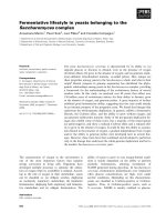
Báo cáo khoa học: Gram-positive bacterial superantigen outside-in signaling causes toxic shock syndrome ppt
Bạn đang xem bản rút gọn của tài liệu. Xem và tải ngay bản đầy đủ của tài liệu tại đây (852.99 KB, 19 trang )
REVIEW ARTICLE
Gram-positive bacterial superantigen outside-in signaling
causes toxic shock syndrome
Amanda J. Brosnahan
1
and Patrick M. Schlievert
2
1 Department of Veterinary and Biomedical Sciences, College of Veterinary Medicine, University of Minnesota, Saint Paul, USA
2 Department of Microbiology, Medical School, University of Minnesota, Minneapolis, USA
Introduction
In 2007 based on 2005 data, the Centers for Disease
Control and Prevention (CDC), together with their
collaborators, reported that Staphylococcus aureus is
the leading cause of serious and fatal infectious dis-
eases in the USA [1]. This results from the organism
making many virulence factors and being a common
commensal organism on human skin and mucosal sur-
faces; as many as 40% of humans may be colonized
on mucosal surfaces, such as anterior nares and vagina
[2]. It is well known that persons with damaged skin,
such as in atopic dermatitis, are nearly always colo-
nized on skin surfaces with the organisms [3]. S. aureus
Keywords
MRSA; mucosal immunity; Staphylococcus
aureus; Streptococcus pyogenes;
superantigen; toxic shock syndrome
Correspondence
P. M. Schlievert, Department of
Microbiology, Medical School, University of
Minnesota, Minneapolis, USA
Fax: 612 626 0623
Tel: 612 624 1484
E-mail:
(Received 7 March 2011, revised 20 April
2011, accepted 27 April 2011)
doi:10.1111/j.1742-4658.2011.08151.x
Staphylococcus aureus and Streptococcus pyogenes (group A streptococci)
are Gram-positive pathogens capable of producing a variety of bacterial exo-
toxins known as superantigens. Superantigens interact with antigen-present-
ing cells (APCs) and T cells to induce T cell proliferation and massive
cytokine production, which leads to fever, rash, capillary leak and subse-
quent hypotension, the major symptoms of toxic shock syndrome. Both
S. aureus and group A streptococci colonize mucosal surfaces, including the
anterior nares and vagina for S. aureus, and the oropharynx and less com-
monly the vagina for group A streptococci. However, due to their abilities to
secrete a variety of virulence factors, the organisms can also cause illnesses
from the mucosa. This review provides an updated discussion of the bio-
chemical and structural features of one group of secreted virulence factors,
the staphylococcal and group A streptococcal superantigens, and their abili-
ties to cause toxic shock syndrome from a mucosal surface. The main focus
of this review, however, is the abilities of superantigens to induce cytokines
and chemokines from epithelial cells, which has been linked to a dodecapep-
tide region that is relatively conserved among all superantigens and is distinct
from the binding sites required for interactions with APCs and T cells. This
phenomenon, termed outside-in signaling, acts to recruit adaptive immune
cells to the submucosa, where the superantigens can then interact with those
cells to initiate the final cytokine cascades that lead to toxic shock syndrome.
Abbreviations
APC, antigen-presenting cell; CA-MRSA, community-associated MRSA; HA-MRSA, hospital-associated MRSA; HLA, human leucocyte
antigen; IFN-c, interferon-c; IL, interleukin; LPS, lipopolysaccharide; MCP-1, monocyte chemotactic protein 1; MHC II, major
histocompatibility complex class II; MIP-3a, macrophage inflammatory protein 3a; MRSA, methicillin-resistant S. aureus; MSSA, methicillin-
sensitive S. aureus; OB, oligosaccharide ⁄ oligonucleotide binding; PANDAS, pediatric auto-immune neuropsychiatric disorders associated
with streptococcal infections; SAg, superantigen; SE, staphylococcal enterotoxin; SLO, streptolysin O; SMEZ, streptococcal mitogenic
exotoxin Z; SPE, streptococcal pyrogenic exotoxin; SSA, streptococcal superantigen; TCR, T cell receptor; TNF, tumor necrosis factor; TSS,
toxic shock syndrome; TSST-1, toxic shock syndrome toxin 1.
FEBS Journal 278 (2011) 4649–4667 ª 2011 The Authors Journal compilation ª 2011 FEBS 4649
strains are not restricted to human colonization and
infection, as many animal species are also infected by
the organisms. However, despite some strain cross-
over, human strains tend to colonize humans and ani-
mal strains tend to colonize animals.
Streptococcus pyogenes (group A streptococcus) also
has a significant disease association with humans.
Unlike S. aureus, group A streptococci are considered
primary pathogens of humans only in that infected
persons typically become ill upon their first exposure
to the organism, most often with either pharyngitis or
impetigo, but more recently also with severe invasive
illnesses, and humans transmit the organism to each
other [4]. Group A streptococci and S. aureus,
although sharing many properties, differ in two impor-
tant aspects of disease causation, in that group A
streptococci are more invasive in causing serious
human illnesses, such as toxic shock syndrome (TSS),
than S. aureus, and group A streptococci strains have
the potential to cause delayed sequelae such as rheu-
matic fever, acute glomerulonephritis and guttate pso-
riasis, whereas S. aureus strains do not [4].
Staphylococcus aureus and group A streptococci are
both Gram-positive cocci, although S. aureus strains
are facultative aerobes whereas group A streptococci
are aerotolerant anaerobes. Both organisms depend on
production of a myriad of cell-surface and secreted vir-
ulence factors for host colonization and disease pro-
duction. The cell-surface virulence factors include a
variety of proteins referred to as microbial surface
components recognizing adhesive matrix molecules [5].
These molecules, including fibronectin-binding proteins
and collagen-binding proteins among many others,
facilitate attachment to host cells, or interfere with
host immune responses through the antiphagocytic
action of proteins such as protein A (S. aureus) or pro-
tein G (group A streptococci). Although important in
disease production, these proteins will not be discussed
further in this review.
Both organisms also produce large numbers of
secreted virulence factors, including families of superan-
tigens (SAgs) and cytolysins. These two major classes of
exoproteins interfere with immune system function sys-
temically (SAgs) and locally (cytolysins). This review
will focus on the role of SAgs in human illnesses, their
structure and function, and will discuss cytolysins in
their role of facilitating SAg penetration of epithelial
barriers and microorganism invasion systemically.
The superantigen family
Nearly all S. aureus strains have the capacity to pro-
duce one or more SAg proteins, including toxic shock
syndrome toxin 1 (TSST-1), staphylococcal enterotox-
ins (SEs) serotypes A, B, C (including multiple SEC
subtypes), D, E and I, and SE-like SAgs, including
SE-like G, H, J–X [6–9]. There is no SE or SE-like F
protein, as this designation was retired when the pro-
tein was renamed TSST-1. SEs and SE-like SAgs differ
in that SEs are emetogenic when given orally to mon-
keys and eaten by humans, whereas SE-like proteins
either lack emetogenic activity or have not been tested
[8]. All of the staphylococcal SAgs, except SE-like X,
are encoded on DNA genetic elements that are consid-
ered variable traits in that their SAg genes are present
in some S. aureus strains but other SAg genes are pres-
ent in other strains. Staphylococcal SAgs are encoded
by genes located on bacteriophages (e.g. SEA), plas-
mids (e.g. SED) or pathogenicity islands (most SAgs),
and in the case of SE-like X, the core chromosome [9].
Group A streptococci also produce numerous SAgs,
including streptococcal pyrogenic exotoxin (SPE, scar-
let fever toxin) serotypes A, C, G–M, streptococcal
superantigen (SSA), and streptococcal mitogenic exo-
toxin Z (SMEZ with numerous subtypes) [4]. With
the exception of SPE G and SMEZ, which are
encoded by genes within the core chromosome, all
remaining streptococcal superantigens are encoded by
genes located on bacteriophages, many of which are
defective and essentially trap the SAg gene in the
chromosome [4].
Although originally described in S. aureus and group
A streptococci, SAgs are increasingly being isolated
from coagulase-negative staphylococci and other
groups of b-hemolytic streptococci, particularly groups
C and G [10–14]. The increasing presence of SAgs in
coagulase-negative staphylococci is particularly discon-
certing, since this gives these normal flora organisms
greater potential to cause serious human illnesses. The
SAgs in coagulase-negative staphylococci and other
groups of b-hemolytic streptococci thus far are related
serologically and biochemically to those present in
either S. aureus or group A streptococci, but there is
ample reason to believe that previously undescribed
SAgs will also be found in such strains, expanding our
horizons on the numbers and types of SAgs and their
associated infections.
Biochemistry of superantigens
SAgs are simple, non-glycosylated proteins that are
secreted from bacterial strains through cleavable signal
peptides [4]. The proteins have relatively low molecular
weights (19 000–30 000) and appear tightly folded.
They are generally resistant to heat (e.g. TSST-1
can be boiled for up to 1 h without loss of biological
Superantigen outside-in signaling A. J. Brosnahan and P. M. Schlievert
4650 FEBS Journal 278 (2011) 4649–4667 ª 2011 The Authors Journal compilation ª 2011 FEBS
activity), proteolysis (this allows SEs to resist gut pro-
teolysis and cause staphylococcal food poisoning),
weak acids (SEs are resistant to 10% bleach for several
minutes) and desiccation (TSST-1 remains completely
biologically active when dried onto Petri dishes for
more than 1 year); the proteins are easily inactivated
by bases, such as 1 m NaOH [4,9].
Structural features of superantigens
SAgs contain two major structural domains: an
amino-terminal oligosaccharide ⁄ oligonucleotide bind-
ing (OB) fold and a carboxy-terminal b-grasp domain.
An epithelial cell binding domain, referred to as the
dodecapeptide region, resides predominantly on the
central a-helix in the b-grasp domain. The region for
T cell receptor (TCR) binding resides in a groove
between both major folds, a low-affinity major histo-
compatibility complex class II (MHC II) site is located
in the OB fold, and a higher-affinity MHC II site is
located in the b-grasp domain. The staphylococcal and
streptococcal SAgs can be divided into five groups
based on their amino acid sequences and shared struc-
tural features (Table 1) [9].
Group I is characterized by TSST-1, which has an
amino acid sequence that is unique compared with
other SAgs. Also included in this group are the variant
TSST-1, referred to as TSST-ovine, and a recently
identified SAg named SE-like X. The dodecapeptide
region of TSST-1 (119-FDKKQLAISTLD-130) is also
unique among SAgs in that it is contained solely on
the central a-helix and a nearby loop, but not involv-
ing b-strands. These group I SAgs have low-affinity
MHC II sites and TCR interaction sites on the back
of the SAgs as seen in the standard view (Fig. 1); these
SAgs lack emetogenic activity.
Group II SAgs are characterized by SEB, SEC and
SPE A and the presence of a cystine loop structure in
which the cystines are separated by 10–19 amino acids
[15]. This cystine loop is required for emetogenic activ-
ity but its presence does not automatically confer
emetogenic ability because SPE A contains the cystine
loop but is not emetogenic [15,16]. It appears that the
structural conformation induced by the loop is more
important than the loop itself for activity. Like
group I SAgs, group II SAgs have only the low-affinity
MHC II binding site, lacking the higher-affinity MHC
II site, and interact with the a-chain of MHC II
through their OB-fold domains. It is important to note
that a zinc binding site has been found in SPE A, but
its role in MHC class II binding remains unclear [17–
19]. Group II SAgs interact with the TCR on the top
front of the SAgs.
Group III SAgs, which include SEA, SED and SEJ,
are similar to group II SAgs due to the presence of the
cystine loop structure, but in the case of group III
SAgs the loop is always nine amino acids long. Group
III SAgs differ from groups I and II SAgs because
group III proteins have both low- and higher-affinity
binding sites for MHC II molecules. These SAgs can
bind either the a-chains of MHC II molecules through
their OB folds (low affinity) or the b-chains through
their b-grasp domains (higher affinity). These SAgs are
emetogenic as well as superantigenic. Interestingly, one
member of this group, SEH, was shown to bind the
TCR a-chain, as opposed to the TCR b-chain that is
used by the other SAgs [20–22].
Group IV SAgs are all produced by group A strep-
tococci. This group includes SPE C, SPE J and SMEZ,
the SAgs do not contain a cystine loop structure, and
the SAgs have both low- and higher-affinity binding
sites for MHC II. These SAgs are non-emetogenic.
Most of the recently characterized SAgs, such as
SE-like K and SE-like Q, are contained within group
V. These SAgs have both low- and higher-affinity
binding sites for MHC II, and do not have cystine
loop structures, similar to group IV SAgs; however,
group V superantigens also have similar 15 amino acid
inserts that are not found in the other SAgs. The
inserts are loop extensions that reside between the
third a-helix and the eighth b-strand, referred to as
the a3-b8 loop. These loops have been shown to be
required for T cell activation for both SPE I and SE-
like K, and are thought to be required for activity of
Table 1. Staphylococcal and streptococcal SAg groups defined by amino acid sequence and three-dimensional structure similarities.
Group Example SAgs MHC II binding Structural features
I TSST-1, TSST-ovine, SE-like X Low-affinity (a-chain) site Unique amino acid sequence, no cystine loop
II SEB, SPE A, SEC, SSA, SEG Low-affinity (a-chain) site Variable length cystine loop
III SEA, SEE, SEJ, SED, SEH Low- and higher-affinity (a- and b-chain) sites Nine amino acid length cystine loop
IV SPE C, SPE J, SPE G, SMEZ-2 Low- and higher-affinity (a- and b-chain) sites No cystine loop
V SEI, SE-like K, SE-like L, SE-like P,
SE-like Q, SPE H
Low- and higher-affinity (a- and b-chain) sites No cystine loop and a 15 amino acid insert
A. J. Brosnahan and P. M. Schlievert Superantigen outside-in signaling
FEBS Journal 278 (2011) 4649–4667 ª 2011 The Authors Journal compilation ª 2011 FEBS 4651
all SAgs that fall into this group [23,24]. Additionally,
it appears that the a3-b8 loop dictates the Vb-TCR
specificity of the SAgs in this group [24]. The SAgs in
this group that have been tested lack emetogenic activ-
ity. For this reason, they are referred to as SE-like.
Disease associations of superantigens
Staphylococcal and streptococcal SAgs unquestionably
cause TSS, and probably contribute significantly to
other illnesses [4,6,7,9,25]. Staphylococcal TSS is an ill-
ness defined by the following criteria: fever, hypoten-
sion, erythematous rash, peeling of the skin upon
recovery, and any three of a multi-organ component,
often seen initially as vomiting and diarrhea (easily
confused with flu-like symptoms) [4,26–28]. If one
defining criterion is absent, the illness is referred to as
probable TSS [29]. If more than one defining symptom
is missing and other causes are ruled out, the illness
can be considered toxin-mediated disease [30]. Staphy-
lococcal TSS is separated into two major categories,
menstrual and non-menstrual illness. Menstrual TSS,
as its name implies, occurs during or within 2 days of
women’s menstrual periods, and the illness is most
often associated with tampon use [26,27]; the tampon
association primarily results from tampon-introduced
oxygen being required for TSST-1 production [31–33].
The human vagina in the absence of tampons is nor-
mally anaerobic. TSST-1 is the cause of nearly all
menstrual cases of TSS, probably because of its greater
capacity than other SAgs to penetrate mucosal surfaces
(discussed later in this review) [9,25,34]. One paper has
suggested other SAgs may occasionally be associated
with menstrual TSS [35]. Non-menstrual TSS takes on
many forms and can be associated with nearly any
type of S. aureus infection [29]. The most common
non-menstrual TSS is illness following upper respira-
tory viral infection, described initially in 1987 by Mac-
Donald et al. [36]. It is suggested that the multiple
proteases produced by S. aureus strains activate the
hemagglutinin of influenza virus, facilitating the ability
of the virus to initiate infection, which in turn creates
a damaged epithelial site allowing secondary S. aureus
infection. We estimate that >10 000 people in the
USA succumb to this infection yearly. Non-menstrual
TSS is primarily caused by TSST-1 (50%) and two
SEs, SEB and SEC (50% together), which are pro-
duced in higher concentrations than other SAgs (up to
10
6
-fold more) [34]. Other SAgs are occasional causes
of non-menstrual TSS, presumably by strains that have
upregulated production of their SAgs. The overall inci-
dence of TSS and probable TSS combined, including
both menstrual and non-menstrual categories, is 1–3
per 100 000 population with a case : fatality rate of
5–10%. Occasionally staphylococcal TSS cases are
now being seen in association with necrotizing fasciitis
Fig. 1. Crystal structures of representative superantigens from each
group. The dodecapeptide region is highlighted in yellow. TSST-1 and
SPE A residues that, when mutated to alanine, result in different lev-
els of IL-8 induction from human vaginal epithelial cells are depicted
as follows: no IL-8 release in red, low IL-8 release in orange, and high
IL-8 release in green. Key residues known to be involved in Vb-TCR
binding are depicted in violet, while key residues involved in MHC II
higher-affinity and low-affinity binding are shown in gray and teal,
respectively (not all residues required for binding are shown). Group
1: TSST-1 (PDB 4TSS [150]); Vb-TCR, H135 ⁄ Q136 [151,152]; MHC II,
G31 ⁄ S32 [153,154]; epithelial, F119–D130 [75]. Group II: SPE A (PDB
1UUP [155]); Vb-TCR, L24, N54 ⁄ Y55, C90, C98 [156]; MHC II, L42–
Y48 [157]; epithelial, T135–D146. The cystine loop is shown in
brown: Y88–A97 (note that for SPE A the cystine loop is apparently
positioned incorrectly to allow SPE A to be emetogenic) [19]. Group
III: SEA (PDB 1ESF [158]); Vb-TCR, S206 ⁄ N207 [158,159]; MHC II,
H187, H225, D227 (high) [158] and F47 (low) [158]; proposed epithe-
lial, T145–D156; cystine loop, A97–A105 [160]. Group IV: SPE C (PDB
1AN8 [137]); Vb-TCR, Y15, R181 [161]; MHC II, H201, D203 (high)
[130,136]; proposed epithelial, L122–D133. Group V: SE-like K (PDB
3EA6, Shi K, Huseby M, Schlievert PM, Ohlendorf DH and Earhart
CA, unpublished results); Vb-TCR, H142, Y158 [24]; MHC II binding
residues have not been defined but proposed important residues
based on SEI, H207, D209, D211 (high) and N98 (low) [24,162]; pro-
posed epithelial, T115–D126. The a3-b8 loop of SE-like K is also
shown in pink: G141–G160 [23,24]. Images generated using
PYMOL
(DeLano Scientific LLC, South San Francisco, CA, USA).
Superantigen outside-in signaling A. J. Brosnahan and P. M. Schlievert
4652 FEBS Journal 278 (2011) 4649–4667 ª 2011 The Authors Journal compilation ª 2011 FEBS
and myositis; this form of the illness appears to be
newly emergent [37,38].
Staphylococcal TSS is managed clinically with use of
antibiotics and fluid, electrolytes and vasopressors as
needed to maintain blood pressure, sometimes acti-
vated protein C in an attempt to manage sepsis-
induced microvascular thrombosis, and intravenous
immunoglobulin to neutralize SAgs in severe cases
[39,40]. It is important to know if TSS cases are associ-
ated with methicillin-resistant S. aureus (MRSA) or
methicillin-sensitive S. aureus (MSSA), as antibiotic
selection for treatment of cases is different. MRSA are
managed with antibiotics such as vancomycin with
clindamycin, whereas MSSA may be managed with
b-lactam antibiotics. Today, MRSA appear in two
forms, hospital-associated (HA-MRSA) and commu-
nity-associated (CA-MRSA) [1,41]. HA-MRSA typi-
cally are multi-antibiotic resistant, but the majority of
these strains do not make large amounts of SAgs [42].
In contrast, CA-MRSA strains, which include pulsed-
field gel electrophoresis types USA200, USA300 and
USA400 (according to CDC designations), typically
are resistant to b-lactam antibiotics and sometimes
clindamycin, and these strains produce high levels of
SAgs [42]. USA200 CA-MRSA strains produce TSST-
1, USA300 CA-MRSA produce SE-like X, and
USA400 CA-MRSA produce either SEB or SEC (it is
important to note that the corresponding MSSA
strains also produce these same high-level SAgs). Thus,
nearly all CA-MRSA, and their MSSA counterparts,
have the ability to cause TSS-like illnesses.
Staphylococcal SAgs are also associated with and
cause a myriad of other illnesses (Table 2) [4]. Note
that each of these illnesses could be the basis of an
entire review, and thus only key references are pro-
vided. Readers are encouraged to perform searches for
additional references. It is particularly noteworthy that
nearly every year since the initial description of staphy-
lococcal TSS in 1980, new illnesses have been associ-
ated with staphylococcal SAgs. Four examples are
presented. In 2005, Kravitz et al. [39] described for the
first time staphylococcal purpura fulminans, which is a
particularly severe form of TSS, characterized by TSS
symptoms and rapidly progressing disseminated intra-
vascular coagulation. When initially described, the ill-
ness was 100% fatal, but now that the treatment
strategies have been modified, including administration
of intravenous immunoglobulin to neutralize SAgs and
activated protein C to alter progression of sepsis-
induced microvascular thrombosis, the case : fatality
rate has fallen to approximately 25%. In 2009, Assi-
macopoulos et al. [43] described extreme pyrexia syn-
drome associated with staphylococcal SAg production,
notably a truncated version of TSST-1, primarily by
CA-MRSA USA300 strains. This illness remains 100%
fatal and is characterized by acute onset of fever in
excess of 108 °F (42 °C). Kotler et al. [44] recently re-
identified the staphylococcal SAg association with
pseudomembranous enterocolitis, an illness that once
was associated with staphylococcal SAgs but then later
nearly always associated with Clostridium difficile
infection, and now re-identified in part associated with
staphylococcal SAgs. One final recent illness associated
with staphylococcal SAgs is atopic dermatitis [3].
Nearly all persons with this very common intensely
itchy skin condition have cutaneous infections due to
SAg-producing S. aureus, particularly TSST-1 and SEs
A–C. It has been shown in both humans and animal
models that SAgs induce skin reactions (rashes) that
can be clinically diagnosed as atopic dermatitis [45,46];
one of the authors (P.M.S.) was diagnosed with atopic
dermatitis after accidentally splashing SEA on his face.
Streptococcal scarlet fever has been known for more
than a century, and in the early 1900s this illness
included a particularly malignant form that today is
recognized as streptococcal TSS [47]. From about 1950
until the 1980s, and for an unknown reason, strepto-
coccal scarlet fever became a mild illness referred to as
scarlatina. Scarlet fever is caused by group A strepto-
cocci that produce SPEs (scarlet fever toxins). In 1987,
Table 2. Superantigen-associated illnesses.
Human illness SAg association
Staphylococcal menstrual TSS TSST-1
Staphylococcal non-menstrual TSS TSST-1, SEB, SEC,
occasionally others1. Soft tissue infection
2. Purpura fulminans
3. Pneumonia
4. Recalcitrant erythematous
desquamating syndrome
of AIDS
5. Anaphylactic
6. Kawasaki-like
7. Scleroderma-like
8. Rheumatoid arthritis like
Staphylococcal extreme pyrexia
syndrome
TSST-1, any SE or SE-like SAg
Staphylococcal food poisoning Any SE
Atopic dermatitis TSST-1, any SE or SE-like SAg
Psaeudomembranous enterocolitis TSST-1, any SE or SE-like SAg
Severe nasal polyposis TSST-1, any SE or SE-like SAg
Perineal erythema TSST-1, any SE or SE-like SAg
Sudden infant death syndrome Any SAg
Streptococcal TSS Any streptococcal SAg
PANDAS SPE C, L and M
Acute rheumatic fever SPE C, L and M
Guttate psoriasis Any SPE, SMEZ or SSA
A. J. Brosnahan and P. M. Schlievert Superantigen outside-in signaling
FEBS Journal 278 (2011) 4649–4667 ª 2011 The Authors Journal compilation ª 2011 FEBS 4653
Cone et al. [48] described two cases of streptococcal
TSS associated with SPEs. Later, in a larger and more
comprehensive study that remains the definitive study
today, Stevens and colleagues [49] described strepto-
coccal TSS in the Rocky Mountain Western USA and
its high association with SPE A. This illness has the
same general features as staphylococcal TSS but there
are some important differences. Streptococcal TSS
nearly always occurs in association with bloodstream
invasion by the causative streptococci and often occurs
in the presence of necrotizing fasciitis and myositis,
whereas S. aureus TSS usually occurs with the organ-
isms remaining localized on mucosal or other body
surfaces [4]. Streptococcal TSS is not tampon associ-
ated since the streptococci are fermentative, and thus
their growth and SAg production are independent of
oxygen. Finally, streptococcal TSS patients often, but
not always, lack the rash that defines staphylococcal
TSS patients. The SAg rash appears to result from
SAg-amplified hypersensitivity, whether type I or type
IV, and streptococcal TSS patients would be expected
to lack multiple exposures to the causative SAg-pro-
ducing organisms [46,50]. In contrast, women having
vaginal colonization with TSST-1 positive strains may
have monthly outgrowths of organisms during their
menstrual periods, giving them significant SAg expo-
sure and risk of development of hypersensitivity. For
as yet unknown reasons, it is easier to develop hyper-
sensitivity to SAgs than it is to develop protective IgG
antibody responses. S. aureus vaginally in women grow
from 10
4
mL
)1
of vaginal secretions at times other
than menstruation to as high as 10
10
mL
)1
of secre-
tions during menstruation [51].
Group A streptococcal TSS is primarily associated
with M protein types 1, 3 and a distant third 18
[49,51–53]. Nearly all M1 and M3 strains produce SPE
A, and thus this SAg’s high association with TSS
[54,55]. M18 strains nearly always produce SPE C, and
this explains its association with TSS [56]; some of
these strains also produce SPE A. Initially, nearly all
group A streptococci associated with TSS produced
one or both of these two SAgs. However, since the ini-
tial association of streptococcal TSS with SPE A and
C, numerous other SAgs have been identified and asso-
ciated with TSS. In a recent key paper, Kotb and her
colleagues [57] showed that severe invasive diseases
caused by group A streptococci, including streptococ-
cal TSS, are linked to the responsiveness of the host to
SAg interaction with MHC II on antigen-presenting
cells (APCs). Exaggerated APC responses, with mas-
sive cytokine production, lead to TSS with or without
necrotizing fasciitis and myositis, whereas attenuated
APC responses to SAgs lead to pharyngitis. The exag-
gerated response of TSS patients to SAgs, whether of
staphylococcal or streptococcal origin, may explain the
unique and characteristic lack of development of pro-
tective antibody responses upon recovery in TSS
patients [58]. There are TSS patients who have had
more than six recurrences of TSS without development
of protective antibodies.
Streptococcal TSS patients are generally managed
clinically similarly to management of staphylococcal
TSS patients with a few exceptions [40]. Standard
treatment will include the use of antibiotics, often
including clindamycin since this antibiotic may have
the added benefit of inhibiting SAg production at sub-
bacterial-growth-inhibiting concentrations [59]. Blood
pressure is stabilized by use of fluids, electrolytes and
vasopressors. Intravenous immunoglobulin has been
shown to increase the survival rates in streptococcal
TSS patients significantly, and activated protein C may
be useful in management of sepsis-induced microvascu-
lar thrombosis seen in some streptococcal TSS patients
[60,61]. Since streptococcal TSS often includes necro-
tizing fasciitis ⁄ myositis, surgical debridement is neces-
sary as part of the management; necrotizing fasciitis
and myositis may occur in staphylococcal TSS, but it
is not common in that illness. Necrotizing fasciitis and
myositis, combined with bloodstream invasion, make
streptococcal TSS much more severe than staphylococ-
cal TSS. Streptococcal TSS may be associated with up
to a 50% or higher case : fatality rate, while strepto-
coccal TSS with necrotizing myositis is closer to 90%
fatal. The incidence of streptococcal TSS appears to be
approximately the same as for staphylococcal TSS –
between 1 and 3 per 100 000 per year.
Streptococcal SAgs have also been associated with
other illnesses. For example, researchers have shown
that streptococcal SAgs are linked to development of
guttate psoriasis [62], a form of psoriasis that recur-
rently follows group A streptococcal infections in
susceptible individuals. Studies have shown the associ-
ation of M18 group A streptococci, and consequently
its associated SAgs, with rheumatic fever [63,64].
Numerous investigators are presently examining the
association of streptococcal SAgs with pediatric auto-
immune neuropsychiatric disorders associated with
streptococcal infections (PANDAS) [65].
Pathogenesis of superantigens:
outside-in signaling from mucosa leads
to TSS
The ability of SAgs to stimulate massive cytokine
production by T cells and APCs, particularly macro-
phages, with resultant TSS has been thoroughly docu-
Superantigen outside-in signaling A. J. Brosnahan and P. M. Schlievert
4654 FEBS Journal 278 (2011) 4649–4667 ª 2011 The Authors Journal compilation ª 2011 FEBS
mented and reviewed, and will be briefly discussed
later. However, their abilities to induce cytokine pro-
duction from epithelial cells lining the mucosal surfaces
that S. aureus and group A streptococci colonize play
a critical role in their abilities to induce TSS from
mucosal surfaces. We refer to this epithelial cell inter-
action as the ‘outside-in’ signaling mechanism, where
SAgs (a) initially stimulate pro-inflammatory cytokine
and chemokine production from epithelial cells to pro-
mote SAg penetration through the mucosa, and (b)
recruit adaptive immune cells (T cells and macrophag-
es) to the submucosa which are critical for cytokine
production and development of TSS.
Although S. aureus can be found vaginally in 20–
30% of all women, only approximately 5% of those
strains are capable of producing TSST-1, the main
cause of menstrual TSS [66,67]. It is known that
S. aureus most often secrete TSST-1 in tampons in
association with the vaginal mucosa to cause TSS
without the organisms themselves penetrating the
mucosa [27]; therefore the ability of TSST-1 to interact
directly with the vaginal epithelium is critical to its
ability to initiate disease from the mucosal surface.
Human vaginal tissue is dominated by non-keratinized
stratified squamous epithelium with intercellular lipids
including ceramides, glucosyl ceramides and cholesterol
located in the surface layers [68–72]. Although den-
dritic APCs are present in the vaginal epithelium, these
cells are vastly outnumbered by epithelial cells. As uni-
formly tight junctions are not present between vaginal
epithelial cells, the intercellular lipids constitute a dom-
inant permeability barrier. Previous work from our
laboratory has shown that other exotoxins secreted by
S. aureus, most importantly the cytolysin a-toxin, are
both cytotoxic to epithelial cells and induce inflamma-
tion from vaginal epithelial cells at sub-cytocidal con-
centrations, which together disrupt the stratified
epithelial barrier and allow TSST-1 to penetrate more
freely through the epithelium [73,74]. Once TSST-1
reaches the lower, more metabolically active layers of
the epithelium, the SAg binds to an unidentified recep-
tor on epithelial cells, possibly CD40. The binding of
TSST-1 to these epithelial cells leads to production of
at least three major pro-inflammatory chemokines and
cytokines, interleukin (IL) 8, macrophage inflamma-
tory protein 3a (MIP-3a) and tumor necrosis factor
(TNF) a, from the cells [73–75]. These chemokines and
cytokines function to recruit adaptive immune cells to
the submucosa where TSST-1 can then initiate the final
cascade of events that leads to TSS.
A similar model has been proposed for streptococcal
TSS since group A streptococci also colonize stratified
squamous epithelia, including the throat and, to a
lesser extent, the vagina [76]. Historically, group A
streptococci are known to cause both puerperal fever,
which is a TSS-like illness that results from vaginal
colonization by the bacterium, and scarlet fever, often
originating in concurrence with pharyngitis. In the past
30 years vulvovaginitis caused by group A streptococci
in prepubescent (and some adult) females has become
more common [77–84]. In the group A streptococcal
model of TSS initiation, the cytolysin streptolysin O
(SLO) may damage the epithelium directly without
eliciting strong inflammatory responses from the cells
[73]. We have shown that the ability of SLO to dam-
age mucosal surfaces directly in the absence of signifi-
cant inflammation may allow both the causative
bacteria and the SPEs to gain access to the submucosa,
and subsequently the bloodstream. This is important
for two reasons: (a) as is the case with TSST-1, SPEs
induce strong pro-inflammatory chemokine and cyto-
kine responses (IL-6, IL-8 and MIP-3a) from stratified
squamous epithelial cells which may also function to
recruit adaptive immune cells to the submucosa, and
(b) in most cases of streptococcal TSS, group A strep-
tococci are found in the bloodstream, contrary to
S. aureus during menstrual TSS where the causative
staphylococci typically remain on the mucosal surfaces.
Supporting this hypothesis, streptococcal SLO aug-
ments bacterial penetration of intact ex vivo porcine
vaginal tissue, which is an excellent model of human
vaginal tissue, whereas the staphylococcal a-toxin does
not [73]. These two models of SAg penetration of
mucosal surfaces are illustrated in Fig. 2. It is impor-
tant to note that streptococcal TSS also occurs in asso-
ciation with breaks in the skin.
Superantigen and epithelial cell
interactions
The ability of SAgs to interact with and elicit immune
responses from epithelial cells has been documented
for various cell lines. Kushnaryov et al. [85,86] first
demonstrated that TSST-1 binded specifically to
human epithelial cells, with 10
4
receptor sites per cell
predicted for the toxin. Moreover, the toxin was visu-
alized in clathrin-coated pits, indicating that it may be
internalized via receptor-mediated endocytosis. Our
laboratory has demonstrated that immortalized human
vaginal epithelial cells have approximately the same
numbers of receptor sites for TSST-1 [74]. We have
also shown that TSST-1, SEB and SPE A induced pro-
inflammatory chemokine and cytokine responses from
two different lines of human vaginal epithelial cells
(obtained from the University of Iowa and ATCC
CRL-2616) [73–75]. More specifically, TSST-1, SEB
A. J. Brosnahan and P. M. Schlievert Superantigen outside-in signaling
FEBS Journal 278 (2011) 4649–4667 ª 2011 The Authors Journal compilation ª 2011 FEBS 4655
and SPE A all induced MIP-3a and IL-8, whereas only
SPE A induced IL-6; TSST-1 also induced low levels
of TNF-a production [73,74]. In addition, TSST-1 has
been shown to elicit TNF-a and IL-8 responses from
primary human bronchial epithelial cells [87]. SEB
induced increased IL-5, IL-6, IL-8 and granulocyte
macrophage colony-stimulating factor responses from
human nasal epithelial cells [88–90]. SEB also elicited
increased IL-1b and IL-8 from corneal epithelial
cells [91].
Mucosal superantigen exposure leads
to systemic inflammation and disease
Many studies have shown that mucosal exposure to
SAgs leads to systemic inflammation and immune acti-
vation. Herz et al. [92] initially demonstrated that low
doses of SEB administered intranasally to BALB ⁄ Cor
C57BL ⁄ 6 mice induced IL-4 production in bronchial
alveolar lavage samples, as well as immune cell recruit-
ment to the airway mucosa. Using a more relevant
‘humanized’ human leucocyte antigen (HLA) trans-
genic mouse model, Rajagopalan et al. [93,94] showed
that intranasal SEB exposure induced the influx of
neutrophils into the lungs and systemic immune activa-
tion, indicated by increased levels of IL-12, IL-6,
TNF-a, interferon-c (IFN-c) and monocyte chemotac-
tic protein (MCP-1) in the serum. Similar results
showed increased serum IL-2 and IL-6 levels in rhesus
macaques exposed to aerosolized SEB [95]. Intrabron-
chial exposure of rabbits to SEB or SEC led to hemor-
rhagic lesions in the lungs and lethality, indicating a
strong systemic response to the SAgs [96]. Even con-
junctival exposure of mice to SEB led to T cell activa-
tion and expansion in the cervical lymph nodes and
spleen [97]. The streptococcal SAg SPE A also induced
Superantigen
signaling
from T cells
and APCs
Toxic
shock
Normal Staphylococcus
aureus
Outside-in
signaling
Streptococcus
pyogenes
Low IL-1β
SLO
Submucosa
Vaginal Surface
APC
T cell
IL-1β
TNF-α
IL-6
IL-8
α toxin
Barrier
penetration
Superantigen
signaling
from the
epithelium
IL-2
IFN-γ
TNF-β
IL-2
IFN-γ
TNF-β
IL-1β
TNF-α
IL-1β
TNF-α
TSST-1
SPE A
IL-6
IL-8
MIP-3α
IL-6
IL-8
MIP-3α
Fig. 2. Outside-in signaling from SAgs at mucosal surfaces leads to TSS: proposed models. In the case of Staphylococcus aureus, bacteria
remain localized on the vaginal epithelium but secrete cytolysins, such as a-toxin, which are pro-inflammatory and act to disrupt the mucosal
barrier. SAgs such as TSST-1 can then more easily penetrate the mucosa to reach the lower levels of the epithelium which are more reac-
tive to the SAg. It is here that we believe SAgs act to stimulate the production of IL-8 and MIP-3a, which in turn function to recruit adaptive
immune cells to the submucosa. The SAgs can then interact with T cells and APCs to stimulate a cytokine cascade that leads to TSS. A sim-
ilar situation is proposed for group A streptococci; however, its main cytolysin, SLO, causes more cell damage in the absence of a strong
pro-inflammatory response. This allows SAgs, such as SPE A, and bacteria to penetrate the mucosal barrier, as is more often the case in
streptococcal TSS. SPE A also stimulates IL-8 and MIP-3a production from epithelial cells to recruit adaptive immune cells to the submucosa
to initiate the cytokine cascade.
Superantigen outside-in signaling A. J. Brosnahan and P. M. Schlievert
4656 FEBS Journal 278 (2011) 4649–4667 ª 2011 The Authors Journal compilation ª 2011 FEBS
airway inflammation and immune cell proliferation in
the spleen when administered intranasally to HLA
transgenic mice [93].
Previous work performed in our laboratory exam-
ined how SAgs cause disease from various mucosal
surfaces using a rabbit endotoxin-enhancement model
of TSS [16]. This model tests the ability of SAgs to
induce cytokine production that synergizes with the
cytokine release caused by lipopolysaccharide (LPS),
thereby rapidly increasing the progression to shock
and death [98,99]. TSST-1, SEC1 and SPE A were all
capable of lethality when administered vaginally (Bro-
snahan and Schlievert, manuscript in preparation for
SPE A; [16,75]). Vaginal exposure of HLA transgenic
mice to SEB in the absence of LPS enhancement led to
systemic immune activation, as evidenced by increased
IFN-c, IL-6 and MCP-1 serum levels and immune cell
infiltration into the lungs and liver [100].
BALB ⁄ C mice fed bolus doses of 50 lg SEB demon-
strated increased TCR variable region b-chain 8
+
(TCRVb8
+
) CD4
+
and CD8
+
T cell numbers in Peyer’s
patches, which are part of the gut-associated lymphoid
tissue, after 12 h [101]. Increased IL-2, IFN-c and TNF-a
mRNA in Peyer’s patches and mesenteric lymph nodes
were also seen in as little as 1.5 h after administration,
indicating localized lymphoid tissue activation after
intestinal mucosal exposure to the SE [101].
A conserved superantigen dodecapeptide
is involved in epithelial signaling
A dodecapeptide (12 amino acids) region that is rela-
tively conserved among staphylococcal and streptococ-
cal SAgs is important for interactions with epithelial
cells. This region was initially identified by Wang et al.
[102] in 1993 as an important region of streptococcal
M5 protein, but was shown to be present in most
SAgs. Although the region is separate from known
MHC II and TCR binding sites on the SAgs, peptides
containing all or part of this region in SEB and SEC
have been shown also to be potentially important for
T cell proliferation and cytokine induction from
peripheral blood mononuclear cells [103,104]. Addi-
tionally, a peptide antagonist (YNKKKATVQELD)
generated against the dodecapeptide region of SEB
was shown to have broad-spectrum anti-inflammatory
activity against multiple superantigens, including SEB,
SEA, SPE A and TSST-1 [105]. The same peptide
antagonist was capable of increasing survival of
d-galactosamine-sensitized mice when administered
prior to challenge with SEB or SPE A. However, it
was more protective when administered after challenge
with TSST-1. Subsequently, the mice given the peptide
antagonist prior to challenge with SEB, SPE A or
TSST-1 developed immunity to the challenging toxin
that allowed them to resist additional challenges (even
to other superantigens) [105–108]. Using HLA trans-
genic ‘humanized’ mice (HLA-DQ8 or HLA-DR3),
however, Rajagopalan et al. [109] demonstrated that
these dodecapeptide antagonists did not prevent T cell
proliferation or protect the mice from developing TSS,
nor were the antagonists able to inhibit SEB binding
to human HLA class II molecules.
Although the dodecapeptide region was disproven to
be relevant for protection in models of TSS resulting
from systemic SAg administration, Shupp et al. [110]
showed that this same region was involved in transcyto-
sis of SEs across intestinal epithelial monolayers. Both
TSST-1 and SEB had been previously shown to trans-
cytose human intestinal epithelial monolayers of Caco-
2 cells, whereas SEA appeared only to diffuse passively
across the barrier [111]. This result was supported in an
in vivo model which demonstrated that SEB could be
found at higher concentrations in the bloodstream than
SEA after oral consumption by mice. However, Shupp
et al. [110] demonstrated different results using the
highly polarized T-84 human intestinal cell line, which
indicated that both SEB and SEA transcytosed tight
monolayers at similar levels, with TSST-1 crossing at a
slightly higher level. Increased penetration of TSST-1
was also noted by our group, compared with SEC1 and
SPE A, when given orally to rabbits [16]. Using a pep-
tide generated against the conserved dodecapeptide
region of SEB (152-KKKVTAQELD-161), Shupp et al.
[110] demonstrated that transcytosis of SEA, SEB, SEE
and TSST-1 through T-84 cells could be inhibited.
Additionally, antisera generated against the peptide
were also inhibitory.
After Shupp et al.’s demonstration that the dodeca-
peptide region was involved in intestinal epithelial
interactions, our laboratory demonstrated that the
Arad et al. [105] dodecapeptide antagonist generated
against SEB (YNKKKATVQELD) did in fact inhibit
TSST-1 induced IL-8 and MIP-3a signaling from
human vaginal epithelial cells, indicating a role for the
dodecapeptide in other epithelial interactions [74].
In order to determine which residues within the
dodecapeptide region were important for SAg-induced
epithelial cytokine responses, single-site alanine muta-
tions were generated along the dodecapeptide of both
TSST-1 and SPE A, and the ability of each mutant to
elicit IL-8 responses from human vaginal epithelial
cells was determined. Whereas residues at the carboxy-
terminal end of the TSST-1 dodecapeptide region
(S127, T128 and D130) were important for stimulation
of IL-8 production by human vaginal epithelial cells,
A. J. Brosnahan and P. M. Schlievert Superantigen outside-in signaling
FEBS Journal 278 (2011) 4649–4667 ª 2011 The Authors Journal compilation ª 2011 FEBS 4657
residues near the amino-terminal end of SPE A (T135,
N136, K137 and M139) proved to be important (Bro-
snahan and Schlievert, manuscript in preparation;
[75]). In each region, two mutants tested (D130A for
TSST-1 and K137A for SPE A) were lethal when
administered intravenously to rabbits in an endotoxin-
enhancement model of TSS, but were unable to induce
TSS after vaginal exposure (Brosnahan and Schlievert,
manuscript in preparation; [75]). Therefore, interfering
with residues within the dodecapeptide region that are
required to elicit pro-inflammatory responses from
vaginal epithelial cells blocks the ability of SAgs to
induce TSS from the vaginal mucosa.
One carboxy-terminal residue, SPE A residue E144,
was important for SPE-A induced epithelial inflamma-
tion, and although this mutant maintained superan-
tigenicity in vitro with human peripheral blood
mononuclear cells, the mutant was unable to induce
TSS after intravenous administration to rabbits. Inter-
estingly, this mutant displayed some lethality when
administered vaginally to rabbits, killing one of three
rabbits tested (Brosnahan and Schlievert, manuscript
in preparation). Similarly, the K121A mutant of
TSST-1, which also induced only low IL-8 levels from
human vaginal epithelial cells and maintained superan-
tigenicity in vitro, was unable to induce TSS after
intravenous exposure, but killed three of three rabbits
after vaginal exposure (Brosnahan and Schlievert,
manuscript in preparation). Thus, although the
mutants stimulated only low IL-8 levels from the
human vaginal epithelial cells, this induction was
enough to initiate the steps required to cause TSS from
mucosal surfaces. These results indicate that signaling
through epithelial cells in the vaginal mucosa by SAgs
is crucial for initiating TSS from mucosal surfaces.
The conserved dodecapeptide sequence was visual-
ized using weblogo 3.0 (eeplu-
sone.com/), which demonstrates the probability of
particular residues occurring at each site based on the
dodecapeptide sequence of 10 different staphylococcal
and streptococcal SAgs (Fig. 3). Highly conserved resi-
dues can be seen as larger letters. Residues 3 and 4
(typically both lysines) and 11 and 12 (typically leucine
and aspartic acid, respectively) are thought to be
strictly conserved among all SAgs, helping to preserve
the overall structure [112]. The dodecapeptide region
of various SAgs has been highlighted in Fig. 1. All
regions are located on b-strands that move into con-
served central a-helices, with the exception of TSST-1
in which the dodecapeptide is located solely in the
a-helix. The highly conserved QELD carboxy-terminal
sequence of the dodecapeptide is located in the cen-
tral a-helix, perhaps providing an explanation for the
conservation of this sequence among most SAgs (note
that SPE C, SMEZ-2, SEK and SEQ have the sequence
QEID instead of QELD).
Superantigen interactions with MHC II
and TCR lead to TSS
The interaction of SAgs with T cells and APCs, such as
macrophages, and the subsequent T cell activation and
massive cytokine production from both cell types cause
the symptoms that characterize TSS. SAgs stimulate T
cells in the absence of proteolytic processing and pre-
sentation by MHC II proteins. Instead, the entire SAg
proteins act as bridges between MHC II molecules
expressed by APCs and the TCR of T cells. Normally,
an antigenic peptide would be expected to stimulate 1
in 10 000 T cells based on specific reactivity to the pep-
tide being presented; however, SAgs have the ability to
stimulate up to 50% of the body’s T cell population
[113]. This relatively non-specific stimulation also leads
to production of cytokines involved in TSS, including
TNF-a and TNF-b that cause capillary leak and con-
sequent hypotension, IL-1b that causes fever, and IL-2
and IFN-c that cause rash [50,114–118].
The development of TSS, in particular streptococcal
TSS, has been linked to the degree of cytokine respon-
siveness to the associated SAgs. A study by Norrby-
Teglund et al. [119] demonstrated that higher levels of
cytokines are produced in those individuals with inva-
sive streptococcal infections compared with those with
non-invasive infections. Subsequently, researchers
showed a link between allelic variation of the HLA
class II haplotypes (the human genes that encode
MHC II molecules) and responsiveness to streptococcal
SAgs, which influenced whether or not a patient would
Fig. 3. Dodecapeptide amino acid sequence conservation. Amino
acid residues within the dodecapeptide region of 10 staphylococcal
and streptococcal SAgs (TSST-1, SEA, SEB, SEC3, SE-like K,
SE-like Q, SPE A, SPE C, SMEZ and SSA) were used to show the
conservation of specific residues among various SAgs. Commonly
used residues appear larger than their less common counterparts.
Image generated using the free
WEBLOGO 3.0 program located at
Hydrophobicity color scheme:
blue is hydrophobic, green is hydrophilic, black is neutral.
Superantigen outside-in signaling A. J. Brosnahan and P. M. Schlievert
4658 FEBS Journal 278 (2011) 4649–4667 ª 2011 The Authors Journal compilation ª 2011 FEBS
develop streptococcal TSS [57,120,121]. Patients with
the DRB1*15 ⁄ DQB1*06 (DR15 ⁄ DQ6) HLA haplotype
had reduced T cell proliferation in response to strepto-
coccal SAgs compared with those with other haplo-
types. The DR15 ⁄ DQ6 haplotype was also associated
with protection from developing more severe strepto-
coccal infections compared with other haplotypes [57].
These findings were confirmed using HLA transgenic
mice. Mice expressing the protective HLA-DQB1*06
(DQ6) showed lower cytokine responses to infection
with M1T1 group A streptococci, which allowed those
mice to survive longer after intravenous infection [122].
Recently, the DR15 ⁄ DQ6 haplotype was shown to
skew the ratio of anti-inflammatory to pro-inflamma-
tory cytokines that are produced in response to SAgs
[123]. The presence of the DR15 ⁄ DQ6 alleles resulted
in increased levels of the anti-inflammatory cytokine
IL-10, which is thought to provide the protective basis
of this haplotype in SAg-mediated illnesses.
Because SAgs are not processed and presented in
the context of MHC II as antigenic peptides, their
ability to act as a bridge between MHC II and the
Vb-TCR can be classified into two different arrange-
ments: (a) the SAg can act as a wedge on the side of
the typical MHC II–Vb-TCR interaction, interacting
with the a-chain of MHC class II and the Vb-TCR
chain, or (b) the SAg interacts with the Vb-TCR but is
then situated between the TCR and the SAg-bound
b-chain of MHC II, appearing to be three beads on a
string [124,125]. Examples for each of these interac-
tions can be seen in Fig. 4. In some cases, contact
occurs between the SAg and the processed antigenic
peptide being presented by MHC II, which affects the
binding arrangement for TSST-1 in particular as it pre-
vents MHC II from contacting the TCR [126,127].
This arrangement can also enhance the presentation of
SAg to TCR; for example, one peptide tested by Wen
et al. [128] enhanced the presentation of TSST-1 up to
Fig. 4. Superantigen interactions with TCR
and MHC II. Ternary structure models of
SPE A and SPE C interacting with TCR and
MHC II molecules. (A) Cartoon and (B) sur-
face views of SPE A ternary complex, based
on the following binary complexes: SPE
A ⁄ TCR-b (PDB 1L0Y [143]), TCR-ab (PDB
1J8H [163]) and SEB ⁄ MHC II (PDB 1SEB
[164]). (C) Cartoon and (D) surface views of
SPE C ternary complex, based on the fol-
lowing binary complexes: SPE C ⁄ TCR-b
(PDB 1KTK [143]), TCR-ab (PDB 1J8H [163])
and SPE C-MHC II (PDB 1HQR [130]).
Notice how SPE A binds the molecules on
the side of the complex, acting as a wedge
between them, while SPE C binds in
between the two molecules, appearing to
be three beads on a string. SAgs are shown
in yellow, with residues critical for MHC II
or TCR binding shown in red and the epithe-
lial binding region highlighted in green. The
epithelial binding site remains distinct from
the MHC II and TCR binding sites and is not
thought to be involved in this interaction.
TCR a-chains are shown in dark blue, TCR
b-chains are shown in light blue, MHC II
a-chains are shown in orange, and MHC II
b-chains are shown in pink. A zinc ion is
shown as a green ball in the SPE C ternary
complex, while peptides in the MHC II
groove are shown in black in all complexes.
Images generated using
PYMOL (DeLano Sci-
entific LLC, South San Francisco, CA, USA).
A. J. Brosnahan and P. M. Schlievert Superantigen outside-in signaling
FEBS Journal 278 (2011) 4649–4667 ª 2011 The Authors Journal compilation ª 2011 FEBS 4659
5000-fold, whereas none of the peptides tested affected
the presentation of SEB. This differential contribution
of the peptide to the presentation of TSST-1 and SEB
may be linked to the fact that, although their MHC II
binding sites overlap, SEB does not interact with the
peptide in the MHC II groove [126].
Interactions with the a-chain of MHC II are rela-
tively low-affinity interactions, whereas interactions
with the b-chain of MHC II are of higher affinity and
require Zn
2+
as a co-factor [129–132]. Interactions
with the low-affinity site on the a-chain of MHC II
involve the amino-terminal OB domain of the SAgs,
whereas interactions with the higher-affinity site on the
b-chain of MHC II involve the carboxy-terminal
domains. Some SAgs interact through both low- and
higher-affinity sites [124,133,134], whereas others inter-
act only through the low-affinity [126] or higher-
affinity [135–137] sites. The higher-affinity interaction
of some SAgs with the b-chain of MHC II results in a
10- to 100-fold increase in activity [138,139], but it is
important to note that those SAgs are usually made in
much smaller quantities by S. aureus and group A
streptococci than those that only interact with the low-
affinity a-chain site.
SAgs were originally characterized by their ability to
stimulate specific groups of T cells based on their
Vb-TCR chains [140]. The Vb-TCR subsets that are
stimulated are based on the amino acid sequence
within the TCR binding site of each SAg. Evidence
has surfaced recently, however, that SEH binds the Va
chain of the TCR instead, and does not seem to bind
the Vb-TCR chain at all [20–22]. An important struc-
tural arrangement to note is that most SAgs bind the
Vb domains of TCRs through the top front groove of
the SAg [141–143], but TSST-1 binds the Vb-TCR
using the top back portion of the molecule [144]. Thus,
there may be considerably more variability in TCR
binding than originally thought. TSST-1 also is the
most specific SAg in that the toxin only binds and
stimulates T cells bearing the Vb2-TCR subtype [113].
For a more in- depth review of superantigen–TCR
binding, see Li et al. [125].
T cell stimulation due to SAgs may involve the Lck-
dependent pathway that is used during normal T cell–
antigenic peptide–MHC II interactions; however, new
evidence has shown that SAgs can also stimulate an
Lck-independent signaling pathway in T cells [145]. In
this case, the SAg SEE activated the pertussis-toxin-
insensitive Ga11 protein, which led to Ca
2+
influx via
the actions of PLC- b, and subsequent activation of
PKC, ERK-1 and ERK-2. The end result was that NF-
AT and NF-jB were translocated to the nucleus, and
IL-2 was made by the T cells. Interestingly, SAgs also
induce a T regulatory phenotype from CD4
+
CD25
)
stimulated T cells [146]. These T
regs
express the classic
CD25 and FOXP3 markers and appear able to inhibit
proliferation of CD4
+
CD25
)
T cells, indicating that
they function as normal T regulatory cells. Thus, the
SAgs may act to skew the immune response by inducing
massive T cell proliferation and cytokine release, fol-
lowed by a T regulatory response that can dampen any
further immune response against bacterial infection.
For the future
Examples are given of important directions of future
SAg research. This list is by no means exhaustive.
Researchers continue to find new SAg-associated ill-
nesses each year; our laboratory is currently in the
process of describing two new SAg illnesses. Moreover,
although we are quite familiar with the role of T cells
and macrophages and their cytokines in TSS, there
remain numerous symptoms of TSS that are of
unknown origin. For example, there is significant mus-
cle damage [26,27] and neurological changes that occur
in TSS, but their origin is unknown [147]. Streptococ-
cal SAgs in particular may be linked to Syndenham’s
chorea, occurring in cases of rheumatic fever, and to
PANDAS. Additionally, characterization is merited
for why some SAgs are produced in high concentra-
tions whereas others are produced in minute amounts.
It is also important to know the biological significance
to human hosts of such differences.
The versatility of SAg interaction with human cells
continues to expand with increasing study. At the pres-
ent time, there are at least five known host cell recep-
tor sites located on some SAgs. These include low- and
higher-affinity MHC II sites, a Vb-TCR site, a cystine
loop structure required for emetogenic activity, and an
epithelial cell binding domain. Our current studies sug-
gest that CD40 molecules on many host cells represent
additional targets of SAg interaction. The potential
biological consequences of SAg binding CD40 are
enormous.
We know much about the interaction of SAgs with
MHC II and Vb-TCRs, but we are only beginning to
understand the significance of epithelial cell interac-
tions and the role of outside-in signaling in human ill-
nesses, not just for SAgs and TSS but for many other
illnesses as well (e.g. HIV infection [148]). Collectively
these early studies suggest that common mechanisms
for microbes in general to cause infections across
mucosal surfaces, counter-intuitively, depend on low-
grade stimulation of the immune system to disrupt the
mucosal barrier. We anticipate that future studies will
lead to development of important, novel therapeutic
Superantigen outside-in signaling A. J. Brosnahan and P. M. Schlievert
4660 FEBS Journal 278 (2011) 4649–4667 ª 2011 The Authors Journal compilation ª 2011 FEBS
strategies to manage infections across these important
barriers.
Lastly, coagulase-negative staphylococci and non-
group A streptococci have been shown recently to pro-
duce SAgs. The full extent of SAg production by these
strains remains to be clarified. However, the importance
of these findings cannot be over-stated. Potentially, the
most effective way for pathogens to cause human dis-
ease, and that creating the most difficulty in demonstra-
tion of causation, is for the pathogen either to mimic
normal flora or simply to be normal flora, but with
addition of one or two important virulence factors. It is
clear from studies of historical S. aureus strains, from
as far back as the 1940s, that the SAg TSST-1 was pres-
ent and certainly contributing to human illnesses [149].
However, the SAg was not identified until 1980 and was
not shown definitively to cause TSS until 1984. It is
likely to be even more difficult to perform similar stud-
ies with new SAgs, if present with coagulase-negative
staphylococci or a-hemolytic oral streptococci which
colonize all humans.
Acknowledgements
This work was supported by grants from the National
Institutes of Health [U54-AI57153 from the Great
Lakes Regional Center of Excellence in Biodefense and
Emerging Infectious Diseases (of which P.M.S. is a
member) and R01-AI06461].
References
1 Klevens RM, Morrison MA, Nadle J, Petit S, Gersh-
man K, Ray S, Harrison LH, Lynfield R, Dumyati G,
Townes JM et al. (2007) Invasive methicillin-resistant
Staphylococcus aureus infections in the United States.
JAMA 298, 1763–1771.
2 Lowy FD (1998) Staphylococcus aureus infections.
N Engl J Med 339, 520–532.
3 Schlievert PM, Strandberg KL, Lin YC, Peterson ML
& Leung DY (2010) Secreted virulence factor compari-
son between methicillin-resistant and methicillin-sensi-
tive Staphylococcus aureus, and its relevance to atopic
dermatitis. J Allergy Clin Immunol 125, 39–49.
4 Schlievert PM & Bohach GA (2007) Staphylococcal
and streptococcal superantigens: an update. In Super-
antigens: Molecular Basis for Their Role in Human
Diseases (Kotb MA & Fraser JD eds), pp. 21–36.
ASM Press, Washington, DC.
5 Patti JM, Allen BL, McGavin MJ & Hook M (1994)
MSCRAMM-mediated adherence of microorganisms
to host tissues. Annu Rev Microbiol 48, 585–617.
6 Bohach GA, Fast DJ, Nelson RD & Schlievert PM
(1990) Staphylococcal and streptococcal pyrogenic
toxins involved in toxic shock syndrome and related
illnesses. Crit Rev Microbiol 17, 251–272.
7 Dinges MM, Orwin PM & Schlievert PM (2000)
Exotoxins of Staphylococcus aureus. Clin Microbiol Rev
13, 16–34.
8 Lina G, Bohach GA, Nair SP, Hiramatsu K, Jouvin-
Marche E & Mariuzza R (2004) Standard nomencla-
ture for the superantigens expressed by Staphylococcus.
J Infect Dis 189, 2334–2336.
9 McCormick JK, Yarwood JM & Schlievert PM (2001)
Toxic shock syndrome and bacterial superantigens: an
update. Annu Rev Microbiol 55, 77–104.
10 Assimacopoulos AP, Stoehr JA & Schlievert PM
(1997) Mitogenic factors from group G streptococci
associated with scarlet fever and streptococcal toxic
shock syndrome. Adv Exp Med Biol 418, 109–114.
11 Shimomura Y, Okumura K, Murayama SY, Yagi J,
Ubukata K, Kirikae T & Miyoshi-Akiyama T (2011)
Complete genome sequencing and analysis of a
Lancefield group G Streptococcus dysgalactiae subsp.
equisimilis strain causing streptococcal toxic shock
syndrome (STSS). BMC Genomics 12, 17.
12 Wagner JG, Schlievert PM, Assimacopoulos AP,
Stoehr JA, Carson PJ & Komadina K (1996) Acute
group G streptococcal myositis associated with strepto-
coccal toxic shock syndrome: case report and review.
Clin Infect Dis 23, 1159–1161.
13 Paillot R, Darby AC, Robinson C, Wright NL, Stew-
ard KF, Anderson E, Webb K, Holden MT, Efstratiou
A, Broughton K et al. (2010) Identification of three
novel superantigen-encoding genes in Streptococ-
cus equi subsp. zooepidemicus, szeF, szeN, and szeP.
Infect Immun 78, 4817–4827.
14 Zell C, Resch M, Rosenstein R, Albrecht T, Hertel C
& Gotz F (2008) Characterization of toxin production
of coagulase-negative staphylococci isolated from
food and starter cultures. Int J Food Microbiol 127,
246–251.
15 Hovde CJ, Marr JC, Hoffmann ML, Hackett SP, Chi
YI, Crum KK, Stevens DL, Stauffacher CV & Bohach
GA (1994) Investigation of the role of the disulphide
bond in the activity and structure of staphylococcal
enterotoxin C1. Mol Microbiol 13, 897–909.
16 Schlievert PM, Jablonski LM, Roggiani M, Sadler I,
Callantine S, Mitchell DT, Ohlendorf DH & Bohach
GA (2000) Pyrogenic toxin superantigen site specificity
in toxic shock syndrome and food poisoning in
animals. Infect Immun 68, 3630–3634.
17 Baker M, Gutman DM, Papageorgiou AC, Collins
CM & Acharya KR (2001) Structural features of a zinc
binding site in the superantigen strepococcal pyrogenic
exotoxin A (SpeA1): implications for MHC class II
recognition. Protein Sci 10, 1268–1273.
18 Earhart CA, Vath GM, Roggiani M, Schlievert PM &
Ohlendorf DH (2000) Structure of streptococcal
A. J. Brosnahan and P. M. Schlievert Superantigen outside-in signaling
FEBS Journal 278 (2011) 4649–4667 ª 2011 The Authors Journal compilation ª 2011 FEBS 4661
pyrogenic exotoxin A reveals a novel metal cluster.
Protein Sci 9, 1847–1851.
19 Papageorgiou AC, Collins CM, Gutman DM, Kline JB,
O’Brien SM, Tranter HS & Acharya KR (1999) Struc-
tural basis for the recognition of superantigen strepto-
coccal pyrogenic exotoxin A (SpeA1) by MHC class II
molecules and T-cell receptors. EMBO J 18, 9–21.
20 Petersson K, Pettersson H, Skartved NJ, Walse B &
Forsberg G (2003) Staphylococcal enterotoxin H
induces V alpha-specific expansion of T cells. J Immu-
nol 170, 4148–4154.
21 Saline M, Rodstrom KE, Fischer G, Orekhov VY,
Karlsson BG & Lindkvist-Petersson K (2010) The
structure of superantigen complexed with TCR and
MHC reveals novel insights into superantigenic T cell
activation. Nat Commun 1, 119.
22 Pumphrey N, Vuidepot A, Jakobsen B, Forsberg G,
Walse B & Lindkvist-Petersson K (2007) Cutting edge:
evidence of direct TCR alpha-chain interaction with
superantigen. J Immunol 179, 2700–2704.
23 Brouillard JN, Gunther S, Varma AK, Gryski I, Herfst
CA, Rahman AK, Leung DY, Schlievert PM, Madre-
nas J, Sundberg EJ et al. (2007) Crystal structure of
the streptococcal superantigen SpeI and functional role
of a novel loop domain in T cell activation by group V
superantigens. J Mol Biol 367, 925–934.
24 Gunther S, Varma AK, Moza B, Kasper KJ, Wyatt
AW, Zhu P, Rahman AK, Li Y, Mariuzza RA,
McCormick JK et al. (2007) A novel loop domain in
superantigens extends their T cell receptor recognition
site. J Mol Biol 371, 210–221.
25 Bergdoll MS & Schlievert PM (1984) Toxic-shock syn-
drome toxin. Lancet ii, 691.
26 Davis JP, Chesney PJ, Wand PJ & LaVenture M
(1980) Toxic-shock syndrome: epidemiologic features,
recurrence, risk factors, and prevention. N Engl J Med
303, 1429–1435.
27 Shands KN, Schmid GP, Dan BB, Blum D, Guidotti
RJ, Hargrett NT, Anderson RL, Hill DL, Broome CV,
Band JD et al. (1980) Toxic-shock syndrome in men-
struating women: association with tampon use and
Staphylococcus aureus and clinical features in 52 cases.
N Engl J Med 303 , 1436–1442.
28 Todd JK, Kapral FA, Fishaut M & Welch TR (1978)
Toxic shock syndrome associated with phage group 1
staphylococci. Lancet 2, 1116–1118.
29 Reingold AL, Hargrett NT, Dan BB, Shands KN,
Strickland BY & Broome CV (1982) Nonmenstrual
toxic shock syndrome: a review of 130 cases.
Ann Intern Med 96, 871–874.
30 Parsonnet J (1998) Case definition of staphylococcal
TSS: a proposed revision incorporating laboratory
findings. Int Congr Symp Ser 229, 15.
31 Hill DR, Brunner ME, Schmitz DC, Davis CC, Flood
JA, Schlievert PM, Wang-Weigand SZ & Osborn TW
(2005) In vivo assessment of human vaginal oxygen
and carbon dioxide levels during and post menses.
J Appl Physiol 99, 1582–1591.
32 Schlievert PM & Blomster DA (1983) Production of
staphylococcal pyrogenic exotoxin type C: influence of
physical and chemical factors. J Infect Dis 147, 236–
242.
33 Yarwood JM & Schlievert PM (2000) Oxygen and car-
bon dioxide regulation of toxic shock syndrome toxin
1 production by Staphylococcus aureus MN8. J Clin
Microbiol 38, 1797–1803.
34 Schlievert PM, Tripp TJ & Peterson ML (2004)
Reemergence of staphylococcal toxic shock syndrome
in Minneapolis-St. Paul, Minnesota, during the 2000–
2003 surveillance period. J Clin Microbiol 42, 2875–
2876.
35 Jarraud S, Cozon G, Vandenesch F, Bes M, Etienne
J & Lina G (1999) Involvement of enterotoxins G
and I in staphylococcal toxic shock syndrome and
staphylococcal scarlet fever. J Clin Microbiol 37,
2446–2449.
36 MacDonald KL, Osterholm MT, Hedberg CW,
Schrock CG, Peterson GF, Jentzen JM, Leonard SA &
Schlievert PM (1987) Toxic shock syndrome. A newly
recognized complication of influenza and influenzalike
illness. JAMA 257, 1053–1058.
37 Kim HJ, Kim DH & Ko DH (2010) Coagulase-posi-
tive staphylococcal necrotizing fasciitis subsequent to
shoulder sprain in a healthy woman. Clin Orthop Surg
2, 256–259.
38 Lalich IJ & Sam-Agudu NA (2010) Community-
acquired methicillin-resistant Staphylococcus aureus
necrotizing fasciitis in a healthy adolescent male. Minn
Med 93, 44–46.
39 Kravitz GR, Dries DJ, Peterson ML & Schlievert PM
(2005) Purpura fulminans due to Staphylococ-
cus aureus. Clin Infect Dis 40, 941–947.
40 Assimacopoulos A & Schlievert PM (2008) Staphylo-
coccal and streptococcal toxic shock and Kawasaki
syndromes. In Clinical Infectious Disease (Schlossberg
D Ed.), pp. 129–134. Cambridge University Press,
New York, NY.
41 Herold BC, Immergluck LC, Maranan MC, Lauderdale
DS, Gaskin RE, Boyle-Vavra S, Leitch CD & Daum
RS (1998) Community-acquired methicillin-resistant
Staphylococcus aureus in children with no identified
predisposing risk. JAMA 279, 593–598.
42 Fey PD, Said-Salim B, Rupp ME, Hinrichs SH,
Boxrud DJ, Davis CC, Kreiswirth BN & Schlievert
PM (2003) Comparative molecular analysis of
community- or hospital-acquired methicillin-resistant
Staphylococcus aureus. Antimicrob Agents Chemother
47, 196–203.
43 Assimacopoulos AP, Strandberg KL, Rotschafer JH &
Schlievert PM (2009) Extreme pyrexia and rapid death
Superantigen outside-in signaling A. J. Brosnahan and P. M. Schlievert
4662 FEBS Journal 278 (2011) 4649–4667 ª 2011 The Authors Journal compilation ª 2011 FEBS
due to Staphylococcus aureus infection: analysis of 2
cases. Clin Infect Dis 48, 612–614.
44 Kotler DP, Sandkovsky U, Schlievert PM & Sordillo
EM (2007) Toxic shock-like syndrome associated with
staphylococcal enterocolitis in an HIV-infected man.
Clin Infect Dis 44, e121–e123.
45 Skov L, Olsen JV, Giorno R, Schlievert PM, Baadsg-
aard O & Leung DY (2000) Application of staphylo-
coccal enterotoxin B on normal and atopic skin
induces up-regulation of T cells by a superantigen-
mediated mechanism. J Allergy Clin Immunol 105,
820–826.
46 John CC, Niermann M, Sharon B, Peterson ML,
Kranz DM & Schlievert PM (2009) Staphylococcal
toxic shock syndrome erythroderma is associated with
superantigenicity and hypersensitivity. Clin Infect Dis
49, 1893–1896.
47 Birkhaug KE (1925) Studies in scarlet fever II. Studies
on the use of convalescent scarlet fever serum and
Dochez’ scarlatinal antistreptococcic serum for the
treatment of scarlet fever. Bull Johns Hopkins Hosp 36,
134–171.
48 Cone LA, Woodard DR, Schlievert PM & Tomory GS
(1987) Clinical and bacteriologic observations of a
toxic shock-like syndrome due to Streptococcus pyoge-
nes. N Engl J Med 317, 146–149.
49 Stevens DL, Tanner MH, Winship J, Swarts R, Ries
KM, Schlievert PM & Kaplan E (1989) Severe group
A streptococcal infections associated with a toxic
shock- like syndrome and scarlet fever toxin A. N Engl
J Med 321, 1–7.
50 Schlievert PM, Bettin KM & Watson DW (1979)
Reinterpretation of the Dick test: role of group A
streptococcal pyrogenic exotoxin. Infect Immun 26 ,
467–472.
51 Schlievert PM, Case LC, Strandberg KL, Tripp TJ,
Lin YC & Peterson ML (2007) Vaginal Staphylococ-
cus aureus superantigen profile shift from 1980 and
1981 to 2003, 2004, and 2005. J Clin Microbiol 45,
2704–2707.
52 Cleary PP, Kaplan EL, Handley JP, Wlazlo A, Kim
MH, Hauser AR & Schlievert PM (1992) Clonal basis
for resurgence of serious Streptococcus pyogenes
disease in the 1980s. Lancet 339, 518–521.
53 Schlievert PM, Assimacopoulos AP & Cleary PP
(1996) Severe invasive group A streptococcal disease:
clinical description and mechanisms of pathogenesis.
J Lab Clin Med 127, 13–22.
54 Hauser AR, Stevens DL, Kaplan EL & Schlievert
PM (1991) Molecular analysis of pyrogenic exotoxins
from Streptococcus pyogenes isolates associated with
toxic shock-like syndrome. J Clin Microbiol 29,
1562–1567.
55 Lee PK & Schlievert PM (1989) Quantification and
toxicity of group A streptococcal pyrogenic exotoxins
in an animal model of toxic shock syndrome-like ill-
ness. J Clin Microbiol 27, 1890–1892.
56 Goshorn SC & Schlievert PM (1989) Bacteriophage
association of streptococcal pyrogenic exotoxin type C.
J Bacteriol 171, 3068–3073.
57 Kotb M, Norrby-Teglund A, McGeer A, El-Sherbini
H, Dorak MT, Khurshid A, Green K, Peeples J, Wade
J, Thomson G et al. (2002) An immunogenetic and
molecular basis for differences in outcomes of invasive
group A streptococcal infections. Nat Med 8, 1398–
1404.
58 Osterholm MT, Davis JP, Gibson RW, Mandel JS,
Wintermeyer LA, Helms CM, Forfang JC, Rondeau J
& Vergeront JM (1982) Tri-state toxic-state syndrome
study. I. Epidemiologic findings. J Infect Dis 145, 431–
440.
59 Schlievert PM & Kelly JA (1984) Clindamycin-induced
suppression of toxic-shock syndrome-associated
exotoxin production. J Infect Dis 149, 471.
60 Cone LA, Stone RA, Schlievert PM, Sneider RA,
Rubin AM, Jesser K & Renker SW (2006) An early
favorable outcome of streptococcal toxic shock syn-
drome may require a combination of antimicrobial and
intravenous gamma globulin therapy together with
activated protein C. Scand J Infect Dis 38, 960–963.
61 Kaul R, McGeer A, Norrby-Teglund A, Kotb M,
Schwartz B, O’Rourke K, Talbot J & Low DE (1999)
Intravenous immunoglobulin therapy for streptococcal
toxic shock syndrome–a comparative observational
study. The Canadian Streptococcal Study Group. Clin
Infect Dis 28, 800–807.
62 Leung DY, Walsh P, Giorno R & Norris DA (1993) A
potential role for superantigens in the pathogenesis of
psoriasis. J Invest Dermatol 100, 225–228.
63 Schlievert PM, Bettin KM & Watson DW (1979) Pro-
duction of pyrogenic exotoxin by groups of streptococci:
association with group A. J Infect Dis 140, 676–681.
64 Smoot LM, McCormick JK, Smoot JC, Hoe NP,
Strickland I, Cole RL, Barbian KD, Earhart CA,
Ohlendorf DH, Veasy LG et al. (2002) Characteriza-
tion of two novel pyrogenic toxin superantigens made
by an acute rheumatic fever clone of Streptococcus
pyogenes associated with multiple disease outbreaks.
Infect Immun 70, 7095–7104.
65 Kim SW, Grant JE, Kim SI, Swanson TA, Bernstein
GA, Jaszcz WB, Williams KA & Schlievert PM (2004)
A possible association of recurrent streptococcal infec-
tions and acute onset of obsessive-compulsive disorder.
J Neuropsychiatry Clin Neurosci 16, 252–260.
66 Strandberg KL, Peterson ML, Schaefers MM, Case
LC, Pack MC, Chase DJ & Schlievert PM (2009)
Reduction in Staphylococcus aureus growth and exo-
toxin production and in vaginal interleukin 8 levels
due to glycerol monolaurate in tampons. Clin Infect
Dis 49, 1711–1717.
A. J. Brosnahan and P. M. Schlievert Superantigen outside-in signaling
FEBS Journal 278 (2011) 4649–4667 ª 2011 The Authors Journal compilation ª 2011 FEBS 4663
67 Schlievert PM, Osterholm MT, Kelly JA & Nishimura
RD (1982) Toxin and enzyme characterization of
Staphylococcus aureus isolates from patients with and
without toxic shock syndrome. Ann Intern Med 96,
937–940.
68 Eschenbach DA, Thwin SS, Patton DL, Hooton TM,
Stapleton AE, Agnew K, Winter C, Meier A & Stamm
WE (2000) Influence of the normal menstrual cycle on
vaginal tissue, discharge, and microflora. Clin Infect
Dis 30, 901–907.
69 Patton DL, Thwin SS, Meier A, Hooton TM, Staple-
ton AE & Eschenbach DA (2000) Epithelial cell layer
thickness and immune cell populations in the normal
human vagina at different stages of the menstrual
cycle. Am J Obstet Gynecol 183, 967–973.
70 van der Bijl P, Thompson IO & Squier CA (1997)
Comparative permeability of human vaginal and
buccal mucosa to water. Eur J Oral Sci 105, 571–575.
71 Wertz PW & van den Bergh B (1998) The physical,
chemical and functional properties of lipids in the skin
and other biological barriers. Chem Phys Lipids 91,
85–96.
72 Thompson IO, van der Bijl P, van Wyk CW & van
Eyk AD (2001) A comparative light-microscopic,
electron-microscopic and chemical study of human
vaginal and buccal epithelium. Arch Oral Biol 46,
1091–1098.
73 Brosnahan AJ, Mantz MJ, Squier CA, Peterson ML &
Schlievert PM (2009) Cytolysins augment superantigen
penetration of stratified mucosa. J Immunol 182, 2364–
2373.
74 Peterson ML, Ault K, Kremer MJ, Klingelhutz AJ,
Davis CC, Squier CA & Schlievert PM (2005) The
innate immune system is activated by stimulation of
vaginal epithelial cells with Staphylococcus aureus and
toxic shock syndrome toxin 1. Infect Immun 73, 2164–
2174.
75 Brosnahan AJ, Schaefers MM, Amundson WH, Mantz
MJ, Squier CA, Peterson ML & Schlievert PM (2008)
Novel toxic shock syndrome toxin-1 amino acids
required for biological activity. Biochemistry 47,
12995–13003.
76 Lancefield RC & Hare R (1935) The serological differ-
entiation of pathogenic and non-pathogenic strains of
hemolytic streptococci from parturient women. J Exp
Med 61, 335–349.
77 Bray S & Morgan J (2006) Two cases of group A
streptococcal vulvovaginitis in premenopausal adults in
a sexual health setting. Sex Health 3 , 187–188.
78 Hansen MT, Sanchez VT, Eyster K & Hansen KA
(2007) Streptococcus pyogenes pharyngeal colonization
resulting in recurrent, prepubertal vulvovaginitis.
J Pediatr Adolesc Gynecol 20, 315–317.
79 Hedlund P (1953) Acute vulvovaginitis in streptococcal
infections. Nord Med 49, 566–568.
80 Heller RH, Joseph JM & Davis HJ (1969) Vulvovagi-
nitis in the premenarcheal child. J Pediatr 74, 370–377.
81 Meltzer MC & Schwebke JR (2008) Lactational amen-
orrhea as a risk factor for group a streptococcal
vaginitis. Clin Infect Dis 46, e112–e115.
82 O’Connor PA & Oliver WJ (1985) Group A beta-
hemolytic streptococcal vulvovaginitis: a recurring
problem. Pediatr Emerg Care 1, 94–95.
83 Schwartz RH, Wientzen RL & Barsanti RG (1982)
Vulvovaginitis in prepubertal girls: the importance of
group A Streptococcus. South Med J 75, 446–447.
84 Sobel JD, Funaro D & Kaplan EL (2007) Recurrent
group A streptococcal vulvovaginitis in adult women:
family epidemiology. Clin Infect Dis 44 , e43–e45.
85 Kushnaryov VM, Bergdoll MS, MacDonald HS,
Vellinga J & Reiser R (1984) Study of staphylococcal
toxic shock syndrome toxin in human epithelial cell
culture. J Infect Dis 150, 535–545.
86 Kushnaryov VM, MacDonald HS, Reiser R &
Bergdoll MS (1984) Staphylococcal toxic shock toxin
specifically binds to cultured human epithelial cells and
is rapidly internalized. Infect Immun 45 , 566–571.
87 Aubert V, Schneeberger D, Sauty A, Winter J,
Sperisen P, Aubert JD & Spertini F (2000) Induction
of tumor necrosis factor alpha and interleukin-8 gene
expression in bronchial epithelial cells by toxic shock
syndrome toxin 1. Infect Immun 68, 120–124.
88 Yu RL & Dong Z (2009) Proinflammatory impact of
Staphylococcus aureus enterotoxin B on human nasal
epithelial cells and inhibition by dexamethasone. Am J
Rhinol Allergy 23, 15–20.
89 Damm M, Quante G, Rosenbohm J & Rieckmann R
(2006) Proinflammatory effects of Staphylococ-
cus aureus exotoxin B on nasal epithelial cells. Otolar-
yngol Head Neck Surg 134, 245–249.
90 O’Brien GJ, Riddell G, Elborn JS, Ennis M & Skibinski
G (2006) Staphylococcus aureus enterotoxins induce
IL-8 secretion by human nasal epithelial cells. Respir
Res 7, 115.
91 Thakur A, Clegg A, Chauhan A & Willcox MD (1997)
Modulation of cytokine production from an EpiOcular
corneal cell culture model in response to Staphylococ-
cus aureus superantigen. Aust N Z J Ophthalmol
25(Suppl 1), S43–S45.
92 Herz U, Ruckert R, Wollenhaupt K, Tschernig T,
Neuhaus-Steinmetz U, Pabst R & Renz H (1999)
Airway exposure to bacterial superantigen (SEB)
induces lymphocyte-dependent airway inflammation
associated with increased airway responsiveness–a
model for non-allergic asthma. Eur J Immunol 29,
1021–1031.
93 Rajagopalan G, Iijima K, Singh M, Kita H, Patel R
& David CS (2006) Intranasal exposure to bacterial
superantigens induces airway inflammation in HLA
class II transgenic mice. Infect Immun 74, 1284–1296.
Superantigen outside-in signaling A. J. Brosnahan and P. M. Schlievert
4664 FEBS Journal 278 (2011) 4649–4667 ª 2011 The Authors Journal compilation ª 2011 FEBS
94 Rajagopalan G, Sen MM, Singh M, Murali NS, Nath
KA, Iijima K, Kita H, Leontovich AA, Gopinathan
U, Patel R et al. (2006) Intranasal exposure to
staphylococcal enterotoxin B elicits an acute systemic
inflammatory response. Shock 25, 647–656.
95 Krakauer T, Pitt L & Hunt RE (1997) Detection of
interleukin-6 and interleukin-2 in serum of rhesus
monkeys exposed to a nonlethal dose of staphylococcal
enterotoxin B. Mil Med 162, 612–615.
96 Strandberg KL, Rotschafer JH, Vetter SM, Buonpane
RA, Kranz DM & Schlievert PM (2010) Staphylococ-
cal superantigens cause lethal pulmonary disease in
rabbits. J Infect Dis 202, 1690–1697.
97 Rajagopalan G, Smart MK, Patel R & David CS
(2006) Acute systemic immune activation following
conjunctival exposure to staphylococcal enterotoxin B.
Infect Immun 74, 6016–6019.
98 Schlievert PM (1982) Enhancement of host susceptibil-
ity to lethal endotoxin shock by staphylococcal
pyrogenic exotoxin type C. Infect Immun 36, 123–128.
99 Kim YB & Watson DW (1970) A purified group A
streptococcal pyrogenic exotoxin. Physiochemical and
biological properties including the enhancement of
susceptibility to endotoxin lethal shock. J Exp Med
131, 611–622.
100 Rajagopalan G, Smart MK, Murali N, Patel R &
David CS (2007) Acute systemic immune activation
following vaginal exposure to staphylococcal entero-
toxin B – implications for menstrual shock. J Reprod
Immunol 73, 51–59.
101 Spiekermann GM & Nagler-Anderson C (1998) Oral
administration of the bacterial superantigen staphylo-
coccal enterotoxin B induces activation and cytokine
production by T cells in murine gut-associated
lymphoid tissue. J Immunol 161, 5825–5831.
102 Wang B, Schlievert PM, Gaber AO & Kotb M
(1993) Localization of an immunologically functional
region of the streptococcal superantigen pepsin-
extracted fragment of type 5 M protein. J Immunol
151, 1419–1429.
103 Hoffmann ML, Jablonski LM, Crum KK, Hackett SP,
Chi YI, Stauffacher CV, Stevens DL & Bohach GA
(1994) Predictions of T-cell receptor- and major histo-
compatibility complex-binding sites on staphylococcal
enterotoxin C1. Infect Immun 62, 3396–3407.
104 Jett M, Neill R, Welch C, Boyle T, Bernton E, Hoover
D, Lowell G, Hunt RE, Chatterjee S & Gemski P
(1994) Identification of staphylococcal enterotoxin B
sequences important for induction of lymphocyte pro-
liferation by using synthetic peptide fragments of the
toxin. Infect Immun 62, 3408–3415.
105 Arad G, Levy R, Hillman D & Kaempfer R (2000)
Superantigen antagonist protects against lethal shock
and defines a new domain for T-cell activation. Nat
Med 6, 414–421.
106 Kaempfer R, Arad G, Levy R & Hillman D (2002)
Defense against biologic warfare with superantigen
toxins. Isr Med Assoc J 4, 520–523.
107 Arad G, Hillman D, Levy R & Kaempfer R (2004)
Broad-spectrum immunity against superantigens is elic-
ited in mice protected from lethal shock by a superan-
tigen antagonist peptide. Immunol Lett 91, 141–145.
108 Arad G, Hillman D, Levy R & Kaempfer R (2001)
Superantigen antagonist blocks Th1 cytokine gene
induction and lethal shock. J Leukoc Biol 69, 921–927.
109 Rajagopalan G, Sen MM & David CS (2004) In vitro
and in vivo evaluation of staphylococcal superantigen
peptide antagonists. Infect Immun 72, 6733–6737.
110 Shupp JW, Jett M & Pontzer CH (2002) Identification
of a transcytosis epitope on staphylococcal enterotox-
ins. Infect Immun 70, 2178–2186.
111 Hamad AR, Marrack P & Kappler JW (1997) Trans-
cytosis of staphylococcal superantigen toxins. J Exp
Med 185, 1447–1454.
112 Mitchell DT, Levitt DG, Schlievert PM & Ohlendorf
DH (2000) Structural evidence for the evolution of
pyrogenic toxin superantigens. J Mol Evol 51, 520–531.
113 Choi Y, Lafferty JA, Clements JR, Todd JK, Gelfand
EW, Kappler J, Marrack P & Kotzin BL (1990) Selec-
tive expansion of T cells expressing V beta 2 in toxic
shock syndrome. J Exp Med 172, 981–984.
114 Schlievert PM, Bettin KM & Watson DW (1978)
Effect of antipyretics on group A streptococcal pyro-
genic exotoxin fever production and ability to enhance
lethal endotoxin shock. Proc Soc Exp Biol Med 157,
472–475.
115 Dinges MM, Jessurun J & Schlievert PM (1998) Com-
parisons of mouse and rabbit models of toxic shock
syndrome. Int Congr Symp Ser 229, 167–168.
116 Dinges MM & Schlievert PM (2001) Comparative
analysis of lipopolysaccharide-induced tumor necrosis
factor alpha activity in serum and lethality in mice and
rabbits pretreated with the staphylococcal superantigen
toxic shock syndrome toxin 1. Infect Immun 69, 7169–
7172.
117 Schlievert PM & Watson DW (1979) Biogenic amine
involvement in pyrogenicity and enhancement of lethal
endotoxin shock by group A streptococcal pyrogenic
exotoxin. Proc Soc Exp Biol Med 162, 269–274.
118 Kotzin BL, Leung DY, Kappler J & Marrack P (1993)
Superantigens and their potential role in human dis-
ease. Adv Immunol 54, 99–166.
119 Norrby-Teglund A, Thulin P, Gan BS, Kotb M,
McGeer A, Andersson J & Low DE (2001) Evidence
for superantigen involvement in severe group a strepto-
coccal tissue infections. J Infect Dis 184, 853–860.
120 Kotb M, Norrby-Teglund A, McGeer A, Green K &
Low DE (2003) Association of human leukocyte anti-
gen with outcomes of infectious diseases: the strepto-
coccal experience. Scand J Infect Dis 35, 665–669.
A. J. Brosnahan and P. M. Schlievert Superantigen outside-in signaling
FEBS Journal 278 (2011) 4649–4667 ª 2011 The Authors Journal compilation ª 2011 FEBS 4665
121 Norrby-Teglund A, Nepom GT & Kotb M (2002)
Differential presentation of group A streptococcal
superantigens by HLA class II DQ and DR alleles.
Eur J Immunol 32, 2570–2577.
122 Nooh MM, El-Gengehi N, Kansal R, David CS &
Kotb M (2007) HLA transgenic mice provide evidence
for a direct and dominant role of HLA class II varia-
tion in modulating the severity of streptococcal sepsis.
J Immunol 178, 3076–3083.
123 Nooh MM, Nookala S, Kansal R & Kotb M (2011)
Individual genetic variations directly effect polarization
of cytokine responses to superantigens associated with
streptococcal sepsis: implications for customized
patient care. J Immunol 186, 3156–3163.
124 Kozono H, Parker D, White J, Marrack P & Kappler
J (1995) Multiple binding sites for bacterial superanti-
gens on soluble class II MHC molecules. Immunity 3,
187–196.
125 Li H, Llera A, Malchiodi EL & Mariuzza RA (1999)
The structural basis of T cell activation by superanti-
gens. Annu Rev Immunol 17, 435–466.
126 Kim J, Urban RG, Strominger JL & Wiley DC (1994)
Toxic shock syndrome toxin-1 complexed with a class
II major histocompatibility molecule HLA-DR1.
Science 266, 1870–1874.
127 Moza B, Varma AK, Buonpane RA, Zhu P, Herfst CA,
Nicholson MJ, Wilbuer AK, Seth NP, Wucherpfennig
KW, McCormick JK et al. (2007) Structural basis of
T-cell specificity and activation by the bacterial superan-
tigen TSST-1. EMBO J 26, 1187–1197.
128 Wen R, Cole GA, Surman S, Blackman MA &
Woodland DL (1996) Major histocompatibility com-
plex class II-associated peptides control the presenta-
tion of bacterial superantigens to T cells. J Exp Med
183, 1083–1092.
129 Sundstrom M, Abrahmsen L, Antonsson P, Mehindate
K, Mourad W & Dohlsten M (1996) The crystal struc-
ture of staphylococcal enterotoxin type D reveals
Zn
2+
-mediated homodimerization. EMBO J 15, 6832–
6840.
130 Li Y, Li H, Dimasi N, McCormick JK, Martin R,
Schuck P, Schlievert PM & Mariuzza RA (2001)
Crystal structure of a superantigen bound to the
high-affinity, zinc-dependent site on MHC class II.
Immunity 14, 93–104.
131 Schad EM, Papageorgiou AC, Svensson LA &
Acharya KR (1997) A structural and functional
comparison of staphylococcal enterotoxins A and C2
reveals remarkable similarity and dissimilarity. J Mol
Biol 269, 270–280.
132 Sundstrom M, Hallen D, Svensson A, Schad E, Dohl-
sten M & Abrahmsen L (1996) The co-crystal structure
of staphylococcal enterotoxin type A with Zn2+ at 2.7
A resolution. Implications for major histocompatibility
complex class II binding. J Biol Chem 271, 32212–32216.
133 Abrahmsen L, Dohlsten M, Segren S, Bjork P, Jonsson
E & Kalland T (1995) Characterization of two distinct
MHC class II binding sites in the superantigen staphy-
lococcal enterotoxin A. EMBO J 14, 2978–2986.
134 Hudson KR, Tiedemann RE, Urban RG, Lowe SC,
Strominger JL & Fraser JD (1995) Staphylococcal
enterotoxin A has two cooperative binding sites on
major histocompatibility complex class II. J Exp Med
182, 711–720.
135 Li PL, Tiedemann RE, Moffat SL & Fraser JD (1997)
The superantigen streptococcal pyrogenic exotoxin C
(SPE-C) exhibits a novel mode of action. J Exp Med
186, 375–383.
136 Tripp TJ, McCormick JK, Webb JM & Schlievert
PM (2003) The zinc-dependent major histocompati-
bility complex class II binding site of streptococcal
pyrogenic exotoxin C is critical for maximal superan-
tigen function and toxic activity. Infect Immun 71,
1548–1550.
137 Roussel A, Anderson BF, Baker HM, Fraser JD &
Baker EN (1997) Crystal structure of the streptococcal
superantigen SPE-C: dimerization and zinc binding
suggest a novel mode of interaction with MHC class II
molecules. Nat Struct Biol 4, 635–643.
138 Proft T, Moffatt SL, Berkahn CJ & Fraser JD (1999)
Identification and characterization of novel superanti-
gens from Streptococcus pyogenes. J Exp Med 189, 89–
102.
139 McCormick JK, Peterson ML & Schlievert PM (2006)
Toxins and superantigens of group A streptococci. In
Gram Positive Pathogens (Fishchetti VA, Novick RP,
Ferretti JJ, Portnoy DA & Rood JI eds), pp. 47–58.
ASM Press, Washington, DC.
140 Marrack P & Kappler J (1990) The staphylococcal
enterotoxins and their relatives. Science 248, 1066.
141 Li H, Llera A, Tsuchiya D, Leder L, Ysern X,
Schlievert PM, Karjalainen K & Mariuzza RA (1998)
Three-dimensional structure of the complex between a
T cell receptor beta chain and the superantigen staphy-
lococcal enterotoxin B. Immunity 9, 807–816.
142 Malchiodi EL, Eisenstein E, Fields BA, Ohlendorf
DH, Schlievert PM, Karjalainen K & Mariuzza RA
(1995) Superantigen binding to a T cell receptor beta
chain of known three-dimensional structure. J Exp
Med 182, 1833–1845.
143 Sundberg EJ, Li H, Llera AS, McCormick JK, Tormo
J, Schlievert PM, Karjalainen K & Mariuzza RA
(2002) Structures of two streptococcal superantigens
bound to TCR beta chains reveal diversity in the archi-
tecture of T cell signaling complexes. Structure 10,
687–699.
144 McCormick JK, Tripp TJ, Llera AS, Sundberg EJ,
Dinges MM, Mariuzza RA & Schlievert PM (2003)
Functional analysis of the TCR binding domain of
toxic shock syndrome toxin-1 predicts further diversity
Superantigen outside-in signaling A. J. Brosnahan and P. M. Schlievert
4666 FEBS Journal 278 (2011) 4649–4667 ª 2011 The Authors Journal compilation ª 2011 FEBS
in MHC class II ⁄ superantigen ⁄ TCR ternary com-
plexes. J Immunol 171, 1385–1392.
145 Bueno C, Lemke CD, Criado G, Baroja ML, Ferguson
SS, Rahman AK, Tsoukas CD, McCormick JK &
Madrenas J (2006) Bacterial superantigens bypass
Lck-dependent T cell receptor signaling by activating a
Galpha11-dependent, PLC-beta-mediated pathway.
Immunity 25, 67–78.
146 Taylor AL & Llewelyn MJ (2010) Superantigen-
induced proliferation of human CD4+CD25) T cells
is followed by a switch to a functional regulatory
phenotype. J Immunol 185, 6591–6598.
147 Rosene KA, Copass MK, Kastner LS, Nolan CM &
Eschenbach DA (1982) Persistent neuropsychological
sequelae of toxic shock syndrome. Ann Intern Med 96,
865–870.
148 Li Q, Estes JD, Schlievert PM, Duan L, Brosnahan
AJ, Southern PJ, Reilly CS, Peterson ML, Schultz-
Darken N, Brunner KG et al. (2009) Glycerol monola-
urate prevents mucosal SIV transmission. Nature 458,
1034–1038.
149 Altemeier WA, Lewis SA, Schlievert PM, Bergdoll
MS, Bjornson HS, Staneck JL & Crass BA (1982)
Staphylococcus aureus associated with toxic shock
syndrome: phage typing and toxin capability testing.
Ann Intern Med 96, 978–982.
150 Prasad GS, Radhakrishnan R, Mitchell DT, Earhart
CA, Dinges MM, Cook WJ, Schlievert PM &
Ohlendorf DH (1997) Refined structures of three
crystal forms of toxic shock syndrome toxin-1 and of a
tetramutant with reduced activity. Protein Sci 6, 1220–
1227.
151 Murray DL, Earhart CA, Mitchell DT, Ohlendorf
DH, Novick RP & Schlievert PM (1996) Localization
of biologically important regions on toxic shock syn-
drome toxin 1. Infect Immun 64, 371–374.
152 Prasad GS, Earhart CA, Murray DL, Novick RP,
Schlievert PM & Ohlendorf DH (1993) Structure of
toxic shock syndrome toxin 1. Biochemistry 32, 13761–
13766.
153 Hurley JM, Shimonkevitz R, Hanagan A, Enney K,
Boen E, Malmstrom S, Kotzin BL & Matsumura M
(1995) Identification of class II major histocompatibil-
ity complex and T cell receptor binding sites in the
superantigen toxic shock syndrome toxin 1. J Exp Med
181, 2229–2235.
154 Kum WW, Wood JA & Chow AW (1996) A mutation
at glycine residue 31 of toxic shock syndrome toxin-1
defines a functional site critical for major histocompati-
bility complex class II binding and superantigenic
activity. J Infect Dis 174, 1261–1270.
155 Baker MD, Gendlina I, Collins CM & Acharya KR
(2004) Crystal structure of a dimeric form of strepto-
coccal pyrogenic exotoxin A (SpeA1). Protein Sci 13 ,
2285–2290.
156 Kline JB & Collins CM (1997) Analysis of the interac-
tion between the bacterial superantigen streptococcal
pyrogenic exotoxin A (SpeA) and the human T-cell
receptor. Mol Microbiol 24, 191–202.
157 Kline JB & Collins CM (1996) Analysis of the
superantigenic activity of mutant and allelic forms of
streptococcal pyrogenic exotoxin A. Infect Immun 64,
861–869.
158 Schad EM, Zaitseva I, Zaitsev VN, Dohlsten M,
Kalland T, Schlievert PM, Ohlendorf DH & Svensson
LA (1995) Crystal structure of the superantigen
staphylococcal enterotoxin type A. EMBO J 14,
3292–3301.
159 Omoe K, Nunomura W, Kato H, Li ZJ, Igarashi O,
Araake M, Sano K, Ono HK, Abe Y, Hu DL et al.
(2010) High affinity of interaction between superanti-
gen and T cell receptor Vbeta molecules induces a high
level and prolonged expansion of superantigen-reactive
CD4+ T cells. J Biol Chem 285, 30427–30435.
160 Grossman D, Van M, Mollick JA, Highlander SK &
Rich RR (1991) Mutation of the disulfide loop in
staphylococcal enterotoxin A. Consequences for T cell
recognition. J Immunol 147, 3274–3281.
161 Rahman AK, Herfst CA, Moza B, Shames SR, Chau
LA, Bueno C, Madrenas J, Sundberg EJ & McCor-
mick JK (2006) Molecular basis of TCR selectivity,
cross-reactivity, and allelic discrimination by a bacte-
rial superantigen: integrative functional and energetic
mapping of the SpeC-Vbeta2.1 molecular interface.
J Immunol 177, 8595–8603.
162 Fernandez MM, Guan R, Swaminathan CP, Malchiodi
EL & Mariuzza RA (2006) Crystal structure of staphy-
lococcal enterotoxin I (SEI) in complex with a human
major histocompatibility complex class II molecule.
J Biol Chem 281, 25356–25364.
163 Hennecke J & Wiley DC (2002) Structure of a complex
of the human alpha ⁄ beta T cell receptor (TCR)
HA1.7, influenza hemagglutinin peptide, and major
histocompatibility complex class II molecule, HLA-
DR4 (DRA*0101 and DRB1*0401): insight into TCR
cross-restriction and alloreactivity. J Exp Med 195,
571–581.
164 Jardetzky TS, Brown JH, Gorga JC, Stern LJ, Urban
RG, Chi YI, Stauffacher C, Strominger JL & Wiley
DC (1994) Three-dimensional structure of a human
class II histocompatibility molecule complexed with
superantigen. Nature 368, 711–718.
A. J. Brosnahan and P. M. Schlievert Superantigen outside-in signaling
FEBS Journal 278 (2011) 4649–4667 ª 2011 The Authors Journal compilation ª 2011 FEBS 4667