Báo cáo khoa học: The lipid translocase, ABCA4: seeing is believing Naomi Laura Pollock and Richard Callaghan ppt
Bạn đang xem bản rút gọn của tài liệu. Xem và tải ngay bản đầy đủ của tài liệu tại đây (219.8 KB, 11 trang )
MINIREVIEW
The lipid translocase, ABCA4: seeing is believing
Naomi Laura Pollock and Richard Callaghan
Nuffield Department of Clinical Laboratory Science, University of Oxford, UK
Introduction
Many members of the A subfamily of ATP binding
cassette (ABC) transporters have crucial roles in lipid
metabolism. Their importance is demonstrated by the
severe consequences of their absence or inability to
function normally. For example, mutations to the glu-
cosylceramide transporter ABCA12 can cause harle-
quin ichthyosis, a potentially lethal condition in which
the epidermal layer of skin is abnormally thickened
and lacks integrity, leaving sufferers vulnerable to
excessive water loss and recurrent infection through
the skin [1,2]. The lack of functional ABCA1 also has
serious clinical implications, namely Tangier disease,
characterized by deposits of cholesterol in peripheral
tissues, resulting from inhibition of the reverse choles-
terol pathway [3–5].
The focus of this review is the protein ABCA4.
Mutations affecting the function of this ABC trans-
porter also lead to the formation of lipid-rich deposits,
but in this case they are limited to a specific region of
one tissue: the macular region of the retina. Malfunc-
tion of ABCA4 can lead to juvenile-onset macular
degeneration, notably the condition Stargardt disease
(SD) [6].
ABCA4 and heritable disorders of
vision
SD is recognized as the most common heritable macu-
lar degenerative disorder, with a prevalence of up to 1
in 8000 [6]. Additional recessively inherited juvenile-
onset retinal degenerative conditions have been
described, including retinitis pigmentosa, cone-rod dys-
trophy [7–10] and age-related macular degeneration
(AMD) [11]. Symptoms shared by these conditions
Keywords
ABC transporter; all-trans-retinal;
phospholipid translocase; Stargardt disease
Correspondence
R. Callaghan, Nuffield Department of
Clinical Laboratory Science, University of
Oxford, UK
Fax: +44 1865 221 834
Tel: +44 1865 221 110
E-mail:
(Received 21 December 2010, revised 28
February 2011, accepted 6 May 2011)
doi:10.1111/j.1742-4658.2011.08169.x
Mutations to members of the A subfamily of ATP binding cassette (ABC)
proteins are responsible for a number of diseases; typically they are associ-
ated with aberrant cellular lipid transport processes. Mutations to the
ABCA4 protein are linked to a number of visual disorders including
Stargardt’s disease and retinitis pigmentosa. Over 500 disease-associated
mutations in ABCA4 have been demonstrated; however, the genotype–
phenotype link has not been firmly established. This shortfall is primarily
because the function of ABCA4 in the visual cycle is not yet fully under-
stood. One hypothesis suggests that ABCA4 mediates the trans-bilayer
translocation of retinal-phosphatidylethanolamine conjugates to facilitate
the retinal regeneration process in the visual cycle. This review examines
the evidence to support, or refute, this working hypothesis on the function
of this clinically important protein.
Abbreviations
ABC, ATP-binding cassette; AMD, age-related macular degeneration; ATR, all-trans-retinal; ATRol, all-trans-retinol; ECD, extracellular domain;
ER, endoplasmic reticulum; NBD, nucleotide binding domain; NrPE, N-retinylidene-phosphatidylethanolamine; OS, outer segment;
PE, phosphatidylethanolamine; PRC, photoreceptor cell; RDH, retinal dehydrogenase; RPE, retinal pigment epithelium; SD, Stargardt
disease; TMD, transmembrane domain; WT, wild-type.
3204 FEBS Journal 278 (2011) 3204–3214 ª 2011 The Authors Journal compilation ª 2011 FEBS
include loss of visual acuity, development of yellow
pigmentation in the retina and loss of central vision
[12].
In 1997, a gene common to SD and some cases of
cone-rod dystrophy and retinitis pigmentosa was iden-
tified [13]. The protein it encoded was homologous to
the Rim protein that had previously been isolated from
Xenopus laevis photoreceptor cells (PRCs) [14–16]. This
large membrane protein comprised 2273 amino acids,
with a predicted molecular weight of 220–250 kDa.
From its primary amino acid sequence, the protein
was identified as an ABC transporter [14,17]. Its topol-
ogy was predicted to include the core ABC transporter
domains of two nucleotide binding domains (NBDs)
and two bundles of six transmembrane helices
(TMDs). Like other proteins in the A subfamily,
ABCA4 has two large extracytoplasmic domains
(ECDs), consisting of almost 40% of its amino acid
residues [18].
Over 500 mutations to the ABCR gene are now
associated with macular degenerative disorders and
extensive screening is available to identify families at
risk from these diseases [8,19–21]. However, we have
relatively little insight into the biochemical conse-
quences of these mutations.
In this review we seek to summarize research to date
on the ABCA4 protein, identify some of the outstand-
ing questions regarding its activity, and set this in the
context of the visual system. For example, what role
does ABCA4 fulfil in the visual cycle? What is the
mechanism which links ABCA4 dysfunction to macu-
lar degeneration? What is the substrate specificity
of this transporter and how does it transport its
substrates?
ABCA4 is involved in the visual cycle
Specialized cell types coordinate vertebrate
vision
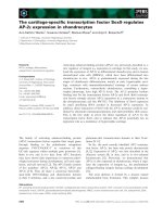
PRCs (Fig. 1A) are a major constituent of the retina.
There are two types of PRCs – rods and cones – which
are adapted to detecting different intensities of light.
Detection of light by PRCs relies on opsin proteins,
localized to the outer segments (OSs) of the cells,
which contain a covalently bound retinoid chromo-
phore [22]. The highest concentration of PRCs is
found in the macula, an oval-shaped region surround-
ing the optic nerve [23]. Loss of photoreceptors from
this region results in the loss of central vision that is
characteristic of SD [24].
Apical to the OSs of the PRCs is the retinal pigment
epithelium (RPE) (Fig. 1B). It is underpinned by a
basement membrane, the Bruch’s membrane, and a
capillary bed, which supplies oxygen and nutrients,
including the precursor of 11-cis-retinal, vitamin A, to
the retina [25]. Another vital function of the RPE is
the engulfment and digestion of old disc membranes.
As new discs bud from the PRC plasma membrane,
older discs are displaced towards the RPE and shed
for phagocytosis by the RPE cells [22]. Compounds
that cannot be digested in this way may accumulate,
either in the RPE or the Bruch’s membrane below it
[26]. These by-products of disc membrane phagocyto-
sis, including cholesterol, cholesteryl-esters and other
lipids, are collectively known as lipofuscin [26–28]. The
build-up of lipofuscin deposits, and the toxic com-
pounds within them, impair the function of the RPE
cells and prevent their metabolic support of PRCs [29].
Fig. 1. Schematic diagrams of PRCs.
ABCA4 is expressed exclusively in the disc
membranes of rod and cone PRCs (A). Villi
extending from the RPE cells intercalate
with PRCs (B).
N. L. Pollock and R. Callaghan ABCA4: seeing is believing
FEBS Journal 278 (2011) 3204–3214 ª 2011 The Authors Journal compilation ª 2011 FEBS 3205
Therefore function of the PRCs is dependent on the
RPE cells.
Trafficking and regeneration of retinoids is
essential to maintain vision
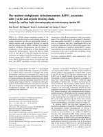
Healthy RPE cells cooperate with PRCs to recycle all-
trans-retinal (ATR) in a process known as the retinoid
cycle (Fig. 2) [25,30,31]. This cycle involves the release
of ATR, a highly reactive molecule, from rhodopsin.
The high concentration of rhodopsin in the disc mem-
branes, up to 3 mm [25], means that in conditions of
high light intensity it is possible that the rate of ATR
release may outstrip the rate of its reduction to all-
trans-retinol (ATRol), necessitating alternative means
of processing or sequestering ATR [32,33].
The aldehyde group of ATR has the potential to
create reactive oxygen species, which can initiate the
oxidation of lipids and induce apoptosis [34]. In
addition, ATR is known to react with phosphatidyleth-
anolamine (PE) to form N-retinylidene-phosphatidyl-
ethanolamine (NrPE) [35,36], which can react with a
further ATR molecule to form toxic bisretinoid com-
pounds [37,38]. The latter cannot be catabolized in the
RPE, accumulate in lipofuscin and cause degeneration
of the RPE [12,27,37,38]. Therefore it is vital for the
PRCs to process ATR as quickly as possible.
It has been suggested that each retinoid in this path-
way has a specific chaperone to prevent unwanted
reactions [25]. For instance rhodopsin has a total of
three binding sites for retinoids, allowing one 11-cis-
retinal to bind an entrance site and another to bind
the active site while ATR remains covalently bound at
an exit site, where it can be reduced to ATRol or
released [25,33,39]. The reversible formation of NrPE
allows PE in the disc membranes to act as a temporary
sink for ATR; subsequent hydrolysis enables ATR to
re-enter the retinoid cycle [40]. However, the reversible
formation of NrPe is the first step towards the forma-
tion of bisretinoids [28,38,41], which makes it a high-
risk strategy for the chaperonage of ATR and unlikely
to be a principal pathway for ATR in the retinoid
cycle. After ATR has left the OS discs, the remaining
steps of the retinoid cycle occur in the RPE cells
(Fig. 2).
Disc membrane composition modulates the
visual cycle
The lipid composition of OS discs is distinct from that
of the plasma membrane from which they are derived,
providing a highly fluid membrane environment to
enable rapid signalling from rhodopsin to the brain
[25,42,43]. Creating this distinct lipid composition
necessitates extensive sorting of phospholipids when
the discs are created, the details of which are not well
understood.
Certain phospholipids and cholesterol associate
with rhodopsin to modulate its activity [44,45],
although cholesterol is progressively lost from the
Fig. 2. Overview of the retinoid cycle. (1)
ATR moves out of the active site of rhodop-
sin into the OS disc (2), where it may be
transported into the PRC cytoplasm by
ABCA4. It is reduced to ATRol by an all-
trans-retinol dehydrogenase. (3) ATRol
moves from the OS disc of the PRC into
the RPE cell layer. (4) Lecithin retinol acyl-
transferase, (5) retinal-pigment-epithelium-
specific 65 kDa protein and (6) 11-cis-retinol
dehydrogenase regenerate ATRol into
11-cis-retinal. (7) 11-cis-retinal moves into
the OS disc, where it (8) binds to rhodopsin
for photoisomerization.
ABCA4: seeing is believing N. L. Pollock and R. Callaghan
3206 FEBS Journal 278 (2011) 3204–3214 ª 2011 The Authors Journal compilation ª 2011 FEBS
ageing OS discs [46,47]. Whether this is a symptom
or a cause of disc ageing and what significance it has
in the visual cycle is unclear. The investment in creat-
ing the unique lipid composition of the OS discs, and
the existence of other visual disorders caused by aber-
rant lipid sorting [19,48], indicate that this is a critical
process to enable vision. As ABCA4 is localized
exclusively to this membrane, it is likely that its activ-
ity is also influenced by the unique membrane envi-
ronment of the OS discs, although there is currently
no evidence for direct involvement of ABCA4 in its
creation.
Experimental evidence for the role of
ABCA4
Retinoid transport by ABCA4 was first proposed in
1997, shortly after the ABCA4 gene was identified
[11,49,50]. This hypothesis was deduced from the local-
ization of the protein to the disc membranes of PRCs,
its ability to bind ATP and its homology with the
ABC transporter family [17,51]. Studies on purified
ABCA4 have enabled this hypothesis to be tested in
some detail, while the creation of ABCA4
) ⁄ )
mice has
provided an in vivo model for macular degenerative
disorders [52–55].
Studies on knockout mice
ABCA4
) ⁄ )
mice enabled detailed characterization of
changes in the retina caused by a lack of ABCA4
activity. Electroretinography, the measurement of the
electrical response of the eye to light, and analysis of
tissue samples taken from eyes have been used to
examine the ABCA4
) ⁄ )
phenotype [52,54].
The first study on ABCA4
) ⁄ )
mice [54] reported
delayed adaptation to dark and delayed clearance of
ATR after photobleaching (the conversion of 11-cis-
retinal to ATR within rhodopsin). The levels of rho-
dopsin and 11-cis-retinal in ABCA4
) ⁄ )
mice were
similar to wild-type (WT) mice, indicating that
ABCA4 is not an essential protein in the retinoid
cycle, nor does its absence alter the availability of
rhodopsin. Rather, the accelerated accumulation of
ATR in the disc membranes of ABCA4
) ⁄ )
mice pro-
vided strong evidence that ABCA4 mediates the pro-
cessing or transport of ATR following its dissociation
from rhodopsin. ABCA4
) ⁄ )
mice also had an
increased rate of lipofuscin accumulation at their reti-
nas and the Bruch’s membrane underlying the RPE
was thicker than in their WT counterparts [54]. This
corresponds to observations of the retinas of human
subjects with retinal degenerative disorders [56],
although this seems to be occur in AMD rather than
in SD.
Finally, ABCA4
) ⁄ )
mice contained at least 10-fold
more A2E, or isoA2E, in retinal extracts than WT
mice of the same age [54]. A2E was detectable only in
RPE extracts, not at the OS discs, suggesting that A2E
was formed rapidly in the RPE of ABCA4
) ⁄ )
mice,
despite the localization of ABCA4 to the OS disc
membranes. This highlights the crux of the ABCA4
question: how is loss of ABCA4 activity in the OS
discs related to changes in the RPE cells, and how
is this effect propagated back to the PRCs to cause
macular degeneration?
Biochemical analyses of purified ABCA4
Some biochemical evidence supports the case for
ABCA4 acting as an ATP-powered retinoid trans-
porter. The first observation relating to this was the
release of ATR from purified ABCA4 upon the addi-
tion of ATP or GTP [57]. ATR appeared to remain
bound to ABCA4 during purification from rod OS
discs, but binding or hydrolysis of ATP altered the
affinity of the protein for ATR, leading to its
release.
The rate of ATP hydrolysis by purified, reconsti-
tuted ABCA4 has also been measured [58–60] to exam-
ine its physiological function. Many ABC transporters
have a background or basal rate of ATPase activity,
which is stimulated when the protein interacts with its
specific transport substrate(s) [61]. In the case of
ABCA4, ATR but no other retinoid compound was
observed to stimulate the ATPase activity of the pro-
tein to an appreciable extent, which led to the conclu-
sion that ATR could be the retinoid substrate
transported by ABCA4 in vivo [60,62]. However, the
presence of PE in the reconstituted proteoliposomes
also enhanced the basal activity of ABCA4. On this
basis, it was proposed that the substrate of ABCA4
could be NrPE, the product of an equilibrium reaction
between ATR and the amine group of PE [57,60,63].
It has also been shown that ATR can quench the
intrinsic tryptophan fluorescence of isolated WT
ECD2, suggesting that ATR binds to ABCA4 at the
ECDs [64]. A dissociation constant (K
D
) of 0.17 lm
for ATR binding to WT ECD2 was inferred from the
data. Moreover, specific mutations to the ECDs, which
are linked to SD, were shown to increase K
D
, indicat-
ing that the binding affinity was lower in the mutant
ECDs. This could account for the poor function of
some mutant forms of ABCA4, which result in the
accumulation of ATR in the OS discs and ultimately
in loss of vision.
N. L. Pollock and R. Callaghan ABCA4: seeing is believing
FEBS Journal 278 (2011) 3204–3214 ª 2011 The Authors Journal compilation ª 2011 FEBS 3207
Defining the orientation of ABCA4 in the disc mem-
brane is also fundamental to our understanding of its
activity. The current topological model [18], based on
analysis of the amino acid sequence and biochemical
data, predicts that the ECDs are located in the disc
lumen, while the NBDs reside in the OS cytoplasm
(Fig. 3A). There is good evidence to support this
model. For example, the endoplasmic reticulum (ER)
lumen and disc lumen are topologically equivalent and
the abundance of glycosylation sites in the ECDs indi-
cates that these domains are located within the ER
lumen during protein synthesis. In addition positively
charged residues at the N-terminal suggest a cytoplas-
mic localization for this region [18]. Combined with
the predicted topology of 12 transmembrane helices,
this gives us a model with the ECDs and NBDs on
opposite sides of the membrane, with the former
located within the disc lumen.
ABCA4 activity – the NrPE flippase model
Based on data from the ABCA4
) ⁄ )
mice and biochemi-
cal experiments, a hypothesis has been proposed indicat-
ing that ABCA4 acts as a transporter of NrPE [63].
Following regeneration of rhodopsin with 11-cis-retinal,
ATR is released from the ‘exit site’ of opsin into the disc
lumen, where a proportion of it reacts with a PE mole-
cule to form NrPE. The selectivity of ECD2 for ATR
suggests that the role of the ECDs is recognition of the
substrate NrPE, which is structurally related to ATR.
Following interaction with the ECDs, the substrate is
flipped or transported across the disc membrane into the
cytoplasmic leaflet, or directly into the cytoplasm.
Translocation is powered by hydrolysis of one or two
ATP molecules at the NBDs, which reside in the
cytoplasm ensuring ready provision of nucleotides. Fol-
lowing release, NrPE can be hydrolysed to PE and
ATR. At this cytosolic location the latter is more acces-
sible to retinal dehydrogenase (RDH), thereby re-form-
ing ATRol and returning to the retinoid cycle.
Loss of function of ABCA4 leads to the accumula-
tion of NrPE in the disc lumen. A subsequent reaction
between NrPE and ATR leads to the irreversible for-
mation of A2E. When discs are shed from the PRCs
and phagocytosed by the RPE cells, A2E cannot be
degraded. Instead it is deposited as lipofuscin in
Bruch’s membrane where it causes RPE cell death and
PRC degeneration, giving rise to the symptoms of SD
and other retinal degenerative disorders.
The role of ABCA4 in disc membranes:
insight or oversight?
The model described above provides a plausible expla-
nation for most of the evidence that we have about the
activity of ABCA4. However, gaps in our understand-
ing of the protein in particular, and the visual cycle in
general, pose a number of intriguing puzzles.
Is ABCA4 really a flippase?
In the absence of a direct functional assay, the fre-
quent assertion that ABCA4 acts as a flippase of NrPE
remains speculative. A number of ABC transporters
have been proposed to act as flippases [65–67], and
although in some cases, for instance the human phos-
phatidylcholine transporter and the Escherichia coli
MsbA protein [68,69], there is reasonable evidence to
support this we have yet to conclusively demonstrate
flippase activity for ABCA4.
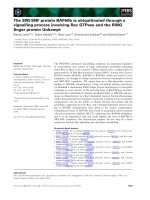
Fig. 3. Orientation of ABCA1 and ABCA4 in the membrane. ABCA4 (A) exists within the disc membranes in PRCs. The ECDs are located
within the disc lumen (L) and substrate (ATR) is hypothesized to travel (arrow A) from the lumen into the cytoplasm. ABCA1 (B) is located in
the plasma membrane and oriented with NBDs in the cytoplasm and the ECDs located extracellularly (EC). Substrates including cholesterol
(Chol) are transported (arrow B) from the cytoplasm to an extracellular acceptor (e.g. apoA-1).
ABCA4: seeing is believing N. L. Pollock and R. Callaghan
3208 FEBS Journal 278 (2011) 3204–3214 ª 2011 The Authors Journal compilation ª 2011 FEBS
Several alternative mechanisms to substrate flipping
have been proposed to describe the protein-mediated
passage of lipids across the bilayer [70]. For example
ABCA1, the protein with most homology to ABCA4,
is thought to mediate transport of cholesterol directly
from the cytoplasmic leaflet to an extracellular binding
protein (Fig. 3B). This is at odds with the classical flip-
pase activity that has been suggested for ABCA4, in
which the substrate would be flipped from the luminal
to the cytoplasmic leaflet of the membrane. Whether
two such closely related proteins could operate by dif-
ferent mechanisms remains an open question.
What is the role of the ECDs?
Accepting the flippase mechanism presents us with
another puzzle with respect to the role of the ECDs.
These domains have been shown to selectively bind
ATR in vitro [71], yet this molecule can also react with
PE to form NrPE in the luminal leaflet of the mem-
brane. If the protein acts as an NrPE flippase, it must
bind NrPE from the luminal leaflet. However, this
undermines the role of the ECDs in ATR binding, as
the ECD region of the protein is soluble and resides in
the disc lumen (Fig. 3). If ABCA4 acts as a flippase, it
would be more logical for an NrPE recognition site to
exist in the TMD of the protein. Even so, the strict
evolutionary conservation within the ECDs [72] and
the grave consequences of mutations in this region
indicate a vital functional role [8,71].
For ABCA1 there is good evidence that the ECDs
interact with lipoproteins to facilitate transport, deliv-
ering substrate from the ABC protein to the soluble
lipoproteins, apoA-1 and apoE-1 [3,70,73] These lipo-
proteins are essential for the efflux of lipids and their
assembly into high-density lipoproteins [70]. Conserved
Cys residues in ECD1 and ECD2 have been shown to
form a disulfide bridge, which is vital for apoA-1 bind-
ing and lipid unloading [74]. This highlights the ques-
tion of whether ABCA4 would behave in the opposite
way, with substrate recognition occurring at the ECDs
[71] (Fig. 3).
Phylogenetic analysis of the ABCA transporters in
Amphioxus, an organism often used as a model of
early vertebrate lineages, has uncovered a close evolu-
tionary relationship between ABCA1, ABCA7 and
ABCA4 [75]. All three are thought to derive from the
same ancestral gene through gene duplication events.
Therefore, it is logical to infer that function as well as
structure of the ECDs may be conserved between these
three proteins [76].
Furthermore, the ECDs comprise nearly 40% of the
molecular mass of ABCA4 and mutations associated
with SD map to amino acid substitutions in the ECDs,
indicating that loss of function here does affect the
function of the protein as a whole [20]. The scarcity of
experimental data describing the ECDs of ABCA4 ren-
ders this a subject for speculation. The role of these
domains requires extensive investigation in order to
fully understand the functional and mechanistic details
of ABCA4.
How significant is the activity of ABCA4 in the
retinoid cycle?
It is generally accepted that the majority of ATR is
processed back to ATRol by an RDH enzyme, possi-
bly while ATR remains bound in the ‘exit site’ of rho-
dopsin [12,33,39]. Both ATR and ATRol can diffuse
through the disc membrane [77], which enables them
to move into the PRC cytoplasm and then to the RPE
cells for conversion back to 11-cis-retinal [40,78].
Based on this ease of diffusion across the disc mem-
brane, one obvious question is whether ATR requires
a transporter at all.
Possibly, a specific transporter is required not to
facilitate pigment regeneration but to facilitate the
reversible sequestration of ATR in a less reactive form,
namely as NrPE. This seems plausible due to the rapid
diffusibility of ATR within the disc. Studies in
ABCA4
) ⁄ )
mice have also estimated that just 30% of
ATR leaves the OS discs as NrPE [25,52]. Hence some
regard ABCA4, although vital, as a minor mechanism
for ATR processing [25,52]: if the formation of NrPE
is inevitable, removing it to the cytoplasm where it
may hydrolyse back to PE and ATR could reduce the
probability of bisretinoid formation.
This would also correspond with the fact that the
pathologies connected to ABCA4 deficiency are degen-
erative. The slow decline of the PRCs and the RPE
layer would actually be the cumulative effect over
many years of relatively small A2E. Even in individu-
als with fully functional ABCA4, lipofuscin deposits
are common in later years [27].
Is disc membrane lipid homeostasis linked to the
function of ABCA4?
The unique lipid composition of OS disc membranes is
achieved by extensive redistribution of lipids after the
creation of the OS discs, but it is not yet clear why, or
even how, this is effected [22]. Flip-flop of lipids
between the leaflets of the disc membranes is rapid
[79,80] and new flippases are still being identified [81].
The similarity between ABCA4 and ABCA1 [75,82], a
cholesterol and phospholipase (PL) efflux pump,
N. L. Pollock and R. Callaghan ABCA4: seeing is believing
FEBS Journal 278 (2011) 3204–3214 ª 2011 The Authors Journal compilation ª 2011 FEBS 3209
suggests that ABCA4 could have a role in this lipid
sorting. Could ATR be exploiting the activity of
ABCA4 as a PE flippase by ‘piggy-backing’ onto the
transbilayer PE movement? Or has ABCA4 evolved
specifically to fulfil this niche role of removing ATR
from the disc lumen?
One of the major observations in ABCA4
) ⁄ )
mice
was the abnormally elevated levels of PE in the disc
membrane [54], which contained 1.6 times the amount
of PE found in WT OS discs. It was assumed that the
lack of ABCA4 disrupted NrPE transport, so the rate
of PE movement to the cytoplasm was reduced and
both this phospholipid and ATR accumulated at the
luminal leaflet of the disc membrane. In WT cells, PE
turnover by a specific phospholipase is relatively rapid
in the cytoplasmically oriented PE [54]. One hypothesis
to account for the elevated level of PE in ABCA4
) ⁄ )
mice is that, without ABCA4, PE does not reach the
cytoplasmic leaflet of the disc membrane and is
trapped in the luminal leaflet.
The functional consequences of the change in the
phospholipid composition of OS discs in ABCA4
) ⁄ )
mice are unknown. Given the sensitivity of rhodopsin
to cholesterol [44,45], it seems likely that an altered
lipid composition (i.e. increase in PE, loss of choles-
terol over time) could affect the kinetics of the visual
cycle in other ways [39,42,83], which may also affect
the kinetics of photobleach recovery.
One comparison between WT and ABCA4
) ⁄ )
mice
noted that, under conditions in which the exposure to
light of the ABCA4
) ⁄ )
mice was varied resulting in
different extents of rhodopsin photobleaching
( 1 · 10
)4
% to 30%), WT mice actually had a slower
recovery than ABCA4
) ⁄ )
mice [52].
The excess of PE in the disc membranes
of ABCA4
) ⁄ )
mice was suggested as an explanation
for this effect: the phospholipid acts as a sink for
ATR allowing more rapid dissociation from rhodop-
sin, despite the lack of functional ABCA4. Alterna-
tively, the ability of ABCA4 to bind 11-cis-retinal
[60] could lead to competition between rhodopsin
and ABCA4 for 11-cis-retinal binding. In the absence
of ABCA4 this competition would be removed,
increasing the availability of 11-cis-retinal to rhodop-
sin and increasing the speed of the photobleach
recovery.
Hence, this study suggested that the role of ABCA4
in OS discs, rather than acting as a major pathway for
ATR reprocessing, is a minor route for ATR out of
the disc membranes, with diffusion playing the signifi-
cant role. ABCA4 would be essential for the removal
of residual amounts of ATR from the OS discs [52].
The slightly reduced efficiency of rhodopsin regenera-
tion would be a small trade-off for ensuring the lon-
gevity of the PRCs.
Therefore, although ABCA4
) ⁄ )
mice have greatly
increased our understanding of the effects of ABCA4
deficiency, they have not provided conclusive evidence
for the exclusive role of ABCA4 as an NrPE trans-
porter. Rather, the implications of lipid sorting in the
discs, and the possible consequences of this, have been
highlighted by these studies.
Lipid homeostasis is clearly vitally important to
maintaining vision; lipofuscin deposits contain not
only the retinoid by-product A2E, but also lipids and
cholesterol derivatives [26–28]. In fact, there is evi-
dence that ABCA4 is not the only ABC transporter
that plays a role in lipofuscin accumulation. Polymor-
phisms in ABCA1 and its partner lipoprotein apoE-1
have recently been linked to an increased risk of AMD
[84,85] and ABCA1 is known to mediate cholesterol
efflux from lysosomes in RPE cells [38]. Inhibition of
this process by A2E has been linked to increased lipo-
fuscin deposits. This is one of the first hints of the
mechanism directly linking A2E accumulation, due to
ABCA4 dysfunction, to the lipofuscin accumulation
which causes macular degeneration.
Import or export?
Finally, perhaps the most intriguing of the conun-
drums about the activity of ABCA4 is the direction of
transport. In the NrPE flippase model of ABCA4
activity, the substrate is transported out of the disc
lumen and into the cytoplasm of the PRC [63]. In
terms of the postulated role of ABCA4 in the visual
cycle, this is a logical suggestion. However, in terms of
our understanding of the mechanism of ABC trans-
porters, this represents a huge departure from the
accepted canon. All eukaryotic ABC transporters are
thought to function in the export direction, with the
exception of Arabidopsis ABCB14, which may act as
an importer [86]. The phenotypical consequence of
deleting ABCB14 was examined in these experiments,
which is analogous to the use of ABCA4
) ⁄ )
mice.
Hence a direct observation of eukaryotic ABC-medi-
ated import has yet to be made.
Most eukaryotic ABC proteins are believed to trans-
port their substrates by an alternating access mecha-
nism [87]: the inward facing protein conformation has
a high affinity binding site to bind the substrate; the
outward facing conformation has a low affinity site,
enabling its release [88,89]. In the case of the prokary-
otic importer ABC proteins, the hypothesis of alternat-
ing access is retained but the high and low affinity
binding sites are reversed [90,91]. The closest
ABCA4: seeing is believing N. L. Pollock and R. Callaghan
3210 FEBS Journal 278 (2011) 3204–3214 ª 2011 The Authors Journal compilation ª 2011 FEBS
homologues of ABCA4 are both believed to transport
phospholipids and cholesterol by an alternating access
mechanism [4,76], while ABCA4 is proposed to act as
a flippase of retinal and PE. The similarity of its sub-
strate to those of ABCA1 and ABCA7 indicates that
the substrate binding sites of all these proteins have
features in common. If we invoke sequence homology
between ABCA1 and ABCA4 as evidence that ABCA4
has capabilities as a lipid transporter [64,92], can we
overlook the fact that transport occurs in opposite
directions?
Identifying an importer amongst the human ABC
transporters would present a major step forward in
our understanding of these proteins. For instance, it
would imply that the distinction between importers
and exporters is more subtle than we assume at pres-
ent, since ABCA1 and ABCA4 have 40% homology
yet are postulated to act in opposite directions. Phylo-
genetic analysis of the ABC transporter superfamily
indicates that importer and exporter function diverged
long before the prokaryotic ⁄ eukaryotic division [93].
The existence of a eukaryotic importer would represent
a new subclass of ABC protein.
Conclusion and perspectives
Despite having posed so many questions about our
understanding of ABCA4, it is important to emphasize
that, at present, all the evidence we have points
towards ABCA4 acting as an import-directed flippase.
None of the questions discussed above necessarily con-
tradicts this model, but we hope that they highlight
gaps in our knowledge which justify investigation.
In this review, we have attempted to describe the
biochemistry of ABCA4 in the context of the visual
cycle. Our current understanding is that ABCA4 is a
transporter of ATR or NrPE. However, its existence in
a specialized lipid environment and its close relation-
ship to other PL and cholesterol transporters also
implicate it in lipid transport. The complexity of inter-
preting recovery after photobleaching and the relative
importance of ABCA4 in ATR regeneration plus lack
of data on the role of ECDs leave some distance until
we fully understand the molecule and its role in main-
taining vision.
Purification and reconstitution of ABCA4 to estab-
lish a full functional assay must be a priority to con-
firm the substrate of this protein. With such a system
in place, one approach is to fluorescently label putative
substrates and measure translocation across a bilayer.
Though technically difficult, this is a more reliable
means of identifying the substrate of an ABC trans-
porter than simply observing stimulation in its rate of
ATP hydrolysis in the presence of the putative sub-
strate. This approach has been successful with other
ABC transporters [68,69] and hence may enable the
categorical classification of ABCA4 as an importer.
With the substrate(s) and direction of transport firmly
established, identifying the location of the binding site
and the mechanism of transport may also be more
straightforward.
References
1 Annilo T et al. (2002) Identification and characteriza-
tion of a novel ABCA subfamily member, ABCA12,
located in the lamellar ichthyosis region on 2q34. Cyto-
genet Genome Res 98, 169–176.
2 Kelsell DP et al. (2005) Mutations in ABCA12 underlie
the severe congenital skin disease harlequin ichthyosis.
Am J Hum Genet 76, 794–803.
3 Oram JF, Lawn RM, Garvin MR & Wade DP (2000)
ABCA1 is the cAMP-inducible apolipoprotein receptor
that mediates cholesterol secretion from macrophages. J
Biol Chem 275, 34508–34511.
4 Oram JF & Vaughan AM (2000) ABCA1-mediated
transport of cellular cholesterol and phospholipids to
HDL apolipoproteins. Curr Opin Lipidol 11, 253–260.
5 Rust S et al. (1999) Tangier disease is caused by muta-
tions in the gene encoding ATP-binding cassette trans-
porter 1. Nat Genet 22, 352–355.
6 Allikmets R et al. (1997) A photoreceptor cell-specific
ATP-binding transporter gene (ABCR) is mutated in
recessive Stargardt macular dystrophy. Nat Genet 15,
236–246.
7 Cremers FP et al. (1998) Autosomal recessive retinitis
pigmentosa and cone-rod dystrophy caused by splice
site mutations in the Stargardt’s disease gene ABCR.
Hum Mol Genet 7, 355–362.
8 Klevering BJ, Deutman AF, Maugeri A, Cremers FP &
Hoyng CB (2005) The spectrum of retinal phenotypes
caused by mutations in the ABCA4 gene. Graefes Arch
Clin Exp Ophthalmol 243, 90–100.
9 Martinez-Mir A, Paloma E, Allikmets R, Ayuso C, del
Rio T, Dean M, Vilageliu L, Gonzalez-Duarte R & Bal-
cells S (1998) Retinitis pigmentosa caused by a homozy-
gous mutation in the Stargardt disease gene ABCR. Nat
Genet 18, 11–12.
10 van Driel MA, Maugeri A, Klevering BJ, Hoyng CB &
Cremers FP (1998) ABCR unites what ophthalmologists
divide(s). Ophthalmic Genet 19, 117–122.
11 Allikmets R et al. (1997) Mutation of the Stargardt dis-
ease gene (ABCR) in age-related macular degeneration.
Science 277, 1805–1807.
12 Rozanowska M & Sarna T (2005) Light-induced
damage to the retina: role of rhodopsin chromophore
revisited. Photochem Photobiol 81, 1305–1330.
N. L. Pollock and R. Callaghan ABCA4: seeing is believing
FEBS Journal 278 (2011) 3204–3214 ª 2011 The Authors Journal compilation ª 2011 FEBS 3211
13 Allikmets R (1997) A photoreceptor cell-specific
ATP-binding transporter gene (ABCR) is mutated in
recessive Stargardt macular dystrophy. Nat Genet 17,
122.
14 Papermaster DS, Converse CA & Zorn M (1976) Bio-
synthetic and immunochemical characterization of large
protein in frog and cattle rod outer segment mem-
branes. Exp Eye Res 23, 105–115.
15 Papermaster DS, Schneider BG, Zorn MA & Kraehen-
buhl JP (1978) Immunocytochemical localization of a
large intrinsic membrane protein to the incisures and
margins of frog rod outer segment disks. J Cell Biol 78,
415–425.
16 Szuts EZ (1985) Light stimulates phosphorylation of
two large membrane proteins in frog photoreceptors.
Biochemistry 24, 4176–4184.
17 Illing M, Molday LL & Molday RS (1997) The 220-
kDa rim protein of retinal rod outer segments is a
member of the ABC transporter superfamily. J Biol
Chem 272, 10303–10310.
18 Bungert S, Molday LL & Molday RS (2001) Membrane
topology of the ATP binding cassette transporter
ABCR and its relationship to ABC1 and related ABCA
transporters: identification of N-linked glycosylation
sites. J Biol Chem 276, 23539–23546.
19 Molday RS & Zhang K (2010) Defective lipid trans-
port and biosynthesis in recessive and dominant
Stargardt macular degeneration. Prog Lipid Res 49,
476–492.
20 Sun H, Smallwood PM & Nathans J (2000) Biochemical
defects in ABCR protein variants associated with
human retinopathies. Nat Genet 26, 242–246.
21 Rivera A et al. (2000) A comprehensive survey of
sequence variation in the ABCA4 (ABCR) gene in Star-
gardt disease and age-related macular degeneration. Am
J Hum Genet 67, 800–813.
22 Boesze-Battaglia K & Schimmel R (1997) Cell mem-
brane lipid composition and distribution: implications
for cell function and lessons learned from photorecep-
tors and platelets. J Exp Biol 200, 2927–2936.
23 Provis JM, Penfold PL, Cornish EE, Sandercoe TM &
Madigan MC (2005) Anatomy and development of the
macula: specialisation and the vulnerability to macular
degeneration. Clin Exp Optom 88, 269–281.
24 Rotenstreich Y, Fishman GA & Anderson RJ (2003)
Visual acuity loss and clinical observations in a large
series of patients with Stargardt disease. Ophthalmology
110, 1151–1158.
25 Lamb TD & Pugh EN Jr (2004) Dark adaptation and
the retinoid cycle of vision. Prog Retin Eye Res 23 ,
307–380.
26 Holz FG, Sheraidah G, Pauleikhoff D & Bird AC
(1994) Analysis of lipid deposits extracted from human
macular and peripheral Bruch’s membrane. Arch
Ophthalmol 112, 402–406.
27 Sparrow JR & Boulton M (2005) RPE lipofuscin and
its role in retinal pathobiology. Exp Eye Res 80, 595–
606.
28 Eldred GE & Lasky MR (1993) Retinal age pigments
generated by self-assembling lysosomotropic detergents.
Nature 361, 724–726.
29 Lakkaraju A, Finnemann SC & Rodriguez-Boulan E
(2007) The lipofuscin fluorophore A2E perturbs choles-
terol metabolism in retinal pigment epithelial cells. Proc
Natl Acad Sci USA 104, 11026–11031.
30 McBee JK, Palczewski K, Baehr W & Pepperberg DR
(2001) Confronting complexity: the interlink of photo-
transduction and retinoid metabolism in the vertebrate
retina. Prog Retin Eye Res 20
, 469–529.
31 Rando RR (2001) The biochemistry of the visual cycle.
Chem Rev 101, 1881–1896.
32 Saari JC, Garwin GG, Van Hooser JP & Palczewski K
(1998) Reduction of all-trans-retinal limits regeneration
of visual pigment in mice. Vision Res 38, 1325–1333.
33 Schadel SA, Heck M, Maretzki D, Filipek S, Teller
DC, Palczewski K & Hofmann KP (2003) Ligand chan-
neling within a G-protein-coupled receptor. The entry
and exit of retinals in native opsin. J Biol Chem 278,
24896–24903.
34 Sparrow JR, Zhou J, Ben-Shabat S, Vollmer H, Itagaki
Y & Nakanishi K (2002) Involvement of oxidative
mechanisms in blue-light-induced damage to A2E-laden
RPE. Invest Ophthalmol Vis Sci 43, 1222–1227.
35 Anderson RE & Maude MB (1970) Phospholipids of
bovine outer segments. Biochemistry 9, 3624–3628.
36 Mata NL, Weng J & Travis GH (2000) Biosynthesis of
a major lipofuscin fluorophore in mice and humans
with ABCR-mediated retinal and macular degeneration.
Proc Natl Acad Sci USA 97, 7154–7159.
37 Sparrow JR, Fishkin N, Zhou J, Cai B, Jang YP,
Krane S, Itagaki Y & Nakanishi K (2003) A2E, a
byproduct of the visual cycle. Vision Res 43, 2983–
2990.
38 Sparrow JR, Wu Y, Kim CY & Zhou J (2010) Phos-
pholipid meets all-trans-retinal: the making of RPE bis-
retinoids. J Lipid Res 51, 247–261.
39 Heck M, Schadel SA, Maretzki D, Bartl FJ, Ritter E,
Palczewski K & Hofmann KP (2003) Signaling states of
rhodopsin. Formation of the storage form, metarhodop-
sin III, from active metarhodopsin II. J Biol Chem 278,
3162–3169.
40 Rattner A, Smallwood PM & Nathans J (2000) Identifi-
cation and characterization of all-trans-retinol dehydro-
genase from photoreceptor outer segments, the visual
cycle enzyme that reduces all-trans-retinal to all-trans-
retinol. J Biol Chem 275, 11034–11043.
41 Ben-Shabat S, Parish CA, Vollmer HR, Itagaki Y, Fish-
kin N, Nakanishi K & Sparrow JR (2002) Biosynthetic
studies of A2E, a major fluorophore of retinal pigment
epithelial lipofuscin. J Biol Chem 277, 7183–7190.
ABCA4: seeing is believing N. L. Pollock and R. Callaghan
3212 FEBS Journal 278 (2011) 3204–3214 ª 2011 The Authors Journal compilation ª 2011 FEBS
42 Niu SL, Mitchell DC, Lim SY, Wen ZM, Kim HY,
Salem N Jr & Litman BJ (2004) Reduced G protein-
coupled signaling efficiency in retinal rod outer seg-
ments in response to n-3 fatty acid deficiency. J Biol
Chem 279, 31098–31104.
43 Niu SL, Mitchell DC & Litman BJ (2002) Manipulation
of cholesterol levels in rod disk membranes by methyl-
beta-cyclodextrin: effects on receptor activation. J Biol
Chem 277, 20139–20145.
44 Albert AD, Boesze-Battaglia K, Paw Z, Watts A &
Epand RM (1996) Effect of cholesterol on rhodopsin
stability in disk membranes. Biochim Biophys Acta
1297, 77–82.
45 Albert AD, Young JE & Yeagle PL (1996) Rhodopsin-
cholesterol interactions in bovine rod outer segment
disk membranes. Biochim Biophys Acta 1285, 47–55.
46 Boesze-Battaglia K & Albert AD (1989) Fatty acid
composition of bovine rod outer segment plasma mem-
brane. Exp Eye Res 49, 699–701.
47 Boesze-Battaglia K, Hennessey T & Albert AD (1989)
Cholesterol heterogeneity in bovine rod outer segment
disk membranes. J Biol Chem 264, 8151–8155.
48 Boesze-Battaglia K, Organisciak DT & Albert AD
(1994) RCS rat retinal rod outer segment membranes
exhibit different cholesterol distributions than those of
normal rats. Exp Eye Res 58, 293–300.
49 Sun H & Nathans J (1997) Stargardt’s ABCR is loca-
lized to the disc membrane of retinal rod outer seg-
ments. Nat Genet 17 , 15–16.
50 Travis GH & Bennett J (1997) The ABCs of AMD. Nat
Med 3, 1196–1197.
51 Travis GH (1997) Insights from a lost visual pigment.
Nat Genet 15, 115–117.
52 Pawar AS, Qtaishat NM, Little DM & Pepperberg
DR (2008) Recovery of rod photoresponses in ABCR-
deficient mice. Invest Ophthalmol Vis Sci 49, 2743–
2755.
53 Radu RA, Yuan Q, Hu J, Peng JH, Lloyd M, Nusino-
witz S, Bok D & Travis GH (2008) Accelerated accu-
mulation of lipofuscin pigments in the RPE of a mouse
model for ABCA4-mediated retinal dystrophies follow-
ing Vitamin A supplementation. Invest Ophthalmol Vis
Sci 49, 3821–3829.
54 Weng J, Mata NL, Azarian SM, Tzekov RT, Birch DG
& Travis GH (1999) Insights into the function of Rim
protein in photoreceptors and etiology of Stargardt’s
disease from the phenotype in abcr knockout mice. Cell
98, 13–23.
55 Wu L, Nagasaki T & Sparrow JR (2010) Photoreceptor
cell degeneration in abcr (- ⁄ -) mice. Adv Exp Med Biol
664, 533–539.
56 Chong NH, Keonin J, Luthert PJ, Frennesson CI,
Weingeist DM, Wolf RL, Mullins RF & Hageman GS
(2005) Decreased thickness and integrity of the macular
elastic layer of Bruch’s membrane correspond to the
distribution of lesions associated with age-related macu-
lar degeneration. Am J Pathol 166, 241–251.
57 Beharry S, Zhong M & Molday RS (2004) N-retinyli-
dene-phosphatidylethanolamine is the preferred retinoid
substrate for the photoreceptor-specific ABC transpor-
ter ABCA4 (ABCR). J Biol Chem 279, 53972–53979.
58 Ahn J & Molday RS (2000) Purification and characteri-
zation of ABCR from bovine rod outer segments.
Methods Enzymol 315, 864–879.
59 Ahn J, Wong JT & Molday RS (2000) The effect of
lipid environment and retinoids on the ATPase activity
of ABCR, the photoreceptor ABC transporter responsi-
ble for Stargardt macular dystrophy. J Biol Chem 275,
20399–20405.
60 Sun H, Molday RS & Nathans J (1999) Retinal stimu-
lates ATP hydrolysis by purified and reconstituted
ABCR, the photoreceptor-specific ATP-binding cassette
transporter responsible for Stargardt disease. J Biol
Chem 274, 8269–8281.
61 Senior AE, al-Shawi MK & Urbatsch IL (1998) ATPase
activity of Chinese hamster P-glycoprotein. Methods
Enzymol 292, 514–523.
62 Sun H & Nathans J (2001) ABCR, the ATP-binding
cassette transporter responsible for Stargardt macular
dystrophy, is an efficient target of all-trans-retinal-
mediated photooxidative damage in vitro. Implications
for retinal disease. J Biol Chem 276, 11766–11774.
63 Molday RS, Beharry S, Ahn J & Zhong M (2006) Binding
of N-retinylidene-PE to ABCA4 and a model for its trans-
port across membranes. Adv Exp Med Biol 572, 465–470.
64 Zhong M & Molday RS (2010) Binding of retinoids to
ABCA4, the photoreceptor ABC transporter associated
with stargardt macular degeneration. Methods Mol Biol
652, 163–176.
65 Doerrler WT, Reedy MC & Raetz CR (2001) An
Escherichia coli mutant defective in lipid export. J Biol
Chem 276, 11461–11464.
66 Romsicki Y & Sharom FJ (2001) Phospholipid flippase
activity of the reconstituted P-glycoprotein multidrug
transporter. Biochemistry 40, 6937–6947.
67 Ruetz S & Gros P (1994) Phosphatidylcholine translo-
case: a physiological role for the mdr2 gene. Cell 77,
1071–1081.
68 Eckford PD & Sharom FJ (2010) The reconstituted
Escherichia coli MsbA protein displays lipid flippase
activity. Biochem J 429, 195–203.
69 Smith AJ, Timmermans-Hereijgers JL, Roelofsen B,
Wirtz KW, van Blitterswijk WJ, Smit JJ, Schinkel AH
& Borst P (1994) The human MDR3 P-glycoprotein
promotes translocation of phosphatidylcholine through
the plasma membrane of fibroblasts from transgenic
mice. FEBS Lett 354, 263–266.
70 Nagao K, Kimura Y, Mastuo M & Ueda K (2010)
Lipid outward translocation by ABC proteins. FEBS
Lett 584, 2717–2723.
N. L. Pollock and R. Callaghan ABCA4: seeing is believing
FEBS Journal 278 (2011) 3204–3214 ª 2011 The Authors Journal compilation ª 2011 FEBS 3213
71 Biswas-Fiss EE, Kurpad DS, Joshi K & Biswas SB
(2010) Interaction of extracellular domain 2 of the
human retina-specific ATP-binding cassette transporter
(ABCA4) with all-trans-retinal. J Biol Chem 285,
19372–19383.
72 Yatsenko AN, Wiszniewski W, Zaremba CM, Jamrich
M & Lupski JR (2005) Evolution of ABCA4 proteins
in vertebrates. J Mol Evol 60, 72–80.
73 Oram JF (2000) Tangier disease and ABCA1. Biochim
Biophys Acta 1529, 321–330.
74 Hozoji M, Kimura Y, Kioka N & Ueda K (2009) For-
mation of two intramolecular disulfide bonds is neces-
sary for ApoA-I-dependent cholesterol efflux mediated
by ABCA1. J Biol Chem 284, 11293–11300.
75 Li G, Zhang QJ, Ji ZL & Wang YQ (2007) Origin and
evolution of vertebrate ABCA genes: a story from
amphioxus. Gene 405, 88–95.
76 Abe-Dohmae S, Ikeda Y, Matsuo M, Hayashi M, Oku-
hira K, Ueda K & Yokoyama S (2004) Human ABCA7
supports apolipoprotein-mediated release of cellular
cholesterol and phospholipid to generate high density
lipoprotein. J Biol Chem 279, 604–611.
77 Wu Q, Chen C & Koutalos Y (2006) All-trans retinol
in rod photoreceptor outer segments moves unrestrict-
edly by passive diffusion. Biophys J 91, 4678–4689.
78 Wu Q, Blakeley LR, Cornwall MC, Crouch RK, Wig-
gert BN & Koutalos Y (2007) Interphotoreceptor reti-
noid-binding protein is the physiologically relevant
carrier that removes retinol from rod photoreceptor
outer segments. Biochemistry 46, 8669–8679.
79 Hessel E, Herrmann A, Muller P, Schnetkamp PP &
Hofmann KP (2000) The transbilayer distribution of
phospholipids in disc membranes is a dynamic equili-
brium evidence for rapid flip and flop movement. Eur J
Biochem 267, 1473–1483.
80 Wu G & Hubbell WL (1993) Phospholipid asymmetry
and transmembrane diffusion in photoreceptor disc
membranes. Biochemistry 32, 879–888.
81 Coleman JA, Kwok MC & Molday RS (2009) Localiza-
tion, purification, and functional reconstitution of the
P4-ATPase Atp8a2, a phosphatidylserine flippase in
photoreceptor disc membranes. J Biol Chem 284,
32670–32679.
82 Dean M & Annilo T (2005) Evolution of the ATP-bind-
ing cassette (ABC) transporter superfamily in verte-
brates. Annu Rev Genomics Hum Genet 6, 123–142.
83 Gibson NJ & Brown MF (1993) Lipid headgroup and
acyl chain composition modulate the MI-MII equili-
brium of rhodopsin in recombinant membranes. Bio-
chemistry 32, 2438–2454.
84 Duncan KG, Hosseini K, Bailey KR, Yang H, Lowe
RJ, Matthes MT, Kane JP, LaVail MM, Schwartz DM
& Duncan JL (2009) Expression of reverse cholesterol
transport proteins ATP-binding cassette A1 (ABCA1)
and scavenger receptor BI (SR-BI) in the retina and ret-
inal pigment epithelium. Br J Ophthalmol 93, 1116–
1120.
85 Neale BM et al. (2010) Genome-wide association study
of advanced age-related macular degeneration identifies
a role of the hepatic lipase gene (LIPC). Proc Natl Acad
Sci USA 107, 7395–7400.
86 Lee M, Choi Y, Burla B, Kim YY, Jeon B, Maeshima
M, Yoo JY, Martinoia E & Lee Y (2008) The ABC
transporter AtABCB14 is a malate importer and modu-
lates stomatal response to CO2. Nat Cell Biol 10, 1217–
1223.
87 Jardetzky O (1966) Simple allosteric model for mem-
brane pumps. Nature 211, 969–970.
88 Pinkett HW, Lee AT, Lum P, Locher KP & Rees DC
(2007) An inward-facing conformation of a putative
metal-chelate-type ABC transporter. Science 315, 373–
377.
89 Zou P & McHaourab HS (2009) Alternating access of
the putative substrate-binding chamber in the ABC
transporter MsbA. J Mol Biol 393, 574–585.
90 Oldham ML, Davidson AL & Chen J (2008) Structural
insights into ABC transporter mechanism. Curr Opin
Struct Biol 18, 726–733.
91 Kerr ID, Jones PM & George AM (2010) Multidrug
efflux pumps: the structures of prokaryotic ATP-bind-
ing cassette transporter efflux pumps and implications
for our understanding of eukaryotic P-glycoproteins
and homologues. FEBS J 277, 550–563.
92 Molday RS, Zhong M & Quazi F (2009) The role of
the photoreceptor ABC transporter ABCA4 in lipid
transport and Stargardt macular degeneration. Biochim
Biophys Acta 1791, 573–583.
93 Saurin W, Hofnung M & Dassa E (1999) Getting in or
out: early segregation between importers and exporters
in the evolution of ATP-binding cassette (ABC) trans-
porters. J Mol Evol 48, 22–41.
ABCA4: seeing is believing N. L. Pollock and R. Callaghan
3214 FEBS Journal 278 (2011) 3204–3214 ª 2011 The Authors Journal compilation ª 2011 FEBS