Báo cáo khoa học: A guide to taming a toxin – recombinant immunotoxins constructed from Pseudomonas exotoxin A for the treatment of cancer ppt
Bạn đang xem bản rút gọn của tài liệu. Xem và tải ngay bản đầy đủ của tài liệu tại đây (361.9 KB, 18 trang )
REVIEW ARTICLE
A guide to taming a toxin – recombinant immunotoxins
constructed from Pseudomonas exotoxin A for the
treatment of cancer
John E. Weldon and Ira Pastan
Laboratory of Molecular Biology, Center for Cancer Research, National Cancer Institute, National Institutes of Health, Bethesda, MD, USA
Introduction
The natural world abounds with an enormous variety
of toxins, comprising poisonous substances that are
naturally produced by living organisms [1]. Typically,
only small quantities of toxins are necessary to damage
cells, although the specific target and the toxic dose
may vary extensively. Microorganisms secrete toxins as
virulence factors during pathogenic infection, and as
secondary metabolites that can contaminate local envi-
ronments. Well known examples include diphtheria
toxin and ergot alkaloids. Higher organisms use toxins
as components in complex venoms and accumulate
them as defense factors to deter predators. Overall,
toxins can take many forms, appear in sizes ranging
from small molecules to large proteins, and have
diverse mechanisms of action, although they normally
serve similar functions related to predation and ⁄ or
defense.
Although frequently hazardous and occasionally
lethal, many toxins have the potential for therapeutic
application by removing the molecule from its natural
Keywords
antibody conjugates; cancer therapy;
intracellular trafficking; moxetumomab
pasudotox; Pseudomonas exotoxin A;
recombinant immunotoxins
Correspondence
I. Pastan, Laboratory of Molecular Biology,
National Cancer Institute, 37 Convent Drive,
Room 5106, Bethesda, MD 20892-4264,
USA
Fax: +1 301 402 1344
Tel: +1 301 496 4797
E-mail:
(Received 6 April 2011, accepted 16 May
2011)
doi:10.1111/j.1742-4658.2011.08182.x
Pseudomonas exotoxin A (PE) is a highly toxic protein secreted by the
opportunistic pathogen Pseudomonas aeruginosa. The modular structure
and corresponding mechanism of action of PE make it amenable to exten-
sive modifications that can redirect its potent cytotoxicity from disease to a
therapeutic function. In combination with a variety of artificial targeting
elements, such as receptor ligands and antibody fragments, PE becomes a
selective agent for the elimination of specific cell populations. This review
summarizes our current understanding of PE, its intoxication pathway, and
the ongoing efforts to convert this toxin into a treatment for cancer.
Abbreviations
aEF2, archaeal translation elongation factor 2; ALL, acute lymphoblastic leukemia; CE, cholera exotoxin; CT, cholera toxin; dsFv, disulfide-
stabilized variable fragment; DT, diphtheria toxin; eEF2, eukaryotic translation elongation factor 2; ER, endoplasmic reticulum; ERAD,
ER-associated degradation; Fv, variable fragment; HCL, hairy cell leukemia; IL, interleukin; KDEL-R, KDEL receptor; LRP, low density
lipoprotein receptor-related protein; PDI, protein disulfide-isomerase; PE, Pseudomonas exotoxin A; RIT, recombinant immunotoxin;
scFv, single-chain Fv.
FEBS Journal 278 (2011) 4683–4700 Journal compilation ª 2011 FEBS. No claim to original US government works 4683
context. Strategies such as altering the route of deliv-
ery, changing the dose, eliminating supporting or syn-
ergizing molecules (e.g. from a complex mixture such
as venom) or even modifying the structure of the mole-
cule may convert a dangerous toxin into a valuable
therapeutic resource. One recent example comprises
the botulinum toxins, which are potent paralytic neu-
rotoxins produced by the microbes of the Clostridium
genus, most notably Clostridium botulinum. Botulinum
toxin type A has been approved as the drug onabotuli-
numtoxinA (Botox
Ò
and Botox Cosmetic
Ò
; Allergan,
Inc., Irvine, CA, USA) for both therapeutic and cos-
metic purposes. Although the toxin has an estimated
human LD
50
of approximately 1 ngÆkg
)1
body weight
[2], the extremely low dose employed clinically and its
delivery via a site-specific injection make the agent safe
for widespread use.
Other toxins must be more heavily modified for
therapeutic purposes. Diphtheria toxin (DT) is an
extremely potent cytotoxic protein that is the primary
virulence factor secreted by the bacterium Corynebacte-
rium diphtheriae, which is the pathogen that causes the
disease diphtheria [3]. The LD
50
of diphtheria toxin in
humans has been reported as £ 100 ng kg
)1
body
weight [2], yet the toxin was converted into the first
recombinant toxin to be approved by the Food and
Drug Administration for the intravenous therapy of
cutaneous T-cell lymphoma. Denileukin diftitox (On-
tak
Ò
; Eisai Inc., Woodcliff Lake, NJ, USA) is a
recombinant form of DT that has been engineered by
replacing the native receptor-binding domain of DT
with interleukin (IL)-2. This substitution alters the tar-
get of the toxin from the membrane-associated hepa-
rin-binding epidermal-growth-factor-like growth factor
[4] to the IL-2 receptor, redirecting its potent cytotoxi-
city toward a therapeutic purpose [5,6].
A comparable strategy to alter the target of an intra-
cellular toxin has been employed for Pseudomonas exo-
toxin A (PE), a protein toxin with many similarities to
DT. PE and DT are only distantly related, although
they both belong to a class of cytotoxic proteins (i.e.
the A-B toxins) that require cellular uptake through
receptor-mediated endocytosis for activity. The overall
structure of these proteins consists of a receptor-binding
domain (B subunit) linked to a domain with cytotoxic
activity (A subunit) that is delivered to the cytosol.
Although their B subunits have very different targets,
the A subunit of both PE and DT is a NAD
+
-diphtha-
mide ADP-ribosyltransferase (EC 2.4.2.36), which tar-
gets and inactivates eukaryotic translation elongation
factor 2 (eEF2). This halts protein synthesis and eventu-
ally leads to cell death. A recently identified third mem-
ber of the NAD
+
-diphthamide ADP-ribosyltransferase
toxin subfamily, cholera exotoxin (CE, also known as
cholix toxin) from Vibrio cholerae, has extensive
sequence (36% identity, 50% similarity) and structural
(2.04 A
˚
C
a
rmsd) resemblance to PE and presumably
utilizes a similar intoxication pathway [7,8]. PE, CE,
DT and other toxins that utilize receptor-meditated
endocytosis can potentially be redirected for therapeutic
purposes by replacing their native receptor-binding
domains with other targeting elements. This review dis-
cusses our current understanding of PE intoxication
and efforts to convert PE into a viable therapeutic
agent.
PE
Pseudomonas aeruginosa is a ubiquitous, Gram-nega-
tive, aerobic bacillus that is often encountered as an
opportunistic human pathogen, although infections in
healthy individuals are rare. Approximately 10% of
hospital-acquired infections are caused by P. aerugin-
osa, and certain patient populations, such as individu-
als with cystic fibrosis or burn wounds, are especially
prone to this infection [9]. The bacterium is known to
possess a number of virulence determinants, the most
toxic of which is the protein PE [10]. Studies in mice
have identified the median lethal dose of PE as being
approximately 200 ng, and evidence suggests that PE
may play a major role in the virulence of P. aeruginosa.
Strains of P. aeruginosa deficient in PE production
are less virulent than strains producing PE, and
patients who survive infection from PE-producing
strains typically have high antibody titers against PE
[3,11].
PE (GenBank accession number AAB59097) is syn-
thesized as a single 638 residues (69 kDa) polypeptide
that is processed by the removal of a 25 residues
N-terminal sequence before secretion as the 613 resi-
dues (66 kDa) native toxin (all sequence numbering in
this review is based on the 613 residues native toxin).
The initial X-ray crystallographic structure of native
PE revealed three major structural domains [12]. The
N-terminal domain I is divided into nonsequential but
structurally adjacent domains Ia (residues 1–252) and
Ib (365–404). The residues between domains Ia and Ib
comprise domain II (253–364) and the remaining
C-terminal residues make up domain III (405–613).
Native PE contains eight cysteines that form four disul-
fide bonds in sequential order: two lie in domain Ia
(C11-C15 & C197-C214), one lies in domain II (C265-
C287) and one lies in domain Ib (C372-C379). Figure 1
illustrates the domain structure of native PE.
Functionally, domain I of PE is the receptor-binding
domain, and is the major component of the B subunit.
Cancer therapy based on Pseudomonas exotoxin A J. E. Weldon and I. Pastan
4684 FEBS Journal 278 (2011) 4683–4700 Journal compilation ª 2011 FEBS. No claim to original US government works
It targets the low density lipoprotein receptor-related
protein (LRP)1 (also known as CD91 or the a
2
-macro-
globulin receptor) or the closely-related variant LRP1B
for subsequent cellular internalization by receptor-
mediated endocytosis [14,15]. Domain III is the cata-
lytically active domain, and is the primary constituent
of the A subunit. It catalyzes the inactivation of eEF2
by transferring an ADP-ribosyl group from NAD
+
to
the diphthamide residue, a highly conserved, post-
translationally modified histidine that is unique to
eEF2. Although domain III is structurally defined by
residues 405–613 of the native toxin, full catalytic
activity requires a portion of domain Ib [16,17]. We
have defined the catalytically functional domain III as
consisting of residues 395–613 [18]. Domain II was
proposed to be involved in toxin translocation and
intracellular trafficking, although supporting evidence
for this function is not consistent.
PE-based therapeutics
PE can be converted into an agent that selectively
eliminates cells by changing its target to a different cell
surface receptor. The new target is typically specified
by attaching either an anti-receptor antibody or a
receptor ligand to PE through chemical conjugation or
recombinant protein engineering. Our laboratory has
focused efforts over many years on the generation of
PE-based recombinant immunotoxins (RITs), which
are recombinant proteins that combine antibodies with
protein toxins. Initial studies in which full-length PE
was chemically conjugated to whole mAbs or receptor
ligands [19,20] gradually gave way to the more efficient
production of recombinant molecules in which domain Ia
of PE was replaced by a ligand [21] or the variable
fragment (Fv) of a mAb [22]. Single-chain Fv (scFv)
molecules, which utilize the heavy chain (V
H
) and light
chain (V
L
) fragments of the Fv covalently connected
with a flexible polypeptide linker sequence [23,24],
were recombinantly inserted at the N-terminus of a
cytotoxic fragment of PE. To enhance the stability of
Native Pseudomonas exotoxin A (PE)
dsFv-PE38 RIT
PE38
PE[LR]
dsFv-PE[LR] RIT
Ia II IIIIb
1 613252/253
364/365
404/405
(1-250)
(365-380)
(1-273) (285-394)
PE38
F
V
PE[LR]
F
V
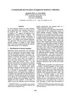
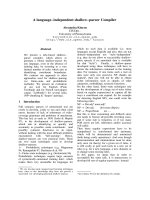
Fig. 1. PE and PE-based RITs. Native PE consists of three struc-
tural domains organized from a single polypeptide sequence.
Domain I is separated into the structurally adjacent but discontinu-
ous domain Ia (blue; residues 1–252) and domain Ib (green; 365–
404) by domain II (yellow; 253–364). Domain III (red; 405–613) lies
at the C-terminus. A cartoon model, created using
VMD [13], based
on the X-ray crystal structure of PE (Protein Data Bank code: 1IKQ)
is shown, excluding those residues absent from the electron den-
sity map (607–613). RITs based on PE are chimeric molecules that
fuse antibodies to fragments of PE, most frequently a 38 kDa
truncation known as PE38 that contains extensive deletions in
domain Ia (D1–250) and domain Ib (D365–380). Recently, a smaller
fragment, PE[LR] (D1–273 and D285–394), has been developed for
use in RITs. Structural models of RITs using a dsFv joined to PE38
or PE[LR] are presented. The Fv is shown in purple. Models are
hypothetical only and do not represent actual structural determina-
tions. The dsFv-PE38 RIT contains a gap in the structure that corre-
sponds to the deletion of residues 365–380 in domain Ib. Disulfide
bonds in PE and the Fv are shown in orange. The site of furin
cleavage is indicated with a black arrow.
J. E. Weldon and I. Pastan Cancer therapy based on Pseudomonas exotoxin A
FEBS Journal 278 (2011) 4683–4700 Journal compilation ª 2011 FEBS. No claim to original US government works 4685
recombinant immunotoxins, disulfide-stabilized Fv
(dsFv) molecules were subsequently developed. The
dsFv divides the V
H
and V
L
into separate polypeptides
that are covalently connected through a disulfide bond
engineered into the framework region of the Fv
[25–27]. A cytotoxic fragment of PE can be inserted at
the C-terminus of one of the two Fv polypeptide
chains (Fig. 1). The generation and production of
PE-based RITs has been described previously [28].
The most commonly employed cytotoxic fragment
of PE in RITs is a 38 kDa version known as PE38 [29]
(Fig. 1). PE38 contains a deletion of the majority of
domain Ia (D1–250) and a portion of domain Ib
(D365–380) from native PE. Several RITs incorporat-
ing a 38 kDa fragment of PE are in preclinical evalua-
tion or have already reached clinical trials (Table 1).
PE38 RITs undergoing preclinical testing include an
antiglycoprotein NMB (scFv) for the treatment of
malignant gliomas and melanomas [30], an anti-HIV-1
gp120 (scFv) for the treatment of HIV [31,32] and a
RIT targeted to osteosarcomas using a dsFv from the
TP-3 mAb [33,34].
RITs that have progressed to clinical trials include the
anti-CD22 RIT RFB4(dsFv)PE38, also known as BL22
or CAT-3888, for the treatment of B-cell malignancies
[35–37]. The RFB4 Fv was subsequently affinity-opti-
mized by phage display selection to create the second-
generation molecule RFB4[GTHW](dsFv)-PE38 [38],
known variously as HA22 or CAT-8015, and now called
moxetumomab pasudotox. Moxetumomab pasudotox is
currently undergoing extensive clinical testing for the
treatment of hematologic malignancies [39,40] (ongoing
studies also can be found under ClinicalTrial.gov identi-
fiers: NCT00462189, NCT00457860, NCT00515892,
NCT01086644, NCT00659425 and NCT00586924).
Other RITs from our laboratory in clinical trials include
the anti-mesothelin SS1(dsFv)PE38, called SS1P, for the
treatment of lung cancer and mesothelioma [41,42]
(ongoing studies also can be found under ClinicalTri-
al.gov identifiers: NCT01041118, NCT00575770 and
NCT01051934) and the anti-TAC(scFv)PE38, called
LMB-2, which targets the IL-2 receptor for the treat-
ment of hematologic malignancies [43] (ongoing studies
also can be found under ClinicalTrial.gov identifiers:
NCT00924170, NCT00077922, NCT00080535 and
NCT00321555). Extensive lists of PE-based therapeutics
at both the preclinical and clinical stages have been pub-
lished [44,45] and additional agents continue to be devel-
oped. We have recently generated a new variant of PE,
PE[LR] (Fig. 1), which shows decreased immunogenic-
ity and nonspecific toxicity in mice at the same time as
retaining cytotoxicity against malignant cells [46].
The strategy of re-routing A-B toxins, such as DT
and PE, through a different cellular target works well
for several reasons. The cytotoxic A domain is stable
and fully active independent of the receptor-binding B
domain, which can be replaced by a component that
confers alternate specificity, such as a ligand or an anti-
body. Additionally, the available tools for recombinant
DNA manipulation and protein expression allow us to
easily generate these chimeric molecules, and protein
engineering techniques provide powerful methods for
developing and selecting improved variants. Further-
more, we can differentiate between normal and malig-
nant cells using tumor-associated cell-surface receptors
as markers. By specifically targeting these receptors
with PE, we can eliminate cancers at the same time as
avoiding toxicities to normal tissue that are frequently
associated with general chemotherapeutic strategies.
Lastly, these proteins are extremely potent toxins that
Table 1. Several PE-based recombinant toxins currently in development for the treatment of cancers.
Agent Alternative names Target Stage of development Cancer
BL22 RFB4(dsFv)-PE38
CAT-3888
CD22 Clinical trials completed;
superseded by
moxetumomab pasudotox
B cell malignancies
Moxetumomab
pasudotox
RFB4[GTHW](dsFv)-PE38
HA22
CAT-8015
CD22 Clinical trials B cell malignancies
LMB-2 anti-TAC(scFv)-PE38 CD25 (IL-2R a chain) Clinical trials T and B cell malignancies
SS1P SS1(dsFv)-PE38 Mesothelin Clinical trials Mesothelioma, lung cancer
MR1-1 MR1-1KDEL
MR1(scFv)-PE38KDEL
Epidermal growth
factor receptor vIII
Clinical trials Brain tumors
Cervene TP-38
TGFa-PE38
Epidermal growth
factor receptor
Clinical trials Brain and central nervous
system tumors
Cintredekin besudotox IL13-PE38QQR Interleukin-13 receptor Clinical trials Glioblastoma multiforme
G49[F6V](scFv)-PE38 – Glycoprotein NMB Preclinical Glioblastoma multiforme
Cancer therapy based on Pseudomonas exotoxin A J. E. Weldon and I. Pastan
4686 FEBS Journal 278 (2011) 4683–4700 Journal compilation ª 2011 FEBS. No claim to original US government works
have been naturally selected for their ability to kill
eukaryotic cells. Their activities typically require no
major enhancement to function at a therapeutic level.
The PE intoxication pathway
A basic outline of the PE intoxication pathway is well
understood. The secreted toxin binds to an LRP1 or
LRP1B cell surface receptor, is internalized by recep-
tor-mediated endocytosis, and undergoes intracellular
trafficking to reach the cytosol. In the cytosol, PE
encounters eEF2 and transfers an ADP-ribosyl group
from NAD
+
to the diphthamide residue. This irrevers-
ibly inactivates eEF2, halts protein synthesis and, ulti-
mately, leads to cell death. A general description of
the pathway is deceptively simple, and many of the
specifics are not clear. Figure 2 attempts to presents a
comprehensive description of PE intoxication, the
details of which are discussed below. The pathway
described in Fig. 2 is not necessarily complete,
although it represents our current understanding of PE
intoxication.
PE in the endocytic pathway
Similar to DT, native PE is a secreted as a proenzyme
that must be activated before it displays catalytic
activity [47]. Full activation can be accomplished
under reducing and denaturing conditions and proteol-
ysis, and appears to involve structural rearrangements
that reveal the previously obscured NAD
+
binding
cleft in domain III [48]. RITs using versions of PE
without domain Ia do not require a structural arrange-
ment to expose the NAD
+
binding site. This differ-
ence is unlikely to affect PE intoxication in RITs,
although it does eliminate the requirement for catalytic
activation.
After endocytosis, PE undergoes an essential proteo-
lytic processing step at a cleavage site between residues
R279 and G280 of domain II [49,50]. Using
SDS ⁄ PAGE, two bands corresponding to the A and B
subunits of PE were initially observed: a 28 kDa N-ter-
minal fragment (B subunit) and a cytotoxic 37 kDa
C-terminal fragment (A subunit), which was enriched
in the cytosolic fraction of treated cells. PE that had
been mutated so that it did not undergo this processing
step failed to kill cells. Subsequent research implicated
the intracellular protease furin (EC 3.4.21.75) in this
process [51–53] and supporting evidence has accumu-
lated [54–59]. PE that is treated with furin before intox-
ication is more active than untreated PE. In addition,
PE is less active on cell lines that are furin deficient or
on cells treated with furin inhibitors.
Furin is a ubiquitous, Ca
2+
-dependent, transmem-
brane serine endoprotease that is a member of the sub-
tilisin-like family of proprotein convertases [60]. It
plays an active role in the maturation of many cellular
proteins, and its prevalence is frequently exploited by
bacterial toxins and viruses during intoxication and
infection. Furin contains a luminal catalytic domain
and a cytoplasmic domain that controls its cycling
between the trans-Golgi network and the plasma mem-
brane. PE could potentially encounter furin at either
of these sites or in the endosomal network during
intracellular trafficking between them.
In addition to furin cleavage of the PE polypeptide
backbone, separation of the A and B fragments must
be preceded by the reduction of a disulfide bond
between residues C265 and C287, which provides a
second covalent linkage. Thus, both a reduction and a
proteolysis step are necessary for PE intoxication [61].
The C265-C287 disulfide bond is buried in the crystal
structure of native PE [12] and must be exposed by
unfolding before it can be reduced [61]. This observa-
tion suggests that furin cleavage precedes reduction,
although the order of events in vivo has not been
established experimentally.
The subcellular location of the reduction event is dif-
ficult to pinpoint. The general redox state of the extra-
cellular environment is normally more oxidizing,
whereas the intracellular environment is more reducing
[62], although numerous factors can influence the redox
balance and different subcellular compartments can
have very different redox potentials. One suggestion
has been that the reduction of PE is accomplished by
protein disulfide-isomerases (PDIs; EC 5.3.4.1) because
in vitro experimental evidence suggests that PE can be
reduced by PDIs [61]. PDIs are a family of enzymes
that catalyze the formation and breakage of disulfide
bonds in proteins [63]. They are abundant not only in
the endoplasmic reticulum (ER) and Golgi, but also in
other intracellular locations and on the cell surface
[64,65]. PE could potentially encounter PDIs at every
stage of the intoxication pathway. The relative abun-
dance of PDIs in the ER, however, suggests that PE
would be more likely to encounter PDIs there.
Indirect support for the involvement of PDIs in PE
intoxication comes from the pathways of other protein
toxins. The protein toxins ricin and cholera toxin (CT)
both follow routes through the ER and into the cyto-
sol after receptor-mediated endocytosis. Evidence
obtained both in vivo and in vitro supports the involve-
ment of PDIs in a reductive separation event essential
to ricin and CT [66–70]. The PDI family of proteins
has additionally been associated with retrograde trans-
port of polypeptides from the ER in the process of
J. E. Weldon and I. Pastan Cancer therapy based on Pseudomonas exotoxin A
FEBS Journal 278 (2011) 4683–4700 Journal compilation ª 2011 FEBS. No claim to original US government works 4687
ER-associated degradation (ERAD), a mechanism
that may be exploited by PE to reach the cytosol, as
discussed below.
The precise role played by intracellular processing of
PE in its intoxication pathway is not entirely clear.
Separation of the A and B subunits serves to activate
the PE proenzyme, although RITs that do not require
activation for catalytic activity still need a cleavable
furin site for full activity (J. E. Weldon, unpublished
results). Separation of the catalytic and binding
domains may therefore serve an additional function,
perhaps by exposing sequences in domain II necessary
for intracellular trafficking. PE38 RITs retain all of
domain II, including the furin cleavage site and C265-
C287 disulfide bond (Fig. 1). Unlike native PE, how-
ever, separation of the catalytic and binding fragments
is not always essential for cytotoxicity. The RIT HA22
(anti-CD22 ⁄ PE38) remains active on CD22-positive
cells even with an R279G mutation that prevents furin
cleavage, although it is three-fold less active than wild-
type HA22 (J. E. Weldon, unpublished results). The
same R279G mutation in the RIT SS1-LR ⁄ GGS (anti-
mesothelin ⁄ PE[LR]) is completely inactive on mesoth-
elin-positive cells. Current research is exploring these
Nucleus
PE
Endoplasmic
reticulum
B
Carboxypeptidase
AB
REDLK
Furin
LRP-1/B
Sec61
A
REDL
REDL
A
B
AB
REDL
AB
REDL
Lysosome
NAD
+
eEF2
ADP-Ribose
eEF2
Extracellular
Intracellular
Early
endosome
A
REDL
Protein synthesis
Apoptosis
AB
REDL
A
B
REDL
PDI
Late
endosome
1
11
10
9
5b
7
6
5a
8
2
3
4
Clathrin-coated
Pit
Tumor-associated receptor
(e.g. CD22)
I
III
Nicotinamide
Golgi
KDEL receptor
REDL
A
B
(dsFv)-PE38 RIT
A
REDLK
II
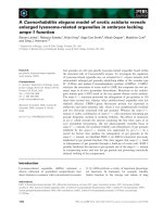
Fig. 2. PE intoxication pathway. Native PE can be divided into two fragments with functions of receptor binding (B) and catalytic activity (A).
After secretion into the extracellular environment, PE is cleaved by a carboxypeptidase (1) to remove the C-terminal lysine residue and
expose the ER localization signal (REDL). The B fragment subsequently recognizes its cell-surface receptor, LRP1 or LRP1B (2), and is inter-
nalized via receptor-mediated endocytosis in clathrin-coated pits (3). Within the endocytic pathway, PE encounters the endoprotease furin,
which cleaves at a site in domain II and separates the polypeptide backbone between the A and B fragments (4). A disulfide bond preserves
a covalent linkage between the two fragments. When in the endocytic pathway, PE can either follow a productive trafficking route to the
Golgi (5b) or continue to the lysosome for terminal degradation (5a). In the Golgi, PE encounters KDEL receptors that recognize the REDL C-
terminal signal and transport PE to the ER in a retrograde manner (6). At an undetermined point in the pathway, possibly by PDI in the ER,
the disulfide bond connecting the A and B fragments is reduced and the two fragments separate (7). The A fragment is subsequently trans-
ported into the cytosol (8), possibly by exploiting the ERAD pathway through the Sec61 translocon. In the cytosol, PE transfers an ADP-ribo-
syl (ADPr) group from NAD
+
to the diphthamide residue of eEF2 (9). This halts protein synthesis (10) and ultimately leads to apoptotic cell
death (11). RITs based on PE (I) target tumor-associated cell surface receptors for internalization (II), and are generally considered to undergo
an intoxication pathway similar to that of PE (III).
Cancer therapy based on Pseudomonas exotoxin A J. E. Weldon and I. Pastan
4688 FEBS Journal 278 (2011) 4683–4700 Journal compilation ª 2011 FEBS. No claim to original US government works
differences. Both the cell line and the target receptor
appear to play major roles in determining the outcome
of intoxication.
PE in the endoplasmic reticulum
The intoxication pathways of DT and PE are remark-
ably similar in several respects [71]. Both are secreted
as proenzymes, internalized by receptor-mediated
endocytosis, processed by furin, and reduced to sepa-
rate the catalytic (A) from the binding (B) fragments.
Subsequent to these steps, however, their respective
pathways diverge dramatically. Although DT pursues
a route directly from acidified endocytic vesicles into
the cytosol [72], PE follows a path through the ER.
The evidence for an ER-dependent PE intoxication
pathway is extensive. It was initially observed that the
R
609
EDL
612
sequence immediately adjacent to the
C-terminal residue of PE was essential for cytotoxicity
[73]. Deletions in the REDL sequence of PE eliminate
its cytotoxicity, although replacement with a similar
sequence, KDEL, restores activity. The KDEL
sequence is a well defined ER retention and retrieval
signal in mammalian cells [74] that is recognized by
integral membrane proteins known as KDEL receptors
(KDEL-R) [75,76]. The subcellular localization of
KDEL-R appears to be a dynamic cycle between the
Golgi and the ER [77,78]. This is consistent with
the proposed function of KDEL-Rs in returning to the
ER proteins that have escaped into the Golgi.
The REDL C-terminal sequence of PE, which also
occurs on several ER-resident proteins, is a variant of
the canonical KDEL sequence and is recognized and
retained in the ER by KDEL-R [79]. As anticipated,
the overexpression of KDEL-R1 (hERD2) sensitizes
cells to PE. Conversely, cells become resistant to PE
when KDEL transport is restricted by microinjected
antibodies to KDEL-R1 or by expression of lysozyme-
KDEL, which competes for binding to free receptor
[80]. Before KDEL-R can recognize PE, however, the
C-terminal residue, K613, must be removed to expose
the REDL signal sequence. Binding to KDEL-R is
seriously impaired if the terminal lysine residue is not
removed [81]. The removal of K613 appears to occur
early in the intoxication process, possibly by plasma
carboxypeptidase(s) in the bloodstream [82].
Analysis of KDEL-R binding to oligopeptides end-
ing with various sequences showed that the REDL
native sequence of PE had an almost 100-fold weaker
affinity than the canonical KDEL sequence [81]. This
result suggests that replacing the native REDL
sequence with KDEL might enhance the cytotoxicity
of PE-based RITs by increasing the efficiency of Golgi
to ER transport, and multiple studies have supported
this hypothesis [81,83]. Unfortunately, the therapeutic
benefit of enhanced cytotoxicity is offset by an accom-
panying increase in nonspecific toxicity in laboratory
animals (R. J. Kreitman, J. E. Weldon and I. Pastan,
unpublished results).
On the basis of the perturbation of different traffick-
ing pathways, it has been suggested that PE can
exploit routes to the ER other than through KDEL-R
[84]. Although alternative pathways to the ER cer-
tainly exist and are used by other toxins, most notably
a KDEL-R-independent lipid transport route used by
Shiga toxin [85,86], the evidence indicates that the vast
majority of PE reaches the ER through KDEL-R.
Deletion of the ER localization signal at the C-termi-
nus of PE reduces its activity by 1000-fold or more
[73]. Our experience with PE-based RITs has shown
that the C-terminal ER localization sequence of PE is
essential for cytotoxicity (J. E. Weldon & I. Pastan,
unpublished observations). An additional mechanism
has been suggested in which PE can translocate
directly from acidified endocytic vesicles into the cyto-
sol, using an approach similar to DT [87]. This
proposal also conflicts with the observation that the
C-terminal ER localization signal of PE is essential. It is
possible that differences between cell lines may account
for the conflicting experimental observations, and more
work needs to be carried out to clarify the matter.
An exit pathway from the ER to the cytosol is sug-
gested by the evidence for an association between PE
and the Sec61p ER translocation pore [88,89]. This
suggests that PE may be exported from the ER into
the cytosol through the Sec61p membrane channel in a
manner similar to the retrotranslocation (also know as
dislocation) of polypeptides destined for proteasomal
degradation by luminal ER-associated degradation
[90]. Presumably, this would entail a chaperone-
assisted unfolding step in the ER followed by translo-
cation and refolding in the cytosol. It is possible that
processed PE and other protein toxins such as CT and
Shiga toxin mimic the presence of a misfolded protein
in the ER to exploit the ERAD system for transport
across the ER membrane to the cytosol [91,92]. To
date, we are unaware of direct evidence for transport
of PE through the Sec61p translocon.
Additional support for the hypothesis that PE
exploits the ERAD system is the amino acid bias
against lysine residues in its catalytic fragment [93].
Sequence analyses of the catalytic (A) fragments of PE
and other protein toxins show that arginine residues
are much more highly preferred over lysine when
examining the occurrence of basic amino acids. Inter-
estingly, this paradigm does not hold true for the B
J. E. Weldon and I. Pastan Cancer therapy based on Pseudomonas exotoxin A
FEBS Journal 278 (2011) 4683–4700 Journal compilation ª 2011 FEBS. No claim to original US government works 4689
fragments, in which lysine residues occur with normal
frequency. In total, there are 15 lysine residues in
native PE but only three lysines in its A fragment (res-
idues 280–613): K590, K606 and K613. All three of
these residues are located near the C-terminus of PE,
and K613 must be removed to expose the C-terminal
REDL ER localization signal. This suggests a selective
pressure against the inclusion of lysine residues in the
protein sequence of the A fragment but not the B
fragment of PE. Because only the A fragment must
traffic to the cytosol for activity, the lack of lysine res-
idues may protect it from the ubiquitin ⁄ proteasome
system, comprising the terminal step of ERAD in
which proteins are targeted for degradation by poly-
ubiquitination of lysine e-amino groups [94]. Both
ricin and abrin toxins engineered to contain additional
lysine residues have shown enhanced ubiquitin-medi-
ated proteasomal degradation [95]. PE may similarly
lack lysine residues to avoid degradation in the cytosol
at the same time as exploiting an ERAD transport
pathway.
PE in the cytosol
Once PE reaches the cytosol, it exerts its catalytic
activity on EF2. The translation factor EF2 [96] is an
essential component of protein synthesis, during which
it catalyzes the coordinated movement of the growing
polypeptide chain along the ribosome. In eukaryotes
(eEF2) and archaea (aEF2), but not bacteria (EF2,
formerly EF-G), the protein contains a unique and rig-
idly conserved post-translationally modified histidine,
known as a diphthamide residue. The purpose of the
diphthamide residue is unclear, although it is strictly
conserved among eukaryotes and archaea. Gene
knockout studies in mice have shown that enzymes in
the diphthamide biosynthesis pathway are essential for
normal development [97,98], although it is not clear if
the diphthamide residue itself is essential. The lack of
a diphthamide did not have a significant impact on the
activity of aEF2 in vitro [99]. In addition, mammalian
and yeast cultured cells lacking the diphthamide modi-
fication on EF2 are viable and resistant to NAD
+
diphthamide ADP-ribosyltransferases, although they
may show effects such as temperature sensitivity and a
decreased growth rate [100–107]. Several hypotheses
for the necessity of the diphthamide have been
proposed, including its involvement in protection from
ribosome-inactivating proteins such as icin [108] or
preservation of translational fidelity [109], although no
consensus has been reached. The existence of bacterial
NAD
+
-diphthamide ADP-ribosyltransferases (PE, DT
and CE), however, demonstrates that bacteria have
found the diphthamide residue an appealing target to
differentiate themselves from archaea and eukaryotes.
Because the initial determination that PE halts pro-
tein synthesis in a manner identical to DT [110], the
catalytic mechanism of PE has been extensively studied
[111–117]. Several residues in domain III of PE have
been identified as playing important roles in catalysis,
including Glu553, His440, Tyr481 and Tyr470. Studies
of the reaction itself indicate that an ADP-ribosyl
group derived from NAD
+
is transferred to the N3
atom of the diphthamide imidazole using a random
third-order S
N
1 mechanism. NAD
+
is cleaved to pro-
duce nicotinamide, which is released, and an ADP-ri-
bosyl oxacarbenium ion intermediate, which contains a
positively charged ribosyl group that reacts with the
diphthamide imidazole N3 atom. The molecular mech-
anism by which the ADP-ribosylation of eEF2 halts
protein synthesis remains unclear, although it is possi-
ble that the ADP-ribose moiety interferes with an
interaction between eEF2 and RNA at the diphtha-
mide site [118].
We also do not know precisely how ADP-ribosyla-
tion of eEF2 leads to cell death, although halting
translation almost certainly leads to growth inhibition
and arrest. Studies that have examined cell death after
treatment with PE or PE-based RITs have reported
results consistent with apoptotic cell death [119–122],
although little is known about the intermediate steps
after ADP-ribosylation of eEF2 and before caspase
activation. Recently, it was reported that apoptosis
induced in mouse embryonic fibroblasts by PE or other
protein synthesis inhibitors was dependent on the
degradation of Mcl-1 and release of Bak [123]. The
anti-apoptotic protein Mcl-1 is rapidly turned over in
the cell, and inhibition of its synthesis may shift the bal-
ance of apoptotic signals towards cell death [124]. It is
possible that this mechanism could be common among
different cell types and protein synthesis inhibitors.
Unanswered questions
At this point, it should be clear that our understanding
of PE intoxication is incomplete. One important miss-
ing element is an understanding of the role of domain
II in PE intoxication. It has been suggested that
domain II assists in the translocation of the toxin into
the cytosol [16,87] and that it plays a role in proper
folding, stability and secretion by P. aeruginosa [125–
127], although there is no consensus. Domains Ia and
III have independent, experimentally verified functions
that can be directly assessed, although speculation con-
cerning the function of domain II has been made pri-
marily by inference. Domain Ib also has no
Cancer therapy based on Pseudomonas exotoxin A J. E. Weldon and I. Pastan
4690 FEBS Journal 278 (2011) 4683–4700 Journal compilation ª 2011 FEBS. No claim to original US government works
independent function but is structurally contiguous
with domain Ia, and a portion of domain Ib is func-
tionally essential to the catalytic activity domain III.
At least a portion of domain II is devoted to maintain-
ing the covalent attachments between the A and B
toxin fragments; it contains the furin protease cleavage
site and flanking cysteines (Cys265-Cys287) that form
a disulfide bond. It is unlikely, however, that the
entirety of domain II exists simply to provide a site for
the separation of the A and B fragments.
Work on PE-based RITs has shown that the major-
ity of domain II is not essential for activity, although
it can have a large influence on cytotoxicity [46].
Depending on the cells examined and the receptor
targeted, mutations that eliminate all of domain II
except for the furin cleavage site can enhance, reduce
or have no impact on cytotoxicity. Eliminating the fu-
rin cleavage site by deletion or preventing cleavage
with a point mutation in the site has either reduced
the cytotoxicity of the RIT or completely abolished it.
An explanation for these differing effects is unknown
and currently under study, although it raises the issue
that our understanding of the PE intoxication path-
way can be complicated by the use of recombinant
immunotoxins. Much of the information accumulated
over years of study concerns PE-based RITs rather
than native PE. Not only is the protein heavily modi-
fied from its native form, but also the target receptor
is changed. This could potentially influence cytotoxic-
ity in a variety of ways, from changing the number of
receptor sites per cell to altering the rate of internali-
zation of the receptor or influencing the intracellular
trafficking. The proteome of the target cell also influ-
ences the pathway. We have observed large differences
in the cytotoxicity of PE and PE-based RITs on dif-
ferent cell lines. The assumption that the route of
trafficking is conserved after internalization in differ-
ent cell lines and through different receptors is not
necessarily accurate, although our understanding of
PE trafficking is currently insufficient to make such
distinctions.
Another unanswered question concerns the fraction
of the internalized PE that productively traffics to
the cytosol. On the basis of studies on DT [128]
and unpublished data from our laboratory using PE
(I. Pastan, unpublished results), it has been proposed
that as few as one molecule of PE in the cytosol may
be sufficient to kill a cell. Typically, cells in culture
require treatment with concentrations of PE greater
than 1000 molecules per cell (approximately
10
)16
gÆcell
)1
) to ensure cell death. This number is close
to an estimate of the toxin load ⁄ cell in a mouse xeno-
graft tumor model. Tumor-bearing mice treated with a
PE-based RIT required 400–750 molecules per cell to
ensure tumor remission [129]. Taken together, these
studies suggest that less than 1% of the internalized
toxin may successfully traffic into the cytosol. The
remainder appears to follow an unproductive path into
lysosomes. This estimate agrees with observations of
cells treated with labeled PE [130,131] (J. E. Weldon,
unpublished observations). The stability of the A frag-
ment of PE in the cytosol has also not been examined,
although its relative lack of lysine residues may hamper
ubiquitination-dependent proteasomal degradation and
enhance cytosolic stability.
Clinical trials of PE-based RITs
Although no PE-based therapies have been approved
by the Food and Drug Administration, several have
reached the point of advanced clinical trials in their
development (Table 1). The examples provided in this
review do not constitute an exhaustive list. At the time
of this review, a search for ‘immunotoxin’ in the NIH
clinical trials database ()
revealed at least 16 active studies involving PE that
has been redirected to selectively eliminate cells. The
majority of these trials involve PE-based RITs devel-
oped in our laboratory, and they are discussed below.
The RIT BL22 (anti-CD22 ⁄ PE38) has undergone
several early-phase clinical trials for the treatment of B
cell malignancies [35–37]. These trials have validated
the use of CD22 as a target and highlighted several
potential problems with this treatment. BL22 was most
effective in patients with drug-resistant hairy cell leuke-
mia (HCL), whose response rates were 81% (25 ⁄ 31) in
a phase I trial [35] and 69% (25 ⁄ 36) in a phase II trial
[36]. Dose-limiting toxicity was related to a completely
reversible hemolytic uremic syndrome resulting from
the destruction of red blood cells. High levels of neu-
tralizing antibodies developed in 24% (11 ⁄ 46) of
patients in the phase I trial and 11% (4 ⁄ 36) of patients
in the phase II trial.
Clinical trials of BL22 have been superseded by
moxetumomab pasudotox, a modified RIT whose Fv
has undergone selection for enhanced CD22 affinity by
phage display [38]. As previously discussed, there are
at least six active clinical trials of moxetumomab pa-
sudotox. Preliminary results from a phase I study in
patients with relapsed or refractory HCL (trial identi-
fier NCT00462189) show a response rate of 81%
(26 ⁄ 32), even though neutralizing antibodies eventually
developed in 44% (14 ⁄ 32) of patients [132]. There is a
notable lack of dose-limiting toxicity as a result of
hemolytic uremic syndrome with moxetumomab
pasudotox, and a maximum tolerated dose has not yet
J. E. Weldon and I. Pastan Cancer therapy based on Pseudomonas exotoxin A
FEBS Journal 278 (2011) 4683–4700 Journal compilation ª 2011 FEBS. No claim to original US government works 4691
been established. An additional phase I clinical trial in
pediatric patients with acute lymphoblastic leukemia
(ALL) or non-Hodgkin’s lymphoma (trial identifier
NCT00659425) shows activity in patients with ALL
[133]. Of the ALL patients evaluated, 25% (3 ⁄ 12) had
complete responses, 50% (6 ⁄ 12) had partial responses
(hematologic activity), 17% (2 ⁄ 12) had stable disease
and 8% (1 ⁄ 12) had progressive disease. Two patients
eventually developed high levels of neutralizing anti-
bodies, and two patients developed a dose-limiting
capillary leak syndrome.
In addition to CD22, CD25 (IL-2 receptor a chain)
has been targeted for the treatment of various leuke-
mias and lymphomas. The anti-CD25 RIT LMB-2 has
undergone a phase I clinical trial [43] showing an over-
all response rate of 23% (8 ⁄ 35), and there are at least
four active clinical trials of LMB-2 (listed above).
Immunogenicity and nonspecific toxicities continue to
be problematic. Of the patients evaluated in the phase I
study, 29% (10 ⁄ 34) showed high levels of neutrali-
zing antibodies to PE38. Toxicities were reversible and
most commonly low level transaminase elevations and
mild fever. LMB-2 has also been used clinically in a par-
tially successful effort to deplete patients of CD25+
regulatory T lymphocytes and thereby enhance the
immune response to vaccination with tumor-specific
antigens [134].
Another PE-based RIT that has reached clinical trials
is the anti-mesothelin SS1P. Two phase I trials treating
patients with mesothelioma, pancreatic cancer or ovar-
ian cancer have been completed [41,42], and at least
two studies are currently active. Patient responses to
SS1P were modest, with a few minor responses. Toxici-
ties associated with treatment were typically mild.
Immunogenicity appears to constitute the major obsta-
cle to SS1P treatment. In the two studies, 88% (30 ⁄ 34)
and 75% (18 ⁄ 24) of patients developed high levels of
neutralizing antibodies to SS1P after a single cycle of
treatment. These rates were significantly higher than the
immunogenicity observed when treating hematologic
malignancies, possibly because patients with blood can-
cers have an immune system that is compromised as a
result of disease and ⁄ or previous chemotherapy. Pri-
marily as a result of the immunogenicity, very few
patients qualified to receive more than a single cycle of
treatment, which might account for the low efficacy of
SS1P. Preliminary results from a phase I clinical trial
combining SS1P with chemotherapy to treat patients
newly diagnosed with advanced-stage pleural mesotheli-
oma (trial identifier NCT00575770) show good results
[135]. SS1P is well tolerated when combined with
pemetrexed and cisplatin, and 50% (7 ⁄ 14) of patients
showed a partial response to treatment.
The future of PE-based RITs
Many obstacles have been overcome in the develop-
ment of RITs for the treatment of cancer, and striking
responses have been observed in many patients with
HCL, although several properties of RITs still need
improvement. One of the most significant problems we
have encountered in the clinical trials is immune
response leading to the generation of neutralizing anti-
bodies. Immunogenicity can be a major difficulty for
protein therapeutics, particularly those derived from
nonhuman sources [136]. For PE-based RITs, neutral-
izing antibodies are a common occurrence and com-
prise a major limitation in patients with solid tumors
who have an intact immune system. Antibody forma-
tion is much less of a barrier to treating patients with
hematologic malignancies, whose immune systems are
typically suppressed, and multiple treatment cycles can
usually be given. Mouse studies show that PE38 RITs
are no more immunogenic than most foreign proteins.
Antibody responses typically do not occur until several
weeks after the initial treatment [137–139]. Neverthe-
less, it is clear that lower immunogenicity would bene-
fit PE-based RITs. This is especially apparent with
SS1P; in approximately 80% of patients, only a single
cycle (three doses) can be administered before the
development of neutralizing antibodies.
Several strategies have been attempted to overcome
the issue of immunogenicity in PE-based RITs.
Poly(ethylene glycol)ylation is a common strategy to
reduce the immunogenicity and alter the pharmacoki-
netics of proteins [140]. We have poly(ethylene gly-
col)ylated various PE RITs [141–143] and found that
their efficacy was greatly diminished. An alternate
strategy is to treat patients with general immunosup-
pressive drugs concurrent with RIT therapy to prevent,
delay or otherwise limit the production of neutralizing
antibodies. This strategy is currently being assessed
clinically using LMB-2 in conjunction with fludarabine
and cyclophosphamide [40] (ClinicalTrials.gov study
identifier: NCT00924170), although previous attempts
to reduce immunogenicity in this manner have been
unsuccessful. Clinical trials using cyclophosphamide
[144] or cyclosporine A [145] in combination with a
ricin-based immunotoxin failed to decrease the anti-
body response. An attempt to treat patients with ritux-
imab (anti-CD20 mAb) before treatment with a PE-
based RIT also failed to suppress the antibody
response [146].
A third strategy is the elimination of immunogenic
epitopes in PE by mutation. The targeted removal of
B cell (antibody) epitopes [147,148] in PE38 has pro-
gressed the furthest [137–139,149]. This strategy has
Cancer therapy based on Pseudomonas exotoxin A J. E. Weldon and I. Pastan
4692 FEBS Journal 278 (2011) 4683–4700 Journal compilation ª 2011 FEBS. No claim to original US government works
been effective at dramatically decreasing immunogenic-
ity in mice, although much work remains to be carried
out to ensure low immunogenicity in humans. The
identification and elimination of T cell epitopes is a
complementary approach [150]. We propose that the
strategy of epitope elimination offers the best promise
for the deimmunization of PE-based RITs.
Other potential strategies to avoid immunogenicity
have a more uncertain forecast. One option is to utilize
human proteins that can act as cytotoxins because
fully human proteins should theoretically not elicit an
immune response in patients. Examples include pro-
apoptotic proteins and RNase. Although these
‘humanized’ RITs [151] are generally beyond the pur-
view of this review, the potential existence of an
endogenous eEF2 ADP-ribosyltransferase in mammals
[152–155] suggests that a human equivalent to PE may
exist. A RIT engineered out of such a protein might be
non-immunogenic, although it must first be identified
and characterized.
Strategies to enhance the efficacy of RITs might also
be practical. If only a single cycle of treatment is
required to achieve a complete tumor remission, the
appearance of neutralizing antibodies after that cycle of
treatment might be irrelevant to the outcome. One
strategy is to combine PE with other chemotherapeutic
drugs to achieve a synergistic effect without nonspecific
toxicity. Combination therapy with standard
chemotherapy drugs has shown promise in preclinical
studies of SS1P [156–158] and clinical trials are cur-
rently underway to test SS1P within a combination
therapy regimen (ClinicalTrials.gov study identifiers:
NCT01041118 and NCT00575770). Research into the
observed synergy suggests that it may be a result of
both chemotherapy-induced tumor cell depletion and
lower levels of free mesothelin shed into the extracellu-
lar space [158]. Free mesothelin competes with cell sur-
face mesothelin for SS1P, acting as an unproductive
sink for the immunotoxin [159]. Other potential combi-
nation drugs include that could act at specific stages in
the PE intoxication pathway to enhance productive
intoxication or those that can enhance the initiation of
apoptotic cell death in PE-treated cells. For example, a
pro-apoptotic BH3 domain mimetic has recently been
shown to synergistically enhance the cytotoxicity of PE-
based recombinant immunotoxins [160].
Further engineering of PE to enhance productive
intoxication might also be possible. As previously
described, the initial development of immunotoxins
utilized full-length native PE chemically conjugated to
mAbs. Improved protein engineering techniques per-
mitted the combination of PE and mAb fragments
(PE38 and Fv) into RITs, which were more efficient to
produce and highly active, with fewer side effects. Var-
iant RITs have subsequently been developed to
enhance ER trafficking by replacing the C-terminal
residues with a KDEL sequence [161] and to limit
endolysosomal proteolytic degradation by deleting pro-
tease-sensitive regions [46]. Other potential engineering
targets include the furin cleavage site of PE, which is
remarkably inefficiently cleaved by furin [162] and may
benefit from enhanced cleavage efficiency.
Additional obstacles exist outside of PE and its
intoxication pathway. Target selection and the target-
ing element are at least as important as the toxin por-
tion of RITs. The ability of a RIT to discriminate
between normal and malignant cells is fundamental to
its success, making the identification and validation of
a target the most important stage in their early devel-
opment. In addition to selectivity, factors such as
receptor site density, internalization rate and internali-
zation route can influence RIT efficacy. For example,
both CD19 and CD22 represent excellent targets for
RITs in the selective elimination of mature B cells
and associated malignancies. Although CD19 is more
heavily expressed than CD22, CD22 internalizes much
more rapidly and is a much better target for PE-based
RITs [163]. The stability of the targeting element is
also essential. The dsFv immunotoxin variants are
typically much more stable than the scFv molecules
originally developed for RITs, and are thus more clin-
ically useful [25]. A detailed discussion of receptor tar-
geting is beyond the scope of this review, although it
plays an essential role in the therapeutic efficacy of
RITs.
Concluding remarks
Substantial progress has been made in the development
of PE-based therapeutics over the past 30 years. Initial
tentative steps to transform a potent bacterial toxin
into a selective agent for the elimination of cells have
become purposeful strides to generate the immunotox-
ins of today and, we anticipate, the medicines of
tomorrow. Advances in our understanding of PE and
its intoxication pathway have fueled the translation of
basic research into clinical therapies that have the
opportunity to make a large positive impact on human
health. High expectations should be tempered by the
realization that obstacles remain to be overcome for
these RITs to achieve their maximum potential. Many
of the details of the PE intoxication process remain
uncertain and must be addressed before we can claim
success. Future advances in PE therapeutics will be
dependent on a clear and comprehensive grasp of PE
and its mechanism.
J. E. Weldon and I. Pastan Cancer therapy based on Pseudomonas exotoxin A
FEBS Journal 278 (2011) 4683–4700 Journal compilation ª 2011 FEBS. No claim to original US government works 4693
Acknowledgements
The authors would like to thank David Fitzgerald and
Dawn Walker for their helpful comments during the
preparation of this manuscript. This work is supported
in part by the Intramural Research Program of the
National Institutes of Health, National Cancer Insti-
tute, Center for Cancer Research. Work on BL22 and
moxetumomab pasudotox is supported in part by
MedImmune, LLC under a Cooperative Research and
Development Agreement.
References
1 Barlow Pugh M et al. (2006) Stedman’s Medical
Dictionary (Barlow Pugh M et al, eds), Lippincott
Williams & Wilkins, Baltimore, MD.
2 Gill DM (1982) Bacterial toxins: a table of lethal
amounts. Microbiol Rev 46, 86–94.
3 Murray PR, Rosenthal KS & Pfaller MA, eds (2009)
Medical Microbiology, 6th edn. Elsevier, Inc.,
Philadelphia, PA.
4 Naglich JG, Metherall JE, Russell DW & Eidels L
(1992) Expression cloning of a diphtheria toxin recep-
tor: identity with a heparin-binding EGF-like growth
factor precursor. Cell 69 , 1051–1061.
5 Manoukian G & Hagemeister F (2009) Denileukin
diftitox: a novel immunotoxin. Expert Opin Biol Ther
9, 1445–1451.
6 Kadin ME & Vonderheid EC (2010) Targeted therapies:
Denileukin diftitox – a step towards a ‘magic bullet’ for
CTCL. Nat Rev Clin Oncol 7, 430–432.
7 Jørgensen R, Purdy AE, Fieldhouse RJ, Kimber MS,
Bartlett DH & Merrill AR (2008) Cholix toxin, a novel
ADP-ribosylating factor from Vibrio cholerae. J Biol
Chem 283, 10671–10678.
8 Sarnovsky R, Tendler T, Makowski M, Kiley M,
Antignani A, Traini R, Zhang J, Hassan R &
FitzGerald DJ (2010) Initial characterization of an
immunotoxin constructed from domains II and III of
cholera exotoxin. Cancer Immunol Immunother 59,
737–746.
9 Driscoll JA, Brody SL & Kollef MH (2007) The epide-
miology, pathogenesis and treatment of Pseudomonas
aeruginosa infections. Drugs 67, 351–368.
10 Liu PV (1966) The roles of various fractions of Pseu-
domonas aeruginosa in its pathogenesis. 3. Identity of
the lethal toxins produced in vitro and in vivo. J Infect
Dis 116, 481–489.
11 Iglewski BH (1996) Pseudomonas. In Medical Microbi-
ology, 4th edn (Baron S ed), 27. University of Texas
Medical Branch at Galveston, Galveston, TX. http://
www.ncbi.nlm.nih.gov/books/NBK7627/
12 Allured VS, Collier RJ, Carroll SF & McKay DB
(1986) Structure of exotoxin A of Pseudomonas
aeruginosa at 3.0-angstrom resolution. Proc Natl Acad
Sci USA 83, 1320–1324.
13 Humphrey W, Dalke A & Schulten K (1996) VMD –
visual molecular dynamics. J Mol Graph 14, 33–38.
14 Kounnas MZ, Morris RE, Thompson MR, FitzGerald
DJ, Strickland DK & Saelinger CB (1992) The alpha
2-macroglobulin receptor ⁄ low density lipoprotein
receptor-related protein binds and internalizes Pseudo-
monas exotoxin A. J Biol Chem 267, 12420–12423.
15 Pastrana DV, Hanson AJ, Knisely J, Bu G & Fitzger-
ald DJ (2005) LRP 1 B functions as a receptor for
Pseudomonas exotoxin. Biochim Biophys Acta 1741,
234–239.
16 Hwang J, Fitzgerald DJ, Adhya S & Pastan I (1987)
Functional domains of Pseudomonas exotoxin identi-
fied by deletion analysis of the gene expressed in
E. coli. Cell 48, 129–136.
17 Siegall CB, Chaudhary VK, FitzGerald DJ & Pastan I
(1989) Functional analysis of domains II, Ib, and III
of Pseudomonas exotoxin. J Biol Chem 264 , 14256–
14261.
18 Kihara A & Pastan I (1994) Analysis of sequences
required for the cytotoxic action of a chimeric toxin
composed of Pseudomonas exotoxin and transforming
growth factor alpha. Bioconjug Chem 5, 532–538.
19 FitzGerald DJ, Padmanabhan R, Pastan I &
Willingham MC (1983) Adenovirus-induced release of
epidermal growth factor and Pseudomonas toxin into
the cytosol of KB cells during receptor-mediated
endocytosis. Cell 32, 607–617.
20 FitzGerald DJ, Trowbridge IS, Pastan I &
Willingham MC (1983) Enhancement of toxicity of
antitransferrin receptor antibody-Pseudomonas
exotoxin conjugates by adenovirus. Proc Natl Acad
Sci USA 80, 4134–4138.
21 Chaudhary VK, FitzGerald DJ, Adhya S & Pastan I
(1987) Activity of a recombinant fusion protein
between transforming growth factor type alpha and
Pseudomonas toxin. Proc Natl Acad Sci USA 84, 4538–
4542.
22 Chaudhary VK, Queen C, Junghans RP, Waldmann
TA, FitzGerald DJ & Pastan I (1989) A recombinant
immunotoxin consisting of two antibody variable
domains fused to Pseudomonas exotoxin. Nature 339,
394–397.
23 Bird RE, Hardman KD, Jacobson JW, Johnson S,
Kaufman BM, Lee SM, Lee T, Pope SH, Riordan GS
& Whitlow M (1988) Single-chain antigen-binding
proteins. Science 242, 423–426 (Erratum: 1989, Science
244, 409).
24 Huston JS, Levinson D, Mudgett-Hunter M, Tai MS,
Novotny´ J, Margolies MN, Ridge RJ, Bruccoleri RE,
Haber E, Crea R et al. (1988) Protein engineering of
antibody binding sites: recovery of specific activity in
an anti-digoxin single-chain Fv analogue produced in
Cancer therapy based on Pseudomonas exotoxin A J. E. Weldon and I. Pastan
4694 FEBS Journal 278 (2011) 4683–4700 Journal compilation ª 2011 FEBS. No claim to original US government works
Escherichia coli. Proc Natl Acad Sci USA 85, 5879–
5883.
25 Brinkmann U, Reiter Y, Jung SH, Lee B & Pastan I
(1993) A recombinant immunotoxin containing a
disulfide-stabilized Fv fragment. Proc Natl Acad Sci
USA 90, 7538–7542.
26 Reiter Y, Brinkmann U, Kreitman RJ, Jung SH, Lee
B & Pastan I (1994) Stabilization of the Fv fragments
in recombinant immunotoxins by disulfide bonds engi-
neered into conserved framework regions. Biochemistry
33, 5451–5459.
27 Reiter Y, Brinkmann U, Webber KO, Jung SH, Lee B
& Pastan I (1994) Engineering interchain disulfide
bonds into conserved framework regions of Fv
fragments: improved biochemical characteristics of
recombinant immunotoxins containing disulfide-stabi-
lized Fv. Protein Eng 7, 697–704.
28 Pastan I, Beers R & Bera TK (2004) Recombinant im-
munotoxins in the treatment of cancer. Methods Mol
Biol 248, 503–518.
29 Kreitman RJ, Siegall CB, Chaudhary VK, FitzGerald
DJ & Pastan I (1992) Properties of chimeric toxins
with two recognition domains: interleukin 6 and trans-
forming growth factor alpha at different locations in
Pseudomonas exotoxin. Bioconjug Chem 3, 63–68.
30 Kuan CT, Wakiya K, Keir ST, Li J, Herndon JE II,
Pastan I & Bigner DD (2010) Affinity-matured anti-
glycoprotein NMB recombinant immunotoxins target-
ing malignant gliomas and melanomas. Int J Cancer
129, 111–121.
31 Berger EA & Pastan I (2010) Immunotoxin comple-
mentation of HAART to deplete persisting HIV-
infected cell reservoirs. PLoS Pathog 6, e1000803.
32 Kennedy PE, Bera TK, Wang QC, Gallo M, Wagner
W, Lewis MG, Berger EA & Pastan I (2006) Anti-
HIV-1 immunotoxin 3B3(Fv)-PE38: enhanced potency
against clinical isolates in human PBMCs and macro-
phages, and negligible hepatotoxicity in macaques.
J Leukoc Biol 2006, 80.
33 Onda M, Olafsen T, Tsutsumi Y, Bruland OS &
Pastan I (2001) Cytotoxicity of antiosteosarcoma
recombinant immunotoxins composed of TP-3 Fv
fragments and a truncated Pseudomonas exotoxin A.
J Immunother 24, 144–150.
34 Onda M, Bruland ØS & Pastan I (2005) TP-3 immu-
notoxins improve antitumor activity in mice with oste-
osarcoma. Clin Orthop Relat Res 430, 142–148.
35 Kreitman RJ, Squires DR, Stetler-Stevenson M, Noel
P, FitzGerald DJ, Wilson WH & Pastan I (2005) Phase
I trial of recombinant immunotoxin RFB4(dsFv)-PE38
(BL22) in patients with B-cell malignancies. J Clin
Oncol 23, 6719–6729.
36 Kreitman RJ, Stetler-Stevenson M, Margulies I, Noel
P, Fitzgerald DJ, Wilson WH & Pastan I (2009) Phase
II trial of recombinant immunotoxin RFB4(dsFv)-
PE38 (BL22) in patients with hairy cell leukemia.
J Clin Oncol 27, 2983–2990.
37 Wayne AS, Kreitman RJ, Findley HW, Lew G,
Delbrook C, Steinberg SM, Stetler-Stevenson M,
Fitzgerald DJ & Pastan I (2010) Anti-CD22 immuno-
toxin RFB4(dsFv)-PE38 (BL22) for CD22-positive
hematologic malignancies of childhood: preclinical
studies and phase I clinical trial. Clin Cancer Res 16,
1894–1903.
38 Salvatore G, Beers R, Margulies I, Kreitman RJ &
Pastan I (2002) Improved cytotoxic activity toward cell
lines and fresh leukemia cells of a mutant anti-CD22
immunotoxin obtained by antibody phage display. Clin
Cancer Res 8, 995–1002.
39 Alderson RF, Kreitman RJ, Chen T, Yeung P, Herbst
R, Fox JA & Pastan I (2009) CAT-8015: a second-gen-
eration Pseudomonas exotoxin A-based immunother-
apy targeting CD22-expressing hematologic
malignancies. Clin Cancer Res 15, 832–839.
40 Kreitman RJ (2009) Recombinant immunotoxins for
the treatment of chemoresistant hematologic malignan-
cies. Curr Pharm Des
15, 2652–2664.
41 Hassan R, Bullock S, Premkumar A, Kreitman RJ,
Kindler H, Willingham MC & Pastan I (2007) Phase I
study of SS1P, a recombinant anti-mesothelin
immunotoxin given as a bolus I.V. infusion to patients
with mesothelin-expressing mesothelioma, ovarian, and
pancreatic cancers. Clin Cancer Res 13, 5144–5149.
42 Kreitman RJ, Hassan R, Fitzgerald DJ & Pastan I
(2009) Phase I trial of continuous infusion anti-mes-
othelin recombinant immunotoxin SS1P. Clin Cancer
Res 15, 5274–5279.
43 Kreitman RJ, Wilson WH, White JD, Stetler-Steven-
son M, Jaffe ES, Giardina S, Waldmann TA & Pastan
I (2000) Phase I trial of recombinant immunotoxin
anti-Tac(Fv)-PE38 (LMB-2) in patients with hemato-
logic malignancies. J Clin Oncol 18, 1622–1636.
44 Wolf P & Elsa
¨
sser-Beile U (2009) Pseudomonas exo-
toxin A: from virulence factor to anti-cancer agent. Int
J Med Microbiol 299 , 161–176.
45 Shapira A & Benhar I (2010) Toxin-based therapeutic
approaches. Toxins 2, 2519–2583.
46 Weldon JE, Xiang L, Chertov O, Margulies I,
Kreitman RJ, Fitzgerald DJ & Pastan I (2009) A pro-
tease-resistant immunotoxin against CD22 with greatly
increased activity against CLL and diminished animal
toxicity. Blood 113, 3792–3800.
47 Leppla SH, Martin OC & Muehl LA (1978) The exo-
toxin P. aeruginosa: a proenzyme having an unusual
mode of activation. Biochem Biophys Res Commun 81,
532–538.
48 Wedekind JE, Trame CB, Dorywalska M, Koehl P,
Raschke TM, McKee M, FitzGerald D, Collier RJ &
McKay DB (2001) Refined crystallographic structure
of Pseudomonas aeruginosa exotoxin A and its
J. E. Weldon and I. Pastan Cancer therapy based on Pseudomonas exotoxin A
FEBS Journal 278 (2011) 4683–4700 Journal compilation ª 2011 FEBS. No claim to original US government works 4695
implications for the molecular mechanism of toxicity.
J Mol Biol 314, 823–837.
49 Ogata M, Chaudhary VK, Pastan I & FitzGerald DJ
(1990) Processing of Pseudomonas exotoxin by a cellu-
lar protease results in the generation of a 37,000-Da
toxin fragment that is translocated to the cytosol.
J Biol Chem 265, 20678–20685.
50 Ogata M, Fryling CM, Pastan I & FitzGerald DJ
(1992) Cell-mediated cleavage of Pseudomonas exo-
toxin between Arg279 and Gly280 generates the enzy-
matically active fragment which translocates to the
cytosol. J Biol Chem 267, 25396–25401.
51 Fryling C, Ogata M & FitzGerald D (1992) Character-
ization of a cellular protease that cleaves Pseudomonas
exotoxin. Infect Immun 60 , 497–502.
52 Moehring JM, Inocencio NM, Robertson BJ &
Moehring TJ (1993) Expression of mouse furin in a
Chinese hamster cell resistant to Pseudomonas exotoxin
A and viruses complements the genetic lesion. J Biol
Chem 268, 2590–2594.
53 Chiron MF, Fryling CM & FitzGerald DJ (1994)
Cleavage of Pseudomonas exotoxin and diphtheria
toxin by a furin-like enzyme prepared from beef liver.
J Biol Chem 269, 18167–18176.
54 Inocencio NM, Moehring JM & Moehring TJ (1994)
Furin activates Pseudomonas exotoxin A by specific
cleavage in vivo and in vitro. J Biol Chem 269, 31831–
31835.
55 Gordon VM, Klimpel KR, Arora N, Henderson MA
& Leppla SH (1995) Proteolytic activation of bacte-
rial toxins by eukaryotic cells is performed by furin
and by additional cellular proteases. Infect Immun 63,
82–87.
56 Gu M, Gordon VM, Fitzgerald DJ & Leppla SH (1996)
Furin regulates both the activation of Pseudomonas exo-
toxin A and the quantity of the toxin receptor expressed
on target cells. Infect Immun 64, 524–527.
57 Sarac MS, Cameron A & Lindberg I (2002) The furin
inhibitor hexa-D-arginine blocks the activation of
Pseudomonas aeruginosa exotoxin A in vivo. Infect
Immun 70, 7136–7139.
58 Shiryaev SA, Remacle AG, Ratnikov BI, Nelson NA,
Savinov AY, Wei G, Bottini M, Rega MF, Parent A,
Desjardins R et al. (2007) Targeting host cell furin
proprotein convertases as a therapeutic strategy
against bacterial toxins and viral pathogens. J Biol
Chem 282, 20847–20853.
59 Ornatowski W, Poschet JF, Perkett E, Taylor-Cousar
JL & Deretic V (2007) Elevated furin levels in human
cystic fibrosis cells result in hypersusceptibility to
exotoxin A-induced cytotoxicity. J Clin Invest 117,
3489–3497.
60 Thomas G (2002) Furin at the cutting edge: from
protein traffic to embryogenesis and disease. Nat Rev
Mol Cell Biol 3
, 753–766.
61 McKee ML & FitzGerald DJ (1999) Reduction of
furin-nicked Pseudomonas exotoxin A: an unfolding
story. Biochemistry 38, 16507–16513.
62 Chaiswing L & Oberley TD (2010) Extracellu-
lar ⁄ microenvironmental redox state. Antioxid Redox
Signal 13, 449–465.
63 Appenzeller-Herzog C & Ellgaard L (2008) The human
PDI family: versatility packed into a single fold.
Biochim Biophys Acta 1783, 535–548.
64 Turano C, Coppari S, Altieri F & Ferraro A (2002)
Proteins of the PDI family: unpredicted non-ER
locations and functions. J Cell Physiol 193, 154–163.
65 Kimura T, Horibe T, Sakamoto C, Shitara Y,
Fujiwara F, Komiya T, Yamamoto A, Hayano T,
Takahashi N & Kikuchi M (2008) Evidence for
mitochondrial localization of P5, a member of the
protein disulphide isomerase family. J Biochem 144,
187–196.
66 Orlandi PA (1997) Protein-disulfide isomerase-medi-
ated reduction of the A subunit of cholera toxin in a
human intestinal cell line. J Biol Chem 272, 4591–4599.
67 Majoul I, Ferrari D & So
¨
ling HD (1997) Reduction of
protein disulfide bonds in an oxidizing environment.
The disulfide bridge of cholera toxin A-subunit is
reduced in the endoplasmic reticulum. FEBS Lett 401,
104–108.
68 Tsai B, Rodighiero C, Lencer WI & Rapoport TA
(2001) Protein disulfide isomerase acts as a redox-
dependent chaperone to unfold cholera toxin. Cell 104,
937–948.
69 Spooner RA, Watson PD, Marsden CJ, Smith DC,
Moore KA, Cook JP, Lord JM & Roberts LM (2004)
Protein disulphide-isomerase reduces ricin to its A and
B chains in the endoplasmic reticulum. Biochem J 383,
285–293.
70 Moore P, Bernardi KM & Tsai B (2010) The Ero1al-
pha-PDI redox cycle regulates retro-translocation of
cholera toxin. Mol Biol Cell 21, 1305–1313.
71 Watson P & Spooner RA (2010) Toxin entry and
trafficking in mammalian cells. Adv Drug Deliv Rev 58,
1581–1596.
72 Beaumelle B, Bensammar L & Bienvenu
¨
e A (1992)
Selective translocation of the A chain of diphtheria
toxin across the membrane of purified endosomes.
J Biol Chem 267, 11525–11531.
73 Chaudhary VK, Jinno Y, FitzGerald D & Pastan I
(1990) Pseudomonas exotoxin contains a specific
sequence at the carboxyl terminus that is required
for cytotoxicity. Proc Natl Acad Sci USA 87, 308–
312.
74 Munro S & Pelham HR (1987) A C-terminal signal
prevents secretion of luminal ER proteins. Cell 48,
899–907.
75 Lewis MJ & Pelham HR (1990) A human homologue
of the yeast HDEL receptor. Nature 348, 162–163.
Cancer therapy based on Pseudomonas exotoxin A J. E. Weldon and I. Pastan
4696 FEBS Journal 278 (2011) 4683–4700 Journal compilation ª 2011 FEBS. No claim to original US government works
76 Capitani M & Sallese M (2009) The KDEL receptor:
new functions for an old protein. FEBS Lett 583,
3863–3871.
77 Tang BL, Wong SH, Qi XL, Low SH & Hong W
(1993) Molecular cloning, characterization, subcellular
localization and dynamics of p23, the mammalian
KDEL receptor. J Cell Biol 120, 325–338.
78 Griffiths G, Ericsson M, Krijnse-Locker J, Nilsson T,
Goud B, Soling HD, Tang BL, Wong SH & Hong W
(1994) Localization of the Lys, Asp, Glu, Leu tetra-
peptide receptor to the Golgi complex and the interme-
diate compartment in mammalian cells. J Cell Biol
127, 1557–1574.
79 Raykhel I, Alanen H, Salo K, Jurvansuu J, Nguyen
VD, Latva-Ranta M & Ruddock L (2007) A molecular
specificity code for the three mammalian KDEL recep-
tors. J Cell Biol 179, 1193–1204.
80 Jackson ME, Simpson JC, Girod A, Pepperkok R,
Roberts LM & Lord JM (1999) The KDEL retrieval
system is exploited by Pseudomonas exotoxin A, but
not by Shiga-like toxin-1, during retrograde transport
from the Golgi complex to the endoplasmic reticulum.
J Cell Sci 112, 467–475.
81 Kreitman RJ & Pastan I (1995) Importance of the glu-
tamate residue of KDEL in increasing the cytotoxicity
of Pseudomonas exotoxin derivatives and for increased
binding to the KDEL receptor. Biochem J 307, 29–37.
82 Hessler JL & Kreitman RJ (1997) An early step in
Pseudomonas exotoxin action is removal of the termi-
nal lysine residue, which allows binding to the KDEL
receptor. Biochemistry 36, 14577–14582.
83 Seetharam S, Chaudhary VK, FitzGerald D & Pastan
I (1991) Increased cytotoxic activity of Pseudomonas
exotoxin and two chimeric toxins ending in KDEL.
J Biol Chem 266, 17376–17381.
84 Smith DC, Spooner RA, Watson PD, Murray JL,
Hodge TW, Amessou M, Johannes L, Lord JM &
Roberts LM (2006) Internalized Pseudomonas exotoxin
A can exploit multiple pathways to reach the endoplas-
mic reticulum. Traffic 7, 379–393.
85 Mallard F, Antony C, Tenza D, Salamero J, Goud B
& Johannes L (1998) Direct pathway from early ⁄
recycling endosomes to the Golgi apparatus revealed
through the study of shiga toxin B-fragment transport.
J Cell Biol 143, 973–990.
86 Girod A, Storrie B, Simpson JC, Johannes L, Goud B,
Roberts LM, Lord JM, Nilsson T & Pepperkok R
(1999) Evidence for a COP-I-independent transport
route from the Golgi complex to the endoplasmic retic-
ulum. Nat Cell Biol 1, 423–430.
87 Me
´
re
´
J, Morlon-Guyot J, Bonhoure A, Chiche L &
Beaumelle B (2005) Acid-triggered membrane insertion
of Pseudomonas exotoxin A involves an original
mechanism based on pH-regulated tryptophan
exposure. J Biol Chem 280, 21194–21201.
88 Koopmann JO, Albring J, Hu
¨
ter E, Bulbuc N, Spee P,
Neefjes J, Ha
¨
mmerling GJ & Momburg F (2000) Export
of antigenic peptides from the endoplasmic reticulum
intersects with retrograde protein translocation through
the Sec61p channel. Immunity 13, 117–127.
89 Wirth A, Jung M, Bies C, Frien M, Tyedmers J,
Zimmermann R & Wagner R (2003) The Sec61p
complex is a dynamic precursor activated channel. Mol
Cell 12, 261–268.
90 Willer M, Forte GM & Stirling CJ (2008) Sec61p is
required for ERAD-L: genetic dissection of the trans-
location and ERAD-L functions of Sec61p using novel
derivatives of CPY. J Biol Chem 283, 33883–33888.
91 Hazes B & Read RJ (1997) Accumulating evidence
suggests that several AB-toxins subvert the endoplas-
mic reticulum-associated protein degradation pathway
to enter target cells. Biochemistry 36, 11051–11054.
92 Lord JM, Roberts LM & Lencer WI (2005) Entry of
protein toxins into mammalian cells by crossing the
endoplasmic reticulum membrane: co-opting basic
mechanisms of endoplasmic reticulum-associated deg-
radation. Curr Top Microbiol Immunol 300, 149–168.
93 London E & Luongo CL (1989) Domain-specific bias
in arginine ⁄ lysine usage by protein toxins. Biochem
Biophys Res Commun 160, 333–339.
94 Hershko A & Ciechanover A (1998) The ubiquitin
system. Annu Rev Biochem 67, 425–479.
95 Deeks ED, Cook JP, Day PJ, Smith DC, Roberts LM
& Lord JM (2002) The low lysine content of ricin A
chain reduces the risk of proteolytic degradation after
translocation from the endoplasmic reticulum to the
cytosol. Biochemistry 41, 3405–3413.
96 Merrick WC & Nyborg J (2000) Protein bioynthesis
elongation cycle. In Translational Control of Gene
Expression (Sonenberg N, Hershey JWB & Mathews
MB eds), pp. 89–126. Cold Spring Harbor Laboratory
Press, Cold Spring Harbor, NY.
97 Liu S, Wiggins JF, Sreenath T, Kulkarni AB, Ward
JM & Leppla SH (2006) Dph3, a small protein
required for diphthamide biosynthesis, is essential in
mouse development. Mol Cell Biol 26, 3835–3841.
98 Webb TR, Cross SH, McKie L, Edgar R, Vizor L,
Harrison J, Peters J & Jackson IJ (2008) Diphthamide
modification of eEF2 requires a J-domain protein and
is essential for normal development. J Cell Sci 121,
3140–3145.
99 De Vendittis E, Amatruda MR, Raimo G & Bocchini
V (1997) Heterologous expression in Escherichia coli of
the gene encoding an archaeal thermoacidophilic
elongation factor 2. Properties of the recombinant
protein. Biochimie 79, 303–308.
100 Foley BT, Moehring JM & Moehring TJ (1992) A
mutation in codon 717 of the CHO-K1 elongation fac-
tor 2 gene prevents the first step in the biosynthesis of
diphthamide. Somat Cell Mol Genet 18, 227–231.
J. E. Weldon and I. Pastan Cancer therapy based on Pseudomonas exotoxin A
FEBS Journal 278 (2011) 4683–4700 Journal compilation ª 2011 FEBS. No claim to original US government works 4697
101 Foley BT, Moehring JM & Moehring TJ (1995)
Mutations in the elongation factor 2 gene which confer
resistance to diphtheria toxin and Pseudomonas exo-
toxin A. Genetic and biochemical analyses. J Biol
Chem 270, 23218–23225.
102 Kimata Y, Harashima S & Kohno K (1993) Expres-
sion of non-ADP-ribosylatable, diphtheria toxin-resis-
tant elongation factor 2 in Saccharomyces cerevisiae.
Biochem Biophys Res Commun 191, 1145–1151.
103 Phan LD, Perentesis JP & Bodley JW (1993) Saccharo-
myces cerevisiae elongation factor 2. Mutagenesis of
the histidine precursor of diphthamide yields a
functional protein that is resistant to diphtheria toxin.
J Biol Chem 268, 8665–8668.
104 Kimata Y & Kohno K (1994) Elongation factor 2
mutants deficient in diphthamide formation show tem-
perature-sensitive cell growth. J Biol Chem 269, 13497–
13501.
105 Liu S, Milne GT, Kuremsky JG, Fink GR & Leppla
SH (2004) Identification of the proteins required for
biosynthesis of diphthamide, the target of bacterial
ADP-ribosylating toxins on translation elongation
factor 2. Mol Cell Biol 24, 9487–9497.
106 Ivankovic M, Rubelj I, Matulic M, Reich E & Brdar
B (2006) Site-specific mutagenesis of the histidine pre-
cursor of diphthamide in the human elongation factor-
2 gene confers resistance to diphtheria toxin. Mutat
Res 609, 34–42.
107 Roy V, Ghani K & Caruso M (2010) A dominant-
negative approach that prevents diphthamide forma-
tion confers resistance to Pseudomonas exotoxin A and
diphtheria toxin. PLoS ONE 5, e15753.
108 Gupta PK, Liu S, Batavia MP & Leppla SH (2008)
The diphthamide modification on elongation factor-2
renders mammalian cells resistant to ricin. Cell
Microbiol 10, 1687–1694.
109 Ortiz PA, Ulloque R, Kihara GK, Zheng H & Kinzy
TG (2006) Translation elongation factor 2 anticodon
mimicry domain mutants affect fidelity and diphtheria
toxin resistance. J Biol Chem 281, 32639–32648.
110 Iglewski BH & Kabat D (1975) NAD-dependent
inhibition of protein synthesis by Pseudomonas
aeruginosa toxin. Proc Natl Acad Sci USA 72, 2284–
2288.
111 Van Ness BG, Howard JB & Bodley JW (1980) ADP-
ribosylation of elongation factor 2 by diphtheria toxin.
NMR spectra and proposed structures of ribosyl-
diphthamide and its hydrolysis products. J Biol Chem
255, 10710–10716.
112 Wilson BA & Collier RJ (1992) Diphtheria toxin and
Pseudomonas aeruginosa exotoxin A: active-site
structure and enzymic mechanism. Curr Top Microbiol
Immunol 175, 27–41.
113 Beattie BK, Prentice GA & Merrill AR (1996)
Investigation into the catalytic role for the tryptophan
residues within domain III of Pseudomonas aeruginosa
exotoxin A. Biochemistry 35, 15134–15142.
114 Armstrong S, Yates SP & Merrill AR (2002) Insight
into the catalytic mechanism of Pseudomonas aerugin-
osa exotoxin A. Studies of toxin interaction with
eukaryotic elongation factor-2. J Biol Chem 277,
46669–46675.
115 Armstrong S & Merrill AR (2004) Toward the
elucidation of the catalytic mechanism of the mono-
ADP-ribosyltransferase activity of Pseudomonas
aeruginosa exotoxin A. Biochemistry 43, 183–194.
116 Jørgensen R, Merrill AR, Yates SP, Marquez VE,
Schwan AL, Boesen T & Andersen GR (2005)
Exotoxin A-eEF2 complex structure indicates ADP
ribosylation by ribosome mimicry. Nature 436, 979–
984.
117 Jørgensen R, Wang Y, Visschedyk D & Merrill AR
(2008) The nature and character of the transition state
for the ADP-ribosyltransferase reaction. EMBO Rep 9,
802–809.
118 Jørgensen R, Yates SP, Teal DJ, Nilsson J, Prentice
GA, Merrill AR & Andersen GR (2004) Crystal
structure of ADP-ribosylated ribosomal translocase
from Saccharomyces cerevisiae. J Biol Chem 279,
45919–45925.
119 Decker T, Oelsner M, Kreitman RJ, Salvatore G,
Wang QC, Pastan I, Peschel C & Licht T (2004)
Induction of caspase-dependent programmed cell death
in B-cell chronic lymphocytic leukemia by anti-CD22
immunotoxins. Blood 103, 2718–2726.
120 Sharma AK & FitzGerald D (2010) Pseudomonas
exotoxin kills Drosophila S2 cells via apoptosis.
Toxicon 56, 1025–1034.
121 Bogner C, Dechow T, Ringshausen I, Wagner M,
Oelsner M, Lutzny G, Licht T, Peschel C, Pastan I,
Kreitman RJ et al. (2010) Immunotoxin BL22 induces
apoptosis in mantle cell lymphoma (MCL) cells depen-
dent on Bcl-2 expression. Br J Haematol 148, 99–109.
122 Mussai F, Campana D, Bhojwani D, Stetler-Stevenson
M, Steinberg SM, Wayne AS & Pastan I (2010) Cyto-
toxicity of the anti-CD22 immunotoxin HA22 (CAT-
8015) against paediatric acute lymphoblastic leukae-
mia. Br J Haematol 150, 352–358.
123 Du X, Youle RJ, FitzGerald DJ & Pastan I (2010)
Pseudomonas exotoxin A-mediated apoptosis is Bak
dependent and preceded by the degradation of Mcl-1.
Mol Cell Biol 30, 3444–3452.
124 Adams KW & Cooper GM (2007) Rapid turnover of
mcl-1 couples translation to cell survival and apopto-
sis. J Biol Chem 282, 6192–6200.
125 Chaudhary VK, Xu YH, FitzGerald D, Adhya S &
Pastan I (1988) Role of domain II of Pseudomonas
exotoxin in the secretion of proteins into the periplasm
and medium by Escherichia coli. Proc Natl Acad Sci
USA 85, 2939–2943.
Cancer therapy based on Pseudomonas exotoxin A J. E. Weldon and I. Pastan
4698 FEBS Journal 278 (2011) 4683–4700 Journal compilation ª 2011 FEBS. No claim to original US government works
126 Kasturi S, Kihara A, FitzGerald D & Pastan I (1992)
Alanine scanning mutagenesis identifies surface amino
acids on domain II of Pseudomonas exotoxin required
for cytotoxicity, proper folding, and secretion into
periplasm. J Biol Chem 267, 23427–23433.
127 Voulhoux R, Taupiac MP, Czjzek M, Beaumelle B &
Filloux A (2000) Influence of deletions within
domain II of exotoxin A on its extracellular secretion
from Pseudomonas aeruginosa. J Bacteriol 182,
4051–4058.
128 Yamaizumi M, Mekada E, Uchida T & Okada Y (1978)
One molecule of diphtheria toxin fragment A introduced
into a cell can kill the cell. Cell 15, 245–250.
129 Kreitman RJ & Pastan I (1998) Accumulation of a
recombinant immunotoxin in a tumor in vivo: fewer
than 1000 molecules per cell are sufficient for complete
responses. Cancer Res 58, 968–975.
130 Fitzgerald D (1996) Why toxins! Semin Cancer Biol 7,
87–95.
131 Bard F, Mazelin L, Pe
´
choux-Longin C, Malhotra V &
Jurdic P (2003) Src regulates Golgi structure and
KDEL receptor-dependent retrograde transport to the
endoplasmic reticulum. J Biol Chem 278, 46601–46606.
132 Kreitman RJ, Tallman MS, Coutre S, Robak T,
Wilson WH, Stetler-Stevenson M, Noel P, Fitzgerald
DJ, Buzoinu M, Lechleider RJ et al. (2010) A phase I
study of Moxetumomab pasudotox, an anti-CD22
recombinant immunotoxin, in relapsed ⁄ refractory
hairy cell leukemia (HCL): updated results. American
Society of Hematology (ASH) annual meeting abstract.
4–7 December 2010. Orlando, FL.
133 Wayne AS, Bhojwani D, Richards K, Stetler-Steven-
son M, Silverman LB, Jeha S, Pui C, McDevitt J, Fitz-
gerald DJ, Kreitman RJ et al. (2011) A phase I study
of Moxetumomab pasudotox, an anti-CD22 recombi-
nant immunotoxin, in patients with pediatric acute
lymphoblastic leukemia (ALL): updated results. Amer-
ican Society of Pediatric Hematology ⁄ Oncology (AS-
PHO) annual meeting abstract. 13–16 April 2011.
Baltimore MD.
134 Powell DJ Jr, Felipe-Silva A, Merino MJ,
Ahmadzadeh M, Allen T, Levy C, White DE,
Mavroukakis S, Kreitman RJ, Rosenberg SA et al.
(2007) Administration of a CD25-directed immunotox-
in, LMB-2, to patients with metastatic melanoma
induces a selective partial reduction in regulatory T
cells in vivo. J Immunol 179, 4919–4928.
135 Hassan R, Sharon E, Schuler B, Mallory Y, Zhang J,
Ling A & Pastan I (2011) Anti-tumor activity of SS1P
with pemetrexed and cisplatin for newly diagnosed
patients with advanced pleural mesothelioma and util-
ity of serum mesothelin as a marker of tumor
response. American Society of Clinical Oncology
(ASCO) annual meeting abstract. 3–7 June 2011.
Chicago, IL.
136 De Groot AS & Scott DW (2007) Immunogenicity of
protein therapeutics. Trends Immunol 28, 482–490.
137 Onda M, Beers R, Xiang L, Nagata S, Wang QC &
Pastan I (2008) An immunotoxin with greatly reduced
immunogenicity by identification and removal of B cell
epitopes. Proc Natl Acad Sci USA 105 , 11311–11316.
138 Hansen JK, Weldon JE, Xiang L, Beers R, Onda M &
Pastan I (2010) A recombinant immunotoxin targeting
CD22 with low immunogenicity, low nonspecific toxic-
ity, and high antitumor activity in mice. J Immunother
33, 297–304.
139 Onda M, Beers R, Xiang L, Lee B, Weldon JE, Kreit-
man RJ & Pastan I (2011) A recombinant immunotox-
in against B cell malignancies with no immunogenicity
in mice by removal of B cell epitopes. Proc Natl Acad
Sci USA 108, 5742–5747.
140 Jevsevar S, Kunstelj M & Porekar VG (2010) PEGyla-
tion of therapeutic proteins. Biotechnol J 5, 113–128.
141 Benhar I, Wang QC, FitzGerald D & Pastan I (1994)
Pseudomonas exotoxin A mutants. Replacement of sur-
face-exposed residues in domain III with cysteine resi-
dues that can be modified with polyethylene glycol in a
site-specific manner. J Biol Chem 269
, 13398–13404.
142 Tsutsumi Y, Onda M, Nagata S, Lee B, Kreitman RJ
& Pastan I (2000) Site-specific chemical modification
with polyethylene glycol of recombinant immunotoxin
anti-Tac(Fv)-PE38 (LMB-2) improves antitumor activ-
ity and reduces animal toxicity and immunogenicity.
Proc Natl Acad Sci USA 97, 8548–8553.
143 Filpula D, Yang K, Basu A, Hassan R, Xiang L,
Zhang Z, Wang M, Wang QC, Ho M, Beers R et al.
(2007) Releasable PEGylation of mesothelin targeted
immunotoxin SS1P achieves single dosage complete
regression of a human carcinoma in mice. Bioconjug
Chem 18, 773–784.
144 Oratz R, Speyer JL, Wernz JC, Hochster H, Meyers
M, Mischak R & Spitler LE (1990) Antimelanoma
monoclonal antibody-ricin A chain immunoconjugate
(XMMME-001-RTA) plus cyclophosphamide in the
treatment of metastatic malignant melanoma: results
of a phase II trial. J Biol Response Mod 9, 345–354.
145 Selvaggi K, Saria EA, Schwartz R, Vlock DR,
Ackerman S, Wedel N, Kirkwood JM, Jones H &
Ernstoff MS (1993) Phase I ⁄ II study of murine mono-
clonal antibody-ricin A chain (XOMAZYME-Mel) im-
munoconjugate plus cyclosporine A in patients with
metastatic melanoma. J Immunother Emphasis Tumor
Immunol 13, 201–207.
146 Hassan R, Williams-Gould J, Watson T, Pai-Scherf L
& Pastan I (2004) Pretreatment with rituximab does
not inhibit the human immune response against the
immunogenic protein LMB-1. Clin Cancer Res 10, 16–
18.
147 Onda M (2009) Reducing the immunogenicity of pro-
tein therapeutics. Curr Drug Targets 10, 131–139.
J. E. Weldon and I. Pastan Cancer therapy based on Pseudomonas exotoxin A
FEBS Journal 278 (2011) 4683–4700 Journal compilation ª 2011 FEBS. No claim to original US government works 4699
148 Nagata S & Pastan I (2009) Removal of B cell epitopes
as a practical approach for reducing the immunogenic-
ity of foreign protein-based therapeutics. Adv Drug
Deliv Rev 61, 977–985.
149 Onda M, Nagata S, FitzGerald DJ, Beers R, Fisher
RJ, Vincent JJ, Lee B, Nakamura M, Hwang J,
Kreitman RJ et al. (2006) Characterization of the B
cell epitopes associated with a truncated form of Pseu-
domonas exotoxin (PE38) used to make immunotoxins
for the treatment of cancer patients. J Immunol 177,
8822–8834.
150 Yeung VP, Chang J, Miller J, Barnett C, Stickler M &
Harding FA (2004) Elimination of an immunodomi-
nant CD4+ T cell epitope in human IFN-beta does
not result in an in vivo response directed at the subdo-
minant epitope. J Immunol 172, 6658–6665.
151 Mathew M & Verma RS (2009) Humanized immuno-
toxins: a new generation of immunotoxins for targeted
cancer therapy. Cancer Sci 100, 1359–1365.
152 Sitikov AS, Davydova EK & Ovchinnikov LP (1984)
Endogenous ADP-ribosylation of elongation factor 2
in polyribosome fraction of rabbit reticulocytes. FEBS
Lett 176, 261–263.
153 Fendrick JL, Iglewski WJ, Moehring JM & Moehring
TJ (1992) Characterization of the endogenous ADP-ri-
bosylation of wild-type and mutant elongation factor 2
in eukaryotic cells. Eur J Biochem 205, 25–31.
154 Bektas¸ M, Akc¸ akaya H, Aroymak A, Nurten R &
Bermek E (2005) Effect of oxidative stress on in vivo
ADP-ribosylation of eukaryotic elongation factor 2.
Int J Biochem Cell Biol 37, 91–99.
155 Ja
¨
ger D, Werdan K & Mu
¨
ller-Werdan U (2011)
Endogenous ADP-ribosylation of elongation factor-2
by interleukin-1b. Mol Cell Biochem 348, 125–128.
156 Hassan R, Broaddus VC, Wilson S, Liewehr DJ &
Zhang J (2007) Anti-mesothelin immunotoxin SS1P in
combination with gemcitabine results in increased
activity against mesothelin-expressing tumor xeno-
grafts. Clin Cancer Res 13, 7166–7171.
157 Zhang Y, Xiang L, Hassan R, Paik CH, Carrasquillo
JA, Jang BS, Le N, Ho M & Pastan I (2006) Syner-
gistic antitumor activity of taxol and immunotoxin
SS1P in tumor-bearing mice. Clin Cancer Res 12,
4695–4701.
158 Zhang Y, Hansen JK, Xiang L, Kawa S, Onda M, Ho
M, Hassan R & Pastan I (2010) A flow cytometry
method to quantitate internalized immunotoxins shows
that taxol synergistically increases cellular immunotox-
ins uptake. Cancer Res 70, 1082–1089.
159 Zhang Y & Pastan I (2008) High shed antigen levels
within tumors: an additional barrier to immunoconju-
gate therapy. Clin Cancer Res 14, 7981–7986.
160 Traini R, Ben-Josef G, Pastrana DV, Moskatel E,
Sharma AK, Antignani A & Fitzgerald DJ (2010)
ABT-737 overcomes resistance to immunotoxin-medi-
ated apoptosis and enhances the delivery of Pseudomo-
nas exotoxin-based proteins to the cell cytosol. Mol
Cancer Ther 9, 2007–2015.
161 Kreitman RJ, Schneider WP, Queen C, Tsudo M,
Fitzgerald DJ, Waldmann TA & Pastan I (1992)
Mik-beta 1(Fv)-PE40, a recombinant immunotoxin
cytotoxic toward cells bearing the beta-chain of the
IL-2 receptor. J Immunol 149, 2810–2815.
162 Izidoro MA, Gouvea IE, Santos JA, Assis DM,
Oliveira V, Judice WA, Juliano MA, Lindberg I & Ju-
liano L (2009) A study of human furin specificity using
synthetic peptides derived from natural substrates, and
effects of potassium ions. Arch Biochem Biophys 487,
105–114.
163 Du X, Beers R, Fitzgerald DJ & Pastan I (2008)
Differential cellular internalization of anti-CD19 and -
CD22 immunotoxins results in different cytotoxic
activity. Cancer Res 68, 6300–6305.
Cancer therapy based on Pseudomonas exotoxin A J. E. Weldon and I. Pastan
4700 FEBS Journal 278 (2011) 4683–4700 Journal compilation ª 2011 FEBS. No claim to original US government works