Báo cáo khoa học: Molecular structures and functional relationships in clostridial neurotoxins potx
Bạn đang xem bản rút gọn của tài liệu. Xem và tải ngay bản đầy đủ của tài liệu tại đây (1.33 MB, 19 trang )
REVIEW ARTICLE
Molecular structures and functional relationships in
clostridial neurotoxins
Subramanyam Swaminathan
Department of Biology, Brookhaven National Laboratory, Upton, NY, USA
Introduction
Clostridium botulinum, an anaerobic bacterium, pro-
duces seven antigenically distinct neurotoxins com-
monly called botulinum neurotoxins (BoNT, A–G) [1].
These neurotoxins are among the most poisonous
known and have an LD
50
of 1–5 ngÆkg
)1
weight of
humans [2]. Botulinum neurotoxins are closely related
to tetanus neurotoxins (TeNT) produced by Clostri-
dium tetani. However, their sites of action and pharma-
cological effects are different [3,4]. BoNTs cause
flaccid paralysis by inhibiting acetycholine release at
Keywords
botulinum neurotoxin; botulism; catalytic
activity; drug discovery; neuroexocytosis;
structure–function; substrate–enzyme
complex; tetanus; translocation; X-ray
crystallography; zinc endopeptidase
Correspondence
S. Swaminathan, Biology Department,
Brookhaven National Laboratory, Upton,
NY 11973, USA
Fax: 1 631 344 3407
Tel: 1 631 344 3187
E-mail:
(Received 28 February 2011, revised 11
May 2011, accepted 17 May 2011)
doi:10.1111/j.1742-4658.2011.08183.x
The seven serotypes of Clostridium botulinum neurotoxins (A–G) are the
deadliest poison known to humans. They share significant sequence homo-
logy and hence possess similar structure–function relationships. Botulinum
neurotoxins (BoNT) act via a four-step mechanism, viz., binding and inter-
nalization to neuronal cells, translocation of the catalytic domain into the
cytosol and finally cleavage of one of the three soluble N-ethylmaleimide-
sensitive factor attachment protein receptors (SNARE) causing blockage of
neurotransmitter release leading to flaccid paralysis. Crystal structures of
three holotoxins, BoNT ⁄ A, B and E, are available to date. Although the
individual domains are remarkably similar, their domain organization is dif-
ferent. These structures have helped in correlating the structural and func-
tional domains. This has led to the determination of structures of individual
domains and combinations of them. Crystal structures of catalytic domains
of all serotypes and several binding domains are now available. The cata-
lytic domains are zinc endopeptidases and share significant sequence and
structural homology. The active site architecture and the catalytic mecha-
nism are similar although the binding mode of individual substrates may be
different, dictating substrate specificity and peptide cleavage selectivity.
Crystal structures of catalytic domains with substrate peptides provide clues
to specificity and selectivity unique to BoNTs. Crystal structures of the
receptor domain in complex with ganglioside or the protein receptor have
provided information about the binding of botulinum neurotoxin to the
neuronal cell. An overview of the structure–function relationship correlating
the 3D structures with biochemical and biophysical data and how they can
be used for structure-based drug discovery is presented here.
Abbreviations
BoNT, botulinum neurotoxin; SNAP-25, synaptosome-associated protein 25 kDa; SNARE complex, soluble N-ethylmaleimide-sensitive factor
attachment protein receptors; SV, synaptic vesicle; Syt, synaptotagmin; TeNT, tetanus neurotoxin; VAMP, vesicle-associated membrane
protein.
FEBS Journal 278 (2011) 4467–4485 Journal compilation ª 2011 FEBS. No claim to original US government works 4467
the neuromuscular junction, whereas TeNT blocks the
release of c-aminobutyric acid and glycine at the inhib-
itory neurons of the spinal chord causing spastic paral-
ysis. BoNTs are produced as single 150 kDa
polypeptide chains and are proteolytically cleaved
before release into two chains, a heavy chain (HC) of
100 kDa and a light chain (LC) of 50 kDa, linked by a
single disulfide bond [5]. Most of the BoNTs are
released as dichains, cleaved by either endogenous or
exogenous proteases. In general, dichain BoNTs are
more potent than single-chain BoNTs. BoNT ⁄ A, B, E
and possibly F are the common source of human
infection [6]. BoNT ⁄ C and BoNT ⁄ D are responsible
for avian and animal infections [7,8].
BoNTs consist of three functional domains. The
HC is made up of two distinct domains, the N-termi-
nal (H
N
) and C-terminal (H
C
) domains, of almost
equal molecular mass. H
C
consists of two subdo-
mains, the N-terminal half, H
CN
and the C-terminal
half H
CC
, each of 25 kDa. BoNTs toxicity is via a
four-step process, binding and internalization to neu-
ronal cell, translocation of the LC into cytosol and
finally the cleavage of one the soluble N-ethylmalei-
mide-sensitive factor attachment protein receptor
(SNARE) proteins at specific sites [9]. The H
C
, and
especially the H
CC
subdomain, is responsible for bind-
ing to presynaptic neuronal target cells, H
N
, for
translocation of the catalytic domain (LC) into the
cytosol. The catalytic domain is a zinc endopeptidase
and possesses a conserved zinc-binding HExxH+E
motif in all BoNTs. Each BoNT has a specific target
in the SNARE complex and cleaves a specific peptide
bond. BoNT⁄ A and E cleave synaptosome-associated
25 kDa protein (SNAP-25) at a specific peptide bond.
BoNT ⁄ B, D, F and G (and also TeNT) cleave
vesicle-associated membrane protein (VAMP), also
known as synaptobrevin. BoNT ⁄ C is unique in that it
cleaves both SNAP-25 and syntaxin [1]. Large sub-
strate peptides and specific scissile bonds are unique
to BoNTs.
The crystal structures of holotoxins BoNT ⁄ A, B
and E have been determined and have given insight
into the function and mechanism of each domain
involved in the four-step process [10–12]. BoNTs share
significant sequence homology [13] and the structures
were expected to be similar. Indeed, individual
domains are similar although E differs from A and B
in the domain organization. The crystal structures of
individual domains and their complexes with substrates
or binding partners give information to analyze and
understand the structure–function relationships. This
review deals with the structure–function relationship of
each individual domain as well as the holotoxin.
Crystal structures of BoNT
⁄
A and B
Crystal structures of BoNT ⁄ A and B have been deter-
mined [11,12]. BoNT ⁄ A and B share significant
sequence homology (39% identity and 56% similarity)
[13] resulting in structural similarity. Because A and B
have similar folds, we describe BoNT ⁄ Basitisa
higher resolution structure (1.8 A
˚
). BoNT ⁄ B consists
of three distinct structural domains corresponding to
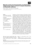
catalytic, translocation and binding domains (Fig. 1).
The catalytic domain (LC) has an a ⁄ b fold, the trans-
location domain (H
N
) is mostly helical with two long
helices ( 100 A
˚
long) forming a coiled-coil. A large
loop, corresponding to residues 481–532 and called the
belt region, wraps around the catalytic domain. This
region corresponds to the translocation domain in the
primary sequence although it is closely associated with
the catalytic domain in three dimensions and is an
intriguing feature unique to BoNTs. The binding
domain consists of two subdomains, H
CN
and H
CC
.
H
CN
consists of a 14-stranded b-barrel in a jelly-
roll motif, commonly associated with lectin-binding
4411290
852
TDBD
S
0441
CD
S
A
B
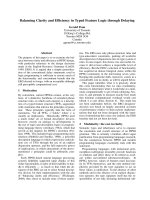
Fig. 1. Clostridium botulinum neurotoxin type B. (A) Linear repre-
sentation of BoNT ⁄ B with the individual domains colored as in (B).
The interchain disulfide bond is also marked. (B) Ribbon representa-
tion of BoNT ⁄ B. BoNT ⁄ A and BoNT ⁄ B are similar in fold and
domain organization. The three functional domains, receptor binding
(BD), translocation (TD) and catalytic (CD) domains are represented
in orange, green and red, respectively. Zinc is shown as cyan ball.
The belt region (also in green) wraps around CD. The N- and C-ter-
minals are marked.
Molecular structures of Clostridial neurotoxins S. Swaminathan
4468 FEBS Journal 278 (2011) 4467–4485 Journal compilation ª 2011 FEBS. No claim to original US government works
proteins [14]. The H
CC
domain is mostly made up of
loops and b strands with a b-trefoil fold [15]. The three
domains are arranged in a linear fashion with the
translocation domain in the middle. The binding
domain is tilted away from the translocation domain
and has only limited interactions with it. The catalytic
domain, which is on the other side of the translocation
domain, is closely associated with it. In BoNT ⁄ AorB
the binding domain and catalytic domains have virtu-
ally no contact. The catalytic zinc is located deep
inside a wide cavity that is partly covered by the belt
region. The cavity in BoNT ⁄ B is wider than that in
BoNT ⁄ A. In the following sections, individual domains
and their functions, as elicited from the 3D structure
and corroborated by biochemical and biophysical
results (or vice versa), are discussed.
Receptor-binding domain
The first step in botulinum toxicity is for the toxin to
bind to the presynaptic membrane of the neuronal tar-
get cell for uptake into neuronal cell. As early as the
1980s, it was shown that the H
C
domain is involved in
neuronal cell binding [16,17]. Also, neuraminidase-trea-
ted cultured cells had reduced affinity for BoNT ⁄ A
and bovine chromaffin cells lacking in polysialogan-
gliosides became sensitive to BoNT ⁄ A when pretreated
with gangliosides [18–20]. Taken together, it was clear
that H
C
domains of BoNTs bind to the neuronal cell
via gangliosides. The presynaptic cell surface is rich in
gangliosides which first bind to the toxin and then
accumulate them on the neuronal surface. Gangliosides
are low-affinity but highly abundant lipids with com-
plex sugar molecules as head groups. Botulinum neu-
rotoxins in general bind to GT1b, GD1b and GD1a
which contain charged sialic acids [21–23].
As described earlier, the receptor-binding domain
consists of two subdomains (Fig. 2). The N-terminal
(H
CN
) and the C-terminal (H
CC
) domains comprising a
jelly-roll motif and a b-trefoil fold, respectively, are
connected by a short helix [14,15]. Binding domains of
all botulinum and tetanus toxins share a similar fold,
even though the sequence identity of the C-terminal
domain is low. The variation in sequence is reflected in
the length of the connecting loops. The function of the
N-terminal domain (H
CN
) is not yet understood. Even
though it has a carbohydrate-binding fold there is no
evidence that it binds to any ganglioside sugar group.
However, recent evidence has shown that BoNT ⁄ A
H
CN
interacts weakly with phosphatidylionositol
phosphates [24].
Mutations in the C-terminal half of tetanus-binding
domain affect ganglioside binding and the 34 residues
(1281–1314) at the C-terminus are enough for ganglio-
side binding in TeNT [25,26]. Photoaffinity labeling
occurred predominantly at His1292 of TeNT and tryp-
tophan fluorescence quenching experiments on ganglio-
side binding implicated tryptophans at the C-terminus
in ganglioside binding [27]. This biochemical evidence
established the importance of H
C
for ganglioside bind-
ing. Crystal structures of BoNTs and TeNT with com-
plex sugars and GT1b analogs have confirmed this and
have mapped the GT1b-binding pocket [12,28,29].
Whereas a single ganglioside-binding site was observed
in BoNT ⁄ B, structure determination of TeNT with
sugars showed two binding sites (Site 1 and Site 2) for
TeNT. Also, when an analog of GT1b was used in
cocrystallization, the branched sugar molecule cross-
linked two TeNT molecules via the two sites.
The crystal structure of BoNT ⁄ B in complex with
sialyllactose identified the ganglioside-binding site in
BoNT ⁄ B [12]. Sialyllactose is a partial mimic of
GT1b and occupies a pocket in the H
CC
domain
(Fig. 3A). The pocket is formed by residues His1240,
Ser1259, Trp1261 and Tyr1262 of the conserved motif
H
CN
H
CC
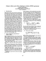
Fig. 2. The receptor binding domain of BoNT ⁄ B. The H
CN
domain
has lectin binding motif and the H
CC
domain contains a b-trefoil fold
which provides binding pockets for the receptors.
S. Swaminathan Molecular structures of Clostridial neurotoxins
FEBS Journal 278 (2011) 4467–4485 Journal compilation ª 2011 FEBS. No claim to original US government works 4469
H…SxWY…G in BoNTs. Residues Glu1188 and
Glu1189 also take part in forming the binding pocket.
The sialic acid sits between His1240 and Trp1261. The
stacking interaction between Trp1261 and the sialic
acid seems to be critical for strong binding. The trisac-
charide molecule forms an extensive hydrogen-bonding
network with the residues forming the pocket. The res-
idues forming this pocket are structurally and sequen-
tially similar in BoNT ⁄ A and TeNT suggesting that
the ganglioside-binding site will be similar in these tox-
ins. This pocket is called Site 1 in this review (also
referred to as the lactose-binding site) [21,28].
Later, the crystal structure of TeNT with a GT1b
analog (GT1b-b) was determined [29]. Gal–GalNAc
moiety of GT1b-b occupies the sialyllactose site of
BoNT ⁄ B. However, the other branch – the disiayllac-
tose moiety (GD3 part) – binds to an adjacent pocket,
called Site 2 in this review (also referred to as the sia-
lic-acid-binding site) [21,28]. This pocket is made up of
Asp1147, Asp1214, Asn1216, Arg1226 and Tyr1229.
Mutational analyses have confirmed that these residues
are important for GT1b binding [30]. In the crystal
structure, GT1b-b links two molecules via Sites 1 and 2.
However, this cross-linking may be an artifact of: (a)
crystal packing, and (b) the b2–3 linkage (different
from the a2–3 linkage in GT1b) of the disialic acid
arm to the central galactose unit. Later mutational
analysis and binding studies on TeNT have shown
that cross-linking does not take place in solution. The
same studies also proved that, whereas there are two
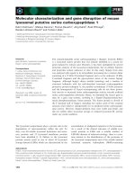
Sialyllactose
GD3
Sialyllactose Tripeptide
Syt IIGT1b
AB
CD
Fig. 3. Binding domain and receptors. (A)
The sialyllactose binding site in the H
CC
domain of BoNT ⁄ B. Only the b-trefoil fold is
shown (view almost normal Fig. 2, down
trefoil fold). (B) GD3, a part of GT1b, binds
at Site 2 in TeNT. The same site is occupied
by the GD3 part of the GT1b-b analog. (C)
A composite figure of the BoNT ⁄ BH
CC
domain with sialylllactose and tripeptide (as
bound to TeNT) shows the double receptor
model. (D) A composite figure of the
BoNT ⁄ BH
CC
domain with GT1b as bound in
Site 1 of BoNT ⁄ A and Syt II peptide as in
Site 2 of BoNT ⁄ B. These two match with
sialyllactose and the tripeptide in (C). Struc-
tures are all in a similar orientation.
Molecular structures of Clostridial neurotoxins S. Swaminathan
4470 FEBS Journal 278 (2011) 4467–4485 Journal compilation ª 2011 FEBS. No claim to original US government works
ganglioside-binding pockets in TeNT, both BoNT ⁄ A
and B have only one binding site corresponding to
Site 1 [30,31]. The second site was later confirmed
using the crystal structure of TeNT cocrystallized with
the carbohydrate part of GD3 [32] (Fig. 3B). However,
the interactions and binding modes are slightly differ-
ent because of the b2–3 linkage in GT1b-b. This causes
the salt bridge between Arg1226 and the sialic moiety
to be different. Whereas the terminal sialic acid is
involved in the salt bridge formation in the GT1b-b
structure, it is the second sialic acid that is involved in
the salt bridge. It is also possible that TeNT binds to
GD3, whereas BoNTs do not.
Recent structural work on BoNT ⁄ A with a ganglio-
side analog has also confirmed the GT1b-binding site,
as identified in BoNT ⁄ B [33] (Fig. 3D). Remarkably,
the binding mode of GT1b to BoNT ⁄ A is similar to
TeNT (at least one branch). Similar to TeNT, it is the
Gal–GalNAc moiety that binds in this site. As in
TeNT, Gal forms a stacking interaction with the con-
served Trp, whereas this interaction is provided by the
sialic acid moiety in the BoNT/B structure. This may
be because sialyllactose is only a partial mimic of one
branch of GT1b. Interestingly, mutational analysis on
the conserved Tyr (1262 in BoNT ⁄ B and 1267 in
BoNT ⁄ A) shows that Tyr is important for binding and
toxicity because when it is mutated to Ala or Phe, the
toxicity of BoNT ⁄ A or B is < 2% compared with the
wild-type [21,31]. Whereas this tyrosine in BoNT ⁄ B
forms two strong hydrogen bonds with sialyllactose,
BoNT ⁄ A complex lacks these contacts. Because
BoNT ⁄ A and B share high sequence homology in the
Site 1 pocket, it is suggested that the GT1b binding
mode will be similar to BoNTA [33]. However, there is
no structural evidence for this.
BoNT ⁄ E has a pocket similar to Site 1 in its H
CC
[10]. However, in the H…SxWY…G motif, H is
replaced by K. There is no structural evidence for
GT1b binding to BoNT ⁄ E although the similarity of
the pockets suggests that it will be the same as
BoNT ⁄ A and B. Crystal structures of the binding
domain of BoNT ⁄ F, C, D and G have been reported
[8,34–37]. BoNT ⁄ F and BoNT ⁄ G have similar GT1b-
binding pockets, except that H in the H…SxWY…G
motif is replaced by G in BoNT ⁄ G. Two binding sites
have been identified in BoNT ⁄ D and they are similar
to Sites 1 and 2 of BoNT ⁄ B (but not exactly the same)
[37]. The crystal structure of BoNT ⁄ C in complex with
sialic acid identifies one site close to Site 2, although
the second possible site has not been identified struc-
turally [8]. Neither BoNT ⁄ C nor D possess the
H…SxWY…G motif found in other BoNTs, but a
Trp is present in a nearby loop called the ganglioside-
binding loop (W1258 in C and W1252 in D-SA) [8].
Mutational studies implicate W1258 of this loop in
GT1b binding but no structural information is avail-
able [38]. The role of this Trp needs further investiga-
tion. In summary, like TeNT, BoNT ⁄ D has two
GT1b-binding sites. Similarly, BoNT ⁄ C may also have
two ganglioside (GT1b ⁄ GD1b) sites.
Double receptor model
Gangliosides are not the sole receptors for BoNTs
because reduction in TeNT binding was observed when
rat brain membranes were treated with proteases
[39,40]. A similar study with BoNT ⁄ B suggested that
proteins may also be involved in BoNT uptake [39–41].
There has also been other biochemical evidence for a
second receptor molecule, specifically a membrane pro-
tein or a glycosylated protein. In view of this, a double
receptor model was proposed [23]. The low-affinity,
high-density gangliosides allow BoNTs to concentrate
on the surface of the cell and move laterally and bind to
a high-affinity, low-density second receptor, a protein.
Although most of the BoNTs require GT1b, each
BoNT has specific protein receptor(s). Because the
binding is a product of the two binding constants, the
overall binding is very high and one of the reasons for
its nanogram level LD
50
. Specific protein receptors have
been identified for BoNT ⁄ A, B, E and G. BoNT ⁄ A uses
the three isoforms of synaptic vesicle 2 proteins (SV2A,
SV2B and SV2C) [42]. BoNT ⁄ B binds to synaptotagmin
(Syt I and Syt II), which also acts as a receptor for
BoNT ⁄ G, although with a lower affinity [43–45].
Recently, glycosylated SV2A and B have also been
identified as receptors for BoNT ⁄ E [46]. BoNT ⁄ F also
requires a second protein receptor and the keratan
sulfate moiety of SV2 probably binds to the second
receptor site [34], although a later study contradicts this
finding [47]. However, BoNT ⁄ C and D are different in
that they do not need a second protein receptor for
binding to the cell membrane, although this is yet to be
confirmed. Instead they may use dual ganglioside
binding [8,37], however, the details are still to emerge.
Although the biochemical evidence has been gaining
ground, structural support is recent. Crystal structures
of TeNT H
C
with a tripeptide (Tyr–Glu–Trp) and
BoNT ⁄ B in complex with Syt II peptide support the
double receptor model [32,48,49].
TeNT H
C
with a tripeptide
The crystal structure of TeNT H
C
complexed with
GD3 sugar group, disialyllactose, showed that this
sugar molecule binds to Site 2 of the binding domain.
S. Swaminathan Molecular structures of Clostridial neurotoxins
FEBS Journal 278 (2011) 4467–4485 Journal compilation ª 2011 FEBS. No claim to original US government works 4471
GD3 is one part of GT1b with two sialic acids and
one lactose. The crystal structure of TeNT complexed
with a tripeptide Tyr–Glu–Trp (YEW) showed that it
binds in exactly the same pocket as GD3. More inter-
estingly, the interactions are also similar, Arg1226
forms strong salt bridge with Glu OE1 and OE2, as
observed with GD3 [32]. The affinity for the tripeptide
(YEW) was also higher than that for the GD3 sugar
because when TeNT was cocrystallized with equimolar
of YEW and GD3, only YEW was found in the crystal
structure. However, the thermal factors were high,
indicating spatial disorder. This gave direct support for
the double receptor model. The two sites (Sites 1 and
2) might initially be occupied by gangliosides but when
the specific protein receptor approaches, the protein
receptor might displace the low-affinity ganglioside.
Such a possibility has been suggested [30]. It is also
possible that the second receptor is a glycosylated pro-
tein whose sugar group might bind to this site by dis-
placing GT1b. Recently, SV2A and SV2B have been
identified as receptor proteins for TeNT [47]. This was
the first structural evidence for a double receptor
model since GT1b binding to Site 1 (Fig. 3C).
BoNT
⁄
B–Syt II structure
Recently, crystal structures of either the holotoxin
BoNT ⁄ B or its H
C
domain have been determined in
complex with Syt II peptide (part of its luminal
domain) [48,49]. The cocrystal structure of BoNT ⁄ B
H
C
with Syt II (8–61) was determined using a fusion
protein with a linker connecting the C-terminus of
BoNT ⁄ BH
C
with the N-terminus of the Syt II peptide.
However, in the crystal structure, the linker peptide
and residues 8–43 of Syt II were not modeled due to
poor electron density. The holotoxin and Syt II pep-
tide (40–60) were cocrystallized to determine the com-
plex structure. In this structure, only the electron
density of residues 45–59 could be modeled. In both
structures, the Syt II peptide occupied the same bind-
ing pocket, namely, Site 2 which is adjacent to Site 1.
The peptide, which is unstructured in the native pro-
tein, is induced to form a helix when it binds to
BoNT ⁄ B and occupies a hydrophobic pocket. Phe47,
Leu50, Phe54, Phe55 and Ile58 of the Syt II peptide
are buried into the binding groove and form hydro-
phobic and stacking interactions with BoNT ⁄ B
residues. Charged residues Glu57 and Lys51 interact
with residues of complementary charges in the protein.
There are other hydrogen bonds and electrostatic inter-
actions enabling the peptide to bind strongly. Taken
together, this Syt II-binding site and the sialyllactose-
binding site support the proposed double receptor
model for BoNT ⁄ B [23]. Comparison of this with the
YEW–TeNT H
C
complex suggests a common binding
site for protein receptors (Fig. 3D). The location of
YEW in TeNT is analogous to Syt II in BoNT ⁄ B,
however, the chemical identities of the interacting resi-
dues are different.
A similar pocket exists in BoNT ⁄ G and Syt I or II
can bind in a similar fashion [44]. It is expected that in
BoNT ⁄ A, E and F their protein receptor will bind
at Site 2. However, no structural information is yet
available.
Translocation domain
Once toxins bind to membranes, a temperature- and
energy-dependent process internalizes them. The neu-
rotoxins have to escape from the vesicles into the cyto-
sol by crossing the hydrophobic vesicle barrier. This is
achieved by decrease in pH to acidic levels, allowing
conformational change of the translocation domain
and leading to penetration into membrane for channel
formation so that the catalytic domain can escape the
endosome. The transmembrane region has been pre-
dicted in BoNTs. In the crystal structures, this region,
653–673 in BoNT ⁄ B and 650–672 in BoNT ⁄ A, does
not take a helical conformation and is at one tip of
the translocation domain apposing one of the long
helices (Fig. 4). It is speculated that the region will
take a helical conformation when the pH becomes
Belt region
Fig. 4. The translocation domain of BoNT ⁄ B. The belt region which
wraps around the catalytic domain loses its hydrophobic interaction
when the catalytic domain separates. The predicted transmem-
brane region is shown in magenta.
Molecular structures of Clostridial neurotoxins S. Swaminathan
4472 FEBS Journal 278 (2011) 4467–4485 Journal compilation ª 2011 FEBS. No claim to original US government works
acidic. However, structural determination of BoNT ⁄ B
at various pH values (as low as 4) did not show any
change in this region [50] although this could be
because of crystal packing. The most intriguing part of
the translocation domain is the N-terminal region
(449–545 in BoNT ⁄ A), especially the 492–545 loop
wrapping around the catalytic domain and hence
called the belt region Although individual catalytic and
binding domains have been crystallized and their struc-
tures determined, this information is lacking for the
translocation domain, partly because it is hydrophobic
and forms aggregates making crystallography a chal-
lenge. Also, without the support of the catalytic
domain, the belt region may not retain its conforma-
tion and may fold back up or down.
Although there is a lack of structural work, recent
biochemical and biophysical studies provide valuable
information. It is postulated that the heavy chain acts
as a chaperone for the light chain to translocate the
catalytic domain [51]. When the pH becomes acidic,
the H
N
domain penetrates the membrane and translo-
cates LC from the N- to the C-terminus, during which
the channel is occluded by the LC. A reduction of
disulfide in the cytosol is required for LC to separate
and then to cleave its target. The interchain disulfide
bond plays a critical role in translocation and must be
intact for translocation but reduced for translocation
to be completed [52]. Recently, it has been shown with
the LC–H
N
complex that: (a) the binding domain is
not required for translocation, and (b) translocation
can take place at neutral pH, unlike with the holotoxin
[53–55]. However, the physiological relevance of this is
not clear. The role of the belt region is not yet well
understood. Recent studies show that the belt plays a
role in translocation. In another study, it was sug-
gested that lowering of the pH neutralizes the acidic
residues in the belt region and nullifies the repulsion
between the negative charge on the membrane and the
protein [56]. However, this does not explain transloca-
tion at neutral pH in the LC–H
N
complex. The belt
region also acts like a pseudosubstrate and inhibits LC
protease activity. The substrate occupies the groove
vacated by the belt region when the light chain sepa-
rates [57,58].
The nature of the channel formed by HC is not
understood although a low-resolution electron micro-
graph shows that BoNT ⁄ B forms a tetrameric channel
[59]. The channel diameter is observed to be 15 A
˚
and
is not large enough for the intact catalytic domain to
enter and exit. The catalytic domain unfolds, escapes
the endosome and refolds in the cytosol. In summary,
structural work on the translocation domain is sparse
and more is needed to understand this process well.
Catalytic domain
This is the most studied domain in BoNTs, both struc-
turally and biochemically. The catalytic domain of
BoNT is a zinc endopeptidase similar to thermolysin
[11,12,60]. Crystal structures of the catalytic domains
(LCs) of all BoNT serotypes and TeNT are available
and they share similar fold [61–71]. The fold is a
compact globule consisting of a mixture of a helices
and b sheets. The characteristic zinc-binding motif,
HExxH+H, is in the middle of the primary sequence
of LC. The active site zinc is bound deep inside a large
open cavity that has a high negative electrostatic
potential (Fig. 5). The zinc ion is coordinated by two
histidines and one glutamate. The fourth coordination
is provided by a water molecule which acts as a nucle-
ophile. The nucleophilic water molecule forms a strong
hydrogen bond with the first Glu in the zinc-binding
motif which acts as a base for the catalytic action.
Remarkably, the active sites of all BoNTs share a simi-
lar architecture and significant sequence conservation.
The conserved residues within 10 A
˚
of zinc form iden-
tical contacts. In BoNT ⁄ E, the zinc ion coordinates
with His211, His215, Glu250 and the nucleophilic
water [61]. His211 forms a hydrogen bond with the
conserved Glu335, which in turn forms hydrogen bond
with the conserved Arg347. The nucleophilic water
forms a hydrogen bond with the conserved Tyr350.
His215 forms a hydrogen bond with the conserved
Fig. 5. Electrostatic potential surface of the catalytic domain of
BoNT ⁄ B. Zinc is in a deep cavity which is highly electronegative.
Zinc is shown as gray sphere.
S. Swaminathan Molecular structures of Clostridial neurotoxins
FEBS Journal 278 (2011) 4467–4485 Journal compilation ª 2011 FEBS. No claim to original US government works 4473
Glu249, which in turn forms a hydrogen bond with
the conserved His218. These interactions are conserved
in all BoNTs, including the nucleophilic water to
Glu212 (Fig. 6). Mutational analyses on these con-
served residues have confirmed that they affect the cat-
alytic activity, some more than the others. Most
importantly, mutation of Tyr350 and Glu212 resulted
in undetectable catalytic activity in the BoNT ⁄ E light
chain. Mutations of Arg347, Glu335, and Glu249 dras-
tically reduce the K
cat
compared with wild-type. This is
equally true for BoNT ⁄ A, and by extension others also
[62,72–74]. The interactions in the vicinity of the zinc
ion are exactly the same, resulting in a common cata-
lytic mechanism. However, unlike other zinc endopep-
tidases, substrates for BoNTs are large polypeptides
and hence have numerous contacts with the enzyme
which are unique for each serotype. This results in
BoNTs exhibiting high specificity for the substrate and
scissile bond selection. Although the active site is con-
served, these interactions away from the active site are
different and dictate the specificity of the substrate.
For example, BoNT ⁄ A and C specifically cleave adja-
cent peptide bonds (Gln197–Arg198 and Arg198–
Ala199, respectively) of the same substrate SNAP-25.
This is true for BoNT ⁄ F and BoNT ⁄ D as well, which
specifically cleave adjacent peptide bonds of VAMP.
This unique specific bond selection is achieved by the
interactions remote from the active site enabling the
specific scissile bond to be positioned for cleavage [1].
More structural work is needed to better understand
this scissile bond selection.
Role of zinc in BoNTs
The role of zinc in proteins could be either structural,
functional or both. A catalytic zinc is normally coordi-
nated by three amino acids and one water, whereas a
structural zinc is coordinated by four amino acids
[75,76]. In BoNT, the zinc is coordinated by three
amino acids and a water molecule. However, it was
thought that its role could be structural from tertiary
structural studies [77]. But structural work has categor-
ically proved that removal of zinc does not change the
conformation and that its role is functional since the
catalytic activity is lost on zinc removal [50,62,78,79].
Enzyme–substrate complex
Most useful information about the enzyme–substrate
interactions and the catalytic mechanism is obtained
from the crystal structures of enzyme–substrate com-
plexes. However, because the substrate is cleaved on
binding to the enzyme a strategy has to be adopted
for forming the complex without cleavage; either an
inactive mutant or an uncleavable mutant substrate is
used to form the complex [57,67,80–82]. An inactive
double-mutant (E224Q, Y366F) of BoNT ⁄ A was
cocrystallized with a SNAP-25 peptide (141–204) for
Glu249
Glu250
2.79
2.75
2.11
3.23
2.05
2.07
2.11
2.85
Glu212
His211
2.59
Glu335
3.04
Arg347
Tyr350
His215
His218
Fig. 6. The interactions at the active site in
the vicinity of zinc. The active site of
BoNT ⁄ E is shown. These interactions
between the conserved residues are con-
served across all serotypes.
Molecular structures of Clostridial neurotoxins S. Swaminathan
4474 FEBS Journal 278 (2011) 4467–4485 Journal compilation ª 2011 FEBS. No claim to original US government works
structure determination [81]. This crystal structure
gave detailed information about the interactions
between the enzyme and the substrate. Because a
mutant was used, reasonable information about the
interactions near the active site could not be obtained
(Fig. 7). However, the interactions away from the
active site were mapped detailing the exosites that
define the specificity of scissile bond. The crystal struc-
ture of BoNT ⁄ A in complex with an hexapeptide,
SNAP peptide
197
QRATKM
202
, containing the scissile
bond clearly demarcated the active site interactions
[82]. Taken together, these two structures faithfully
map out the interactions between the enzyme and the
C-terminal region (amino acids 141–204) of the sub-
strate and provide invaluable information for design-
ing substrate-based inhibitors.
Although the overall conformation of the two
enzyme–substrate peptide complexes is very similar
(RMSD 1A
˚
for 400 Ca atoms), loops 200, 250 and
370 vary significantly. This conformational change
may be because of either the recognition of a-exosites
in the complex with a larger peptide or an artifact of
crystal packing. In the structure with the hexapeptide,
loops 200, 250 and 370 pack together tightly, whereas
in the structure with a larger peptide, loop 200 moved
away. This also points to the induced fit when the lar-
ger substrate peptide is used.
In the hexapeptide (QRATKM)–BoNT⁄ A complex,
the carbonyl oxygens of P1 (Gln197) and P1¢ (Arg198)
form strong hydrogen bonds with the side chains of
Tyr366 and Arg363, respectively. The amino nitrogen
of P1 displaces the nucleophilic water and coordinates
with zinc. Also, P1¢ (Arg198) forms a salt bridge with
Asp370 of the enzyme (Fig. 8). These interactions dem-
onstrate the critical role played by these residues in
addition to the zinc-coordinating residues, and explain
the mutational analyses [62,72]. Based on this, a cata-
lytic mechanism has been proposed (see Fig. 6 in Ref.
[67]). This is supported by mutagenesis studies on
BoNTs. Conserved Tyr and Arg help to position, ori-
ent and stabilize the substrate for cleavage. Glu224
acts as a general base by absorbing a proton from the
nucleophilic water. The nucleophilic water attacks the
carbonyl carbon of the scissile bond, which forms a
tetrahedral transition intermediate. The zinc ion and
Tyr might stabilize this intermediate transition state.
The shuttling of protons with the help of Glu224
assists the subsequent formation of a stable leaving
amino group. This model is consistent with that pro-
posed for BoNT ⁄ B, E and F [57,62,83] and will hold
good for all BoNTs. The proposed noncanonical self
protease activity could be due to the high concentra-
tion of protein ⁄ substrate and low pH used in crystalli-
zation and may not be physiologically relevant [68].
Crystal structures of BoNT ⁄ F in complex with two
VAMP peptides, VAMP 22–58 ⁄ Gln58D-cysteine and
VAMP 27–58 ⁄ Gln58D-cysteine, use an active enzyme
with uncleavable substrate inhibitor peptides with
K
i
1nm [57]. These crystal structures mapped out
the interactions between BoNT ⁄ F and the VAMP sub-
strate. Three exosites were identified which may govern
substrate specificity. Interestingly, conformational
changes involving rotamer positions of side chains of
enzyme residues were observed when the substrate
binds. These changes, which are due to induced fitting
when a complex is formed, either open up the site for
substrate to enter or reorient to make better contact
with the substrate. The movement of loop 370
observed in BoNT ⁄ A is not seen in this structure. Bio-
chemical and mutational studies confirmed that
BoNT ⁄ F recognizes VAMP via unique exosites. This
structure established that Arg133, Glu164 and Arg171
are important residues determining substrate specific-
ity. The biochemical and structural results agree well
[57,84]. Extending substrate beyond the C-terminal of
the inhibitor peptide improved hydrolysis, suggesting
additional interactions of the region 59–65 [85]. How-
ever, the substrate inhibitor in the BoNT ⁄ F complex
structure stops at P1 and does not provide any
information in this regard (Fig. 9).
Fig. 7. SNAP-25 peptide bound to BoNT ⁄ A catalytic domain. The
enzyme is shown in light blue, SNAP-25 in green and the zinc ion
in magenta as a sphere. Coordinates were taken from PDB 1XTG.
S. Swaminathan Molecular structures of Clostridial neurotoxins
FEBS Journal 278 (2011) 4467–4485 Journal compilation ª 2011 FEBS. No claim to original US government works 4475
Although VAMP is small compared with SNAP-25
used in the BoNT ⁄ A complex, it showed distinct exosite
interactions. The orientation in which the substrate
binds to the enzyme is the same in both and the active
site interactions are conserved. However, VAMP is dif-
ferently positioned compared with SNAP-25. The three
major exosites in BoNT ⁄ F are completely different from
those in BoNT ⁄ A. By comparison with SNAP-25 in the
complex, it is concluded that the substrate takes the
place of the belt region (Fig. 10). Again as in BoNT ⁄ A,
the unstructured substrate takes a helical conformation
induced by contact with the enzyme. Exosite 1 forms a
short helix and its hydrophobic side chains point
towards the hydrophobic core of the enzyme. Surpris-
ingly, the V1 SNARE motif (
38
QVDEVVDIMR
47
)is
not a helix, but the adjacent region (N-terminal side) is
helical. In summary, the enzyme–complex structures
help in understanding the interactions between the two,
leading to clues for drug design against botulism.
Whether the exosites, alone or in conjunction with the
active site, can be used as targets for drug design should
be explored.
Structure-based drug discovery for
botulism
Even though botulinum neurotoxins are potential bio-
warfare agents and a public health hazard, effective
drugs are yet to be developed, especially for post
intoxication with botulinum toxins. Antibody thera-
peutics is emerging, but more than one antibody may
be needed to contain the effect of a single serotype
and there are limitations [86]. An equine antitoxin is
also available for post-exposure therapeutics. Small
molecules can be used effectively to treat botulinum
poisoning both before and after exposure and research
in this direction is expanding fast. Botulinum neuro-
toxin could be deactivated by targeting any one of the
three major steps in its toxicity pathway, binding,
translocation and catalytic activity. This can be done
His227
His223
2.97
2.12
2.11
2.10
2.07
2.00
Glu262
Tyr366
2.37
2.75
3.26
Gln197
Glu224
Glu164
Phe163
Gln162
Ile161
Phe194
2.90
3.05
Lys201
Thr200
Rg
Ala199
2.95
3.32
2.81
Arg198
3.04
63
2.58
P370
Met202
Fig. 8. BoNT ⁄ A and substrate peptide inter-
actions. Interactions between the main
chain atoms with the conserved residues
are maintained across serotypes. The car-
bonyl oxygen of P1 and P1¢ hydrogen bond
with the conserved Tyr and Arg (Tyr366 and
Arg363 in BoNT ⁄ A). These interactions help
to position the substrate and stabilize the
intermediate transition state.
Molecular structures of Clostridial neurotoxins S. Swaminathan
4476 FEBS Journal 278 (2011) 4467–4485 Journal compilation ª 2011 FEBS. No claim to original US government works
by designing small molecules to block the catalytic site,
binding site or the channel formed by translocation
domain. The following section discusses how structural
information could be exploited to achieve this goal.
Binding domain as target
Both the ganglioside and the protein receptor (at least
for some) binding sites of BoNT H
C
have been charac-
terized and the interactions between the receptors and
the enzyme analyzed. The first study was performed on
the binding domain of tetanus toxin (TeNT H
C
). Com-
putational methods and virtual screening were used to
identify small molecules that would bind to H
C
and were
subsequently confirmed by ESI-MS and NMR spectros-
copy [87]. This study revealed the presence of the second
binding site (Site 2, in addition to Site 1) which has now
been shown to be the protein receptor site. Of the many
compounds identified and tested for TeNT, doxorubi-
cin, a DNA-intercolator, was identified as binding with
constant of 9.4 lm. The ESI-MS experiments also
showed that doxorubicin binds to a hydrophobic pocket
and competes with gangliosides for binding. Because the
binding domains of TeNT and BoNTs are similar, this
molecule was cocrystallized with the holotoxin BoNT ⁄ B
for crystallographic studies. Doxorubicin binds in a
cavity formed by Glu1189, Glu1190, His1240, Trp1261
and Tyr1262, which also form the conserved sequence
SxWY…G in BoNTs [88] (Fig. 11). This site is the same
as that occupied by sialyllactose in earlier studies and
also the GT1b-binding site. Doxorubicin has numerous
contacts with the protein molecule and shows good
binding. Because GT1b and doxorubicin bind at the
same site, it is believed that doxorubicin might become a
lead molecule for developing drugs against botulism. It
would be helpful to study more derivatives of doxorubi-
cin to find a potential inhibitor. Unfortunately, this
study has not been continued.
Recently, interest in the binding domain has gained
momentum because the binding sites (both ganglio-
sides and protein receptors) have been identified [21].
These sites will be good targets for developing small
molecules to prevent the toxin binding to neuronal
membranes. Because these two sites are independent,
adjacent and nonoverlapping, two molecules connected
by a linker to block both sites will be a good
approach. To date, there are no published results in
this direction.
Translocation domain could be a target
A second approach would be to block the transloca-
tion channel and prevent translocation of the catalytic
Fig. 9. VAMP-based inhibitor bound to BoNT ⁄ F catalytic domain.
The inhibitor stops at P1 which also is mutated (Asp58Cys) and in
the D form. BoNT ⁄ F and VAMP are shown in blue and green,
respectively. Zinc is shown as a sphere in magenta.
Fig. 10. The belt regions and substrates (SNAP-25 and VAMP)
superposed on BoNT ⁄ A. The substrates occupy the same groove as
the belt region when the light chain separates. The belts of BoNT ⁄ A
and E are shown in magenta and blue. SNAP-25 and VAMP are in
yellow and grey. QRATKM of BoNT ⁄ A and RIME of BoNT ⁄ E are in
orange and green. All of them occupy the same groove.
S. Swaminathan Molecular structures of Clostridial neurotoxins
FEBS Journal 278 (2011) 4467–4485 Journal compilation ª 2011 FEBS. No claim to original US government works 4477
domain into the cytosol. Triterpenoid toosendanin, a
natural product from the bark of the tree Melia toos-
endan, has been used in China to protect monkeys
against botulism [89]. More recently, using single cur-
rent measurement and a semisynthetic strategy, it was
shown that toosendanin blocks the channel formed by
HC and inhibits LC translocation [90]. For the first
time, small molecules have been identified that prevent
the toxicity of botulism by blocking the translocation
channel. Interestingly, this could be a pan-active drug
because it blocks both BoNT ⁄ A and E. However,
structural details of the channel formation are yet to
emerge. Such information, when available, would
speed up the development of drugs using the transloca-
tion mechanism as a target.
Substrate-based inhibitors to block catalytic
activity
Crystal structures of enzyme–substrate peptide com-
plexes provide enough information to design structure-
based inhibitors. The tetra- and hexapeptide complexes
with BoNT LC have identified the subsites occupied
by each of the residues at and near the scissile bond
[57,67,80,82]. This offers a starting point for designing
serotype-specific peptide inhibitors that could be
transformed into effective drugs for BoNTs. These
structures also provide a model for a pharmacophore
based on which, either small molecule, peptidomimetic
or non-peptidomimetic inhibitors, could be developed.
Various groups are working towards this goal and at
some point all use structural information to optimize
the lead molecules. Bavari and co-workers started with
a ligand-free BoNT ⁄ A LC (PDB: 1E1H) model to
design small molecules, found that the model was not
suitable for virtual screening and had to use molecular
dynamics to identify different loop conformers [91–95].
The loop conformation was a major reason for this. It
may have been better to start with a substrate–peptide-
bound experimental structure in which the loops would
have taken viable conformations for virtual screening.
Pang has worked with a BoNT ⁄ A LC–SNAP-25(141-
204) model to successfully identify a few small mole-
cule inhibitors which have been subsequently opti-
mized [96,97]. Smith and co-workers used BoNT ⁄ A
LC (1E1H) for virtual screening to search the NCI
open repository and have identified and optimized a
few small molecules [98,99]. Janda and co-workers
have combined synthetic chemistry and high-through-
put screening, identified small molecules (based on
hydroxamates) and optimized them using structure–
activity relationships [69,100]. Brunger and co-workers
started from the CRATKML heptapeptide spanning
the scissile bond and developed peptidomimetics
[101,102]. Swaminathan and co-workers used their
structure of BoNT ⁄ A with hexa- and terapeptides to
develop substrate peptide inhibitors [67,82,103]. Many
tetrapeptides have been identified and modification of
these into peptide or peptidomimetic inhibitors is in
progress. Swaminathan’s group is also working on
small molecule inhibitors based on virtual screening.
Currently, most of the inhibitor work is focused on
BoNT ⁄ A.
The subsite S1¢ of BoNT ⁄ A is large and could
accommodate bigger molecules than arginine. Also,
this site formed by three loops adjusts to accommodate
different molecules by induced fitting. Interestingly,
when the complex structure of Ac-CRATKML, a
moderate inhibitor (K
i
=2lm), is compared with
QRATKM complex, the peptide as a whole slides by
one residue but still Arg occupies P1¢ site, although it
takes a different rotamer position [78]. This fact may
be exploited in designing peptidomimetics. A number
of tetra, hexa and hepta substrate peptide complexes
with BoNT ⁄ A LC have added more information about
the interactions. These structures have shown that the
P1 residue could be changed to Arg without affecting
the binding efficiency, and in fact it has proved to be a
better inhibitor because it complements the charge in
that region. It is known that changing P1 to cysteine
improves binding [104] but oxidation of Cys may cause
Dox
Fig. 11. Doxorubicin (DOX) binding to H
CC
of BoNT ⁄ B. Doxorubicin
is shown in ball and stick model. Only the H
CC
domain is shown.
Doxorubicin binds in the same place (Site 1) as sialyllactose and
could be used as a lead molecule for drug discovery.
Molecular structures of Clostridial neurotoxins S. Swaminathan
4478 FEBS Journal 278 (2011) 4467–4485 Journal compilation ª 2011 FEBS. No claim to original US government works
a problem. The structural environment of P1 and P1¢
also suggests that an amino acid containing an aro-
matic ring may be better suited because it would
improve stacking interactions. The hexapeptide could
be extended by one residue at the N-terminus. How-
ever, this might affect the chelation of zinc by P1
amino group. The requirement of P1¢ Arg is crucial
for BoNT ⁄ A activity. However, the optimal size of the
peptide inhibitor is a tetrapeptide (Swaminathan,
unpublished). It is possible to introduce modifications
in the peptides to bring rigidity, specificity and resis-
tance from proteases. There are endless possibilities
that can be tried with the information provided by
these structures. More importantly, it will be worth-
while designing a common molecule that could act as
pan-active inhibitor.
Chimeric BoNTs for therapeutics
The binding domain of BoNT is also being targeted
for vaccine development. Foster and co-workers are
developing LC-H
N
as vaccine candidates. Chimeric
molecules of individual domains of various BoNTs are
being developed for therapeutic use [105]. Barbieri has
engineered the catalytic domain by mutation to target
cells other than neuronal cells for therapeutic use
[106]. The chimeric molecule will expand the use of
BoNTs in therapeutics to other ailments.
Crystal structure of BoNT
⁄
E
The crystal structure of the holotoxin BoNT ⁄ E (un-
nicked, single chain) has provided information on its
novel domain organization [10]. BoNT ⁄ E has 39.8%
and 37.2% identity with BoNT ⁄ A and BoNT ⁄ B,
respectively. In view of this, it was expected that the
overall fold and domain organization will be similar
like their function. Indeed, the individual domains of
BoNT ⁄ A, B and E are similar with RMSD values of
1.2 A
˚
. The binding, translocation and catalytic
domains have characteristic features of BoNTs
although the loop regions are slightly different, proba-
bly because of the differences in lengths caused by the
amino acid sequence. The active site in the holotoxin
structure contains one extra water molecule and is
reminiscent of what has been observed in recombinant
BoNT ⁄ A catalytic domain structure [78].
Surprisingly, although the individual domains are
similar, the domain organization in BoNT ⁄ E is different
from in A or B. Whereas the translocation domain in A
or B is flanked by catalytic and binding domains on
either side in a linear fashion, in BoNT ⁄ E, they are on
the same side of the translocation domain making the
molecule more globular (Fig. 12). In BoNT ⁄ A and B
there are only limited interactions between the translo-
cation and binding domains and no interaction between
the binding and catalytic domains. In BoNT ⁄ E, all three
domains have contacts with one another. The belt
region in E is similar to that in A or B, but makes con-
tact with both the binding and catalytic domains. Two
possibilities for this difference may be ruled out. First,
both BoNT ⁄ A and E were crystallized at pH 7, whereas
BoNT ⁄ B was crystallized at pH 6. So it is not the crys-
tallization condition that is responsible for this differ-
ence. Second, whereas BoNT ⁄ A and B are nicked
dichain molecules, BoNT ⁄ E is an un-nicked single-chain
molecule. However, a low-resolution image obtained
with electron microscopy using nicked, dichain BoNT ⁄ E
shows a somewhat similar arrangement [107]. So the dif-
ference is not because of the single chain molecule. The
position of the binding domain of E could be obtained
by rotating the binding domain of A or B about the lin-
ker region connecting the translocation and binding
domains. This linker region (858–870, BoNT ⁄ A) takes a
helical conformation, is normal to the long axis of the
translocation domain and allows the binding domain to
traverse to the other side. However, in BoNT⁄ E this
region is a loop (830–845) and allows the binding
BD
Linker
CD
TD
Fig. 12. Ribbon representation of BoNT ⁄ E. The binding (BD), trans-
location (TD) and catalytic (CD) domains are shown in orange, green
and red. The active site zinc is shown in sphere model (cyan). The
linker region (830–845) is colored blue and shown by an arrow
mark. The domain organization is different from BoNT ⁄ A or B. Both
BD and CD are on the same side of the translocation domain.
S. Swaminathan Molecular structures of Clostridial neurotoxins
FEBS Journal 278 (2011) 4467–4485 Journal compilation ª 2011 FEBS. No claim to original US government works 4479
domain to veer around to be on the side of catalytic
domain. Because of this unique arrangement, the buried
surfaces between the domains are fairly large. The belt
region in BoNT ⁄ E is similar to that in A or B and may
be substituted by the substrate molecule when the cata-
lytic domain separates from HC after disulfide reduc-
tion or when the substrate approaches the toxin.
The receptor domain contains two receptor binding
pockets corresponding to ganglioside and protein
receptor similar to A or B. BoNT⁄ E binds to GT1b
and glycosylated SV2A and SV2B. The transmembrane
region in E is similar to A and B and is not helical in
the structure. Remarkably, while this region and the
receptor binding pockets are at the opposite ends of
the molecule in A or B, they are on the same end
(assuming the receptor binding is similar to BoNT ⁄ B)
because of the orientation of the binding domain.
Implication of different domain
organization
Although BoNT ⁄ A is the most potent of BoNTs,
BoNT ⁄ E acts more quickly, which has been attributed
to its fast translocation. The new domain organization
gives some support to this. The transmembrane region
(638–658) and the receptor binding regions are on the
same side of the molecule, point in the same direction
and are close to the endosomal membrane. Because
receptor binding is independent of pH, the receptor
remains bound and anchored to the endosomal wall.
Because of this, if the transmembrane region is at the
other end it has to reorient itself to get closer to the
wall. Accordingly, in BoNT ⁄ A or B the transmem-
brane region has to reorient, whereas it is already stra-
tegically positioned for immediate entry into the
membrane in BoNT ⁄ E. We speculate that in BoNT ⁄ A
or B, insertion into the membrane is a two-step pro-
cess. First, the translocation and catalytic domains
rotate around the linker region (as described earlier)
and move closer to the endosome wall, in contrast to
BoNT ⁄ E which is already strategically positioned for
translocation (see Fig. 7 in Ref. [10]). This may be one
reason for the faster action of BoNT ⁄ E. However, this
model needs to be further established using biophysical
and biochemical experiments (Fig. 13).
Studies with chimeric molecules showed that chi-
mera EA containing the catalytic and translocation
domains of type E and the receptor domain of type A
(1–844 of E and 871–1296 of A) acts as fast as
BoNT ⁄ E, whereas chimera AE containing the catalytic
and translocation domains of type A and the binding
domain of type E (1–874 of A and 845–1252 of E) is the
slowest (E > EA > A > AE) [108]. Thus the speed of
translocation does not depend on the binding domain,
because swapping the receptor binding domain of E
onto A did not speed up the translocation. The speed of
translocation depends only on the catalytic and translo-
cation domains. Comparing A or B with E, the only dif-
ference is the conformation of the linker region between
the binding and translocation domains. In BoNT ⁄ E this
region is 830–845 of the translocation domain and
forms interactions with both the other domains.
Remarkably, chimera EA contains this region. It will be
worthwhile to study a chimera of E with this linker pep-
tide replaced by that corresponding to A.
Conclusion
The structural, biochemical and biophysical data on
botulinum neurotoxins have helped in understanding
the mechanism of action of every domain. However,
further structural work on chimeric molecules and
complexes is needed to understand subtle differences.
These will help in developing counter measures for
these toxins using every stage of toxicity as a target.
Acknowledgements
The author thanks Drs S. Eswaramoorthy, D. Kuma-
ran and R. Agarwal and other collaborators for their
GT1b
Receptor
protein
Fig. 13. A composite figure of BoNT ⁄ E with GT1b as bound in A
and protein receptor as bound in B. BoNT ⁄ E is shown in surface
representation with the binding (BD), translocation (TD) and cata-
lytic (CD) domains shown in orange, green and red, respectively.
The transmembrane region is shown in light pink. In BoNT ⁄ E all
these three are at the same end of the molecule strategically posi-
tioned near and facing the endosome membrane wall helping faster
translocation.
Molecular structures of Clostridial neurotoxins S. Swaminathan
4480 FEBS Journal 278 (2011) 4467–4485 Journal compilation ª 2011 FEBS. No claim to original US government works
contribution in this research. Research was supported
by award from DTRA BO74208I under DOE prime
contract No. DEAC02-98CH10886 with Brookhaven
National Laboratory.
References
1 Schiavo G, Matteoli M & Montecucco C (2000)
Neurotoxins affecting neuroexocytosis. Physiol Rev 80,
717–766.
2 Gill DM (1982) Bacterial toxins: a table of lethal
amounts. Microbiol Rev 46, 86–94.
3 Montecucco C & Schiavo G (1994) Mechanism of
action of tetanus and botulinum neurotoxins. Mol
Microbiol 13, 1–9.
4 Schiavo G, Rossetto O, Benfenati F, Poulain B &
Montecucco C (1994) Tetanus and botulinum neuro-
toxins are zinc proteases specific for components of
the neuroexocytosis apparatus. Ann NY Acad Sci 710,
65–75.
5 Montecucco C & Schiavo G (1995) Structure and
function of tetanus and botulinum neurotoxins. Q Rev
Biophys 28, 423–472.
6 Sobel JD (2005) Botulism. Clin Infect Dis 41, 1167–
1173.
7 Oguma K, Fujinaga Y & Inoue K (1995) Structure
and function of Clostridium botulinum toxins.
Microbiol Immunol 39, 161–168.
8 Karalewitz AP, Kroken AR, Fu Z, Baldwin MR, Kim
JJ & Barbieri JT (2010) Identification of a unique gan-
glioside binding loop within botulinum neurotoxins C
and D-SA. Biochemistry 49, 8117–8126.
9 Montecucco C, Papini E & Schiavo G (1994) Bacterial
protein toxins penetrate cells via a four-step mecha-
nism. FEBS Lett 346, 92–98.
10 Kumaran D, Eswaramoorthy S, Furey W, Navaza J,
Sax M & Swaminathan S (2009) Domain organization
in Clostridium botulinum neurotoxin type E is unique:
its implication in faster translocation. J Mol Biol 386,
233–245.
11 Lacy DB, Tepp W, Cohen AC, DasGupta BR &
Stevens RC (1998) Crystal structure of botulinum
neurotoxin type A and implications for toxicity.
Nat Struct Biol 5, 898–902.
12 Swaminathan S & Eswaramoorthy S (2000) Structural
analysis of the catalytic and binding sites of Clostrid-
ium botulinum neurotoxin B. Nat Struct Biol 7,
693–699.
13 Lacy DB & Stevens RC (1999) Sequence homology
and structural analysis of clostridial neurotoxins.
J Mol Biol 291, 1091–1104.
14 Umland TC, Wingert LM, Swaminathan S, Furey
WF, Schmidt JJ & Sax M (1997) Structure of the
receptor binding fragment H
c
of tetanus neurotoxin.
Nat Struct Biol 4, 788–792.
15 Murzin AG, Lesk AM & Chothia C (1992) beta-
Trefoil fold. Patterns of structure and sequence in
the Kunitz inhibitors interleukins-1beta and 1alpha
and fibroblast growth factors. J Mol Biol 223,
531–543.
16 Simpson LL (1984) The binding fragment from tetanus
toxin antagonizes the neuromuscular blocking actions
of botulinum toxin. J Pharmacol Exp Ther 229,
182–187.
17 Simpson LL (1984) Botulinum toxin and tetanus toxin
recognize similar membrane determinants. Brain Res
205, 177–180.
18 Bigalke H, Muller H & Dreyer F (1986) Botulinum A
neurotoxin unlike tetanus toxin acts via a neuramini-
dase sensitive structure. Toxicon 24, 1065–1074.
19 Marxen P & Bigalke H (1989) Tetanus toxin: inhibi-
tory action in chromaffin cells is initiated by specified
types of gangliosides and promoted in low ionic
strength solution. Neurosci Lett 107, 261–266.
20 Marxen P, Fuhrmann U & Bigalke H (1989) Ganglio-
sides mediate inhibitory effects of tetanus and botu-
linum A neurotoxins on exocytosis in chromaffin cells.
Toxicon 27, 849–859.
21 Binz T & Rummel A (2009) Cell entry strategy of
clostridial neurotoxins. J Neurochem 109, 1584–1595.
22 Kitamura M, Takamiya K, Aizawa S, Furukawa K &
Furukawa K (1999) Gangliosides are the binding sub-
stances in neural cells for tetanus and botulinum toxins
in mice. Biochim Biophys Acta 1441 , 1–3.
23 Montecucco C (1986) How do tetanus and botulinum
toxins bind to neuronal membranes? Trends Biochem
Sci 11, 314–317.
24 Muraro L, Tosatto S, Motterlini L, Rossetto O &
Montecucco C (2009) The N-terminal half of the
receptor domain of botulinum neurotoxin A binds
to microdomains of the plasma membrane. Biochem
Biophys Res Commun 380, 76–80.
25 Halpern JL & Loftus A (1993) Characterization of the
receptor-binding domain of tetanus toxin. J Biol Chem
268, 11188–11192.
26 Shapiro RS, Specht CD, Collins BE, Woods AS,
Cotter RJ & Schnaar RL (1997) Identification of a
ganglioside recognition domain of tetanus toxin using
a novel ganglioside photoaffinity ligand. J Biol Chem
272, 30380–30386.
27 Kamata Y, Yoshimoto M & Kozaki S (1997) Interac-
tion between botulinum neurotoxin type A and gangli-
oside: ganglioside inactivates the neurotoxin and
quenches its tryptophan fluorescence. Toxicon 35,
1337–1340.
28 Emsley P, Fotinou C, Black I, Fairweather NF,
Charles IG, Watts C, Hewitt E & Isaacs NW (2000)
The structures of the H(C) fragment of tetanus toxin
with carbohydrate subunit complexes provide insight
into ganglioside binding. J Biol Chem 275, 8889–8894.
S. Swaminathan Molecular structures of Clostridial neurotoxins
FEBS Journal 278 (2011) 4467–4485 Journal compilation ª 2011 FEBS. No claim to original US government works 4481
29 Fotinou C, Emsley P, Black I, Ando H, Ishida H,
Kiso M, Sinha KA, Fairweather NF & Isaacs NW
(2001) The crystal structure of tetanus toxin Hc frag-
ment complexed with a synthetic Gt1b analogue sug-
gests cross-linking between ganglioside receptors and
the toxin. J Biol Chem 276, 32274–32281.
30 Rummel A, Bade S, Alves J, Bigalke H & Binz T
(2003) Two carbohydrate binding sites in the H(CC)-
domain of tetanus neurotoxin are required for toxicity.
J Mol Biol 326, 835–847.
31 Rummel A, Mahrhold S, Bigalke H & Binz T (2004)
The HCC-domain of botulinum neurotoxins A and B
exhibits a singular ganglioside binding site displaying
serotype specific carbohydrate interaction. Mol Micro-
biol 51, 631–644.
32 Jayaraman S, Eswaramoorthy S, Kumaran D &
Swaminathan S (2005) Common binding site for disial-
yllactose and tri-peptide in C-fragment of tetanus neu-
rotoxin. Proteins 61, 288–295.
33 Stenmark P, Dupuy J, Imamura A, Kiso M & Stevens
RC (2008) Crystal structure of botulinum neurotoxin
type A in complex with the cell surface co-receptor
GT1b-insight into the toxin-neuron interaction. PLoS
Pathog 4, e1000129.
34 Fu Z, Chen C, Barbieri JT, Kim JJ & Baldwin MR
(2009) Glycosylated SV2 and gangliosides as dual
receptors for botulinum neurotoxin serotype F.
Biochemistry 48, 5631–5641.
35 Schmitt J, Karalewitz AP, Benefield DA, Mushrush DJ,
Pruitt RN, Spiller BW, Barbieri JT & Lacy DB (2010)
Structural analysis of botulinum neurotoxin type G
receptor binding. Biochem Cell Biol 49, 5200–5205.
36 Stenmark P, Dong M, Dupuy J, Chapman ER &
Stevens RC (2010) Crystal structure of the botulinum
neurotoxin type G binding domain: insight into cell
surface binding. J Mol Biol 397, 1287–1297.
37 Strotmeier J, Lee K, Volker AK, Mahrhold S, Zong
Y, Zeiser J, Zhou J, Pich A, Bigalke H, Binz T et al.
(2010) Botulinum neurotoxin serotype D attacks neu-
rons via two carbohydrate-binding sites in a ganglio-
side-dependent manner. Biochem J 431, 207–216.
38 Tsukamoto K, Kozai Y, Ihara H, Kohda T, Mukam-
oto M, Tsuji T & Kozaki S (2008) Identification of the
receptor-binding sites in the carboxyl-terminal half of
the heavy chain of botulinum neurotoxin types C and
D. Microb Pathog 44, 484–493.
39 Lazarovici P & Yavin E (1986) Affinity-purified teta-
nus neurotoxin interaction with synaptic membranes:
properties of a protease-sensitive receptor component.
Biochem Cell Biol 25, 7047–7054.
40 Pierce EJ, Davison MD, Parton RG, Habig WJ &
Cruitchley DR (1986) Characterization of tetanus
toxin binding to rat brain membranes. Evidence for a
high-affinity proteinase-sensitive receptor. Biochem J
236, 845–852.
41 Evans DM, Williams RS, Shone CC, Hambleton P,
Melling J & Dolly JO (1986) Botulinum neurotoxin
type B. Its purification, radioiodination and interaction
with rat-brain synaptosomal membranes. Eur J
Biochem 154, 409–416.
42 Dong M, Yeh F, Tepp WH, Dean C, Johnson EA, Janz
R & Chapman ER (2006) SV2 is the protein receptor
for botulinum neurotoxin A. Science 312, 592–596.
43 Dong M, Richards DA, Goodnough MC, Tepp WH,
Johnson EA & Chapman ER (2003) Synaptotagmins I
and II mediate entry of botulinum neurotoxin B into
cells. J Cell Biol 162, 1293–1303.
44 Rummel A, Eichner T, Weil T, Karnath T, Gutcaits
A, Mahrhold S, Sandhoff K, Proia RL, Acharya KR,
Bigalke H et al. (2007) Identification of the protein
receptor binding site of botulinum neurotoxins B and
G proves the double-receptor concept. Proc Natl Acad
Sci USA 104, 359–364.
45 Dong M, Tepp WH, Liu H, Johnson EA & Chapman
ER (2007) Mechanism of botulinum neurotoxin B and
G entry into hippocampal neurons. J Cell Biol 179,
1511–1522.
46 Dong M, Liu H, Tepp WH, Johnson EA, Janz R &
Chapman ER (2008) Glycosylated SV2A and SV2B
mediate the entry of botulinum neurotoxin E into
neurons. Mol Biol Cell 19
, 5226–5237.
47 Yeh FL, Dong M, Yao J, Tepp WH, Lin G, Johnson
EA & Chapman ER (2010) SV2 mediates entry of teta-
nus neurotoxin into central neurons. PLoS Pathog 6,
e1001207.
48 Chai Q, Arndt JW, Dong M, Tepp WH, Johnson EA,
Chapmann ER & Stevens RC (2006) Structural basis
of cell surface receptor recognition by botulinum
neurotoxin B. Nature (Lond) 444, 1019–1020.
49 Jin R, Rummel A, Binz T & Brunger AT (2006)
Botulinum neurotoxin B recognizes its protein receptor
with high affinity and specificity. Nature (Lond) 444,
1092–1095.
50 Eswaramoorthy S, Kumaran D, Keller J & Swamina-
than S (2004) Role of metals in the biological activity
of Clostridium botulinum neurotoxins. Biochemistry 43,
2209–2216.
51 Koriazova L & Montal M (2003) Translocation of
botulinum neurotoxin light chain protease through the
heavy chain channel. Nat Struct Biol 10, 13–18.
52 Fischer A & Montal M (2007) Crucial role of the
disulfide bridge between botulinum neurotoxin light
and heavy chains in protease translocation across
membranes. J Biol Chem 282, 29604–29611.
53 Fischer A, Mushrush DJ, Lacy DB & Montal M
(2008) Botulinum neurotoxin devoid of receptor bind-
ing domain translocates active protease. PLoS Pathog
4, e1000245.
54 Masuyer G, Thiyagarajan N, James PL, Marks PHH,
Chaddock J & Acharya KR (2009) Crystal structure
Molecular structures of Clostridial neurotoxins S. Swaminathan
4482 FEBS Journal 278 (2011) 4467–4485 Journal compilation ª 2011 FEBS. No claim to original US government works
of a catalytically active, non-toxic endopeptidase deriv-
ative of Clostridium botulinum toxin A. Biochem
Biophys Res Commun 381, 50–53.
55 Montal M (2009) Translocation of botulinum neuro-
toxin light chain protease by the heavy chain protein-
conducting channel. Toxicon 54, 565–569.
56 Galloux M, Vitrac H, Montagner C, Raffestin S,
Popoff MR, Chenal A, Forge V & Gillet D (2008)
Membrane interaction of botulinum neurotoxin A
translocation (T) domain. The belt region is a regula-
tory loop for membrane interaction. J Biol Chem 283,
27668–27676.
57 Agarwal R, Schmidt JJ, Stafford RG & Swaminathan
S (2009) Mode of VAMP substrate recognition and
inhibition of Clostridium botulinum neurotoxin F.
Nat Struct Mol Biol 16, 789–794.
58 Brunger AT, Breidenbach MA, Jin R, Fischer A,
Santos JS & Montal M (2007) Botulinum neurotoxin
heavy chain belt as an intramolecular chaperone for
the light chain. PLoS Pathog 3, 1191–1194.
59 Schmid MF, Robinson JP & DasGupta BR (1993)
Direct visualization of botulinum neurotoxin induced
channels in phospholipid vesicles. Nature 364, 827–
830.
60 Matthews BW (1988) Structural basis of the action of
thermolysin and related zinc peptides. Acc Chem Res
21, 333–340.
61 Agarwal R, Eswaramoorthy S, Kumaran D, Binz T &
Swaminathan S (2004) Structural analysis of botu-
linum neurotoxin type E catalytic domain and its
mutant Glu212 fi Gln reveals the pivotal role of the
Glu212 carboxylate in the catalytic pathway. Biochem-
istry 43, 6637–6644.
62 Agarwal R, Binz T & Swaminathan S (2005) Analysis
of active site residues of botulinum neurotoxin E by
mutational, functional and structural studies:
Glu335Gln is an apoenzyme. Biochemistry 44,
8291–8302.
63 Agarwal R, Binz T & Swaminathan S (2005) Struc-
tural analysis of botulinum neurotoxin serotype f light
chain: implications on substrate binding and inhibitor
design. Biochemistry 44, 11758–11765.
64 Arndt JW, Chai Q, Christian T & Stevens RC (2006)
Structure of botulinum neurotoxin type D light chain
at 1.65 A
˚
resolution: repercussions for VAMP-2
substrate specificity. Biochem Cell Biol 45, 3255–3262.
65 Arndt JW, Yu W, Bi F & Stevens RC (2005) Crystal
structure of botulinum neurotoxin type G light chain:
serotype divergence in substrate recognition. Biochem-
istry 44, 9574–9580.
66 Jin R, Sikorra S, Stegmann CM, Pich A, Binz T &
Brunger AT (2007) Structural and biochemical studies
of botulinum neurotoxin serotype C1 light chain prote-
ase: implications for dual substrate specificity.
Biochemistry 46, 10685–10693.
67 Kumaran D, Rawat R, Ludivico ML, Ahmed SA &
Swaminathan S (2008) Structure and substrate based
inhibitor design for Clostridium botulinum neurotoxin
serotype A. J Biol Chem 283, 18883–18891.
68 Segelke B, Knapp M, Kadkhodayan S, Balhorn R &
Rupp B (2004) Crystal structure of Clostridium botu-
linum neurotoxin protease in a product-bound state:
evidence for noncanonical zinc protease activity. Proc
Natl Acad Sci USA 101, 6888–6893.
69 Silvaggi NR, Boldt GE, Hixon MS, Kennedy JP,
Tzipori S, Janda KD & Allen KN (2007) Structures of
Clostridium botulinum neurotoxin serotype A light
chain complexed with small-molecule inhibitors high-
light active-site flexibility. Chem Biol 14, 533–542.
70 Rao KN, Kumaran D, Binz T & Swaminathan S
(2005) Structural studies on the catalytic domain of
clostridial tetanus toxin. Toxicon 45, 929–939.
71 Breidenbach MA & Brunger AT (2005) 2.3 A
˚
crystal
structure of tetanus neurotoxin light chain. Biochemis-
try 44, 7450–7457.
72 Binz T, Bade S, Rummel A, Kollewe A & Alves J
(2002) Arg
362
and Tyr
365
of the botulinum neurotoxin
type A light chain are involved in transition state sta-
bilization. Biochemistry 41, 1717–1723.
73 Li L, Binz T, Niemann H & Singh BR (2000) Probing
the mechanistic role of glutamate residues in the zinc-
binding motif of type A botulinum neurotoxin light
chain. Biochemistry 39, 2399–2405.
74 Li Y, Foran P, Fairweather NF, Paiva AD, Weller U,
Dougan G & Dolly JO (1994) A single mutation in the
recombinant light chain of tetanus toxin abolishes its
proteolytic activity and removes the toxicity seen after
reconstitution with native heavy chain. Biochemistry
33, 7014–7020.
75 Vallee BL & Auld DS (1990) Active-site zinc ligands
and activated H
2
O of zinc enzymes. Proc Natl Acad
Sci USA 87, 220–224.
76 Vallee BL & Auld DS (1990) Zinc coordination, func-
tion, and structure of zinc enzymes and other proteins.
Biochemistry 29, 5647–5659.
77 Fu F, Lomneth RB, Cai S & Singh BR (1998) Role of
zinc in the structure and toxic activity of botulinum
neurotoxin. Biochemistry 37, 5267–5278.
78 Silvaggi NR, Wilson D, Tzipori S & Allen KN
(2008) Catalytic features of the botulinum neurotoxin
a light chain revealed by high resolution structure
of an inhibitory peptide complex. Biochemistry 47,
5736–5745.
79 Simpson LL, Maksymowych AB & Hao S (2001) The
role of zinc binding in the biological activity of botu-
linum toxin. J Biol Chem 276, 27034–27041.
80 Agarwal R & Swaminathan S (2008) SNAP-25 sub-
strate peptide (residues 180–183) binds to but bypasses
cleavage by catalytically active Clostridium botulinum
neurotoxin E. J Biol Chem 283, 25944–25951.
S. Swaminathan Molecular structures of Clostridial neurotoxins
FEBS Journal 278 (2011) 4467–4485 Journal compilation ª 2011 FEBS. No claim to original US government works 4483
81 Breidenbach MA & Brunger A (2004) Substrate recog-
nition strategy for botulinum neurotoxin serotype A.
Nature 432, 925–929.
82 Kumaran D, Rawat R, Ahmed SA & Swaminathan S
(2008) Substrate binding mode and its implication on
drug design for botulinum neurotoxin A. PLoS Pathog
4, e1000165.
83 Swaminathan S, Eswaramoorthy S & Kumaran D
(2004) Structure and enzymatic activity of botulinum
neurotoxins. Mov Disord 19(Suppl. 8), S17–S22.
84 Sikorra S, Henke T, Galli T & Binz T (2008) Substrate
recognition mechanism of VAMP ⁄ synaptobrevin-
cleaving clostridial neurotoxins. J Biol Chem 283,
21145–21152.
85 Schmidt JJ & Stafford RG (2005) Botulinum neuro-
toxin serotype F: identification of substrate recognition
requirements and development of inhibitors with low
nanomolar affinity. Biochemistry 44, 4067–4073.
86 Marks JD (2004) Deciphering antibody properties that
lead to potent botulinum neurotoxin neutralization.
Mov Disord 19(Suppl. 8), S101–S108.
87 Lightstone FC, Prieto MC, Singh AK, Piqueras MC,
Whittal RM, Knapp MS, Balhorn R & Roe DC
(2000) Identification of novel small molecule ligands
that bind to tetanus toxin. Chem Res Toxicol 13,
356–362.
88 Eswaramoorthy S, Kumaran D & Swaminathan S
(2001) Crystallographic evidence for doxorubicin bind-
ing to the receptor-binding site in Clostridium botu-
linum neurotoxin B. Acta Crystallogr D 57, 1743–1746.
89 Zou J, Miao WY, Ding FH, Meng JY, Ye HJ, Jia
GR, He XY, Sun GZ & Li PZ (1985) The effect of
toosendanin on monkey botulism. J Tradit Chin Med
5, 29–30.
90 Fischer A, Nakai Y, Eubanks LM, Clancy CM, Tepp
WH, Pellett S, Dickerson TJ, Johnson EA, Janda KD
& Montal M (2009) Bimodal modulation of the botu-
linum neurotoxin protein-conducting channel. Proc
Natl Acad Sci USA 106, 1330–1335.
91 Burnett JC, Henchal EA, Schmaljohn AL & Bavari S
(2005) The evolving field of biodefence: therapeutic
developments and diagnostics. Nat Rev Drug Discov 4,
281–297.
92 Burnett JC, Opsenica D, Sriraghavan K, Panchal
RG, Ruthel G, Hermone AR, Nguyen TL, Kenny
TA, Lane DJ, McGrath CF et al. (2007) A refined
pharmacophore identifies potent 4-amino-7-chloro-
quinoline-based inhibitors of the botulinum neuro-
toxin serotype A metalloprotease. J Med Chem 50,
2127–2136.
93 Burnett JC, Ruthel G, Stegmann CM, Panchal RG,
Nguyen TL, Hermone AR, Stafford RG, Lane DJ,
Kenny TA, McGrath CF et al. (2007) Inhibition of
metalloprotease botulinum serotype A from a pseudo-
peptide binding mode to a small molecule that is
active in primary neurons. J Biol Chem 282, 5004–
5014.
94 Burnett JC, Schmidt JJ, McGrath CF, Nguyen TL,
Hermone AR, Panchal RG, Vennerstrom JL, Koduku-
la K, Zaharevitz DW, Gussio R et al. (2005) Confor-
mational sampling of the botulinum neurotoxin
serotype A light chain: implications for inhibitor bind-
ing. Bioorg Med Chem 13, 333–341.
95 Burnett JC, Schmidt JJ, Stafford RG, Panchal RG,
Nguyen TL, Hermone AR, Vennerstrom JL, McGrath
CF, Lane DJ, Sausville EA et al. (2003) Novel small
molecule inhibitors of botulinum neurotoxin A metal-
loprotease activity. Biochem Biophys Res Commun
310,
84–93.
96 Pang YP, Vummenthala A, Mishra RK, Park JG,
Wang S, Davis J, Millard CB & Schmidt JJ (2010)
Potent new small-molecule inhibitor of botulinum neu-
rotoxin serotype A endopeptidase developed by syn-
thesis-based computer-aided molecular design. PLoS
ONE 4, e7730.
97 Tang J, Park JG, Millard CB, Schmidt JJ & Pang
YP (2007) Computer-aided lead optimization:
improved small-molecule inhibitor of the zinc endo-
peptidase of botulinum neurotoxin serotype A. PLoS
ONE 2, e761.
98 Lai H, Feng M, Roxas-Duncan V, Dakshinamurthy S,
Smith LA & Yang DC (2009) Quinolinol and peptide
inhibitors of zinc protease in botulinum neurotoxin A:
effects of zinc ion and peptides on inhibition. Arch
Biochem Biophys 491, 75–84.
99 Roxas-Duncan V, Enyedy I, Montgomery VA, Eccard
VS, Carrington MA, Lai H, Gul N, Yang DC &
Smith LA (2009) Identification and biochemical char-
acterization of small-molecule inhibitors of Clostrid-
ium botulinum neurotoxin serotype A. Antimicrob
Agents Chemother 53, 3478–3486.
100 Capkova K, Salzameda NT & Janda KD (2009)
Investigations into small molecule non-peptidic inhibi-
tors of the botulinum neurotoxins. Toxicon 54,
575–582.
101 Zuniga JE, Hammill JT, Drory O, Nuss JE, Burnett
JC, Gussio R, Wipf P, Bavari S & Brunger AT (2010)
Iterative structure-based peptide-like inhibitor design
against the botulinum neurotoxin serotype A. PLoS
ONE 5, e11378.
102 Zuniga JE, Schmidt JJ, Fenn T, Burnett JC, Arac D,
Gussio R, Stafford RG, Badie SS, Bavari S & Brunger
AT (2008) A potent peptidomimetic inhibitor of
botulinum neurotoxin serotype A has a very different
conformation than SNAP-25 substrate. Structure
(Camb) 16, 1588–1597.
103 Hale M, Oyler G, Swaminathan S & Ahmed SA
(2010) Basic tetrapeptides as potent intracellular inhib-
itors of type A botulinum neurotoxin protease activity.
J Biol Chem 286, 1802–1811.
Molecular structures of Clostridial neurotoxins S. Swaminathan
4484 FEBS Journal 278 (2011) 4467–4485 Journal compilation ª 2011 FEBS. No claim to original US government works
104 Schmidt JJ & Stafford RG (2002) A high-affinity
competitive inhibitor of type A botulinum neurotoxin
protease activity. FEBS Lett 532, 423–426.
105 Foster KA (2009) Engineered toxins: new therapeutics.
Toxicon 54, 587–592.
106 Chen S & Barbieri JT (2009) Engineering botulinum
neurotoxin to extend therapeutic intervention. Proc
Natl Acad Sci 106, 9180–9184.
107 Fischer A, Garcia-Rodriguez C, Geren I, Lou J,
Marks JD, Nakagawa T & Montal M (2008)
Molecular architecture of botulinum neurotoxin E
revealed by single particle electron microscopy. J Biol
Chem 283, 3997–4003.
108 Wang J, Meng J, Lawrence GW, Zurawski TH, Sasse
A, Bodeker MO, Gilmore MA, Fernandez-Salas E,
Francis J, Steward LE et al. (2008) Novel chimeras of
botulinum neurotoxin ⁄ A and ⁄ E unveil contributions
from the binding, translocation and protease domains
to their functional characteristics. J Biol Chem 283,
16993–17002.
S. Swaminathan Molecular structures of Clostridial neurotoxins
FEBS Journal 278 (2011) 4467–4485 Journal compilation ª 2011 FEBS. No claim to original US government works 4485