Báo cáo khoa học: Dynamics in electron transfer protein complexes potx
Bạn đang xem bản rút gọn của tài liệu. Xem và tải ngay bản đầy đủ của tài liệu tại đây (316.34 KB, 10 trang )
MINIREVIEW
Dynamics in electron transfer protein complexes
Qamar Bashir, Sandra Scanu and Marcellus Ubbink
Leiden Institute of Chemistry, Leiden University, Gorlaeus Laboratories, The Netherlands
Introduction
Protein–protein interactions form the basis of biologi-
cal processes. The strength, duration and nature of
protein interactions are correlated with their biological
function, making it very relevant to understand the
biophysical aspects of protein complex formation. A
key thermodynamic property is the affinity between
two proteins, given by the dissociation constant (K
d
).
The K
d
is equal to k
off
⁄ k
on
, where k
off
and k
on
are the
dissociation and association rate constants, respec-
tively. Values of dissociation constants cover a wide
range, from 10
)2
to 10
)16
m [1,2], depending on the
biological function.
Protein complexes can be classified as static or tran-
sient. Static complexes are characterized by slow disso-
ciation (k
off
<1s
)1
), and the partners in the complex
usually bind strongly in a single, well-defined orienta-
tion. The dissociation constant in these complexes
can be as low as 10
)15
to 10
)16
m [2,3]. Such an
affinity is equivalent to a free energy of binding of
) 21 kcalÆmol
)1
, which means that the complex is sta-
ble and highly selective. Tight binding is required for
the biological function of these complexes. Examples
include complexes of antigens and antibodies, as well
as of enzymes and inhibitors.
In contrast, transient complexes form when a high
turnover is required, such as in signal transduction cas-
cades or electron transfer chains. Electron transfer
reactions are found in many metabolic processes, such
Keywords
cytochrome; encounter complex; NMR;
plastocyanin; transient complex
Correspondence
M. Ubbink, Leiden Institute of Chemistry,
Leiden University, Gorlaeus Laboratories,
P.O. Box 9502, 2300 RA Leiden,
The Netherlands
Fax: +31 71527 5856
Tel: +31 7152 74628
E-mail:
(Received 23 November 2010, revised 8
February 2011, accepted 22 February 2011)
doi:10.1111/j.1742-4658.2011.08062.x
Electron transfer proteins transport electrons safely between large redox
enzymes. The complexes formed by these proteins are among the most
transient. The biological function requires, on the one hand, sufficient spec-
ificity of the interaction to allow for rapid and selective electron transfer,
and, on the other hand, a fast turnover of the complex. Recent progress in
the characterization of the nature of these complexes has demonstrated that
the encounter state plays an important role. This state of initial binding is
dominated by electrostatic interactions, and consists of an ensemble of ori-
entations. Paramagnetic relaxation enhancement NMR and chemical shift
perturbation analysis provide ways for the experimental characterisation of
the encounter state. Several studies that have used these techniques have
shown that the surface area sample in the encounter state can be limited to
the immediate environment of the final, specific complex. The encounter
complex can represent a large fraction and, in some small complexes, no
specific binding is detected at all. It can be concluded that, in electron
transfer protein complexes, a fine balance is sought between the low-speci-
ficity encounter state and the high-specificity productive complex to meet
the opposing requirements of rapid electron transfer and a high turnover
rate.
Abbreviations
CcP, cytochrome c peroxidase; Mb, myoglobin; Pc, plastocyanin; PCS, pseudocontact shift; PRE, paramagnetic relaxation enhancement;
RDC, residual dipolar coupling.
FEBS Journal 278 (2011) 1391–1400 ª 2011 The Authors Journal compilation ª 2011 FEBS 1391
as photosynthesis and respiration [4]. Such complexes
are characterized by low binding affinities, with K
d
val-
ues in the micromolar to millimolar range [5], and life-
times down to the millisecond timescale. With a free
energy of binding of 8 kcalÆmol
)1
or less, the specificity
of the interaction cannot be high. Also, these proteins
participate in transient interactions with several part-
ners using a single interaction site, compromising the
specificity. According to the Marcus theory [6,7], the
rate of electron transfer (k
et
) falls off exponentially
with the distance between the redox centres. Thus, to
bring the redox centres sufficiently close to allow elec-
tron transfer to occur, the partners need to associate
with some degree of specificity, at least for larger elec-
tron transfer proteins. In electron transfer complexes,
a delicate balance between specificity and turnover rate
needs to be found [8]. This article aims to review some
recent insights into the process of electron transfer
protein complex formation, illustrated by several well-
studied examples.
A two-step model for complex
formation
A biological message is transferred from one protein
to the next via physical interactions between their
binding sites. In order to convey the message, the pro-
teins must approach each other by diffusion and bind
through specific surface patches. As the patch consti-
tutes just a small part of the total protein surface, only
a fraction of the collisions will bring proteins into the
proper orientation, resulting in low association rates.
However, in many biological processes a quick transfer
of the message is crucial, requiring fast association and
dissociation of the proteins. Association is defined as
the rate for the formation of a productive complex
(k
on
), and the chance of forming a productive complex
from a diffusional collision is very small, owing to the
small reactive patches. This chance can be increased by
extension of the lifetime of the collision and reduction
of the surface area searched by the proteins to find the
interface. This is achieved by the formation of an
encounter complex [9], prior to the formation of the
well-defined complex.
Thus, the association of proteins to form a complex
is a multistep process, which starts with random colli-
sions of the individual proteins. The proteins first asso-
ciate to form an encounter complex. This part of the
process is diffusion-controlled and dominated by non-
specific electrostatic interactions [10–12]. These interac-
tions keep the macromolecules in proximity for a
prolonged time, allowing a more extensive two-
dimensional search of the surface of the partner by
translational and rotational movements. In the encoun-
ter complex, the proteins can reorient their interaction
patches, which is required for formation of the bound
complex. The encounter complex either proceeds
towards the final complex or dissociates again (Fig. 1).
The well-defined complex is dominated by short-range
interactions, such as van der Waals forces and hydrogen
bonding. The dominant role of electrostatic forces in
the initial stage of complex formation is a consequence
of their long-distance nature, in contrast to the short-
range forces that are responsible for specificity [13].
The encounter complex has been visualized experi-
mentally for several protein–protein [14,15] and pro-
tein–DNA [16,17] complexes. Experimental and
theoretical studies have provided evidence that tran-
sient nonspecific encounter complexes play an impor-
tant role in protein binding and function. The nature
and, in particular, the fraction of the encounter state
differ between complexes, depending on the biological
role of the complex. Tight complexes are likely to have
the equilibrium shifted towards the productive com-
plex, and exchange between productive and encounter
complex could be slow. For weak complexes, the
encounter complex represents a larger fraction of the
complex [8,14,18,19], and may be in fast exchange with
the productive complex, maintaining the correct bal-
ance between specificity and fast association. In some
cases, the complex can have a larger fraction of the
encounter complex than the specific complex, or even
be a pure encounter complex [20–24]. Mutations in the
interface may shift the equilibrium between the encoun-
ter complex and the specific complex [18,25,26]. Several
of these studies will be discussed below, but the meth-
ods used to study the encounter complex based on
NMR spectroscopy will first be described. It must be
noted that kinetic approaches can also yield valuable
information about the process of complex formation.
These studies have been reviewed recently [27].
NMR spectroscopy
NMR spectroscopy has proven to be a very useful
technique for studying protein complexes. Various
AB C
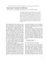
Fig. 1. Model for protein complex formation. Free proteins (A)
associate to form an encounter complex (B), consisting of multiple
protein orientations, which leads to the formation of the single-ori-
entation, specific complex (C). Reprinted with permission from [26].
Copyright 2008 American Chemical Society.
Dynamics in electron transfer protein complexes Q. Bashir et al.
1392 FEBS Journal 278 (2011) 1391–1400 ª 2011 The Authors Journal compilation ª 2011 FEBS
NMR methods have been developed to investigate pro-
tein structure, binding and dynamics in solution. The
word dynamics is used to describe both motions within
a protein, of backbone, side chains, and domains, and
the movement of one protein around the other in tran-
sient complexes. Here, the latter meaning is used.
One of the commonly applied NMR methods used
to probe protein–protein interactions is chemical shift
perturbation analysis [28]. This helps to delineate the
binding interface and to estimate the association and
dissociation rates of the protein complexes [29]. In this
method, usually a series of heteronuclear single quan-
tum coherence (HSQC) or transverse relaxation opti-
mized (TROSY) spectra of a
15
N-labelled protein are
recorded during a titration with a partner protein.
Each peak in the spectrum reports on the chemical
environment of one of the amide groups in the pro-
tein. The nuclei at the interface sense the binding
event, resulting in chemical shift changes, provided
that the dissociation rate of the complex is high in
comparison with the chemical shift difference between
the free and bound states (the fast-exchange regime).
The size of the changes can be fitted to determine the
K
d
. The average size of the shift changes also provides
information on the degree of dynamics in the protein
complex [23,24]. The more dynamic the complex, the
smaller the chemical shift changes are. In this way, the
average size of the chemical shift changes represents
the relative populations of the encounter complex and
the specific complex. The explanation for this observa-
tion is that a specific complex has a well-defined orien-
tation and is stabilized by short-range interactions
such as hydrogen bonding and salt bridges, resulting
in large chemical shift changes. The encounter com-
plex exists in multiple orientations, and it is assumed
that at least a single solvation layer remains. As a con-
sequence, the chemical shift changes are small and
averaged over all orientations. Chemical shift pertur-
bations fail to provide accurate information about the
binding interface when proteins undergo large confor-
mational changes upon complex formation. In such
cases, the conformational changes can result in chemi-
cal shift changes of nuclei far from the interface. How-
ever, the method is very useful for studying the
interfaces of transient complexes, which do not
undergo such changes.
Paramagnetic relaxation enhancement (PRE) is
another NMR method used to study the dynamics in
protein complexes [14,15]. Paramagnetic effects arise
from an unpaired electron on a metal ion or stable
organic radical. The unpaired electron increases the
relaxation rate of the nuclei in its vicinity, owing to
the large magnetic moment of the unpaired electron.
The effect depends on the sixth power of the distance
between the nucleus and the unpaired electron. PRE
provides unique distance information, in the range of
10–35 A
˚
[30]. Most proteins are not paramagnetic, and
require the introduction of a paramagnetic centre on
the protein surface, such as a nitroxide spin label or a
metal-chelating tag [31]. These probes can be covalently
attached to the protein surface via cysteines. For the
study of protein interactions, the paramagnetic centre is
attached to one protein, and the relaxation rates of the
nuclei in the other protein are measured. PRE has pro-
ven to be a useful technique for the visualization of the
encounter complex. Only nuclei that are close to
the paramagnetic centre are strongly affected, owing to
the sixth power distance dependence. Therefore, this
approach can be used to detect minor orientations that
represent only a few per cent of the complex, as has
been demonstrated for protein–protein [14,15] and pro-
tein–DNA [16,17] complexes, as well as macromolecu-
lar self-association [32,33] and state equilibria [34,35].
Several approaches have so far been proposed to visual-
ize the encounter complex by combining modelling and
PRE data, including explicit ensemble refinement
[15,36], empirical ensemble simulations [20,26], and
Brownian dynamics ⁄ Monte Carlo simulations
[8,18,19,25,37]. The first two are, in essence, fitting pro-
cedures that introduce no assumptions about the
encounter complex. The last one is not a fitting but a
simulation procedure, independent of the experimental
data, that introduces the reasonable assumption that
the interactions between the proteins in the encounter
complex are dominated by electrostatic forces.
Pseudocontact shifts (PCSs) and residual dipolar
couplings (RDCs) can also be employed to study the
dynamics in protein complexes [38–42]. PCSs arise
from the anisotropy of the paramagnetic effects of the
unpaired electron of a metal ion on the nuclei of pro-
teins. PCSs provide long-range distance restraints that
can be used to determine the orientations of the two
proteins in the complex. RDCs are obtained by partial
alignment of protein molecules resulting in incomplete
averaging of anisotropic dipolar interactions. The par-
tial alignment of protein molecules can be achieved by
using external alignment media or by the strongly
paramagnetic metals. RDCs were initially employed to
obtain distance-independent information for the struc-
ture refinement. Recently, RDCs have also been used
to study the dynamics of proteins and protein com-
plexes. Both PCSs and RDCs have been applied to
estimate the maximum percentage of the favourable
orientations of flexible protein domains [43–45],
an approach that could also be applied to transient
protein complexes.
Q. Bashir et al. Dynamics in electron transfer protein complexes
FEBS Journal 278 (2011) 1391–1400 ª 2011 The Authors Journal compilation ª 2011 FEBS 1393
Cytochrome c and cytochrome c
peroxidase
Cytochrome c is found loosely associated with the
inner membrane of the mitochondrion. It is small, with
a molecular mass of 12 kDa, and comprises 100–108
amino acids and a c-type haem group. Its main func-
tion in cellular respiration is to transport electrons
from cytochrome c reductase (complex III) to cyto-
chrome c oxidase (complex IV), embedded in the inner
membrane of the mitochondrion [46]. In yeast, cyto-
chrome c has other physiological partners as well, such
as cytochrome c peroxidase (CcP) and cytochrome b
2
.
CcP is a water-soluble haem-containing enzyme of the
peroxidase family that takes electrons from cyto-
chrome c and reduces hydrogen peroxide to water [47].
Yeast CcP is a monomer of molecular mass 34 kDa,
containing 294 amino acids and a b-type haem group.
The cytochrome c–CcP system has been extensively
investigated as a model for long-range interprotein
electron transfer. The crystal structure of the complex
[48] shows that it is mainly stabilized by van der Waals
interactions and a single hydrogen bond between an
asparagine (Asn70) of cytochrome c and a glutamic
acid (Glu290) of CcP. The orientations of cyto-
chrome c and CcP in the complex in solution have also
been determined by NMR [14], showing that the crys-
tal structure represents the dominant form present in
solution. However, this study also provided strong evi-
dence that other orientations of cytochrome c and CcP
are present in the solution complex, as had been sug-
gested already by the Brownian dynamics simulations
by Northrup et al. [49]. The PRE data showed that
certain regions of cytochrome c experience relaxation
effects from spin-labelled CcP, despite being far from
the site of spin label attachments. Apparently, other
orientations occur in the complex, in which those parts
of cytochrome c are close to the spin label, at least for
a fraction of the time. On the basis of this finding, a
spin label was linked to CcP at 10 positions, covering
nearly the entire surface of CcP, and the area sampled
by cytochrome c in the encounter complex was estab-
lished [8]. The encounter complex was also simulated
by Monte Carlo calculations, considering the electro-
static interactions at the atomic level (Fig. 2), and it
was shown that the simulation yields an encounter
complex that represents the experimental data well. It
was demonstrated that, in solution, the complex exists
as an equilibrium of 30% of the encounter complex
and 70% of the specific complex. The results also show
that cytochrome c samples only 15% of the surface
area of CcP, in the immediate surroundings of the
specific binding site.
In another study [18] of the yeast cytochrome c–CcP
complex, it has been shown that the equilibrium of the
encounter complex and the specific complex can be
modulated by single point mutations in the interface.
Both the PRE analysis and the average size of the
chemical shift perturbations of cytochrome c mutants
in complex with CcP showed that the interface muta-
tions can make the complex either more or less
dynamic. Clearly, it is possible to remodel the energy
landscape of the complex and tune its binding specific-
ity with subtle changes in the interface, indicating the
delicate balance between the encounter and specific
forms of the complex.
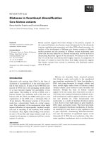
AB
Fig. 2. Simulated encounter ensemble of the cytochrome c–CcP complex. Representations of the ensemble structures with CcP (A) and
cytochrome c (B) superimposed are shown as ribbons, with the haems in cyan. The centres of mass of cytochrome c (A) and CcP (B) are
shown as spheres, coloured to indicate the density of the distributions, decreasing from red to blue. The highest densities denote the most
favourable electrostatic orientations. Densities were determined by counting the number of neighbours within 2 A
˚
. Reprinted in part with
permission from [8]. Copyright 2010 American Chemical Society.
Dynamics in electron transfer protein complexes Q. Bashir et al.
1394 FEBS Journal 278 (2011) 1391–1400 ª 2011 The Authors Journal compilation ª 2011 FEBS
Cytochrome f and plastocyanin
Cytochrome f and plastocyanin (Pc) are electron trans-
fer partners in oxygenic photosynthesis in plants,
algae, and cyanobacteria. Pc acts as electron shuttle
between the cytochrome b
6
f complex and photosys-
tem I. It is an 11-kDa protein with a b-sandwich struc-
ture that accommodates a type I copper centre. The
metal ion is coordinated by a methionine, a cysteine,
and two histidines, one of which is partly solvent-
exposed. This histidine side chain is surrounded by
nonpolar residues, forming a hydrophobic surface
patch that is involved in protein–protein interactions.
The composition of this region differs between eukary-
otic and cyanobacterial Pc, with the latter containing
more long-chain aliphatic residues, such as methionine
and leucine [50].
Cytochrome f belongs to the c-type cytochrome fam-
ily, because the haem is covalently attached to two
cysteines, in the characteristic CXXCH motif. It has a
large soluble part (28 kDa), attached to the membrane
via a single a-helix at the C-terminus. The protein con-
sists mostly of b-sheets, and has a distinctive elongated
shape, with an upper, small domain and a lower, lar-
ger one, which contains the haem. As in Pc, the sur-
face area near the metal is hydrophobic, apparently to
enhance the formation of a specific complex and allow
for rapid electron transfer [50].
Ubbink et al. [51] derived the solution structure of
the complex of spinach Pc and turnip cytochrome f
by paramagnetic NMR, taking advantage of the inter-
molecular PCSs of Pc amide nuclei caused by the
Fe(III) in the cytochrome f haem. Later, the struc-
tures of this complex of the cyanobacterium Phormidi-
um laminosum [52], Nostoc sp. PCC 7119 [53], and
Prochlorothrix hollandica [26], as well as of poplar Pc
and turnip cytochrome f [54], were determined in a
similar fashion. Some interesting differences, in both
structure and dynamics, were observed between these
complexes.
In the spinach Pc–turnip cytochrome f complex, the
size of the chemical shift perturbations upon binding
and the presence of intermolecular PCSs were taken to
indicate that the complex is predominantly in a specific
state. The structure suggested that electron transfer
occurs via Tyr1 (cytochrome f) and His87 (Pc), and
this pathway was further supported by analysis of Pc
side chain chemical shift changes [55]. The larger
chemical shift perturbations were observed in the
hydrophobic patch, suggesting exclusion of water mol-
ecules from the interface, in line with a tight fit for fast
electron transfer. The charge–charge interactions were
accompanied by small chemical shift perturbations,
suggesting that the charges remain solvent-exposed.
Similar conclusions were reached for the Nostoc and
poplar ⁄ turnip structures.
The complex of P. laminosum displays a much
weaker affinity, in the millimolar range, and the inter-
action is dominated by hydrophobic interactions. The
observed PCSs were small, and did not result in a con-
verged structure, strongly suggesting that this complex
is more dynamic in nature. The data suggested an ori-
entation of Pc in which only the hydrophobic patch is
in contact with cytochrome f, without a charge–charge
interaction, in contrast to the other structures. Exten-
sive kinetic measurements have also been performed,
demonstrating a weak, but nonzero, dependence on
electrostatic interactions, implying that orientations
other than those observed in the NMR structure also
occur in the complex [56,57]. The large viscosity
dependence of the reaction rate was interpreted to
indicate that both the association and an intracomplex
rearrangement step influence the overall rate of Pc
reduction [58]. The NMR and kinetic data in combi-
nation suggest that the encounter complex may play
an especially prominent role in this cytochrome f–Pc
interaction. Also, the complex of Pr. hollandica [26]
appeared to be rather dynamic, although a structure
could be determined, and charge interactions are
important in this case. The Pc from this cyanobacte-
rium features two deviations from the otherwise con-
served residues in the hydrophobic patch, one of
which is an exposed and solvent-protruding tyrosine
[59]. A double mutation of this Pc (Y12G ⁄ P14L)
results in a flattened interface and a complex with
cytochrome f that is even more dynamic than the
wild-type form (Fig. 3). It can be concluded that the
cytochrome f–Pc complex is similar to the CcP–cyto-
chrome c complex, in that it seems to be borderline
specific, with the balance of specific and encounter
complexes being shifted between complexes from dif-
ferent species. The approach developed for the CcP
complex [8] could be applied to the cytochrome f–Pc
complex to quantify this balance and characterize the
encounter complex.
Highly dynamic complexes of small
electron transfer proteins
In small proteins, electron transfer over a sufficiently
short distance is possible in multiple orientations, and
the requirement to form a specific complex is less strin-
gent. This conclusion is based on work on several
small electron transfer complexes that are capable of
rapid electron transfer, but have been shown to be
highly dynamic.
Q. Bashir et al. Dynamics in electron transfer protein complexes
FEBS Journal 278 (2011) 1391–1400 ª 2011 The Authors Journal compilation ª 2011 FEBS 1395
Cytochromes b
5
are ubiquitous electron transport
proteins found in animals, plants, and fungi [60]. Cyto-
chrome b
5
is involved in several electron transfer pro-
cesses with a variety of redox partners, among which
the cytochrome b
5
–cytochrome c complex has been
extensively studied. Many experimental and theoretical
studies have been performed to characterize the inter-
action between these two proteins, and have indicated
that the electrostatic interactions are important for the
association of the electron transfer complex [61–64].
Spectroscopic measurements have established that the
two proteins form a complex with 1 : 1 stoichiometry
[65]. Shao et al. [66] have investigated the interaction
between bovine cytochrome b
5
and horse heart cyto-
chrome c by NMR spectroscopy. They have performed
chemical shift perturbation analysis,
15
N-relaxation
experiments and cross-saturation experiments to study
the dynamic behaviour of the complex and to map out
the binding interface. Their results have demonstrated
that the conserved negatively charged region of cyto-
chrome b
5
surrounding the solvent-exposed haem edge
is involved in the interaction with cytochrome c, sug-
gesting a 1 : 1 stoichiometry. However, in another
NMR study [67] of the complex between rabbit cyto-
chrome b
5
and yeast cytochrome c, it has been shown
that, at a high molar ratio, a weak ternary complex of
one molecule of cytochrome b
5
and two molecules of
cytochrome c exists. Some other studies have also sug-
gested the formation of a ternary complex [63,68].
Brownian dynamics simulation of the complex
between yeast cytochrome c and bovine cytochrome b
5
predicted that the two proteins would dock essentially
through a single binding domain but not in a single
conformation [64]. Volkov et al. [69] have investigated
the complexes of ferric bovine cytochrome b
5
with fer-
ric and ferrous yeast cytochrome c by NMR, and
docking simulations of the binary cytochrome b
5
–cyto-
chrome c and cytochrome b
5
–(cytochrome c)
2
ternary
complexes. Chemical shift perturbation analysis indi-
cated that cytochrome c uses a confined surface patch
for interaction with a much more extensive surface
area of cytochrome b
5
, and that the complex formation
is not influenced by the oxidation state of cyto-
chrome c. The results suggested the presence of a
dynamic ensemble of conformations for the proteins in
the complex [69].
Cytochrome b
5
acts as a repair protein in muscle
cells, where it reduces the accidently oxidized form of
myoglobin (Mb). The oxidized Fe(III)Mb is unable to
bind oxygen. Transient absorption kinetic experiments
with cytochrome b
5
and Mb have shown that the two
proteins form a weak complex [70,71]. Studies of elec-
tron transfer between Mb and cytochrome b
5
sup-
ported the view of a highly dynamic complex, which
was dubbed ‘dynamic docking’ [21,22,72]. The complex
comprises an ensemble of nearly isoenergetic configu-
rations, only few of which are electron transfer active.
In the ensemble, cytochrome b
5
binds to a large area
on the surface of Mb in a wide variety of conforma-
tions. The binding is weak, and does not involve the
formation of a single, well-defined complex. The NMR
chemical shift mapping studies by Worrall et al. [24]
also support the highly dynamic nature of the cyto-
chrome b
5
–Mb complex. In these NMR studies, com-
plex formation was shown by the chemical shift
perturbations and the increase in the overall correla-
tion time of cytochrome b
5
in the presence of Mb.
However, the chemical shift changes were 10-fold
smaller than in other transient redox protein com-
plexes. The smaller size of the chemical shift perturba-
tions suggests a highly dynamic complex. The
perturbed residues map over a wide surface area of
cytochrome b
5
, with patches of residues located around
the exposed haem 6-propionate as well as at the back
of the protein. Recently, it has been shown that the
highly dynamic cytochrome b
5
–Mb complex can be
converted into a more specific one by introducing three
charge reversal mutations around the front face of Mb
[25,37].
Finally, a recent study on the nonphysiological, but
highly electron transfer active, complex of the iron–sul-
phur protein adrenodoxin and cytochrome c with a
Fig. 3. Representation of the dynamics in the Pr. hollandica Pc
Y12G ⁄ P14L–cytochrome f complex. Cytochrome f is shown as a
red ribbon, the haem as sticks, and the iron ion as a sphere. The
copper ion in a set of 50 Pc molecules is shown as magenta
spheres. The two most extreme orientations of Pc are shown as
blue ribbons. Reprinted with permission from [26]. Copyright 2008
American Chemical Society.
Dynamics in electron transfer protein complexes Q. Bashir et al.
1396 FEBS Journal 278 (2011) 1391–1400 ª 2011 The Authors Journal compilation ª 2011 FEBS
variety of paramagnetic NMR approaches clearly indi-
cated that this complex is also highly dynamic (Fig. 4)
and can be considered to be entirely in an encounter
state [20,39].
Conclusions
Electron transfer reactions require a high turnover
rate, and therefore fast dissociation. To achieve suffi-
cient affinity, the association rate also needs to be
high. The affinity cannot be very high, because that
would limit the dissociation rate, and thus the specific-
ity is inherently limited. Furthermore, electron transfer
proteins react through conserved patches with several
partners, also compromising the specificity. The con-
flicting requirements for specificity and turnover result
in a delicate balance between a specific orientation and
the more dynamic encounter state. The encounter com-
plex is dominated by long-range electrostatic interac-
tions that keep the protein molecules in close
proximity, thus increasing the lifetime of the associa-
tion and allowing a more extensive two-dimensional
search for the binding site, increasing the chance of the
productive complex being formed. The highly stabi-
lized encounter state and the moderate affinity of the
specific complex result in nearly equal free energies for
both states, allowing the encounter state to represent a
significant fraction of the complex. However, the rela-
tive populations of the encounter and specific com-
plexes vary among complexes. It appears that the
larger complexes require a relatively stable specific
complex, because only in that state can rapid electron
transfer occur. In small complexes, multiple orienta-
tions may be compatible with electron transfer, and
the complexes remain highly dynamic. It has been
demonstrated that single-point interfacial mutations
can shift the equilibrium of the encounter complex and
the specific complex towards either side. Thus, the resi-
dues in the binding sites are optimized for providing
just sufficient affinity to ensure the right balance
between the encounter complex and the specific
complex.
The conformational space searched by the proteins
in the encounter complex may also vary between dif-
ferent protein complexes. It has been shown for the
yeast cytochrome c–CcP complex that this area is
small in relation to the total protein surface, and is
restricted to the region around the specific binding site.
This sampling in the encounter complex, and the rela-
tive populations of both states, can now be determined
experimentally, and data for more complexes are
expected to become available.
Chemical shift perturbation analysis serves as a diag-
nostic tool with which to study dynamics in protein
complexes. The size of chemical shift perturbations
correlates with the fraction of the encounter complex.
The striking variation in the size of chemical shift
changes suggests that some complexes exist entirely as
ensembles of nonspecific complexes. However, this
approach merely provides a qualitative measure of the
dynamic nature of a complex. PRE can complement
the perturbation analysis. It is sensitive to lowly popu-
lated states, enabling the determination of the surface
area sampled by the proteins in the dynamic encounter
complex. It should be noted that an observed PRE is a
weighted average over space and time of different ori-
entations, and provides little information about the
individual protein orientations in the ensemble. For
the visualization of the encounter complex and to
investigate the role of interface residues in protein
complex formation, the experimental methods still
need to be combined with the theoretical modelling
techniques.
Acknowledgements
Q. Bashir was supported by a fellowship from the
Higher Education Commission of Pakistan. S. Scanu
and M. Ubbink received financial support from the
Netherlands Organization for Scientific Research,
grants 700.57.011 (ECHO) and 700.58.441 (VICI),
respectively.
References
1 Lee FS, Shapiro R & Vallee BL (1989) Tight-binding
inhibition of angiogenin and ribonuclease-A by placen-
tal ribonuclease inhibitor. Biochemistry 28, 225–230.
2 Janin J (2000) Kinetics and thermodynamics of protein–
protein interactions. In Protein–Protein Recognition
Fig. 4. The dynamic complex of adrenodoxin and cytochrome c.
Adrenodoxin is shown as a surface coloured to indicate the electro-
static potential: red for negative and blue for positive. The FeS-bind-
ing loop is shown in yellow. The distribution of cytochrome c
is shown as centres of mass around adrenodoxin. Reprinted with
permission from [20]. Copyright 2008 American Chemical Society.
Q. Bashir et al. Dynamics in electron transfer protein complexes
FEBS Journal 278 (2011) 1391–1400 ª 2011 The Authors Journal compilation ª 2011 FEBS 1397
(Kleanthous C ed), pp. 1–32. Oxford University Press,
Oxford.
3 Shapiro R & Vallee BL (1991) Interaction of human
placental ribonuclease with placental ribonuclease inhib-
itor. Biochemistry 30, 2246–2255.
4 Martı
´
nez-Fa
´
bregas J, Rubio S, Dı
´
az-Quintana A,
Dı
´
az-Moreno I & De la Rosa MA (2011) Proteomic
tools for the analysis of transient interactions between
metalloproteins. FEBS J 278 , 1401–1410.
5 Crowley PB & Ubbink M (2003) Close encounters of
the transient kind: protein interactions in the photosyn-
thetic redox chain investigated by NMR spectroscopy.
Acc Chem Res 36, 723–730.
6 Marcus RA (1956) On the theory of oxidation–reduc-
tion reactions involving electron transfer. 1. J Chem
Phys 24, 966–978.
7 Marcus RA & Sutin N (1985) Electron transfers in
chemistry and biology. Biochim Biophys Acta 811,
265–322.
8 Bashir Q, Volkov AN, Ullmann GM & Ubbink M
(2010) Visualization of the encounter ensemble of the
transient electron transfer complex of cytochrome c and
cytochrome c peroxidase. J Am Chem Soc 132, 241–247.
9 Ubbink M (2009) The courtship of proteins: under-
standing the encounter complex. FEBS Lett 583,
1060–1066.
10 Schreiber G & Fersht AR (1996) Rapid, electrostatically
assisted association of proteins. Nat Struct Biol 3, 427–
431.
11 Sheinerman FB, Norel R & Honig B (2000) Electro-
static aspects of protein–protein interactions. Curr Opin
Struct Biol 10, 153–159.
12 Suh JY, Tang C & Clore GM (2007) Role of electro-
static interactions in transient encounter complexes in
protein–protein association investigated by paramag-
netic relaxation enhancement. J Am Chem Soc 129,
12954–12955.
13 Ly HK, Sezer M, Wisitruangsakul N, Feng JJ, Kranich
A, Millo D, Wideinger IM, Zebger I, Murgida DH &
Hildebrandt P (2011) Surface-enhanced vibrational
spectroscopy for probing transient interactions of pro-
teins with biomimetic interfaces: electric field effects on
structure, dynamics and function of cytochrome c.
FEBS J 278, 1382–1390.
14 Volkov AN, Worrall JAR, Holtzmann E & Ubbink M
(2006) Solution structure and dynamics of the complex
between cytochrome c and cytochrome c peroxidase
determined by paramagnetic NMR. Proc Natl Acad Sci
USA 103, 18945–18950.
15 Tang C, Iwahara J & Clore GM (2006) Visualization of
transient encounter complexes in protein–protein associ-
ation. Nature 444, 383–386.
16 Iwahara J & Clore GM (2006) Detecting transient inter-
mediates in macromolecular binding by paramagnetic
NMR. Nature 440
, 1227–1230.
17 Clore GM (2008) Visualizing lowly-populated regions
of the free energy landscape of macromolecular com-
plexes by paramagnetic relaxation enhancement. Mol
Biosyst 4, 1058–1069.
18 Volkov AN, Bashir Q, Worrall JAR, Ullmann GM &
Ubbink M (2010) Shifting the equilibrium between the
encounter state and the specific form of a protein com-
plex by interfacial point mutations. J Am Chem Soc
132, 11487–11495.
19 Kim YC, Tang C, Clore GM & Hummer G (2008)
Replica exchange simulations of transient encounter
complexes in protein–protein association. Proc Natl
Acad Sci USA 105, 12855–12860.
20 Xu XF, Reinle WG, Hannemann F, Konarev PV,
Svergun DI, Bernhardt R & Ubbink M (2008) Dynam-
ics in a pure encounter complex of two proteins studied
by solution scattering and paramagnetic NMR spectros-
copy. J Am Chem Soc 130, 6395–6403.
21 Liang ZX, Nocek JM, Huang K, Hayes RT, Kurnikov
IV, Beratan DN & Hoffman BM (2002) Dynamic dock-
ing and electron transfer between Zn-myoglobin and
cytochrome b
5
. J Am Chem Soc 124, 6849–6859.
22 Liang ZX, Kurnikov IV, Nocek JM, Mauk AG, Bera-
tan DN & Hoffman BM (2004) Dynamic docking and
electron-transfer between cytochrome b
5
and a suite of
myoglobin surface-charge mutants. Introduction of a
functional-docking algorithm for protein–protein com-
plexes. J Am Chem Soc 126, 2785–2798.
23 Worrall JAR, Reinle W, Bernhardt R & Ubbink M
(2003) Transient protein interactions studied by NMR
spectroscopy: The case of cytochrome c and adreno-
doxin. Biochemistry 42, 7068–7076.
24 Worrall JAR, Liu YJ, Crowley PB, Nocek JM, Hoff-
man BM & Ubbink M (2002) Myoglobin and cyto-
chrome b
5
: A nuclear magnetic resonance study of a
highly dynamic protein complex. Biochemistry 41,
11721–11730.
25 Xiong P, Nocek JM, Griffin AKK, Wang JY & Hoff-
man BM (2009) Electrostatic redesign of the [myoglo-
bin, cytochrome b
5
] interface to create a well-defined
docked complex with rapid interprotein electron trans-
fer. J Am Chem Soc 131, 6938–6939.
26 Hulsker R, Baranova MV, Bullerjahn GS & Ubbink M
(2008) Dynamics in the transient complex of plastocya-
nin–cytochrome f from Prochlorothrix hollandica. JAm
Chem Soc 130, 1985–1991.
27 Schreiber G, Haran G & Zhou HX (2009) Fundamental
aspects of protein–protein association kinetics. Chem
Rev 109, 839–860.
28 Zuiderweg ERP (2002) Mapping protein–protein inter-
actions in solution by NMR spectroscopy. Biochemistry
41, 1–7.
29 Pellecchia M (2005) Solution nuclear magnetic reso-
nance spectroscopy techniques for probing intermolecu-
lar interactions. Chem Biol 12, 961–971.
Dynamics in electron transfer protein complexes Q. Bashir et al.
1398 FEBS Journal 278 (2011) 1391–1400 ª 2011 The Authors Journal compilation ª 2011 FEBS
30 Battiste JL & Wagner G (2000) Utilization of site-direc-
ted spin labeling and high-resolution heteronuclear
nuclear magnetic resonance for global fold determina-
tion of large proteins with limited nuclear Overhauser
effect data. Biochemistry 39, 5355–5365.
31 Keizers PHJ & Ubbink M (2011) Paramagnetic tagging
for protein structure and dynamics analysis. Prog Nucl
Magn Reson Spectrosc 58, 88–96.
32 Tang C, Ghirlando R & Clore GM (2008) Visualization
of transient ultra-weak protein self-association in solu-
tion using paramagnetic relaxation enhancement. JAm
Chem Soc 130, 4048–4056.
33 Tang C, Louis JM, Aniana A, Suh JY & Clore GM
(2008) Visualizing transient events in amino-terminal
autoprocessing of HIV-1 protease. Nature 455, 693–696.
34 Tang C, Schwieters CD & Clore GM (2007) Open-to-
closed transition in apo maltose-binding protein
observed by paramagnetic NMR. Nature 449, 1078–
1082.
35 Henzler-Wildman KA, Thai V, Lei M, Ott M, Wolf-
Watz M, Fenn T, Pozharski E, Wilson MA, Petsko
GA, Karplus M et al. (2007) Intrinsic motions
along an enzymatic reaction trajectory. Nature 450,
838–844.
36 Volkov A, Ubbink M & Van Nuland NAJ (2010) Map-
ping the encounter state of a transient protein complex
by PRE NMR spectroscopy. J Biomol NMR 48, 225–236.
37 Nocek JM, Knutson AK, Xiong P, Co NP & Hoffman
BM (2010) Photoinitiated singlet and triplet electron
transfer across a redesigned [myoglobin, cytochrome b
5
]
interface. J Am Chem Soc 132, 6165–6175.
38 Tolman JR, Flanagan JM, Kennedy MA & Prestegard
JH (1997) NMR evidence for slow collective motions in
cyanometmyoglobin. Nat Struct Biol 4, 292–297.
39 Xu XF, Keizers PHJ, Reinle W, Hannemann F, Bern-
hardt R & Ubbink M (2009) Intermolecular dynamics
studied by paramagnetic tagging. J Biomol NMR 43,
247–254.
40 Clore GM & Schwieters CD (2004) Amplitudes of pro-
tein backbone dynamics and correlated motions in a
small alpha ⁄ beta protein: correspondence of dipolar
coupling and heteronuclear relaxation measurements.
Biochemistry 43, 10678–10691.
41 Clore GM & Schwieters CD (2004) How much back-
bone motion in ubiquitin is required to account for
dipolar coupling data measured in multiple alignment
media as assessed by independent cross-validation?
J Am Chem Soc 126, 2923–2938.
42 Tolman JR & Ruan K (2006) NMR residual dipolar
couplings as probes of biomolecular dynamics. Chem
Rev 106, 1720–1736.
43 Bertini I, Giachetti A, Luchinat C, Parigi G, Petoukhov
MV, Pierattelli R, Ravera E & Svergun DI (2010) Con-
formational space of flexible biological macromolecules
from average data. J Am Chem Soc 132, 13553–13558.
44 Bertini I, Gupta YK, Luchinat C, Parigi G, Peana M,
Sgheri L & Yuan J (2007) Paramagnetism-based NMR
restraints provide maximum allowed probabilities for
the different conformations of partially independent
protein domains. J Am Chem Soc 129, 12786–12794.
45 Longinetti M, Luchinat C, Parigi G & Sgheri L (2006)
Efficient determination of the most favoured orienta-
tions of protein domains from paramagnetic NMR
data. Inverse Probl 22, 1485–1502.
46 Saraste M (1999) Oxidative phosphorylation at the fin
de sie
`
cle. Science 283, 1488–1493.
47 Yonetani T (1965) Studies on cytochrome c peroxidase.
2. Stoichiometry between enzyme H
2
O
2
and ferrocyto-
chrome c and enzymic determination of extinction coef-
ficients of cytochrome c. J Biol Chem 240, 4509–4514.
48 Pelletier H & Kraut J (1992) Crystal structure of a com-
plex between electron transfer partners, cytochrome c
peroxidase and cytochrome c. Science 258, 1748–1755.
49 Northrup SH, Boles JO & Reynolds JCL (1988) Brown-
ian dynamics of cytochrome c and cytochrome c peroxi-
dase association. Science 241, 67–70.
50 Ubbink M (2004) Complexes of photosynthetic redox
proteins studied by NMR. Photosynth Res 81, 277–287.
51 Ubbink M, Ejdeba
¨
ck M, Karlsson BG & Bendall DS
(1998) The structure of the complex of plastocyanin
and cytochrome f, determined by paramagnetic NMR
and restrained rigid-body molecular dynamics. Structure
6, 323–335.
52 Crowley PB, Otting G, Schlarb-Ridley BG, Canters
GW & Ubbink M (2001) Hydrophobic interactions in a
cyanobacterial plastocyanin–cytochrome f complex.
J Am Chem Soc 123, 10444–10453.
53 Diaz-Moreno I, Diaz-Quintana A, De la Rosa MA &
Ubbink M (2005) Structure of the complex between
plastocyanin and cytochrome f from the cyanobacte-
rium Nostoc sp. PCC 7119 as determined by paramag-
netic NMR – the balance between electrostatic and
hydrophobic interactions within the transient complex
determines the relative orientation of the two proteins.
J Biol Chem 280, 18908–18915.
54 Lange C, Cornvik T, Diaz-Moreno I & Ubbink M
(2005) The transient complex of poplar plastocyanin
with cytochrome f: effects of ionic strength and pH.
Biochim Biophys Acta 1707, 179–188.
55 Ejdeback M, Bergkvist A, Karlsson BG & Ubbink M
(2000) Side-chain interactions in the plastocyanin–cyto-
chrome f complex. Biochemistry 39, 5022–5027.
56 Schlarb-Ridley BG, Bendall DS & Howe CJ (2002)
Role of electrostatics in the interaction between cyto-
chrome f and plastocyanin of the cyanobacterium Pho-
rmidium laminosum. Biochemistry 41, 3279–3285.
57 Hart SE, Schlarb-Ridley BG, Delon C, Bendall DS &
Howe CJ (2003) Role of charges on cytochrome f from
the cyanobacterium Phormidium laminosum in its inter-
action with plastocyanin. Biochemistry 42, 4829–4836.
Q. Bashir et al. Dynamics in electron transfer protein complexes
FEBS Journal 278 (2011) 1391–1400 ª 2011 The Authors Journal compilation ª 2011 FEBS 1399
58 Schlarb-Ridley BG, Mi HL, Teale WD, Meyer VS,
Howe CJ & Bendall DS (2005) Implications of the
effects of viscosity, macromolecular crowding, and
temperature for the transient interaction between
cytochrome f and plastocyanin from the cyanobacte-
rium Phormidium laminosum. Biochemistry 44, 6232–
6238.
59 Babu CR, Volkman BF & Bullerjahn GS (1999) NMR
solution structure of plastocyanin from the photosyn-
thetic prokaryote, Prochlorothrix hollandica. Biochemis-
try 38, 4988–4995.
60 Lederer F (1994) Cytochrome b
5
-fold – an adaptable
module. Biochimie 76, 674–692.
61 Rodgers KK, Pochapsky TC & Sligar SG (1988) Prob-
ing the mechanisms of macromolecular recognition –
the cytochrome b
5
–cytochrome c complex. Science 240,
1657–1659.
62 Burch AM, Rigby SEJ, Funk WD, Macgillivray RTA,
Mauk MR, Mauk AG & Moore GR (1990) NMR char-
acterization of surface interactions in the cytochrome b
5
cytochrome c complex. Science 247, 831–833.
63 Mauk AG, Mauk MR, Moore GR & Northrup SH
(1995) Experimental and theoretical analysis of the
interaction between cytochrome c and cytochrome b
5
.
J Bioenerg Biomembr 27, 311–330.
64 Northrup SH, Thomasson KA, Miller CM, Barker PD,
Eltis LD, Guillemette JG, Inglis SC & Mauk AG
(1993) Effects of charged amino-acid mutations on the
bimolecular kinetics of reduction of yeast iso-1-ferricy-
tochrome c by bovine ferrocytochrome b
5
. Biochemistry
32, 6613–6623.
65 Mauk MR, Reid LS & Mauk AG (1982) Spectrophoto-
metric analysis of the interaction between cyto-
chrome b
5
and cytochrome c. Biochemistry 21, 1843–
1846.
66 Shao WP, Im SC, Zuiderweg ERP & Waskell L (2003)
Mapping the binding interface of the cytochrome b
5
–
cytochrome c complex by nuclear magnetic resonance.
Biochemistry 42, 14774–14784.
67 Banci L, Bertini I, Felli IC, Krippahl L, Kubicek K,
Moura JJG & Rosato A (2003) A further investigation
of the cytochrome b
5
–cytochrome c complex. J Biol
Inorg Chem 8, 777–786.
68 Whitford D, Concar DW, Veitch NC & Williams RP
(1990) The formation of protein complexes between
ferricytochrome b
5
and ferricytochrome c studied using
high-resolution 1H-NMR spectroscopy. Eur J Biochem
192, 715–721.
69 Volkov AN, Ferrari D, Worrall JAR, Bonvin AMJJ &
Ubbink M (2005) The orientations of cytochrome c in
the highly dynamic complex with cytochrome b5 visual-
ized by NMR and docking using HADDOCK. Protein
Sci 14, 799–811.
70 Nocek JM, Sishta BP, Cameron JC, Mauk AG & Hoff-
mann BM (1997) Cyclic electron transfer within the
[Zn-myoglobin, cytochrome b
5
] complex. J Am Chem
Soc 119, 2146–2155.
71 Liang ZX, Nocek JM, Kurnikov IV, Beratan DN &
Hoffman BM (2000) Electrostatic control of electron
transfer between myoglobin and cytochrome b
5
: effect
of methylating the heme propionates of Zn-myoglobin.
J Am Chem Soc 122, 3552–3553.
72 Wheeler KE, Nocek JM, Cull DA, Yatsunyk LA,
Rosenzweig AC & Hoffman BM (2007) Dynamic
docking of cytochrome b
5
with myoglobin and alpha-
hemoglobin: heme-neutralization ‘squares’ and the
binding of electron-transfer-reactive configurations.
J Am Chem Soc 129, 3906–3917.
Dynamics in electron transfer protein complexes Q. Bashir et al.
1400 FEBS Journal 278 (2011) 1391–1400 ª 2011 The Authors Journal compilation ª 2011 FEBS