Báo cáo khoa học: Surface-enhanced vibrational spectroscopy for probing transient interactions of proteins with biomimetic interfaces: electric field effects on structure, dynamics and function of cytochromec doc
Bạn đang xem bản rút gọn của tài liệu. Xem và tải ngay bản đầy đủ của tài liệu tại đây (257.1 KB, 9 trang )
MINIREVIEW
Surface-enhanced vibrational spectroscopy for probing
transient interactions of proteins with biomimetic
interfaces: electric field effects on structure, dynamics
and function of cytochrome c
Hong Khoa Ly
1
, Murat Sezer
1
, Nattawadee Wisitruangsakul
1,2
, Jiu-Ju Feng
1,3
, Anja Kranich
1
,
Diego Millo
1
, Inez M. Weidinger
1
, Ingo Zebger
1
, Daniel H. Murgida
4
and Peter Hildebrandt
1
1 Technische Universita
¨
t Berlin, Institut fu
¨
r Chemie, Germany
2 Iron and Steel Institute of Thailand, Bangkok, Thailand
3 School of Chemistry and Environmental Science, Henan Normal University, Xinxiang, China
4 Departamento de Quı
´
mica Inorga
´
nica, Analı
´
tica y Quı
´
mica Fı
´
sica ⁄ INQUIMAE-CONICET, Facultad de Ciencias Exactas y Naturales, Universi-
dad de Buenos Aires, Argentina
Introduction
Transient interactions of proteins with reaction part-
ners play a key role in biochemical and biophysical
processes [1–3]. They govern the formation of encoun-
ter complexes between proteins, preceding, for
instance, interprotein electron transfer reactions. These
interactions may include different elementary steps,
such as lateral diffusion of the reactant along the
surface of a macromolecular target, a reorientation
corresponding to a rotational diffusion within the
interaction domain, and eventually mutual conforma-
tional changes opening favourable reaction pathways
for the subsequent processes. Additional constraints
exist for transient interactions at membranes, where
most of the biological processes take place. Here, high
Keywords
apoptosis; cytochrome c; electric field;
electron transfer; protein dynamics; surface-
enhanced infrared spectroscopy; surface-
enhanced resonance Raman spectroscopy
Correspondence
P. Hildebrandt, Technische Universita
¨
t
Berlin, Institut fu
¨
r Chemie, Sekr. PC 14,
Straße des 17 Juni 135, D-10623 Berlin,
Germany
Fax: +49 30 31421122
Tel: +49 30 31421419
E-mail:
(Received 23 November 2010, revised 21
January 2011, accepted 22 February 2011)
doi:10.1111/j.1742-4658.2011.08064.x
Most of the biochemical and biophysical processes of proteins take place
at membranes, and are thus under the influence of strong local electric
fields, which are likely to affect the structure as well as the reaction mecha-
nism and dynamics. To analyse such electric field effects, biomimetic inter-
faces may be employed that consist of membrane models deposited on
nanostructured metal electrodes. For such devices, surface-enhanced reso-
nance Raman and IR absorption spectroscopy are powerful techniques to
disentangle the complex interfacial processes of proteins in terms of rota-
tional diffusion, electron transfer, and protein and cofactor structural
changes. The present article reviews the results obtained for the haem pro-
tein cytochrome c, which is widely used as a model protein for studying
the various reaction steps of interfacial redox processes in general. In addi-
tion, it is shown that electric field effects may be functional for the natural
redox processes of cytochrome c in the respiratory chain, as well as for the
switch from the redox to the peroxidase function, one of the key events
preceding apoptosis.
Abbreviations
RR, resonance Raman; SAM, self-assembled monolayer; SEIRA, surface-enhanced infrared absorption; SERR, surface-enhanced resonance
Raman; TR, time-resolved.
1382 FEBS Journal 278 (2011) 1382–1390 ª 2011 The Authors Journal compilation ª 2011 FEBS
local electric fields constitute reaction conditions that
differ substantially from those in the solution phase
[4]. Specifically, in the interfacial region between the
hydrophobic core of the lipid bilayer and the polar or
charged head groups, large changes of the potential
over a short distance lead to local electric fields as high
as 10
9
VÆm
)1
[5], which are expected to have a strong
impact on proteins transiently bound at membrane
interfaces or to integral membrane proteins. Such high
electric fields may perturb acid–base equilibrium, and
induce and align molecular dipoles in the macromole-
cule [6], thereby causing structural changes within the
macromolecule that may eventually affect the reaction
mechanism and dynamics. This may be particularly
true for processes involving the translocation of
charges, such as proton or electron transfer.
It is not surprising that our understanding of the
biomolecular processes under the influence of electric
fields is in its infancy, as dedicated experimental tech-
niques are required. This is particularly true for pro-
cesses at membranes, as high demands are imposed on
the sensitivity and selectivity of the methodology,
which should provide molecular structure and dynam-
ics information.
In this article, we present the potential of Raman
and IR spectroscopic techniques for probing surface-
confined processes in membrane models that are
designed to mimic important properties of biological
interfaces. The first part of this minireview is thus ded-
icated to the methodology and the concept for study-
ing electric field effects of immobilized proteins. The
second part summarizes the results that have been
obtained for the mammalian (horse heart) haem pro-
tein cytochrome c. Cytochrome c is a soluble protein
exerting its functions at the interface of the inner mito-
chondrial membrane. It primarily acts as an electron
carrier, delivering electrons from complex III to com-
plex IV, which are both integral membrane enzyme
complexes [7]. In addition, cytochrome c has been
shown to play a crucial role in apoptotic processes [8],
presumably initiated by a structural transition of the
protein that abolishes its redox function and strongly
increases peroxidase activity [9]. In the third part, we
will discuss the impact of the present biomimetic
approach on understanding the physiological processes
of cytochrome c.
Methodology
Raman and IR spectroscopy are molecular structure-
sensitive techniques, as the frequencies and band
intensities of vibrational transitions represent a unique
fingerprint of the specific conformation of a molecule
[10]. Both techniques, however, are associated with low
sensitivity and selectivity. For Raman spectroscopy,
this drawback can be overcome by choosing excitation
lines in resonance with an electronic transition of the
cofactor of the protein, to selectively enhance those
Raman bands that originate from the chromophoric
part of the macromolecule [resonance Raman (RR)].
IR spectroscopy is preferentially employed in the dif-
ference mode, such that only those IR bands are moni-
tored that are different between two protein states. In
this way, both the sensitivity and the selectivity are
substantially increased, as IR difference and RR spec-
troscopy solely probe the vibrational modes reflecting
a reaction of the protein and originating from its
active site, respectively. An additional increase in sensi-
tivity, required to probe proteins bound to or inte-
grated in membrane models, is achieved by exploiting
the enhancement of optical processes via coupling of
the radiation field with surface plasmons of nanostruc-
tured Ag or Au. These surface-enhanced RR (SERR)
and surface-enhanced IR absorption (SEIRA) differ-
ence spectroscopies allow probing molecules even at
submonolayer coverages of surfaces [10].
A particular advantage of SERR and SEIRA spec-
troscopy is that the signal-amplifying support material
may also be used as a working electrode when inte-
grated in an electrochemical cell [10–12]. Then, the
metal support may serve as an electron supply or sink
for electron transfer reactions to or from an immobi-
lized redox protein. In addition, variation of the elec-
trode potential is also one parameter that can alter the
local electric field strength experienced by the bound
proteins. For such spectroelectrochemical applications,
Au would be the metal of choice, as the applicable
potential range is distinctly wider than for Ag. In fact,
the optical properties, which control the surface
enhancement, are very good in the IR region, such
that SEIRA experiments are usually carried out on
thin Au films [10,12]. However, on the short-wave-
length side of the spectrum, optical excitation of sur-
face plasmons of Au is restricted to the region above
550 nm [10]. This limitation has severe consequences
for SERR spectroscopy: since the electronic transitions
of most of the protein cofactors (e.g. haem) of proteins
are at shorter wavelengths, the combination of molecu-
lar RR and the surface-enhanced Raman effect that
provides the unique selectivity and sensitivity for the
cofactor of the immobilized proteins is only possible
with nanostructured Ag for direct excitation of surface
plasmons down to 400 nm. With the recent discovery
of the coupling of surface plasmons in layered systems,
however, it is now possible to use Ag solely as a signal
amplifier while the redox protein interacts with a
H. Khoa Ly et al. SERR and SEIRA spectroscopy of cytochrome c
FEBS Journal 278 (2011) 1382–1390 ª 2011 The Authors Journal compilation ª 2011 FEBS 1383
different material such as Au, which also serves as the
(primary) electrochemical reaction partner [13–15].
These layered hybrid electrodes are based on a nano-
structured Ag support, covered with a dielectric thin
film (2–20 nm) made of a self-assembled monolayer
(SAM) of mercaptans or of silica. This film is then
coated by an Au layer (circa 20 nm in thickness).
Recent studies have demonstrated, that in such
devices, the RR signal amplification of the redox pro-
teins immobilized on the outer Au film is only slightly
lower than that determined for adsorption on the Ag
support [14].
However, regardless of the type of metal, the direct
binding of proteins on solid supports bears the risk of
irreversible denaturation. Biocompatible coatings on
the metal surface can avoid these unwanted side reac-
tions [4,16]. Although such coatings increase the dis-
tance of the redox protein from the metal surface, the
attenuation of the spectroscopic signals does not usu-
ally impair the measurement of SERR and SEIRA
spectra with high signal-to-noise ratios.
Concept
Particularly interesting biocompatible coating materials
for metal electrodes are monolayers and bilayers of
lipid analogues, as they allow mimicking biological
surfaces appropriate for protein binding [16]. For solu-
ble proteins, SAMs of x-functionalized alkanethiols or
disulphides represent the most versatile immobilization
platform [17]. These amphiphiles can form densely
packed layers, specifically on Au or Ag. SAMs carry-
ing an excess of negative or positive charges can be
created by using protonable tail groups, and are par-
ticularly suited for electrostatic immobilization of solu-
ble proteins. In the case of the highly cationic
cytochrome c, SAMs containing carboxyl-terminated
mercaptans are preferentially employed [16]. In this
sense, SAM-coated electrodes are able to mimic some
basic features of biological interfaces as far as Cou-
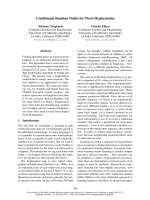
lombic interactions are concerned. Specifically, the
interfacial potential distribution is likely to be very
similar for the electrode–SAM and the bilayer systems
(Fig. 1) [4,5]. In both cases, the region between the
hydrophobic core and the polar ⁄ charged head groups
is characterized by a steep potential gradient corre-
sponding to high local electric fields. Furthermore, the
local electric field strength in the electrochemical sys-
tems can readily be controlled by changing various
parameters. An increase in the electric field strength at
the SAM–solution interface, i.e. at the position of pro-
tein binding, is achieved by: (a) increasing the differ-
ence between the electrode potential and the potential
of zero charge; (b) decreasing the SAM thickness; and
(c) increasing the charge density on the SAM surface
[4,17].
For a comprehensive analysis of electric field effects,
a quantification of the field strength is highly desirable.
The direct experimental determination of local electric
field strengths is possible on the basis of the vibra-
tional Stark effect [18,19]. A particularly appropriate
vibrational Stark effect probe is the nitrile function, as
the electric field response of the respective stretching
frequency and its sensitivity towards environmental
factors such as hydrogen bond interactions are well
understood [20,21]. SAMs containing nitrile-terminated
mercaptans may be used for coating Ag and Au, such
that the nitrile stretching can be monitored by surface-
enhanced vibrational spectroscopy [22]. Preliminary
results obtained for mixed SAMs of nitrile-terminated
and carboxyl-terminated mercaptans have afforded
local electric field strengths that are comparable to
those estimated on the basis of simple electrostatic cal-
culations [23]. In addition, the nitrile function may be
attached to cysteines of the protein introduced at
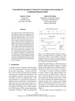
Fig. 1. Schematic presentation of the potential distribution at the
membrane–solution interface (top) and at the electrode–SAM inter-
face (bottom). w
S
, w
D
and Dw denote the surface potential, dipole
potential and transmembrane potential, respectively; D/ is the dif-
ference between electrode potential (E ) and the potential of zero
charge (E
pzc
).
SERR and SEIRA spectroscopy of cytochrome c H. Khoa Ly et al.
1384 FEBS Journal 278 (2011) 1382–1390 ª 2011 The Authors Journal compilation ª 2011 FEBS
selected positions by site-directed mutagenesis [19]. IR
and SEIRA spectroscopy then allow probing the local
electric field strength of the protein in solution and in
the immobilized state, respectively. In this way, it is
possible to map the electric field strength across the
electrode–SAM–protein interface, which is a prerequi-
site for a comprehensive quantitative description of
electric field effects on protein structure, dynamics,
and function.
Information provided by surface-
enhanced vibrational spectroscopy
Redox proteins immobilized on SAM-coated Ag and
Au electrodes are usually studied by cyclic voltamme-
try [24]. This technique monitors the current flow as a
function of the electrode potential, and thus probes the
processes of redox-active proteins. However, it does
not provide information about the molecular mecha-
nism of the interfacial processes, which, on the other
hand, is accessible with the structure-sensitive SERR
and SEIRA spectroscopy. To probe the dynamics of
molecular structure changes during the redox process,
these techniques may be coupled with the potential
jump technique [25]. In this approach, a rapid poten-
tial jump is applied to the working electrode, leading
to a perturbation of the equilibrium at the initial
potential. The subsequent relaxation processes that
restore thermodynamic equilibrium at the final poten-
tial may then be probed by SERR and SEIRA spec-
troscopy, the latter being operated in the step scan or
rapid scan mode for probing processes faster or slower
than 100 ms, respectively [26].
SERR and SEIRA spectroscopic techniques provide
different kinds of information about the interfacial
processes of cytochrome c. First, the unique vibra-
tional signatures of reduced and oxidized haems allows
the two oxidation states of the immobilized cyto-
chrome c to be distinguished. The respective marker
bands mainly originate from totally symmetric modes
that are selectively enhanced when the excitation line is
in resonance with the strongly allowed Soret transition
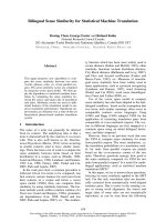
[10,23,25]. Thus, SERR spectra obtained with 413-nm
excitation allow for the determination of the redox
equilibria and, in the time-resolved (TR) mode, the
electron transfer kinetics of the immobilized cyto-
chrome c (Fig. 2).
Second, the frequencies of these marker bands not
only respond to changes in the oxidation state of the
central haem iron, but also reflect alterations in the
coordination sphere, such that these marker bands
allow monitoring equilibria and dynamics of the spin,
coordination and ligation configuration of cyto-
chrome c [10,27].
Third, when excitation lines close to the weak
Q-transition of the haem are used, the surface
enhancement of totally symmetric (A
1g
) and non-
totally symmetric (e.g. B
1g
) modes of the haem
depends on its orientation relative to the surface [28].
As the haem is fixed in the protein matrix, the relative
intensities of B
1g
and A
1g
modes in these Q-band-
excited SERR spectra may be used as a spectral
Fig. 2. Schematic presentation of the potential jump time-resolved SERR experiment for a potential jump from negative to positive poten-
tials. Left: temporal relationship between potential jump, concentration changes, and measurements. Middle: SERR spectra measured at the
initial potential E
i
(top), the final potential E
f
(bottom), and after a delay time d following the potential jump from E
i
to E
f
; the red and blue
lines refer to the component spectra of the reduced and oxidized cytochrome c (Cyt c), respectively. Right: results of the spectral analysis
showing the relaxation of the reduced cytochrome c following the potential jump. Further details are given in [25].
H. Khoa Ly et al. SERR and SEIRA spectroscopy of cytochrome c
FEBS Journal 278 (2011) 1382–1390 ª 2011 The Authors Journal compilation ª 2011 FEBS 1385
marker for tracing changes in the average orientation
of the immobilized protein.
Fourth, SEIRA spectroscopy provides complemen-
tary information about redox-linked structural changes
of the protein and orientation changes of individual
peptide segments [12,26]. SEIRA experiments are car-
ried out in the difference mode. The spectra measured
at various electrode potentials are related to a refer-
ence spectrum obtained at a fixed potential, such that
the difference spectra display only those bands that
undergo potential-dependent changes. The characteris-
tic marker bands for this technique are the amide I
modes, the frequencies of which are indicative for the
various secondary structure elements of cytochrome c.
Dynamics of the interfacial redox
process
For 20 years, SAM-coated electrodes have been used
as a convenient platform for studying biological elec-
tron transfer reactions [17]. Special attention has been
paid to the analysis of the distance dependence of the
heterogeneous electron transfer upon variation of the
SAM thickness, using cytochrome c as a model protein
[25,26,28–37]. It was found that, with decreasing dis-
tance, the electron transfer rate constant first increases
exponentially, as expected for long-range electron tun-
nelling, but then levels off to reach a plateau. Qualita-
tively, the same findings were obtained with both
electrochemical methods such as cyclic voltammetry,
which probe the electron flow between the immobilized
cytochrome c and the electrode, and SERR spectros-
copy with Soret band excitation, which monitors the
change in the oxidation state of the haem (Fig. 3).
However, by means of the various surface-enhanced
vibrational spectroscopic approaches, it is possible to
monitor further elementary reaction steps of the immo-
bilized protein that are coupled to electron tunnelling.
TR-SEIRA spectroscopy reveals that small protein
structural changes occur concomitantly with electron
transfer [26]. These changes are reflected by bands that
have also been detected in redox-induced IR difference
spectra of cytochrome c in solution. The most promi-
nent spectral changes have been attributed to the
b-turn III peptide segment 67–70 [26]. However, no
structural changes that might account for the unique
kinetic behaviour are detectable by SERR or SEIRA
spectroscopy. On the other hand, protein orientation
changes show a distance dependence that deviates from
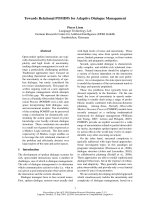
the electron tunnelling kinetics [28]. At SAMs of mer-
captohexadecanoic acid including 15 methylene groups
in the alkyl chain (C15-SAM), potential jump-induced
protein reorientation, as probed by TR-SERR
experiments with Q-band excitation, is much faster
than electron tunnelling (Fig. 3). With a decreasing
number of methylene groups, the rate of protein reori-
entation decreases until it approaches the rate of
electron tunnelling. At SAMs of mercaptohexanoic
acid (C5-SAM), the rate constants determined for ori-
entation changes and electron tunnelling are essentially
the same.
Accordingly, one may distinguish two different
regimes for the interfacial electron transfer of the
immobilized cytochrome c: at long distances, at SAMs
with 10 or more methylene groups, electron transfer is
solely controlled by electron tunnelling, whereas at
shorter distances, orientation changes of the immobi-
lized protein appear to be rate-limiting. In view of the
distance dependence of the interfacial electric field, one
may alternatively classify the redox process in terms
of a low-field (long distances) and a high-field (short
distances) regime.
Electron transfer in the low-field
regime
Despite the low number of experimental data points in
the low-field regime, both in electrochemical and
in spectroelectrochemical measurements, one may
conclude that the kinetic data follow the expected
exponential distance dependence for electron tunnel-
ling. Furthermore, overpotential-dependent and
Fig. 3. Rate constants for reorientation (red) and reduction (blue) of
cytochrome c immobilised on Ag electrodes coated with carboxyl-
terminated SAMs of different chain lengths, determined by time-
resolved SERR spectroscopy [25,28,40]. The bottom axis indicates
the electric field strength at the SAM–cytochrome c interface as
estimated on the basis of an electrostatic model [23]. The straight
line represents the exponential distance dependence of electron
tunnelling, extrapolated from rate constants determined for C15-
SAM and C10-SAM. The dotted lines are included to guide the
eyes.
SERR and SEIRA spectroscopy of cytochrome c H. Khoa Ly et al.
1386 FEBS Journal 278 (2011) 1382–1390 ª 2011 The Authors Journal compilation ª 2011 FEBS
temperature-dependent measurements for cytochrome c
immobilized on C15-SAM-coated Ag electrode (TR-
SERR) are consistent with the Marcus theory for
long-range electron tunnelling [38]. The reorganization
energy derived from these studies is distinctly lower
than that determined for cytochrome c in solution [39],
indicating a strongly reduced contribution of the sol-
vent reorganization in the immobilized state [38]. How-
ever, much weaker overpotential dependencies,
corresponding to physically meaningless low values for
the reorganization energy, are obtained for SAM-
coated Au and Au–Ag hybrid instead of Ag electrodes
[40]. In an attempt to reconcile these conflicting results,
it has been proposed that, also in the low-field regime,
the local electric field at the SAM–cytochrome c inter-
face affects the electron transfer step. Assuming that
the local electric field strength is proportional to the
difference between the actual electrode potential and
the metal-specific potential of zero charge, an empirical
linear correction has been included in the free-energy
term of the Marcus equation, in analogy to previous
approaches employed for describing field effects on
intramolecular electron transfer reactions [41]. This
rather simple approximation allows for a consistent
description of the overpotential dependencies on Ag,
Au–Ag hybrid and Au electrodes by TR-SERR and
TR-SEIRA experiments [40], but further experimental
studies and a refinement of the theoretical model are
required.
Electron transfer in the high-field
regime
Molecular dynamics simulations of cytochrome c
immobilized on electrodes coated with carboxyl-termi-
nated SAMs have identified two main binding
domains, differing with respect to the haem plane ori-
entation relative to the surface normal [42,43]. The
thermodynamically preferred high-affinity binding
domain, composed of Lys72, Lys73, Lys79, Lys86, and
Lys87, is associated with a distinctly weaker average
electronic coupling of the haem with the electrode than
the medium-affinity binding domain involving Lys25
and Lys27. As long as the rotational diffusion of the
protein on the SAM surface is fast in comparison with
electron tunnelling, electron transfer is expected to
occur mainly via the orientation of the highest electron
tunnelling probability. This is the case in the low-field
regime, as demonstrated by the comparison of the elec-
tron transfer and reorientation rate constants [28].
With increasing field strength, protein reorientation is
increasingly restricted, due to an electric field-depen-
dent increase in the activation barrier for the transition
between the various protein orientations. Thus, elec-
tron transfer is modulated by the orientation dynamics
of the immobilized protein, as reflected by the viscosity
dependence of the experimentally determined overall
rate constant. Upon further increasing of the field
strength, and thus increasing mobility restrictions of
the protein, the contribution of orientational dynamics
decreases, and electron transfer will largely occur via
all orientations that the protein can adopt upon elec-
trostatic binding. Thus, the heterogeneous electron
transfer is a convolution of the orientation-dependent
electron tunnelling and the orientational distribution
of the immobilized protein, and is thus characterized
by an apparent electron transfer rate constant, k
app
.In
addition, the electric field effect on electron tunnelling
itself, which is not negligible even in the low-field
regime, is expected to play a dominant role at high
electric fields, and may account for the decrease in k
app
when the SAM thickness is reduced below that of a
C5-SAM [40], or when the charge density in the inter-
face is increased [16]. Under these extreme conditions,
k
app
displays a kinetic isotope effect that is tentatively
attributed to the field-dependent reorganization of the
hydrogen bond network in the protein–SAM interface
[40]. Altogether, the interfacial redox processes in the
high-field regime reflect a complex interplay of various
(field-dependent) elementary steps that should lead to
nonexponential kinetics of the overall electron transfer
[44]. Unfortunately, the accuracy, time resolution and
dynamic range of TR-SERR and TR-SEIRA spectros-
copy are currently not sufficient to disentangle the
overall kinetics in this regime, which are therefore
approximated by monoexponential behaviour.
Electric field effects on the function of
cytochrome c
The electric field-dependent modulation of the electron
transfer mechanism and dynamics has been suggested
to play a role in the natural redox processes of cyto-
chrome c with the mitochondrial membrane-bound
enzyme complexes III and IV [7]. It has been proposed
that, in general, these processes take place under low-
field conditions that ensure rapid interprotein electron
transfer [16]. However, a transient increase in the
transmembrane potential may cause an intermediate
transition to the high-field regime, such that the redox
reactions of cytochrome c, and possibly also intramo-
lecular charge transfer processes in complexes III and
IV, are slowed down. Such an increase in the potential
may occur if the transmembrane proton gradient
produced during the enzymatic process, inter alia,of
complex IV is not immediately degraded by ATPase,
H. Khoa Ly et al. SERR and SEIRA spectroscopy of cytochrome c
FEBS Journal 278 (2011) 1382–1390 ª 2011 The Authors Journal compilation ª 2011 FEBS 1387
thereby constituting a feedback inhibition to avoid
unproductive consumption of molecular oxygen. This
hypothesis is difficult to check, although previous stud-
ies on complex IV reconstituted in liposomes have
shown that both the intramolecular charge transfer
processes and the redox reaction with cytochrome c
can be significantly retarded upon raising of the trans-
membrane ion gradient [45–47].
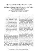
SERR spectroscopic studies on SAM-coated elec-
trodes have demonstrated that very high electric fields
induce a conformational transition from the native
form (denoted as state B1) to the conformational state
(state B2) (Fig. 4) [27]. In this state, the axial Met80
ligand is removed from the haem iron, leading to a
coordination equilibrium between a five-coordinated
and a six-coordinated species, in which this axial coor-
dination site remains vacant or is occupied by a histi-
dine (His33 or His26) [48]. This structural transition,
which is associated with a decrease in the redox poten-
tial by 300–400 mV, also occurs when cytochrome c
binds to liposomes of negatively charged phospholip-
ids, specifically at low protein ⁄ lipid ratios correspond-
ing to high local electric fields (Fig. 4) [27,48,49]. It is
therefore possible that state B2 may also be formed
when cytochrome c binds to the inner mitochondrial
membrane, which includes the anionic cardiolipin as
the main lipid component. Under physiological condi-
tions, this conformational transition would be associ-
ated with a change in protein function, because, owing
to the low redox potential, state B2 cannot be reduced
by complex III, abolishing cytochrome c’s function as
an electron carrier. On the other hand, loss of the
Met80 ligand strongly increases the peroxidase activity
[50], which may account for the cytochrome c-depen-
dent peroxidation of cardiolipin and the resultant
increase in the permeability of the inner mitochondrial
membrane [9]. This process is considered to be func-
tional for the release of cytochrome c to the cytosol,
where the protein may be involved in caspase-depen-
dent apoptotic pathways [8]. Even though other factors
may contribute to the transformation of cytochrome c
from an electron carrier to a peroxidase, local electric
fields are likely to promote this transition. Indeed, com-
putational studies have shown that, although the intrin-
sic stability of the Fe–S(Met) bond is not significantly
affected by biologically meaningful electric fields [51],
homogeneous fields of 25 mVÆA
˚
)1
are able to perturb
flexible segments of the protein, favouring the transi-
tion [43].
Acknowledgements
This work was supported by the Cluster of Excel-
lence ‘UniCat’, funded by the DFG (P. Hildebrandt),
ANPCyT (PICT2006-459), UBA (UBACyT 200200
90100094) (D. H. Murgida), and the National Science
foundation of China (Nos. 20905021) (J J. Feng).
References
1 Prudencio M & Ubbink M (2004) Transient complexes
of redox proteins: structural and dynamic details from
NMR studies. J Mol Recognit 17, 524–539.
2 Bashir Q, Scanu S & Ubbink M (2011) Dynamics in
electron transfer protein complexes. FEBS J 278, 1391–
1400.
3 Martı
´
nez-Fa
´
bregas L, Rubio S, Dı
´
az-Quintana A,
Dı
´
az-Moreno I & De la Rosa M (2011) Proteomic tools
for the analysis of transient interactions between
metalloproteins. FEBS J 278, 1401–1410.
4 Murgida DH & Hildebrandt P (2008) Disentangling
interfacial redox processes of proteins by SERR spec-
troscopy. Chem Soc Rev 37, 937–945.
Fig. 4. Relative contributions of the B2 states for ferric cyto-
chrome c (Cyt c) bound to dioleoyl-phosphatidylglycerol (DOPG)
vesicles as a function of the protein ⁄ lipid ratio, determined by RR
spectroscopy (top) [49], and for cytochrome c bound to coated Ag
electrodes as a function of the electric field strength, determined
by SERR spectroscopy (bottom) [23]. The latter plot includes data
from electrodes with carboxyl-terminated SAMs (Cx-SAM, with
x = 15, 10, 5, 2, 1; light blue), and sulphate and C
11
-PO
3
coatings
(dark blue) [16]. The solid lines are included to guide the eyes.
SERR and SEIRA spectroscopy of cytochrome c H. Khoa Ly et al.
1388 FEBS Journal 278 (2011) 1382–1390 ª 2011 The Authors Journal compilation ª 2011 FEBS
5 Clarke RJ (2001) The dipole potential of phospholipid
membranes and methods for its detection. Adv Colloid
Interface Sci 89, 263–281.
6 Neumann E (1986) Chemical electric effects in biological
macromeolecules. Prog Biophys Mol Biol 47, 197–231.
7 Scott RA & Mauk AG, eds (1995) Cytochrome c –A
Multidisciplinary Approach. University Science Books,
Sausalito.
8 Jiang X & Wang X (2004) Cytochrome c-mediated
apoptosis. Annu Rev Biochem 73, 87–106.
9 Kagan VE, Tyurin VA, Jiang J, Tyurina YY, Ritov
VB, Amoscato AA, Osipov AN, Belikova NA, Kapra-
lov AA, Kini V et al. (2005) Cytochrome c acts as a
cardiolipin oxygenase required for release of proapop-
totic factors. Nat Chem Biol 1, 223–232.
10 Siebert F & Hildebrandt P (2007) Vibrational Spectros-
copy in Life Science. Wiley-VCH, Weinheim.
11 Murgida D & Hildebrandt P (2001) Active site structure
and dynamics of immobilized cytochrome c on self-
assembled monolayers – a time-resolved surface
enhanced resonance spectroscopic study. Angew Chem
Int Ed 40, 728–731.
12 Ataka K & Heberle J (2003) Electrochemically induced
surface enhanced infrared difference absorption
(SEIDA) spectroscopy of a protein monolayer. JAm
Chem Soc 125, 4986–4987.
13 Feng JJ, Gernert U, Sezer M, Kuhlmann U, Murgida
DH, David C, Richter M, Knorr A, Hildebrandt P &
Weidinger I (2009) A novel Au–Ag hybrid device for
surface enhanced (resonance) Raman spectroscopy.
Nano Lett 9, 298–303.
14 Sezer M, Feng JJ, Ly KH, Shen Y, Nakanishi T, Kuhl-
mann U, Mo
¨
hwald H, Hildebrandt P & Weidinger I
(2010) Multi-layer electron transfer across nanostruc-
tured Ag-SAM–Au-SAM junctions probed by surface
enhanced Raman spectroscopy. Phys Chem Chem Phys
12, 9822–9829.
15 Feng JJ, Gernert U, Hildebrandt P & Weidinger IM
(2010) Novel nano-sandwiched Ag–silica–Au supports
with high SER-activity via long range plasmon cou-
pling. Adv Funct Mat 20, 1954–1961.
16 Murgida DH & Hildebrandt P (2005) Redox and
redox-coupled processes of heme proteins and enzymes
at electrochemical interfaces. Phys Chem Chem Phys 7,
3773–3784.
17 Love JC, Estroff LA, Kriebel JK, Nuzzo RG & White-
sides GM (2005) Self-assembled monolayers of thiolates
on metals as a form of nanotechnology. Chem Rev 105,
1103–1169.
18 Suydam IT, Snow CD, Pande VS & Boxer SG (2006)
Electric fields at the active site of an enzyme: direct
comparison of experiment with theory. Science 313,
200–204.
19 Fafarman AT, Webb LJ, Chuang JI & Boxer SG (2006)
Site-specific conversion of cysteine thiols into thiocya-
nate creates an IR probe for electric fields in proteins.
J Am Chem Soc 128, 13356–13357.
20 Suydam IT & Boxer SG (2003) Vibrational Stark effects
calibrate the sensitivity of vibrational probes for electric
fields in proteins. Biochemistry 42, 12050–12055.
21 Farfarman AT, Sigala PA, Herschlag D & Boxer SG
(2010) Decomposition of vibrational shifts of nitriles
into electrostatic and hydrogen bonding effects. JAm
Chem Soc 132, 12811–12813.
22 Oklejas V & Harris JM (2003) In-situ investigation of
binary-component self-assembled monolayers: a SERS-
based spectroelectrochemical study of the effects of
monolayer composition on interfacial structure.
Langmuir 19, 5794–5801.
23 Murgida DH & Hildebrandt P (2001) The heteroge-
neous electron transfer of cytochrome c adsorbed on
coated silver electrodes. Electric field effects on struc-
ture and redox potential. J Phys Chem B 105,
1578–1586.
24 Armstrong FA (2005) Recent developments in dynamic
electrochemical studies of adsorbed enzymes and their
active sites. Curr Opin Chem Biol 9, 110–117.
25 Murgida DH & Hildebrandt P (2001) Proton coupled
electron transfer in cytochrome c. J Am Chem Soc 123,
4062–4068.
26 Wisitruangsakul N, Zebger I, Ly KH, Murgida DH,
Egkasit S & Hildebrandt P (2008) Redox-linked protein
dynamics probed by time-resolved surface enhanced
infrared absorption spectroscopy. Phys Chem Chem
Phys 10, 5276–5286.
27 Wackerbarth H & Hildebrandt P (2003) Redox and
conformational equilibria and dynamics of cyto-
chrome c at high electric fields. ChemPhysChem 4 ,
714–724.
28 Kranich A, Ly HK, Hildebrandt P & Murgida DH
(2008) Direct observation of the gating step in protein
electron transfer: electric field controlled protein dynam-
ics. J Am Chem Soc 130, 9844–9848.
29 Feng ZQ, Imabayashi S, Kakiuchi T & Niki K (1997)
Long-range electron-transfer reaction rates to cyto-
chrome c across long- and short-chain alkanethiol
self-assembled monolayers: electroreflectance studies.
J Chem Soc Faraday Trans 93, 1367–1370.
30 Avila A, Gregory BW, Niki K & Cotton TM (2000) An
electrochemical approach to investigate gated electron
transfer using a physiological model system: cyto-
chrome c immobilized on carboxylic acid-terminated al-
kanethiol self-assembled monolayers on gold electrodes.
J Phys Chem B 104, 2759–2766.
31 Niki K, Hardy WR, Hill MG, Li H, Sprinkle JR,
Margoliash E, Fujita K, Tanimura R, Nakamura N,
Ohno H et al. (2003) Coupling to lysine-13 promotes
electron tunneling through carboxylate-terminated
alkanethiol self-assembled monolayers to cytochrome c.
J Phys Chem B 107, 9947–9949.
H. Khoa Ly et al. SERR and SEIRA spectroscopy of cytochrome c
FEBS Journal 278 (2011) 1382–1390 ª 2011 The Authors Journal compilation ª 2011 FEBS 1389
32 Wei JJ, Liu HY, Khoshtariya DE, Yamamoto H,
Dick A & Waldeck DH (2002) Electron-transfer
dynamics of cytochrome c: a change in the reaction
mechanism with distance. Angew Chem Int Ed 41,
4700–4703.
33 Murgida DH, Hildebrandt P, Wei J, He YF, Liu HY &
Waldeck DH (2004) SERR and electrochemical study
of cytochrome c bound on electrodes through coordina-
tion with pyridinyl-terminated SAMs. J Phys Chem B
108, 2261–2269.
34 Wei JJ, Liu HY, Niki K, Margoliash E & Waldeck DH
(2004) Probing electron tunneling pathways: electro-
chemical study of rat heart cytochrome c and its mutant
on pyridine-terminated SAMs. J Phys Chem B 108,
16912–16917.
35 Yue HJ & Waldeck DH (2005) Understanding interfa-
cial electron transfer to monolayer protein assemblies.
Curr Opin Solid State Mater Sci 9, 28–36.
36 Khoshtariya DE, Wei JJ, Liu HY, Yue HJ & Waldeck
DH (2003) Charge-transfer mechanism for cyto-
chrome c adsorbed on nanometer thick films. Distin-
guishing frictional control from conformational gating.
J Am Chem Soc 125, 7704–7714.
37 Yue HJ, Khoshtariya D, Waldeck DH, Grochol J,
Hildebrandt P & Murgida DH (2006) On the electron
transfer mechanism between cytochrome c and metal
electrodes. Evidence for dynamic control at short
distances. J Phys Chem B 110, 19906–19913.
38 Murgida D & Hildebrandt P (2002) Electrostatic-field
dependent activation energies control biological electron
transfer. J Phys Chem B 106, 12814–12819.
39 Cheng J, Terrettaz S, Blankman JI, Muller CJ, Dangi B
& Guiles RD (1997) Electrochemical comparison of
heme proteins by insulated electrode voltammetry. Isr J
Chem 37, 259–266.
40 Ly KH, Wisitruangsakul N, Sezer M, Feng JJ, Kranich
A, Weidinger I, Zebger I, Murgida DH & Hildebrandt
P (2010) Electric field effects on the interfacial electron
transfer and protein dynamics of cytochrome c. J Elec-
troanal Chem, in press, doi:10.1016/j.jelechem.2010.12.
020.
41 Lockhart DJ, Kirmaier C, Holten D & Boxer SG
(1990) Electric field effects on the initial electron-trans-
fer kinetics in bacterial photosynthetic reaction centers.
J Phys Chem 94, 6987–6995.
42 Paggi D, Martı
´
n DF, Kranich A, Hildebrandt P,
Martı
´
M & Murgida DH (2009) Computer simulation
and SERR detection of cytochrome c dynamics at
SAM-coated electrodes. Electrochim Acta 54,
4963–4970.
43 De Biase PM, Paggi DA, Doctorovich F, Hildebrandt
P, Estrin DA, Murgida DH & Marti MA (2009) Molec-
ular basis for the electric field modulation of cyto-
chrome c structure and function. J Am Chem Soc 131,
16248–16256.
44 Georg S, Kabuss J, Weidinger IM, Murgida DH, Hilde-
brandt P, Knorr A & Richter M (2010) On the distance
dependent electron transfer rate of immobilised redox
proteins – a statistical physics approach. Phys Rev E 81,
046101(1–10).
45 Moroney PM, Scholes TA & Hinkle PC (1984) Effect
of membrane-potential and pH gradient on electron-
transfer in cytochrome-oxidase. Biochemistry
23,
4991–4997.
46 Gregory LS & Ferguson-Miller S (1989) Independent
control of respiration in cytochrome-c oxidase vesicles
by pH and electrical gradients. Biochemistry 28, 2655–
2662.
47 Sarti P, Antonini G, Malatesta F & Brunori M (1992)
Respiratory control in cytochrome-oxidase vesicles is
correlated with the rate of internal electron-transfer.
Biochem J 284, 123–127.
48 Oellerich S, Wackerbarth H & Hildebrandt P (2002)
Spectroscopic characterization of non-native states of
cytochrome c. J Phys Chem B 106, 6566–6580.
49 Oellerich S, Lecomte S, Paternostre M, Heimburg T &
Hildebrandt P (2004) Peripheral and integral binding of
cytochrome c to phospholipid vesicles. J Phys Chem B
108, 3871–3878.
50 Basova LV, Kurnikov IV, Wang L, Ritov VB, Belikova
NA, Vlasova II, Pacheco AA, Winnica DE, Peterson J,
Bayir H et al. (2007) Cardiolipin switch in mitochon-
dria: shutting off the reduction of cytochrome c and
turning on the peroxidase activity. Biochemistry 2007,
46.
51 De Biase PM, Doctorovich F, Murgida DH & Estrin
DA (2007) Electric field effects on the reactivity of heme
model systems. Chem Phys Lett 434, 121–126.
SERR and SEIRA spectroscopy of cytochrome c H. Khoa Ly et al.
1390 FEBS Journal 278 (2011) 1382–1390 ª 2011 The Authors Journal compilation ª 2011 FEBS