Báo cáo khoa học: Structural basis for substrate recognition by Erwinia chrysanthemi GH30 glucuronoxylanase potx
Bạn đang xem bản rút gọn của tài liệu. Xem và tải ngay bản đầy đủ của tài liệu tại đây (1.06 MB, 12 trang )
Structural basis for substrate recognition by
Erwinia chrysanthemi GH30 glucuronoxylanase
L
ˇ
ubica Urba
´
nikova
´
1
,Ma
´
ria Vrs
ˇ
anska
´
2
, Kristian B. R. Mørkeberg Krogh
3
, Tine Hoff
3
and Peter Biely
2
1 Institute of Molecular Biology, Slovak Academy of Sciences, Bratislava, Slovakia
2 Institute of Chemistry, Center of Glycomics, Slovak Academy of Sciences, Bratislava, Slovakia
3 Novozymes A ⁄ S, Bagsvaerd, Denmark
Introduction
The important industrial enzyme endo-b-1,4-xylanase
(
EC 3.2.1.8) has been placed into several glycoside
hydrolase (GH) families on the basis of hydrophobic
cluster analysis, 3D, and mode of action [1] (carbohy-
drate-active enzymes server at ).
The best-characterized xylanases belong to GH fami-
lies 10 and 11. These enzymes do not seem to be spe-
cialized for hydrolysis of a particular xylan, because
they are capable of degrading hardwood acetyl glucu-
ronoxylans, cereal arabinoxylans, and even algal
b-1,4-b-1,3-xylan (rhodymenan) [2–4]. The activity of
xylanases belonging to these two families does not
appear to be dependent on the type of side chain dec-
orations of the xylan main chain, but is strongly
dependent on the density of substituents [2,5]. The
cleavage of the xylan main chain by GH10 xylanases
Keywords
crystal structure with ligand;
Erwinia chrysanthemi; GH30;
glucuronoxylan-specific xylanase; substrate
recognition
Correspondence
P. Biely, Institute of Chemistry, Center of
Glycomics, Slovak Academy of Sciences,
Du
´
bravska
´
cesta 9, SK-845 38 Bratislava,
Slovakia
Fax: +421 2 5941 0222
Tel: +421 2 5941 0275
E-mail:
(Received 17 December 2010, revised 10
April 2011, accepted 13 April 2011)
doi:10.1111/j.1742-4658.2011.08127.x
Xylanase A from the phytopathogenic bacterium Erwinia chrysanthemi is
classified as a glycoside hydrolase family 30 enzyme (previously in family 5)
and is specialized for degradation of glucuronoxylan. The recombinant
enzyme was crystallized with the aldotetraouronic acid b-
D-xylopyranosyl-
(1 fi 4)-[4-O-methyl-a-
D-glucuronosyl-(1 fi 2)]-b-D-xylopyranosyl-(1 fi 4)-
D-xylose as a ligand. The crystal structure of the enzyme–ligand complex
was solved at 1.39 A
˚
resolution. The ligand xylotriose moiety occupies sub-
sites )1, )2 and )3, whereas the methyl glucuronic acid residue attached to
the middle xylopyranosyl residue of xylotriose is bound to the enzyme
through hydrogen bonds to five amino acids and by the ionic interaction
of the methyl glucuronic acid carboxylate with the positively charged guan-
idinium group of Arg293. The interaction of the enzyme with the methyl
glucuronic acid residue appears to be indispensable for proper distortion of
the xylan chain and its effective hydrolysis. Such a distortion does not
occur with linear b-1,4-xylooligosaccharides, which are hydrolyzed by the
enzyme at a negligible rate.
Database
Structural and experimental data are available in the Protein Data Bank database under
accession number
2y24 [45].
Abbreviations
GH, glycoside hydrolase; GlcA,
D-glucuronic acid; MeGlcA, 4-O-methyl-D-glucuronic acid; MeGlcA
2
Xyl
2
,4-O-methyl-a-D-glucuronosyl-(1 fi 2)-
b-
D-xylopyranosyl-(1 fi 4)-D-xylose; MeGlcA
2
Xyl
3
, b-D-xylopyranosyl-(1 fi 4)-[4-O-methyl-a-D-glucuronosyl-(1 fi 2)]-b-D-xylopyranosyl-(1 fi 4)-D-
xylose; MeGlcA
3
Xyl
3
,4-O-methyl-a-D-glucuronosyl-(1 fi 2)-b-D-xylopyranosyl-(1 fi 4)-b-D-xylopyranosyl-(1 fi 4)-D-xylose; MeGlcA
3
Xyl
4
,
b-
D-xylopyranosyl-(1 fi 4)-[4-O-methyl-a-D-glucuronosyl-(1 fi 2)]-b-D-xylopyranosyl-(1 fi 4)-b-D-xylopyranosyl-(1 fi 4)-D-xylose; MeXyl
3
Xyl
3
,
4-O-methyl-a-
D-glucuronosyl-(1 fi 2)-b-D-xylopyranosyl-(1 fi 4)-b-D-xylopyranosyl-(1 fi 4)-D-xylose; MeXyl
3
Xyl
4
, b-D-xylopyranosyl-(1 fi 4)-[4-O-
methyl-a-
D-glucuronosyl-(1 fi 2)]-b-D-xylopyranosyl-(1 fi 4)-b-D-xylopyranosyl-(1 fi 4)-D-xylose; VS, virtual screening; Xyl, xylose; XynA,
Erwinia chrysanthemi GH30 xylanase.
FEBS Journal 278 (2011) 2105–2116 Journal compilation ª 2011 FEBS. No claim to original Slovakian government works 2105
requires at least two consecutive unsubstituted xylo-
pyranosyl residues, whereas hydrolysis by GH11
xylanases requires three consecutive unsubstituted
xylopyranosyl residues [2–6]. Heavily substituted
xylan, such as corn fiber xylan [7], is completely resis-
tant to the action of members of these two xylanase
families (P. Biely, unpublished results). An interesting
endoxylanase, classified in GH family 8, was found to
be produced by an Antarctic bacterium, Pseudoaltero-
monas haloplanktis [8]. This enzyme showed the high-
est activity on rhodymenan [8], which indicates that
the enzyme might be specialized for hydrolysis of the
linear xylan present in algae.
Unique xylanases are found in GH family 30 [1,9].
These enzymes were originally classified in GH fam-
ily 5. Some bacterial GH30 xylanases are specialized
for the hydrolysis of xylans that contain d-glucuronic
acid (GlcA) or 4-O-methyl-d-glucuronic acid (MeG-
lcA) side residues. However, not all GH30 xylanases
show this specificity. The recently described GH30 xy-
lanase from the fungus Bispora sp. does not show such
a requirement for these side residues [10]. With Bacil-
lus subtilis GH30 xylanase and Erwinia chrysanthemi
GH30 xylanase (XynA), it was clearly demonstrated
that cleavage of the xylan main chain is dependent on
the presence of MeGlcA side residues [11–14]. A simi-
lar enzyme from another Bacillus species was recently
described [15]. The cleavage of the main xylan chain
takes place at the second glycosidic linkage from the
MeGlcA side group towards the reducing end of the
xylan chain. The elucidation of the three-dimensional
structure of the E. chrysanthemi GH30 enzyme [16],
together with its established mode of action [14],
allowed us to present a hypothesis for the basis of sub-
strate recognition in this group of so-called ‘append-
age-dependent xylanases’ [11]. Examination of the
structure of XynA [14] for the presence of aromatic
amino acids and positively charged amino acid groups
in the vicinity of the identified catalytic glutamic acids
(Glu253, nucleophile; Glu165, acid ⁄ base) indicated
that the substituted xylopyranosyl residue should be
accommodated at the hypothetical subsite )2. Tyr290
and Trp289 near subsite )2 were considered to consti-
tute a suitable place for binding of MeGlcA. However,
the space between the two aromatic amino acids was
too narrow to accommodate the uronic acid. An ionic
interaction between the negatively charged MeGlcA
carboxylate and the positively charged Arg293
(pK
a
> 12) occurring in the vicinity of the
Tyr290 ⁄ Trp289 sandwich was also proposed to play an
important role in uronic acid binding [14]. It became
clear that definite understanding of the recognition of
uronic acid by GH30 xylanases would require X-ray
crystallographic studies of the enzyme–ligand complex.
Preliminary data on the crystallization of the B. subtilis
GH30 xylanase have also been released, but as yet
without a proper ligand [17].
Here we report an X-ray structure of the complex
of XynA with the aldotetraouronic acid b-d-xylopyr-
anosyl-(1 fi 4)-[4-O-met hyl-a-d-glucuronosyl-(1 fi 2)]-b-d-
xylopyranosyl-(1 fi 4)-d-xylose (MeGlcA
2
Xyl
3
)(Fig.S1).
The ligand filled three of the hypothetical subsites on
the glycone (subsites with negative designation) side of
the substrate-binding site [18,19]. Subsite )2 accommo-
dates the xylopyranosyl residue substituted with MeG-
lcA. A detailed analysis of the enzyme–ligand complex
confirmed the ionic interaction of the substrate carbox-
ylate group with the enzyme. Furthermore, it pointed
to a number of hydrogen bonds formed between the
enzyme and its substrate.
Results
Crystallization and data collection
Electrophoretically homogeneous recombinant XynA
was subjected to dynamic light scattering analysis
before crystallization. This method gives information
on the homogeneity and size distribution of particles
in solution [20]. Despite a relatively high measured
polydispersity (Fig. S2), the protein crystallized rela-
tively easily and produced high-quality crystals.
Attempts were made to obtain crystals of the pro-
tein–MeGlcA
2
Xyl
3
complex by diffusion of the ligand
into pregrown crystals of the ligand-free enzyme or by
cocrystallization. Crystals of ligand-free XynA were
obtained under several conditions, which were further
optimized to give diffraction-quality crystals. Crystals
of two distinct habits were obtained (Fig. S3A,B);
however, they were found to belong to the same P3
2
21
space group.
Crystals of the XynA–MeGlcA
2
Xyl
3
complex were
obtained by both methods tested; however, the best
diffraction data were recorded with the crystal of the
complex obtained by cocrystallization (Fig. S3C).
These data are reported here.
The crystals of the complex belonged to the P3
2
21
space group, with dimensions a = b = 59.578 A
˚
and
c = 168.296 A
˚
, c = 120°. Crystal symmetry, unit cell
dimensions and the molecular mass of the protein gave
a Matthews coefficient of 2.05 and a 40% solvent con-
tent in the crystal for one protein molecule in the
asymmetric unit [21]. Diffraction data statistics are
shown in Table 1.
For a comparison, the first structure of XynA, crys-
tallized without any ligand, belonged to the monoclinic
X-ray structure of xylanase A–ligand complex L
ˇ
. Urba
´
nikova
´
et al.
2106 FEBS Journal 278 (2011) 2105–2116 Journal compilation ª 2011 FEBS. No claim to original Slovakian government works
P2
1
space group [16,22]. The authors reported multiple
crystal forms, including hexagonal crystals with a
P6
n
space group and unit cell dimensions of
a = b = 60.32 A
˚
and c = 165.78 A
˚
, which are very
close to the unit cell dimensions reported here. In all
cases, only one protein molecule was found in the
asymmetric unit. As expected, the crystal contacts in
the monoclinic and trigonal forms were different.
Structure description
The structure of XynA in complex with MeGlcA
2
Xyl
3
was solved by molecular replacement at resolution
1.39 A
˚
, with the original XynA crystal structure as a
search model (1NOF) [16]. The final R-factor and R
free
-
factor were 12.2% and 16.9%, respectively. The refine-
ment statistics are shown in Table 1. The model consists
of 383 amino acids (numbered 31–413 in the sequence),
a single MeGlcA
2
Xyl
3
ligand, an imidazole, three mole-
cules of poly(ethylene glycol), and 571 water molecules.
MeGlcA
2
Xyl
3
, imidazole and poly(ethylene glycol) mol-
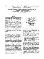
ecules were modeled at later stages of refinement, when
the electron density was unambiguous (Fig. 1) [the
poly(ethylene glycol) molecules are not shown].
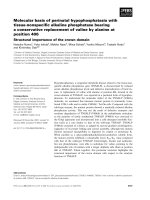
The overall structure of XynA in the complex with
MeGlcA
2
Xyl
3
(Fig. 2A,B) is nearly identical to the
1NOF structure described previously by Larson et al.
[16]. The enzyme consists of a (b ⁄ a)
8
-barrel catalytic
domain and a b-sheet immunoglobulin-like C-terminal
domain (a potential xylan-binding module) connected
by amino acids 45 and 317–322. One cis-peptide bond
has been found between Val200 and Ala201.
Superposition of the structure of the ligand-free
enzyme with the structure of the enzyme in the complex
using 378 CA atoms (CA atoms with two alternative
conformations were omitted) resulted in root mean
square, average and maximum xyz displacements
of 0.275 A
˚
, 0.241 A
˚
, and 0.849 A
˚
, respectively. It is
Table 1. Data collection and refinement statistics. R
merge
=
P
hkl
P
i
|I
i
(hkl)–ÆI(hkl)æ| ⁄
P
hkl
P
i
I
i
(hkl), where I
i
(hkl) is the intensity measure-
ment for the ith observation of reflection hkl and ÆI(hkl)æ is the average intensity for multiple measurements for this reflection.
R =
P
||F
obs
| ) |F
calc
|| ⁄
P
|F
obs
|, where F
obs
and F
calc
are observed and calculated structure factor amplitudes. A random subset (5%) of data
excluded from the refinement was used for R
free
factor calculation.
XynA–MeGlcA
2
Xyl
3
Data collection
Beamline X13 EMBL Hamburg
Wavelength (nm) 0.831
Space group P3
2
21 (No. 154)
Unit cell dimensions
a, b, c (A
˚
) 59.578, 59.578, 168.296
a, b, c (°) 90, 90, 120
Resolution range, overall ⁄ outer shell (A
˚
) 1.39–20.0 ⁄ 1.388–1.395
No. of observed reflections, overall ⁄ outer shell 468 272 ⁄ 3188
No. of unique reflections, overall ⁄ outer shell 70 207
Completeness, overall ⁄ outer shell (%) 98.6 ⁄ 98.6
Mean I ⁄ r (I ), overall ⁄ outer shell 8.5 ⁄ 1.3
Wilson B-factor (A
˚
) 18.89
R
merge
, overall ⁄ outer shell (%) 8.0 ⁄ 41.8
Refinement
R overall ⁄ R working ⁄ R
free
(%) 12.20 ⁄ 11.96 ⁄ 16.93
Asymmetric unit content (No. of molecules)
Protein ⁄ MeGlcA
2
Xyl
3
⁄ imidazole ⁄ poly(ethylene glycol) ⁄ water 1 ⁄ 1 ⁄ 1 ⁄ 3 ⁄ 571
B average (A
˚
2
)
Main chain ⁄ side chain ⁄ ligands ⁄ water 11.14 ⁄ 13.24 ⁄ 22.96 ⁄ 29.74
Model quality
Ramachandran plot
Preferred region [% (number of residues)] 90.1 (301 ⁄ 334
a
)
Allowed region [% (number of residues)] 9.6 (32 ⁄ 334
a
)
Generously allowed region [% (number of residues)] 0.3 (1 ⁄ 334
a
)
Geometry
Rmsd bond distances (A
˚
) 0.024
Rmsd bond ⁄ torsion angles (°) 1.973 ⁄ 6.841
Estimated standard uncertainties based on R-value ⁄ R
free
(A
˚
) 0.056 ⁄ 0.056
a
Number of nonglycine and nonproline residues.
L
ˇ
. Urba
´
nikova
´
et al. X-ray structure of xylanase A–ligand complex
FEBS Journal 278 (2011) 2105–2116 Journal compilation ª 2011 FEBS. No claim to original Slovakian government works 2107
interesting that the side chain conformations of amino
acids interacting with MeGlcA
2
Xyl
3
did not change as
a result of binding. The superposition of both structures
also showed that the acetate anion observed in the first
published structure (1NOF) [16] interacts with the posi-
tively charged guanidinium group of Arg293 in a man-
ner similar to the carboxylate group of MeGlcA
2
Xyl
3
.
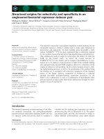
The protein substrate-binding site has a total area of
321.9 A
˚
2
, and is composed of 17 amino acids; 44.4%
of the binding site surface is hydrophobic, i.e. covered
by carbon atoms. The remaining 55.6% is polar, cov-
ered by nitrogen and oxygen atoms (Table S1;
Fig. 3A). Thirteen of the 17 amino acids form 174 van
der Waals contacts and 10 hydrogen bonds with MeG-
lcA
2
Xyl
3
(Table 2; Fig. 3B). The three xylose (Xyl)
units of the ligand take part in the stacking interac-
tions with the aromatic rings of Trp289, Tyr172 and
Trp55 in subsites ) 1, )2, and )3, as shown in detail in
Fig. 4A,B. The Xyl in subsite )1 is also coordinated
with Trp113, Asn164, and the catalytic Glu165 and
Glu253 (Fig. 4B). The MeGlcA moiety interacts with
the edges of the aromatic rings of the Trp289 and
Tyr290 side chains, and also forms one hydrogen bond
with the Trp289 amide nitrogen, NE1. The most
important interaction for substrate recognition appears
to be an ionic interaction between the positively
charged Arg293 guanidinium group and the negatively
charged carboxylate of MeGlcA (Fig. 4C).
An electron density found in the proximity of the
catalytic amino acids Glu165 and Glu253 was ascribed
to imidazole, a component of the crystallization buffer.
Imidazole interacts with Tyr168 and Trp232, and is
also electrostatically bound to the catalytic Glu165
(Fig. 4D). Thus, imidazole appears to occupy sub-
site +1, interacting with the Xyl or xylosyl residues of
the enzyme-cleaved substrates. Depending on the char-
acter of the substrate, this Xyl becomes the product of
hydrolysis or the nonreducing end of the leaving
group. A stereo view of the mode of binding of
MeGlcA
2
Xyl
3
is shown in Fig. 5A. The interactions of
the enzyme with MeGlcA
2
Xyl
3
and imidazole are sum-
marized in Table 2.
Binding energy calculations and ligand-docking
studies
The energy of ligand binding was estimated with lead-
finder [24]. The scoring functions of leadfinder are
based on a semiempirical molecular mechanical
approach that explicitly accounts for various types of
molecular interaction. The DG-scoring is a measure of
binding energy, and the virtual screening (VS) scoring
corresponds to the ligand-binding potency.
The experimentally determined structure of the pro-
tein–MeGlcA
2
Xyl
3
complex was used for calculating
the binding energy at pH 5.5, which is the pH
MeGlcA
2
Fig. 1. MeGlcA
2
Xyl
3
and imidazole in the 2F
o
) F
c
electron density
map (gray mesh), contoured at the 1.0 r level. Atoms are shown as
sticks and colored as follows: C, green; O, red; N, blue. Three Xyl resi-
dues with the MeGlcA moiety are bound in subsites )1, )2, and )3.
A
B
Fig. 2. The arrangement of protein and ligand molecules in the
XynA–MeGlcA
2
Xyl
3
crystal asymmetric unit. (A) A direct view of
the structure and (B) a view of the structure rotated 90° around the
y-axes, showing the active site of XynA. The catalytic (b ⁄ a)
8
-barrel
domain is in red, the C-terminal b
9
-barrel domain is in blue, the con-
necting region is in orange, the catalytic amino acids Glu165 and
Glu253 are in ball-and-stick representations, and MeGlcA
2
Xyl
3
, imid-
azole and three poly(ethylene glycol) molecules are in ball-and-stick
representations with bonds in bold green, yellow, and turquoise,
respectively.
X-ray structure of xylanase A–ligand complex L
ˇ
. Urba
´
nikova
´
et al.
2108 FEBS Journal 278 (2011) 2105–2116 Journal compilation ª 2011 FEBS. No claim to original Slovakian government works
optimum of the enzyme [14]. Similar calculations were
performed for its three analogs. For the first one, b-d-
xylopyranosyl-(1 fi 4)-[4-O-methyl-a-d-xy lopyranosyl-
(1 fi 2)]-b-d-xylopyranosyl-(1 fi 4)- d-xylose (MeXyl
2
Xyl
3
), the carboxyl group of MeGlcA was replaced
by hydrogen; that is, MeGlcA was converted to
4-O-methyl-d-xylose. For the second one, 4-O-methyl-
a-d-glucuronosyl-(1 fi 2)-b-d-xylopyranosyl-(1 fi 4)-
d-xylose (MeGlcA
2
Xyl
2
), the nonreducing xylosyl
residue of the ligand was replaced by hydrogen. The
third compound examined in this regard was Xyl
3
, the
core xylooligosaccharide.
In addition to the above calculations, the Xyl mono-
mer was docked into the hypothetical subsite +1,
which is occupied by imidazole in the crystal structure.
The program offered several different positions for Xyl
bound in subsite +1. The position displayed in Fig. 5B
corresponds to the lowest DG and VS scores, and is also
optimal from the structural point of view. The Xyl O4
atom appears to be hydrogen bonded to the catalytic
Glu165 and positioned in a relatively short distance
(2.83 A
˚
) from the glycosidic oxygen (O1) of the reduc-
ing-end xylosyl residue bound in subsite )1 (Fig. 5B).
The results of the binding energy calculations and
molecular docking are summarized in Table 3. The dif-
ference between the binding energies of MeGlcA
2
Xyl
3
and its virtual analog MeXyl
2
Xyl
3
indicates that the
ionic interaction of the ligand carboxyl group with
Arg293 corresponds to about 36% of the total binding
energy of MeGlcA
2
Xyl
3
()2.29 kcalÆmol
)1
versus
Table 2. Protein–MeGlcA
2
Xyl
3
and protein–imidazole interactions. The numbers in parentheses correspond to Xyl-binding subsites.
Protein atom Ligand atom ⁄ group Distance (A
˚
) Type of interaction
MeGlcA
2
Xyl
3
Arg293 NH1 MeGlcA COOH group 2.86 Salt bridge
Arg293 NE MeGlcA COOH group 2.93 Salt bridge
Tyr295 OH MeGlcA O6 2.65 Hydrogen bond
Ser258 OG MeGlcA O3 3.38 Hydrogen bond
Tyr255 OH MeGlcA O2 2.69 Hydrogen bond
Trp289 NE1 MeGlcA O5 3.36 Hydrogen bond
Tyr295 OH MeGlcA O5 3.14 Hydrogen bond
Trp113 NE1 Xyl (–1) O3 2.86 Hydrogen bond
Glu253 OE2 Xyl (–1) O2 2.76 Hydrogen bond
Asn164 ND2 Xyl (–1) O2 2.95 Hydrogen bond
Glu165 OE1 Xyl (–1) O1 2.60 Hydrogen bond
Glu165 OE2 Xyl (–1) O1 2.64 Hydrogen bond
Trp289 aromatic ring Xyl (–1) 3.86–6.05 Stacking
Tyr172 aromatic ring Xyl (–2) 4.07–4.59 Stacking
Trp55 aromatic ring Xyl (–3) 3.56–4.78 Stacking
Imidazole
Glu165 OE1 Imidazole N1 2.70 Hydrogen bond
Glu165 OE2 Imidazole N1 3.47 Hydrogen bond
Trp168 aromatic ring Imidazole ring 3.7–4.7 Stacking
Tyr232 aromatic ring Imidazole ring 3.5–4.6 Stacking
A
B
Fig. 3. Details of the interactions of XynA
with MeGlcA
2
Xyl
3
and imidazole. (A) Stick
representation of MeGlcA
2
Xyl
3
and imidaz-
ole (atoms: green, C; red, O; blue, N).
Amino acids involved into substrate binding
are in ball-and-stick representations (atoms:
gray, C; red, O; blue, N). Hydrogen bonds
are marked by dashed lines. (B) The van der
Waals surface representation of the
enzyme, showing the active site cleft filled
by MeGlcA
2
Xyl
3
and imidazole.
L
ˇ
. Urba
´
nikova
´
et al. X-ray structure of xylanase A–ligand complex
FEBS Journal 278 (2011) 2105–2116 Journal compilation ª 2011 FEBS. No claim to original Slovakian government works 2109
)6.22 kcalÆmol
)1
). The sum of both ionic and nonionic
enzyme–MeGlcA interactions corresponds to about
55% of the total binding energy ()3.47 kcalÆmol
)1
ver-
sus )6.22 kcalÆmol
)1
). The binding energy of the non-
reducing xylosyl residue in subsite )3 is only 9% of
the total DG ()0.55 kcalÆmol
)1
versus )6.22 kcalÆ-
mol
)1
). The calculated binding energies for the ligand,
its two virtual analogs and Xyl
3
(Table 3) correspond
to specific enzyme activities on different substrates (see
below).
Specific activity on aldouronic acids and linear
xylooligosaccharides
Two aldotetraouronic acids differing in the presence of
the nonreducing xylopyranosyl residue filling subsite )3
were available: 4-O-methyl-a-d-glucuronosyl-(1 fi 2)-b-
d-xylopyranosyl-(1 fi 4)-b-d-xylopyranosyl-(1 fi 4)-d-
xylose (MeGlcA
3
Xyl
3
), the shortest acidic oligosac
charide liberated from glucuronoxylan by endoxylanas-
es of GH10 [4], and aldopentaouronic acid, b-d-xylo
pyranosyl-(1 fi 4)-[4-O-methyl- a-d-glucuronosyl-(1 fi 2)]-
b-d-xylopyranosyl-(1 fi 4)-b-d-xylopyranosyl-(1 fi 4)-
d-xylose (MeGlcA
3
Xyl
4
), the shortest acidic oligosaccha
ride liberated from glucuronoxylan by endoxylanases of
GH family 11 [4] (Fig. S1). XynA hydrolyzed the ald-
opentaouronic acid more efficiently. Specific activities at
4mm substrate were 42 mmolÆmin
)1
Æmg
)1
for the pent-
amer and 13 mmolÆmin
)1
Æmg
)1
for the tetramer. These
data suggest that subsite )3 also contributes to sub-
strate binding. In view of the recent information that
a Bacillus GH30 xylanase shows activity on linear
b-1,4-xylooligosaccharides [15], we also examined the
rate of hydrolysis of xylotetraose and xylopentaose.
AB
C
D
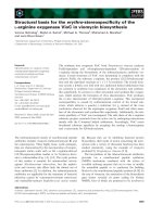
Fig. 4. Detailed view of the interaction of the enzyme with individual carbohydrate residues of MeGlcA
2
Xyl
3
derived from the enzyme–ligand
complex with imidazole bound in aglycone subsite +1. Amino acids and ligands are in ball-and-stick representations, with sticks colored gold
and green, respectively. The atoms are colored as follows: red, O; blue, N; gold and green, C. Hydrogen bonds are marked by dashed lines.
Stacking interactions are also highlighted as dashed lines connecting the centers of interacting groups marked by asterisks. The distances
are in A
˚
. (A) Stacking interactions of Tyr172 and Trp55 with xylosyl residues in subsites )2 and )3. (B) Hydrogen bonds between the
enzyme and xylosyl residue in subsite )1. The stacking interaction with Trp289 is also indicated. (C) Coordination of the MeGlcA residue of
the ligand with Tyr255, Ser258, Trp289, Arg293, and Tyr295. There is no stacking interaction of MeGlcA with the sandwich of
Trp289 ⁄ Tyr290. (D) Stacking interactions of imidazole in subsite +1 with Trp168 and Tyr232, and its hydrogen bond with the catalytic
Glu165. The six-membered aromatic ring of Trp168 and Leu204 might be involved in binding of Xyl in subsite +2.
X-ray structure of xylanase A–ligand complex L
ˇ
. Urba
´
nikova
´
et al.
2110 FEBS Journal 278 (2011) 2105–2116 Journal compilation ª 2011 FEBS. No claim to original Slovakian government works
At 4 mm, both oligomers served as enzyme substrates,
but with specific activities three orders of magnitude
lower than those on aldouronic acids. These observa-
tions point again to a crucial role for the MeGlcA
carboxylate in enzyme substrate recognition and the role
of MeGlcA as an essential specificity determinant. We
should mention that xylopentaose was hydrolyzed about
three times faster than xylotetraose, which is also in
accord with the results of the docking experiments and
calculated binding energies (Table 3).
Discussion
The xylanase investigated in this work is one of the
appendage-dependent endoxylanases, which are of
bacterial origin and can be found in the GH30
A
B
Fig. 5. Stereoview of the interactions of XynA with MeGlcA
2
Xyl
3
and imidazole (IMD). (A) Enzyme–MeGlcA
2
Xyl
3
interactions (for clarity,
Ser258, forming a hydrogen bond to MeGlcA, is not shown). Ligands and amino acids involved in ligand binding are in ball-and-stick represen-
tations (atoms: black, C; red, O; blue, N) with sticks colored green and light gray, respectively. Hydrogen bonds and ionic interactions are
marked by dashed lines. Glu253 is marked by an asterisk. (B) Interactions of the enzyme with Xyl docked at subsite +1. For comparison,
Xyl (derived from the MeGlcA
2
Xyl
3
structure) in subsite )1 and imidazole are also shown. The length of the hydrogen bonds is indicated in A
˚
.
Table 3. Summary of molecular modeling experiments and binding energy calculation.
Ligand Calculation based on
Binding energy,
DG (kcalÆmol
)1
) VS score
Difference in binding energies, DG
1
) DG
2
Ligands kcalÆmol
)1
Functional group
of ligand
MeGlcA
2
Xyl
3
Crystal structure )6.22 )10.10 – – –
MeXyl
2
Xyl
3
a
Crystal structure )3.93 )8.15 MeGlcA
2
Xyl
3
– MeXyl
2
Xyl
3
)2.29 COOH group
MeGlcA
2
Xyl
2
b
Crystal structure )5.67 )9.18 MeGlcA
2
Xyl
3
– MeGlcA
2
Xyl
2
)0.55 Xyl at subsite )3
Xyl
3
Crystal structure )2.75 )6.03 MeGlcA
2
Xyl
3
– Xyl
3
)3.65 MeGlcA
Xyl
1
at subsite +1 Docking )3.51 )5.06 – – –
a
COOH group of MeGlcA was replaced by hydrogen.
b
Nonreducing Xyl was replaced by hydrogen.
L
ˇ
. Urba
´
nikova
´
et al. X-ray structure of xylanase A–ligand complex
FEBS Journal 278 (2011) 2105–2116 Journal compilation ª 2011 FEBS. No claim to original Slovakian government works 2111
(formerly GH5) family [1,8,13–15]. These enzymes are
specialized for depolymerization of xylans that contain
GlcA or MeGlcA side substituents. They exhibit a
unique mode of action. The cleavage of the glucuron-
oxylan main chain takes place exclusively at the second
glycosidic linkage from the branch towards the reduc-
ing end of the polysaccharide chain. In other words,
the cleavage occurs during the formation of the pro-
ductive enzyme–substrate complex, in which the substi-
tuted xylopyranosyl residue is bound in the
hypothetical subsite )2. In this way, the MeGlcA or
GlcA residues determine the site of substrate cleavage,
and the content of these uronic acids determines the
xylan chain cleavage frequency. In this work, we con-
firm the hydrolysis of linear xylooligosaccharides by a
GH30 xylanase [15]; however, the rate of their hydro-
lysis with XynA was negligible in comparison with the
rate of hydrolysis of aldouronic acids.
After the 3D structure of the enzyme became
known [16] and the mode of GH30 xylanase action
had been elucidated [13,14], a question emerged con-
cerning the basis for the recognition of the MeGlcA
and GlcA residues by the enzyme. We have postu-
lated an ionic interaction between the uronic acid car-
boxylate and the positively charged Arg293 occurring
in the vicinity of a sandwich of two aromatic amino
acids, Tyr290 ⁄ Trp289, that could interact with the
uronic acid [14]. However, because the space between
Tyr290 and Trp289 in the published crystal structure
was too narrow to accommodate the uronic acid, it
became clear that the enzyme should be crystallized
in a complex with a suitable ligand and that the
structure of the complex could provide the required
information.
We have succeeded obtaining crystals of XynA with
the aldotetraouronic acid MeGlcA
2
Xyl
3
, which is a
product of the cleavage of MeGlcA
3
Xyl
4
by the same
enzyme. In the crystal structure, the ligand was found
to be bound in a manner similar to the one that we
have predicted [14]. The xylopyranosyl residue substi-
tuted by MeGlcA was bound in subsite )2, and MeG-
lcA was in a position that clearly indicates an ionic
interaction between its carboxyl group and the posi-
tively charged Arg293. However, MeGlcA was not
sandwiched between Tyr290 and Trp289, as proposed
earlier [14]. Instead, in addition to the ionic interaction
with Arg293, it interacts with the side chains of the
aromatic amino acids Tyr255, Trp289, and Tyr295,
and with Ser258, through several hydrogen bonds,
which are listed in Table 2 and shown in Figs 4C and
5A.
It is interesting that the ligand occurs in the complex
with XynA in the form of its a-anomer. Such a config-
uration corresponds to the enzyme a-glycosyl ester
intermediate with the catalytic glutamate Glu165. This
is interesting in light of the fact that the enzyme is a
retaining GH [1]. At this stage of our work, we do not
have any explanation for this observation.
An important question to be answered in connec-
tion with the mode of action of GH30 xylanases is
why the enzymes do not efficiently attack linear b-
1,4-linked xylooligosaccharides. The MeGlcA carbox-
ylate is involved in binding by Arg293. According to
the calculations of the binding energies of the ligand
and its virtual analogs (Table 3), the interaction of
the enzyme with MeGlcA is stronger than with the
xylopyranosyl residues in the negatively numbered
subsites. The ionic interaction could also be impor-
tant for the first contact of the enzyme with sub-
strates, and also indispensable for creating a stable
enzyme–substrate complex. In the next steps, the
enzyme–substrate complex formation could be based
on stacking interactions between aromatic amino
acids covering the enzyme binding site and Xyl resi-
dues of the xylan main chain. The final step could be
the locking of the substrate, namely MeGlcA and a
xylosyl or xylobiosyl moiety, at subsites +1 and +2,
in a proper position for cleavage. The importance of
Xyl binding at subsite +1 is supported by calcula-
tions of the binding energy of free Xyl in subsite +1
(Table 3; Fig. 5B). One can envisage strong bending
of the xylan chain as a consequence of both ionic
and stacking interactions. This apparently cannot
occur with linear oligosaccharides or a xylan main
chain that is either unsubstituted or carries uncharged
side substituents such as l-arabinose. The strong
bending could be the reason why the enzyme hardly
recognizes linear xylooligosaccharides as substrates
and does not attack arabinoxylan [14]. To learn more
about the enzyme–substrate interactions, complexes of
the enzyme with larger, nonhydrolyzable ligands
should be crystallized and their structure elucidated.
An alternative approach could include the preparation
of inactive enzyme mutants and crystallization of
these mutants with natural substrates.
Conclusions
The crystal structure of XynA with MeGlcA
2
Xyl
3
shows that the unique substrate specificity and mode
of action of bacterial GH30 xylanases on xylans with
MeGlcA and GlcA side substituents is achieved
mainly by recognition of the uronic acid side residue.
A crucial role in this recognition is ascribed to ionic
interaction of the enzyme with the uronic acid
carboxylate. Lack of the uronic acid renders the
X-ray structure of xylanase A–ligand complex L
ˇ
. Urba
´
nikova
´
et al.
2112 FEBS Journal 278 (2011) 2105–2116 Journal compilation ª 2011 FEBS. No claim to original Slovakian government works
xylan main chain virtually resistant to the enzyme’s
action. The specific activities on unsubstituted b-1,4-
xylooligosaccharides are three orders of magnitude
lower than those on similar substrates containing
MeGlcA.
Experimental procedures
Cloning and expression of XynA
Recombinant XynA was obtained by expressing its
synthetic gene in B. subtilis A164delta5 [25]. The synthetic
gene, based on the published gene sequence (Swiss-Prot:
Q46961), was generated by the company DNA2.0 (Menlo
Park, CA, USA) and delivered as a cloned fragment in
their standard cloning vector (kanamycin-resistant).
The synthetic gene sequence (Fig. S3A) was codon-opti-
mized for expression in B. subtilis following the recommen-
dations of Gustafsson et al. [26]. The expressed DNA
sequence can be found in Fig. S3B. The xylanase gene was
cloned with the signal peptide from Savinase [26] (included
in the vector), replacing the native secretion signal. The
coding region without the native signal was amplified by
PCR from the plasmid containing the synthetic gene, and
cloned into the expression vector pDG268neo [25]). The
PCR primers contained an N-terminal ClaI site and a
C-terminal Mlu I site. The PCR fragment and vector were
digested with ClaI and MluI. The vector and fragment were
ligated and transformed into Escherichia coli. Several rec-
ombinants were obtained. A plasmid containing the correct
gene sequence was transformed into B. subtilis, following
the methods in Widner et al. [27]. A recombinant B. subtilis
clone containing the integrated expression construct was
grown in PS-1 liquid culture medium [27]. The enzyme was
purified from the culture supernatant.
Purification of recombinant XynA
The culture supernatant, collected by centrifugation
(17 700 g for 30 min), was filtered (0.22 lm), and the filtrate
was adjusted to pH 8.5 and subsequently loaded onto an
MEP HyperCel (Pall, East Hills, NY, USA) XK 26 ⁄ 20 col-
umn (GE Healthcare Bio-Sciences, Piscataway, NJ, USA).
The column (60 mL) was equilibrated in 50 mm Tris ⁄ HCl
buffer (pH 8.5) (buffer A). Unbound protein was washed off
with 300 mL of buffer A. The proteins were eluted with
50 mm sodium acetate buffer (pH 4.5) (buffer B). Fractions
were analyzed by SDS ⁄ PAGE, and fractions containing the
enzyme were combined and their pH was adjusted to pH 6.0.
The combined fractions were diluted five times in 25 mm Mes
buffer (pH 6.0) (buffer C) and applied to a cation exchange
SP Sepharose Fast Flow (GE Healthcare Biosciences,
Uppsala, Sweden) XK 26 ⁄ 20 column (GE Healthcare Bio-
Sciences, Piscataway, NJ). The cation exchanger (20 mL)
was equilibrated in buffer C. Unbound protein was
washed off with 100 mL of buffer C. The XynA was
eluted with a linear gradient of NaCl (0–0.5 m) in buf-
fer C, using five column volumes. Fractions were analyzed
by SDS ⁄ PAGE, and those containing XynA were
combined.
Other enzymes
GH3 b-xylosidase was a product of a recombinant
Saccharomyces cerevisiae strain expressing a plasmid-borne
Aspergillus niger XlnD gene [28], GH67 a-glucuronidase
was obtained from R. P. deVries and J. Visser (Agricultural
University of Wageningen, The Netherlands), and GH115
a-glucuronidase was a product of Pichia stipitis [29].
Substrates and oligosaccharide ligand
The ligand used for cocrystallization with XynA was
MeGlcA
2
Xyl
3
. This aldotetraouronic acid was prepared
from MeGlcA
2
Xyl
4
, the shortest acidic product generated
from hardwood glucuronoxylan by a family 11 endo-b-
1,4-xylanase [27] by the action of recombinant XynA.
The enzyme catalyzed the reaction MeGlcA
3
Xyl
4
fi
MeGlcA
2
Xyl
3
+ Xyl [14]. MeGlcA
3
Xyl
4
(20 mg), isolated
from glucuronoxylan-spent medium of Thermomyces la-
nuginosus [30], was incubated in 2 mL of water with
0.3 mg of purified recombinant XynA at 30 °C. After the
hydrolysis was completed (examined by TLC), the prod-
uct was isolated from the reaction mixture by preparative
paper chromatography on Whatman No. 3 (prewashed
with deionized water) in the solvent system ethyl ace-
tate ⁄ acetic acid ⁄ water (18 : 7 : 8, v ⁄ v ⁄ v) for 17 h.
The sugars on guide strips were localized with the silver
nitrate reagent. The water eluate of the desired product
was filtered and freeze-dried. The structure of the product
as MeGlcA
2
Xyl
3
was confirmed enzymatically (Fig. S1).
The compound was resistant to GH67 a-glucuronidase
but served as a substrate for GH115 a-glucuronidase to
yield MeGlcA and xylotriose. It was hydrolyzed by the
GH3 b-xylosidase [28] to Xyl and MeGlcA
2
Xyl
2
, giving
MeGlcA and xylobiose with both types of a-glucuroni-
dase. MeGlcA
3
Xyl
3
was isolated from glucuronoxylan
hydrolysate by endoxylanase of GH10 as the shortest
acidic oligosaccharide [4]. Xylotetraose and xylopentaose
were from Megazyme (Ireland).
Crystallization
An enzyme solution was prepared by concentrating the pro-
tein in 25 mm MES buffer (pH 6.0), containing150 mm
NaCl, to a concentration of 20 mgÆmL
)1
, using an Amicon
stirred cell and a Biomax membrane with cutoff 5 kDa.
Fifty-microliter aliquots of the concentrated solution were
L
ˇ
. Urba
´
nikova
´
et al. X-ray structure of xylanase A–ligand complex
FEBS Journal 278 (2011) 2105–2116 Journal compilation ª 2011 FEBS. No claim to original Slovakian government works 2113
stored at )20 °C until use. Crystals were prepared by the
vapor diffusion method in a hanging drop, with XRL
plates and plastic coverslips (Molecular Dimensions,
Suffolk, UK). The drops were composed of the protein
stock solution and precipitant solution at a 1 : 1 ratio in a
final volume of 2 lL, and equilibrated against 500 lLof
precipitant solution. In the case of the cocrystallization, the
3-lL drops were prepared by mixing the protein, ligand
and precipitant solutions at a 1 : 1 : 1 ratio. An aqueous
solution of MeGlcA
2
Xyl
3
(20 mm) was used as the ligand
solution. Pact Premier I and II and Crystal Clear I crystalli-
zation kits (Molecular Dimensions) were used for prelimin-
ary crystallization screening. Clusters of thin and fragile
needle crystals were obtained under 19 conditions, which
were further optimized. Diffraction-quality crystals were
prepared by crystal seeding. Data were collected from the
crystal of the XynA–MeGlcA
2
Xyl
3
complex obtained by
cocrystallization with 0.1 m imidazole ⁄ d,l-malic acid buffer
(pH 7.5) and 20% (w ⁄ v) poly(ethylene glycol) 1500 as a
precipitant solution.
Data collection and structure determination
The crystals were tested and data were collected at the X13
beamline at EMBL c ⁄ o DESY, Hamburg, Germany. The
crystals were mounted on the loops, soaked in a cryopro-
tectant solution, and flash cooled in a stream of cold nitro-
gen gas (100 K) directly at the goniometer head.
As cryoprotectants, Paratone-N, perfluoropolyether, paraf-
fin oil and precipitant solution enriched with glycerol to a
final concentration of 20% were tested. The best results
were obtained with paraffin oil. Data were collected at
100 K, according to the strategy proposed by best [31], and
processed by xds [32], and scala [33], reindex and com-
bat from ccp4 suite 6.1.3 (Collaborative Computational
Project Number 4, 1994 [23]), using ccp4i Interface 2.0.6
[34] running under Windows. The structure was solved by
the molecular replacement method with molrep [35] and
xylanase A (Protein Data Bank code 1NOF [16]) as a
model structure. The structure was refined with ref-
mac 5.5.01 [36] in combination with coot-findwaters,
and the model was visualized and rebuilt with coot [37].
All electron density maps were calculated by FFT [38]. For
structure validation, procheck was used [39,40], and for
structure analyses, areaimol, contact and other programs
of the ccp4 suite were used with the default parameters.
Figures were prepared with molscript [41] and pymol [42].
Molecular modeling
Docking experiments and binding energy calculations were
performed with leadfinder [23]. This program was also
used for the preparation of the protein structure for dock-
ing by addition of hydrogen atoms according to optimal
ionization states of protein residues at a given pH. The
ligand structures were prepared for molecular modeling in
their optimal protonation state with ChemAxon marvin
suite [43].
Specific activity on aldouronic acids and linear
xylooligosaccharides
Four-millimolar solutions of the compounds in 50 mm
sodium acetate buffer (pH 5.5) were incubated at 40 °C
with XynA at various dilutions. At time intervals, aliquots
were taken to determine the reducing sugars by the Somo-
gyi–Nelson procedure [44]. The high background of the
substrates reduced the accuracy of the measurements,
particularly at early stages of hydrolysis.
Acknowledgements
The authors are grateful to M. Czisza
´
rova
´
for excellent
technical assistance. This work was supported by
VEGA grants 2 ⁄ 0001 ⁄ 10 and 2 ⁄ 0165 ⁄ 08 from the Slo-
vak Academy of Sciences. We acknowledge the EMBL
X13 beamline at the DORIS storage ring, DESY,
Hamburg for providing us with synchrotron source
facilities. We thank M. Groves (EMBL Hamburg) for
his help with data processing, and O. Stroganov
(BioMolTech) for technical help with leadfinder.
Note added in proof
During processing of this article for publication we
have learned about the appearence of the paper
describing similar substrate recognition mechanism by
a GH30 xylanase from Bacillus subtilis using a crystal
structure of the complex of the enzyme with different
ligand (St John FJ, Hurlbert JC, Rice JD, Preston JF
& Pozharski E (2011) Ligand bound structures of a
glycosyl hydrolase family 30 glucuronoxylan xylanohy-
drolase. J Mol Biol 407, 92–109).
References
1 Henrissat B & Davies GJ (1997) Structural and
sequence-based classification of glycoside hydrolases.
Curr Opin Struct Biol 7, 637–644.
2 Biely P (2003) Xylanolytic enzymes. In Handbook of
Food Enzymology (Whitaker JR, Voragen AGJ & Wond
DWS eds), pp. 879–915. Marcel Dekker, New York.
3 Collins T, Gerday C & Feller G (2005) Xylanases,
xylanase families and extremophilic xylanases. FEMS
Microbiol Rev 29, 3–23.
4 Biely P, Vrs
ˇ
anska
´
M, Tenkanen M & Kluepfel D (1997)
Endo-b-1,4-xylanase families: differences in catalytic
properties. J Biotechnol 57, 151–166.
X-ray structure of xylanase A–ligand complex L
ˇ
. Urba
´
nikova
´
et al.
2114 FEBS Journal 278 (2011) 2105–2116 Journal compilation ª 2011 FEBS. No claim to original Slovakian government works
5 Pell G, Taylor EJ, Gloster TM, Turkenburg JP, Fontes
CMGA, Ferreira LMA, Nagy T, Clark SJ, Davies GJ
& Gilbert HJ (2004) The mechanism by which family 10
glycoside hydrolases bind decorated substrates. J Biol
Chem 279, 9597–9605.
6 Pollet A, Delcour JA & Courtin CM (2010) Structural
determinants of the substrate specificities of xylanases
from different glycoside hydrolase families. Crit Rev
Biotechnol 30, 176–191.
7 Hespell RB (1998) Extraction and characterization of
hemicellulose from the corn fiber produced by corn
wet-milling processes. J Agric Food Chem 46, 2615–
2619.
8 Collins T, Meuwis M-A, Stals I, Claeyssens M, Feller
G & Gerday C (2002) A novel family 8 xylanase, func-
tional and physicochemical characterization. J Biol
Chem 277, 35133–35139.
9 St John FJ, Gonzalez JM & Pozharski E (2010) Consol-
idation of glycosyl hydrolase family 30: a dual
domain 4 ⁄ 7 hydrolase family consisting of two structur-
ally distinct groups. FEBS Lett 584, 4435–4441.
10 Luo H, Yang J, Li J, Shi P, Huang H, Bai Y, Fan Y &
Yao B (2010) Molecular cloning and characterization of
the novel acidic xylanase XYLD from Bispora sp.
MEY-1 that is homologous to family 30 glycosyl hydro-
lases. Appl Microbiol Biotechnol 86, 1829–1839.
11 Nishitani K & Nevins DJ (1991) Glucuronoxylan xylan-
ohydrolase. A unique xylanase with the requirement for
appendant glucuronosyl units. J Biol Chem 266, 6539–
6543.
12 Hurlbert JC & Preston JF (2001) Functional character-
ization of a novel xylanase from a corn strain of
Erwinia chrysanthemi. J Bacteriol 183, 2093–2100.
13 St John FJ, Rice JD & Preston JF (2006) Characteriza-
tion of XynC from Bacillus subtilis subsp. Subtilis
strain 168 and analysis of its role in depolymerization
of glucuronoxylan. J Bacteriol 188, 8617–8626.
14 Vrs
ˇ
anska
´
M, Kolenova
´
K, Puchart V & Biely P (2007)
Mode of action of glycoside hydrolase family 5 glucu-
ronoxylan xylanohydrolases from Erwinia chrysanthemi.
FEBS J 274, 1666–1677.
15 Gallardo O, Fernandez-Fernandez M, Valls C, Valenzue-
la SV, Roncero MB, Vidal T, Diaz P & Pastor FIJ (2010)
Characterization of a family GH5 xylanase with activity
on neutral oligosaccharides and evaluation as a pulp
bleaching aid. Appl Environ Microbiol 76, 6290–6294.
16 Larson SB, Day J, Barba de la Rosa AP, Keen NT
& McPherson A (2003) First crystallographic struc-
ture of a xylanase from glycoside hydrolase
family 5: implication for catalysis. Biochemistry 42,
8411–8422.
17 St John FJ, Godwin DK, Preston JF, Pozharski E &
Hulbert JC (2009) Crystallization and crystallographic
analysis of Bacillus subtilis xylanase C. Acta Crystallogr
F65, 499–503.
18 Biely P, Kra
´
tky Z & Vrs
ˇ
anska
´
M (1981) Substrate-bind-
ing site of endo-1,4-
b-xylanase of the yeast Cryptococcus
albidus. Eur J Biochem 119, 559–564.
19 Davies GJ, Wilson KS & Henrissat B (1997) Nomencla-
ture for sugar-binding subsites in glycosyl hydrolases.
Biochem J 321, 557–559.
20 D’Arcy A (1994) Crystallizing proteins: a rational
approach? Acta Crystallogr D50, 469–471.
21 Matthews BW (1968) Solvent content of protein crys-
tals. J Mol Biol 33, 491–497.
22 Barba de la Rosa AP, Day J, Larson SB, Keen NT &
McPherson A (1997) Crystallization of xylanase from
Erwinia chrysanthemi: influence of heat and polymeric
substrate. Acta Crystallogr D53, 256–261.
23 Collaborative Computational Project Number 4 (1994)
The CCP4 suite: programs for protein crystallography.
Acta Crystallogr D50, 760–763.
24 Stroganov VO, Novikov FN, Stroylov VS, Kulkov V &
Chilov GG (2008) LeadFinder: an approach to improve
accuracy of protein–ligand docking, binding energy esti-
mation, and virtual screening. J Chem Inf Mode 48,
2371–2385.
25 Betzel C, Klupsch S, Papendorf G, Hastrup S, Branner
S & Wilson KS (1992) Crystal structure of the alkaline
proteinase Savinase from Bacillus lentus at 1.4 A
˚
resolu-
tion. J Mol Biol 223, 427–445.
26 Gustafsson C, Govindarajan S & Minshull J (2004)
Codon bias and heterologous protein expression. Trends
Biotechnol 22, 346–353.
27 Widner B, Thomas M, Sternberg D, Lammon D, Behr
R & Sloma A (2000) Development of marker-free
strains of Bacillus subtilis capable of secreting high
levels of industrial enzymes. J Ind Microbiol Biotechnol
25, 204–212.
28 Biely P, Hirsch J, la Grange D, van Zyl WH & Prior
BA (2000) A chromogenic substrate for a b-xylosidase-
coupled assay of a-glucuronidase. Anal Biochem 286,
289–294.
29 Ryabova O, Vrs
ˇ
anska
´
M, Kaneko S, van Zyl WH &
Biely P (2009) Novel family of hemicellulolytic a-glucu-
ronidase. FEBS Lett 583, 1457–1462.
30 Puchart V & Biely P (2008) Simultaneous production
of endo-b-1,4-xylanase and branched xylooligosaccha-
rides by Thermomyces lanuginosus. J Biotechnol 137,
34–43.
31 Bourenkov GP & Popov AN (2006) A quantitative
approach to data-collection strategies.
Acta Crystallogr
D62, 58–64.
32 Kabsch WJ (1993) Automatic processing of rotation
diffraction data from crystals of initially unknown sym-
metry and cell constants. J Appl Crystallogr 26, 795–
800.
33 Evans PR (2006) Scaling and assessment of data qual-
ity. Acta Crystallogr D62, 72–82.
L
ˇ
. Urba
´
nikova
´
et al. X-ray structure of xylanase A–ligand complex
FEBS Journal 278 (2011) 2105–2116 Journal compilation ª 2011 FEBS. No claim to original Slovakian government works 2115
34 Potterton E, Briggs P, Turkenburg M & Dodson EJ
(2003) A graphical user interface to the CCP4 program
suite. Acta Crystallogr D59, 1131–1137.
35 Vagin A & Teplyakov A (1997) MOLREP: an auto-
mated program for molecular replacement. J Appl Crys-
tallogr 30, 1022–1025.
36 Murshudov GN, Vagin AA & Dodson EJ (1997)
Refinement of macromolecular structures by the maxi-
mum-likelihood method. Acta Crystallogr D53, 240–
255.
37 Emsley P, Lohkamp B, Scott WG & Cowtan K (2010)
Features and development of Coot. Acta Crystallogr
D66, 486–501.
38 Read RJ & Schierbeek AJ (1988) A phased translation
function. J Appl Crystallogr 21, 490–495.
39 Laskowski RA, MacArthur MW, Moss DS & Thornton
JM (1993) PROCHECK: a program to check the ste-
reochemical quality of protein structures. J Appl Crys-
tallogr 26, 283–291.
40 Ramachandran GN & Sasisekharan V (1968) Confor-
mation of polypeptides and proteins. Adv Protein Chem
23, 283–438.
41 Kraulis PJ (1991) MOLSCRIPT: a program to produce
both detailed and schematic plots of protein structures.
J Appl Crystallogr 24, 946–950.
42 DeLano WL (2008) The PyMOL Molecular Graphics
System. DeLano Scientific LLC, Palo Alto, CA, USA.
43 Marvin 5.3.8. (2010) ChemAxon (maxon.
com).
44 Paleg LG (1959) Citric acid interference in the estima-
tion of reducing sugars with alkaline copper reagents.
Anal Chem 31, 1092–1094.
45 Berman HP, Henrick K & Nakamura H (2003)
Announcing the worldwide Protein Data Bank. Nat
Struct Biol 10, 980.
Supporting information
The following supplementary material is available:
Fig. S1. Conversion of aldopentaouronic acid gener-
ated from glucuronoxylan by GH11 endoxylanases to
aldotetraouronic acid used as the ligand (framed struc-
ture) for cocrystallization of E. chrysanthemi GH30
xylanase.
Fig. S2. Dynamic light scattering profile of E. chry-
santhemi GH30 xylanase.
Fig. S3. Examples of distinct habits of E. chrysanthemi
GH30 xylanase crystals.
Fig. S4. The gene and the protein sequences of
E. chrysanthemi GH30 xylanase.
Table S1. Characterization of the binding site surface
and the list of the interactions between E. chrysanthemi
GH30 xylanase and MeGlcA
2
Xyl
3
ligand.
This supplementary material can be found in the
online version of this article.
Please note: As a service to our authors and readers,
this journal provides supporting information supplied
by the authors. Such materials are peer-reviewed and
may be re-organized for online delivery, but are not
copy-edited or typeset. Technical support issues arising
from supporting information (other than missing files)
should be addressed to the authors.
X-ray structure of xylanase A–ligand complex L
ˇ
. Urba
´
nikova
´
et al.
2116 FEBS Journal 278 (2011) 2105–2116 Journal compilation ª 2011 FEBS. No claim to original Slovakian government works