Báo cáo khoa học: MNB⁄ DYRK1A as a multiple regulator of neuronal development pdf
Bạn đang xem bản rút gọn của tài liệu. Xem và tải ngay bản đầy đủ của tài liệu tại đây (326.81 KB, 13 trang )
MINIREVIEW
MNB
⁄
DYRK1A as a multiple regulator of neuronal
development
Francisco J. Tejedor
1
and Barbara Ha
¨
mmerle
2
1 Instituto de Neurociencias, CSIC and Universidad Miguel Hernandez, Alicante, Spain
2 Centro de Investigacio
´
n Prı
´
ncipe Felipe, Valencia, Spain
Introduction
MNB ⁄ DYRK1A is a protein kinase that belongs to
the dual-specificity tyrosine phosphorylation-regulated
kinase (DYRK) family. MNB ⁄ DYRK1A is highly
conserved from insects to humans [1] and it displays
characteristic properties that are discussed in detail
in one of the three minireviews in this series [2].
Orthologous genes have been cloned independently in
various organisms and named Minibrain (Mnb) or
Dyrk1A.
The evidence from diverse experimental systems has
shown various possible functions of MNB ⁄ DYRK1A
in central nervous system (CNS) development, includ-
ing its influence on proliferation, neurogenesis, neuro-
nal differentiation, cell death and synaptic plasticity
(see Table 1). These data, together with the localiza-
tion of the human MNB ⁄ DYRK1A gene on chromo-
some 21 [3,4] and its overexpression in the brain of
fetuses with Down syndrome (DS, trisomy 21) [5],
have provided support to several hypotheses implicat-
ing MNB ⁄ DYRK1A in neurodevelopmental altera-
tions underlying the cognitive deficits of DS
(previously reviewed in [6,7]). These facts have cer-
tainly stimulated and conditioned the research into the
neurobiological functions of MNB ⁄ DYRK1A. More
recently, the observation that MNB ⁄ DYRK1A is over-
expressed in the adult DS brain [8], together with bio-
chemical data, also implicated MNB ⁄ DYRK1A in
various neurodegenerative processes. This issue is
extensively covered in the second accompanying paper
of this minireview series [9].
Here we will focus on the neurodevelopmental func-
tions of MNB ⁄ DYRK1A. We will discuss the data
revealing the main roles interpreted by MNB ⁄ DYRK1A
during brain development and their possible molecular
Keywords
Down syndrome; neural proliferation;
neurogenesis; neuronal differentiation;
protein kinase
Correspondence
F. J. Tejedor, Instituto de Neurociencias,
CSIC and Universidad Miguel Hernandez,
Alicante, Spain
Fax: 34 965919561
Tel: 34 965919423
E-mail:
(Received 20 July 2010, revised 13 September
2010, accepted 23 September 2010)
doi:10.1111/j.1742-4658.2010.07954.x
MNB ⁄ DYRK1A is a member of the dual-specificity tyrosine phosphoryla-
tion-regulated kinase (DYRK) family that has been strongly conserved
across evolution. There are substantial data implicating MNB ⁄ DYRK1A
in brain development and adult brain function, as well as in neurodegener-
ation and Down syndrome pathologies. Here we review our current under-
standing of the neurodevelopmental activity of MNB ⁄ DYRK1A. We
discuss how MNB ⁄ DYRK1A fulfils several sequential roles in neuronal
development and the molecular mechanisms possibly underlying these func-
tions. We also summarize the evidence behind the hypotheses to explain
how the imbalance in MNB ⁄ DYRK1A gene dosage might be implicated in
the neurodevelopmental alterations associated with Down syndrome.
Finally, we highlight some research directions that may help to clarify the
mechanisms and functions of MNB ⁄ DYRK1A signalling in the developing
brain.
Abbreviations
CNS, central nervous system; DS, Down syndrome; DYRK, dual-specificity tyrosine phosphorylation-regulated kinase;
NRSF, neuron-restrictive silence factor.
FEBS Journal 278 (2011) 223–235 ª 2010 The Authors Journal compilation ª 2010 FEBS 223
Table 1. Substrates and proteins that interact with MNB ⁄ DYRK1A in relation to its neuronal functions. Because the spatiotemporal regulation of its expression appears to be critical to
understanding MNB ⁄ DYRK1A’s roles in neuronal development, we have also included possible regulators of Mnb ⁄ Dyrk1A expression and of MNB ⁄ DYRK1A kinase activity. For each pro-
tein we show: its main molecular properties, the molecular relationship with MNB ⁄ DYRK1A, the phosphorylation sites (if experimentally determined), the experimental system used to
define this relationship, the possible function in neuronal development (if any), and the literature showing the relationship to MNB ⁄ DYRK1A. This list has been restricted to those
genes ⁄ proteins for which there is evidence in the literature of a neuronal-related activity. Additionally, we highlight (*) those cases in which there is evidence (or strong indications) that
the interaction with MNB ⁄ DYRK1A is involved in neuronal functions. ActR, regulator of activity; ExpR, regulator of expression; I, interacting protein; S, substrate; ND, not determined; Cult-
Neu, cultured neurons; ivCNS, CNS in vivo; NCL, neural cell line; nNCL, non-neural cell line; Dif, differentiation; Other, nondevelopmental neuronal function; Prol, proliferation; Syn, synapse
related; UF, unknown function; $, MNB ⁄ DYRK1A kinase primes the phosphorylation of several substrates by glycogen synthase kinase 3.
Protein or signalling pathway Molecular nature
Molecular
relationship
with MNB ⁄
DYRK1A Phosphorylation sites Experimental system Function Reference
Amphiphysin Protein associated with the
cytoplasmic surface of synaptic
vesicles
S Ser293 NCL, ivCNS Syn [54]
b-Amyloid Peptide derived from amyloid
precursor protein. Main
component of amyloid plaques in
Alzheimer’s disease
ExpR ivCNS, NCL Other [78]
Arip4 (androgen receptor
interacting protein 4)
Steroid hormone receptor cofactor I nNCL, CultNeu, ivCNS UF [79]
APP (amyloid precursor protein) Amyloid precursor protein S Thr668 nNCL Other [80]
ASF (alternative splicing factor) Splicing factor S, I Ser227, Ser234, Ser238 NCL, nNCL Other [81]
bFGF Growth factor ActR NCL Dif [38]
Caspase 9* Cystein aspartyl protease S Thr125 nNCL, ivCNS Cell death [59,82,83]
Cyclin D1* Cell cycle regulator ? ivCNS, NCL Prol [26]
CREB (cAMP responsive element
binding protein)
Transcription factor S Ser133 NCL Dif [38]
CRY2 (cryptochrome 2) Flavoprotein, involved in circadian
rhythm
S Ser553, Ser557 nNCL, ivCNS Other [84]
DNM1 (dynamin 1)* Cytoplasmic protein, involved in
membrane trafficking
S Ser857 nNCL, ivCNS Dif [14,16,50,51]
Endophilin 1 Cytoplasmic protein involved in
membrane trafficking
I ivCNS Syn [55]
E2F1 Transcription factor, involved in cell
cycle regulation
ExpR NCL, nNCL Prol, Dif [22]
FKHR ⁄ FOXO1 (forkhead in
rhabdoyosarcoma)
Transcription factor S, I Ser329 nNCL UF [85,86]
GLI1 (glioma-associated
oncogene 1)
Transcription factor involved in SHH
signalling
S Multiple sites (ND) NCL, nNCL Prol ⁄ Dif [36,87]
GSK-3 (glycogen synthase
kinase 3)*
Protein kinase involved in multiple
cellular processes
$ nNCL, CultNeu, ivCNS Dif, other [43,88,89]
MNB ⁄ DYRK1A in neuronal development F. J. Tejedor and B. Ha
¨
mmerle
224 FEBS Journal 278 (2011) 223–235 ª 2010 The Authors Journal compilation ª 2010 FEBS
Table 1. (Continued).
Protein or signalling pathway Molecular nature
Molecular
relationship
with MNB ⁄
DYRK1A Phosphorylation sites Experimental system Function Reference
Hip1 (huntingtin interacting protein 1) Accessory protein of the
clathrin-mediated endocytosis
pathway
S ND NCL Dif [90]
INI1 ⁄ SNF5; SNR1 Chromatin modifying proteins I NCL, CultNeu, ivCNS Prol [23,45]
MAP1B* Microtubule-associated protein S Ser1392 nNCL, CultNeu Dif [43]
NFAT (nuclear factor of activated T-cells*) Transcription factor S ND ivCNS, NCL Dif [46,47]
Notch* Cell–cell signalling transmembrane
receptor protein
S Multiple sites (ND) NCL, nNCL, ivCNS Prol, Dif [31]
NRSF ⁄ REST (neuron-restrictive
silence factor)
Transcriptional repressor ExpR nNCL, ivCNS Prol ⁄ Dif [33]
p53* Transcription factor S Ser15 NCL, ivCNS [27]
PAHX-AP1 Phytanoyl-CoA a-hydroxylase
associated protein 1, brain-specific
protein
I NCL UF [91]
Presenilin1 Catalytic subunit of c-secretase S Thr354 NCL, nNCL, ivCNS UF [92]
Ras ⁄ Map kinase signalling Transmembrane signalling pathway I NCL Dif [39]
SEPT4 (septin 4)* GTPase and cytoskeletal
scaffolding protein
S ND nNCL, ivCNS Syn [49]
SIRT1 NAD-dependent protein
deacetylase
S Thr522 nNCL Cell death [74]
SPRY2 (sprouty2)* Negative modulator of growth
factor-mediated tyrosine kinase
receptor signalling
S Thr75 CultNeu, ivCNS Prol, Dif [19,93]
STAT3 Signal transducer and activator of
transcription
S Ser727 nNCL UF [94,95]
SJI1 (synaptojanin 1) Phosphoinositide phosphatase S Multiple sites (ND) ivCNS Syn [53]
a-synuclein Cytoplasmic protein, major
component of Lewy bodies
S Ser87 NCL, ivCNS Other [96]
TAU* Cytoskeletal protein, microtubule
associated
S Thr212 nNCL, ivCNS Other [78,88,97]
14-3-3 14-3-3 family of regulating proteins I, ActR NCL, nNCL UF [98,99]
F. J. Tejedor and B. Ha
¨
mmerle MNB ⁄ DYRK1A in neuronal development
FEBS Journal 278 (2011) 223–235 ª 2010 The Authors Journal compilation ª 2010 FEBS 225
mechanisms. Additionally, and given the extensive
repertoire of putative substrates and proteins with
which the MNB ⁄ DYRK1A kinase may interact, we will
try to highlight the genes ⁄ proteins related to its neuro-
developmental activities. We will also discuss the
possible implications of MNB ⁄ DYRK1A in the neuro-
developmental alterations associated with DS. Finally,
we will highlight some directions for future research that
we think may help to clarify the mechanisms and func-
tions of MNB ⁄ DYRK1A signalling in the developing
brain.
The diverse functions of MNB ⁄ DYRK1A
in neuronal development
The initial evidence for the involvement of
MNB ⁄ DYRK1A in neurodevelopment was provided
by the analysis of mnb mutants of Drosophila. These
flies develop a smaller adult brain, particularly in the
optic lobes, which appears to be caused by altered
proliferation in the neuroepithelial primordia of the
larval CNS. This phenotype suggests a key function
of MNB ⁄ DYRK1A in the regulation of neural prolif-
eration and neurogenesis [10]. The highly conserved
structure of this kinase [1] prompted extensive studies
to be carried out on its vertebrate homologues.
Indeed, a smaller brain with fewer neurons in certain
regions was described in haploinsufficient Dyrk1A
+ ⁄ )
mice [11], strongly suggesting an evolutionary con-
served function of MNB ⁄ DYRK1A in brain develop-
ment. This idea is also supported by the fact that
truncation of the human MNB ⁄ DYRK1A gene causes
microcephaly [12].
Although in mammals Mnb ⁄ Dyrk1A is expressed in
most adult tissues [5,13], its expression seems to be
prevalent during embryonic brain development and it
gradually decreases during postnatal periods to reach
low levels in the adult [13,14]. Mnb ⁄ Dyrk1A is specifi-
cally expressed in four sequential phases during the
development of the mouse brain: transient expression
in preneurogenic progenitors; cell cycle-regulated
expression in neurogenic progenitors; transient expres-
sion in recently born neurons; and persistent expres-
sion in late differentiating neurons ([14]; summarized
in Fig. 1). This rather dynamic cellular ⁄ temporal
expression strongly suggests that MNB ⁄ DYRK1A
plays several sequential roles in neuronal development,
which we shall discuss in this section. These roles seem
to be neuron specific, as the analysis of the developing
chick [15,16] and mouse CNS [14] show that
MNB ⁄ DYRK1A expression is restricted to neuronal
lineages, although its expression in glia has been
reported in primary cultures [17].
Proliferation and neurogenesis
There is strong evidence that Mnb ⁄ Dyrk1A is tran-
siently expressed during the single cell cycle of preneur-
ogenic chick and mouse embryonic neuroepithelial
progenitors that precedes the onset of neurogenesis
[14,15]. This expression is of particular interest as
Mnb ⁄ Dyrk1A mRNA is asymmetrically segregated
during cell division and it is inherited by only one of the
daughter cells [15] (Fig. 1). These data, together with
its co-expression in preneurogenic mouse neuroepithelia
with Tis21 [15], an antiproliferative gene that is upregu-
lated in neural progenitors that make the switch from
proliferative to neuron-generating divisions [18], sug-
gest that Mnb ⁄ Dyrk1A may act as a cell determinant of
neurogenesis. Accordingly, Mnb ⁄ Dyrk1A could induce
the switch from proliferative to neurogenic cell divi-
sions in neuronal progenitors. Interestingly, it has been
recently shown that MNB ⁄ DYRK1A protein is actively
distributed during adult neural stem cell division. Con-
sequently, the inherited MNB ⁄ DYRK1A kinase acts as
an inhibitor of epidermal growth factor receptor degra-
dation by phosphorylating sprouty2, a modulator of
tyrosine kinase receptor signalling [19]. In accordance
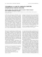
Fig. 1. Schematic representation of the sequential expression of
Mnb ⁄ Dyrk1A during the transition from neural proliferation to neu-
ronal differentiation. In the vertebrate neuroepithelia, Mnb ⁄ Dyrk1A
mRNA is first transiently expressed in preneurogenic progenitors,
before it is asymmetrically segregated during cell division and it is
inherited by only one of the daughter progenitor cells, triggering
the onset of neurogenic divisions. Its expression is maintained in
neurogenic progenitors although at a lower level. Later,
Mnb ⁄ Dyrk1A is also transiently upregulated in postmitotic precur-
sors (newborn neurons) and downregulated as the neuron begins
to migrate away from the ventricular zone (VZ). Once the migrating
neuron reaches its target position, Mnb ⁄ Dyrk1A is again expressed
and it translocates transiently into the nucleus preceding the onset
of dendrite formation. As dendrites begin to grow, MNB ⁄ DYRK1A
localizes to the apical side of the growing dendrites.
MNB ⁄ DYRK1A in neuronal development F. J. Tejedor and B. Ha
¨
mmerle
226 FEBS Journal 278 (2011) 223–235 ª 2010 The Authors Journal compilation ª 2010 FEBS
with this, adult neural stem cells derived from
Dyrk1A
+ ⁄ )
mice exhibit defects in self-renewal.
Noteworthy, the activity of Pom1p, an
MNB ⁄ DYRK1A-related kinase from Schizosacchar-
omyces pombe, is cell cycle regulated in relation to
symmetric growth and division [20]. However, Pom1p
activity is high during symmetric cell division and
when lost cells undergo asymmetric growth and divi-
sion, the opposite to what appears to occur with
MNB ⁄ DYRK1A in neural progenitors [14,15]. More-
over, mutants of mbk-1, the closest Mnb ⁄ Dyrk1A-
related gene in Caenorhabditis elegans, do not show
neurodevelopmental alterations [21]. Thus, new func-
tions have probably been acquired by DYRK kinases
during evolution to adapt to the new morphogenetic
requirements of complex nervous systems.
MNB ⁄ DYRK1A is also expressed in neurogenic
progenitors in the Drosophila larval optic lobe [10] and
in the embryonic mouse brain [14]. Although this
expression seems to occur throughout the cell cycle, it
is possible that the intensity of Mnb⁄ Dyrk1A expres-
sion might vary at different cell cycle stages. Indeed,
the expression of Mnb ⁄ Dyrk1A can be regulated by
E2F1 [22], a transcription factor that plays a key role
in the control of cell proliferation. Conversely, there is
also evidence that MNB ⁄ DYRK1A may participate in
the regulation of the cell cycle. For instance, it has
been reported that MNB ⁄ DYRK1A interacts with
SNR1 in Drosophila [23], a chromatin remodelling
factor with a relevant role in cell cycle regulation
[24]. Interestingly, increased levels of cyclin B1 have
been detected in transgenic mice overexpressing
Mnb ⁄ Dyrk1A [25] and it has recently been proposed
that MNB ⁄ DYRK1A regulates the nuclear export and
degradation of cyclin D1 in neurogenic mouse neuro-
epithelia [26]. Another very recent report has shown
that the overexpression of MNB ⁄ DYRK1A induced
impaired G1 ⁄ G0–S phase transition in immortalized
rat hippocampal progenitor cells [27]. The proposed
mechanism is mediated by the phosphorylation of p53,
which led to the induction of p21CIP1. There are also
indications that MNB ⁄ DYRK1A is involved in the
mitosis of non-neural cell lines [28]. These data estab-
lish a rather complex scenario with MNB⁄ DYRK1A
potentially fulfilling multiple actions in cell cycle regu-
lation for which we have very little understanding of
the molecular details.
Interestingly, important evidence has recently
emerged regarding the role of MNB ⁄ DYRK1A in ter-
minating proliferation. Thus, based on the transient
co-expression of MNB ⁄ DYRK1A with p27KIP1, the
main cyclin-dependent kinase inhibitor in the mamma-
lian forebrain [29], we proposed that MNB ⁄ DYRK1A
is involved in the developmental signals that control
cell cycle exit and early events of neuronal differentia-
tion [14]. Indeed, it was recently reported that the
overexpression of MNB ⁄ DYRK1A in the embryonic
mouse telencephalon inhibits proliferation and induces
premature neuronal differentiation of neural progeni-
tors [26]. This gain of function was proposed to be
driven through cyclin D1 nuclear export and
degradation. Nevertheless, it has still to be proven
whether the effect on cyclin D1 is a direct effect of
MNB ⁄ DYRK1A or an indirect consequence of cell
cycle withdrawal. Thus, confirmation of this mecha-
nism by loss of function experiments would be impor-
tant, especially as MIRK ⁄ DYRK1B, the closest
homologue of MNB ⁄ DYRK1A, enhances cyclin D1
turnover [30].
Neuronal differentiation
In terms of the possible role of MNB ⁄ DYRK1A in
early stages of neuronal differentiation, a recent report
shows that the interaction and phosphorylation of the
intracellular domain of NOTCH by MNB ⁄ DYRK1A
attenuates NOTCH signalling in transfected neural cell
lines [31]. NOTCH-mediated lateral inhibition is a key
mechanism to regulate neuronal differentiation in the
vertebrate CNS (reviewed in [32]). During neurogene-
sis, the cells in which NOTCH signalling is activated
remain as progenitors, whereas those in which
NOTCH activity diminishes differentiate into neurons.
Thus, although the possible effects of MNB ⁄ DYRK1A
kinase, as well as the underlying molecular mecha-
nisms, need to be assessed in adequate models of the
developing CNS, it is tempting to hypothesize that the
MNB ⁄ DYRK1A kinase may regulate the onset of neu-
ronal differentiation by inhibiting NOTCH signalling.
Another rather interesting possibility is that
MNB ⁄ DYRK1A influences neuronal differentiation
through the transcriptional regulator neuron-restrictive
silence factor (REST ⁄ NRSF). Using genetic
approaches, transchromosomic models of DS, embry-
onic stem cells with partial trisomy 21 and transgenic
Mnb ⁄ Dyrk1A mice, it has been shown that an imbalance
in Mnb ⁄ Dyrk1A dosage perturbs Rest ⁄ Nrsf levels, alter-
ing gene transcription programmes of early embryonic
development [33]. REST ⁄ NRSF is expressed strongly
during early brain development in non-neuronal tissues
and in neural progenitors, cells in which it represses
fundamental neuronal genes [34]. Furthermore, activa-
tion of REST ⁄ NRSF target genes is both necessary
and sufficient for the transition from pluripotent
embryonic stem cells to neural progenitor cells, and
from these to mature neurons [35]. In addition,
F. J. Tejedor and B. Ha
¨
mmerle MNB ⁄ DYRK1A in neuronal development
FEBS Journal 278 (2011) 223–235 ª 2010 The Authors Journal compilation ª 2010 FEBS 227
phosphorylation by MNB ⁄ DYRK1A also regulates
the transcriptional activity of glioma-associated onco-
gene 1 [36], a major effector of SHH signalling, which
is a key pathway in the regulation of proliferation ⁄ dif-
ferentiation during vertebrate CNS development [37].
Given the roles played by MNB ⁄ DYRK1A in
sequential steps of neurogenesis and its capacity to
interact with and ⁄ or modulate different signalling
pathways (EGF, FGF, NGF, SHH, NFAT, etc), it is
tempting to hypothesize that MNB ⁄ DYRK1A plays a
key role in co-ordinating neural proliferation and neu-
ronal differentiation. Such co-ordination is crucial for
proper brain development, as premature differentiation
or overproliferation can alter the balance between neu-
ronal populations, leading to mental disorders and
neuropathologies.
MNB ⁄ DYRK1A has also been implicated in various
aspects of late neuronal differentiation. Thus,
MNB ⁄ DYRK1A kinase activity was upregulated in
response to bFGF during the differentiation of immor-
talized hippocampal progenitor cells. Blockade of this
upregulation inhibited neurite formation. The mecha-
nism proposed implicates phosphorylation of the tran-
scription factor cAMP responsive element binding
protein [38]. MNB ⁄ DYRK1A overexpression also pot-
entiates nerve growth factor-mediated neuronal differ-
entiation of PC12 cells by facilitating the formation of a
Ras ⁄ B-Raf ⁄ MEK1 multiprotein complex in a manner
independent of MNB ⁄ DYRK1A kinase activity [39].
Furthermore, the upregulation of MNB ⁄ DYRK1A
expression and its translocation to the nucleus precedes
the onset of dendrite formation in several differentiating
neuronal populations ([14,16]; see also Fig. 1). Indeed,
the number of neurites developed by newborn mouse
hippocampal pyramidal neurons in culture is dimin-
ished when MNB ⁄ DYRK1A kinase activity is inhibited
[40], indicating that MNB ⁄ DYRK1A kinase activity is
required for neurite formation. So far, the mechanisms
underlying this role of MNB ⁄ DYRK1A remain
unclear. In addition, we observed that MNB ⁄ DYRK1A
concentrates on the apical side of dendrites in differenti-
ating neurons [14,16], suggesting a possible role in den-
drite growth. The fact that cortical pyramidal cells from
haploinsuffcient Dyrk1A
+ ⁄ )
mice were considerably
smaller and less branched than those of control litter-
mates further supports this idea [41].
Although the mechanisms underlying the effects of
MNB ⁄ DYRK1A in dendritogenesis remain unknown,
several possibilities might be considered in future
studies. First, a kinome RNAi screen implicated
MNB ⁄ DYRK1A in the regulation of actin-based pro-
trusions in CNS-derived Drosophila cell lines [42].
Thus, MNB ⁄ DYRK1A could be involved in regulating
actin dynamics, an important process in the regulation
of neuronal morphology. Second, it has been shown
that MNB ⁄ DYRK1A primes specific sites of MAP1B
for glycogen synthase kinase 3b phosphorylation, an
event that seems to be associated with alterations in
microtubule stability [43]. It has also been shown that
Drosophila MNB interacts with SNR1 [23], a member
of the SWI ⁄ SNF complex, which is involved in the
morphogenesis of dendritic arbors in Drosophila sen-
sory neurons [44]. Moreover, MNB ⁄ DYRK1A inter-
acts with INI1 (the SNR1 mammalian orthologue) in
transfected neural cell lines [45]. In addition, the
MNB ⁄ DYRK1A kinase has been shown to be a nega-
tive regulator of nuclear factor of activated T-cell
signalling [46,47], which plays an important role in
axonal growth during vertebrate development [48].
Finally, it is worth mentioning that two known sub-
strates of the MNB ⁄ DYRK1A kinase colocalize with
MNB ⁄ DYRK1A on the apical side of growing den-
drites in several groups of neurons [14,16,49]: dynamin
1 [50,51], an important element in membrane traffick-
ing; and septin 4 [49], a cytoskeletal scaffolding
component implicated in neurodegeneration [52].
There are also some indications that MNB ⁄ DYRK1A
might be involved in synaptic functions. At the molecu-
lar level, it has been shown that MNB ⁄ DYRK1A binds
to, phosphorylates and ⁄ or modulates the interaction of
several components of the endocytic protein complex
machinery, such as amphiphysin, dynamin 1, endophilin 1
and synaptojanin 1 [50,51,53–55], suggesting that it is
involved in synaptic vesicle recycling. Transgenic mice
overexpressing Mnb ⁄ Dyrk1A exhibit altered synaptic
plasticity associated to learning and memory defects
[56], whereas haploinsufficient Dyrk1A
+ ⁄ )
mice have a
reduced number of spines in the dendrites of cortical
pyramidal cells [41] and show alterations in the pre- and
postsynaptic components of dopaminergic transmission
[57]. Thus, although these phenotypes may be due to
changes in synaptic plasticity related to MNB ⁄
DYRK1A function in the adult brain, we should not
rule out that these phenotypes might reflect impaired
synapse formation during development, particularly as
dendritogenesis and synaptogenesis are two processes
that are tightly co-ordinated during brain development
[58].
Finally, we must stress that although MNB ⁄
DYRK1A is widely expressed in the developing CNS,
there are clear indications that MNB⁄ DYRK1A does
not affect neuronal proliferation ⁄ differentiation in all
CNS structures. For instance, regional morphological
phenotypes have been reported in the brain of
Mnb ⁄ Dyrk1A mutant flies [10] and mice [11].
Furthermore, the effect of Mnb ⁄ Dyrk1A loss of func-
MNB ⁄ DYRK1A in neuronal development F. J. Tejedor and B. Ha
¨
mmerle
228 FEBS Journal 278 (2011) 223–235 ª 2010 The Authors Journal compilation ª 2010 FEBS
tion and gain of function in the developing mouse
retina indicates that the main role of MNB ⁄ DYRK1A
in this tissue may be related to cell death ⁄ survival
rather than to cell proliferation ⁄ differentiation [59].
Possible implications of MNB
⁄
DYRK1A
in the neurodevelopmental alterations
associated with DS
The human MNB ⁄ DYRK1A orthologue was initially
localized in the so-called DS critical region [3,4], the
minimal region of chromosome 21 that when tripli-
cated confers most DS phenotypes [60]. This finding,
together with its overexpression in fetuses with DS [5],
initially suggested the implication of MNB ⁄ DYRK1A
in a broad range of DS phenotypes. However, a recent
more refined genetic analysis of numerous HSA21 seg-
mental trisomies has generated a high-resolution
genetic map of DS phenotypes [61]. According to this
study, there is not a single DS critical region, but
rather different ones for the diverse phenotypic fea-
tures. Thus, the extra dosage of MNB ⁄ DYRK1A
appears to be associated with a more restricted reper-
toire of DS phenotypes than previously thought,
including mental retardation but excluding congenital
heart disease.
The brains of individuals with DS are characterized
by their reduced size and a decrease in neuronal den-
sity in certain regions (reviewed in [62]). This neuronal
deficit most probably originates through alterations in
neurogenesis during development, as it is already
detected in fetuses and children with DS [63,64].
Accordingly, altered neural proliferation and neuro-
genesis have been found in the forebrain of fetuses
with DS and in trisomic DS mouse models [65–67].
Based on the previously described functions of
MNB ⁄ DYRK1A in the transition from proliferation
to differentiation during neurogenesis, we predict that
overexpression of MNB ⁄ DYRK1A in the developing
brain of fetuses with DS could contribute to this neu-
ronal deficit in several ways. First, through its role as
an asymmetric determinant of neurogenesis, the over-
expression of MNB ⁄ DYRK1A may cause the preco-
cious onset of neurogenesis in progenitors and the
concomitant depletion of the proliferating progenitor
pool (Fig. 2). Second, due to its role in regulating the
cell cycle exit of neurons, the overexpression of
MNB ⁄ DYRK1A may induce premature cell cycle
arrest of neurogenic progenitors leading to a decrease
in the number of neurons generated by each progeni-
tor. Thus, the combined effects of impairing these two
activities could result in a decrease in the production
of neurons (Fig. 2). Considering the effect of
MNB ⁄ DYRK1A on cell cycle regulators like cyclin D1
[26] and p21CIP1 [27], a third possible effect of the
overexpression of MNB ⁄ DYRK1A might be to modu-
late the cell cycle of neuronal progenitors. For
instance, extended cell cycles have been found in a DS
mouse model [65,66]. This may be relevant as neuro-
genic progenitors have a longer cell cycle than prolifer-
ative progenitors, and a lengthening cell cycle could
contribute to a switch from proliferative to neurogenic
divisions [68]. Further work will be required to assess
these hypotheses.
Surprisingly, despite all the evidence pointing to var-
ious roles of MNB ⁄ DYRK1A in neural proliferation,
neurogenesis and neuronal differentiation, no strong
CNS developmental phenotypes have so far been
described for most transgenic mice overexpressing
Mnb ⁄ Dyrk1A. Nevertheless, all these transgenic mice
exhibit learning ⁄ memory impairments [25,56,69,70].
It is possible that moderate increases in MNB ⁄ DYRK1 A
could produce subtle phenotypes that would require a
DSNormal
Proliferating progenitor
Transition progenitor
Neurogenic progenitor
Postmitotic precursor
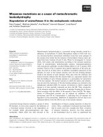
Fig. 2. A working model for the involvement of MNB ⁄ DYRK1A
overexpression in the neuronal deficit of DS. A schematic represen-
tation of the pattern of progenitor division and neuronal generation
in a normal brain, and the possible consequences that
MNB ⁄ DYRK1A overexpression might cause during neurogenesis in
the DS brain. During normal neurogenesis, the transient expression
of Mnb ⁄ Dyrk1A in preneurogenic progenitors triggers the onset of
neurogenic divisions and consequently the production of neurons.
The increase in the level of Mnb ⁄ Dyrk1A expression in DS may
produce the precocious onset of neurogenic progenitors and a con-
comitant loss of proliferating progenitors, leading to a reduction in
the total number of neurogenic lineages. Additionally, the over-
expression of MNB ⁄ DYRK1A might induce premature cell cycle
arrest of neurogenic progenitors, leading to a decrease in the
number of neurogenic divisions undertaken by each neurogenic
progenitor. Thus, the consequences of these alterations in neuro-
genesis would be a decrease in the production of neurons.
F. J. Tejedor and B. Ha
¨
mmerle MNB ⁄ DYRK1A in neuronal development
FEBS Journal 278 (2011) 223–235 ª 2010 The Authors Journal compilation ª 2010 FEBS 229
more detailed analysis to detect. However, we should
not rule out the possibility that due to the activities of
MNB ⁄ DYRK1A in several sequential phases in prolif-
eration ⁄ neurogenesis ⁄ differentiation, a maintained
overexpression in the trangenic mice could result in
compensatory phenotypes. Strikingly, the brains of
152F7 mice, which carry a YAC mouse line with three
copies of at least two neighbouring HSA21 genes in
addition to MNB ⁄ DYRK1A, are enlarged [25,69], a
phenotype that apparently contradicts with the
expected antiproliferative effect of MNB ⁄ DYRK1A
[26,27].
It is also well known that cortical neurons of brains
with DS exhibit dendritic shortening or atrophy
(reviewed in [71]). Thus, another developmental pro-
cess that could be impaired through the overexpression
of MNB ⁄ DYRK1A in DS is dendritogenesis. Indeed,
cultured cortical neurons of Mnb ⁄ Dyrk1A transgenic
mice exhibit poorer dendrite arborization [45]. More-
over, overexpression of MNB ⁄ DYRK1A in wild-type
primary mouse cortical neurons leads to similar
changes [45], strongly suggesting that MNB ⁄ DYRK1A
triploidy can impair dendrite development in DS.
Increased cell death is also associated with DS. For
instance, cultured human cortical DS neurons exhibit
intracellular oxidative stress and increased apoptosis
[72]. Furthermore, increased cell death has been
observed in the forebrain of fetuses with DS [67]. The
involvement of MNB ⁄ DYRK1A in the regulation of
caspase 9-mediated apoptosis in differentiating neurons
of the developing retina has generated some specula-
tion about the effects of MNB ⁄ DYRK1A gene-dosage
imbalance in deregulating the apoptotic response in
DS [59]. However, it seems unlikely that the over-
expression of MNB ⁄ DYRK1A can contribute to the
neuronal deficit of DS by stimulating developmentally
regulated cell death as several studies have related
increased MNB ⁄ DYRK1A levels to antiapoptotic or
cell survival effects rather than to the induction cell
death [59,73,74].
Concluding remarks and perspectives
As summarized in Table 1, many proteins have been
identified as possible substrates and ⁄ or interacting pro-
teins of the MNB ⁄ DYRK1A kinase. Nevertheless, we
know very little about the actual physiological sub-
strates ⁄ interacting partners of MNB ⁄ DYRK1A in neu-
ronal development. In large, this is due to the fact that
most molecular studies have been carried out in non-
neuronal cells. Thus, efforts should be made to address
the true specificity of these putative MNB ⁄ DYRK1A-
related proteins in adequate neuronal systems and in
suitable functional contexts. Also, given the wide
molecular repertoire of substrates (transcription fac-
tors, translation factors, cytoskeletal proteins, mem-
brane receptors, regulators of membrane dynamics,
etc), it is possible that MNB⁄ DYRK1A kinase could
act at several levels in a multifaceted manner, integrat-
ing several cellular responses within a given neuronal
process.
MNB ⁄ DYRK1A also displays a rather varied sub-
cellular distribution during neurodevelopment [14–16].
The early literature classified MNB ⁄ DYRK1A as a
nuclear protein kinase because it contained a bipartite
nuclear translocation signal and MNB ⁄ DYRK1A-
tagged peptides indeed localized in the nucleus of
transfected cell lines [75]. However, immunocytochemi-
cal analysis by high-resolution confocal microscopy
has since shown that the endogenous MNB ⁄ DYRK1A
protein has a mainly cytoplasmic and perinuclear
localization in differentiating mammalian neurons [14].
Nevertheless, MNB ⁄ DYRK1A has also been detected
in the form of speckles in neuronal nuclei at given
developmental stages [14,16]. Thus, a working hypoth-
esis is that MNB ⁄ DYRK1A is normally concentrated
in the perinuclear area and that it translocates into the
nucleus to regulate transcription factors in response to
certain stimuli. It will therefore be very interesting to
study the mechanisms that regulate this translocation
process (see also the interesting comments about the
distribution of MNB ⁄ DYRK1A in the adult mamma-
lian brain in the accompanying review [9]).
As previously discussed, there is also compelling evi-
dence for the very precise spatiotemporal regulation
of Mnb ⁄ Dyrk1A expression during brain development
[13–16], which appears to be crucial for MNB ⁄
DYRK1A function. For example, it has been reported
that the transient expression ⁄ activation of MNB ⁄
DYRK1A induces neuronal differentiation [38,39], but
this is impaired by its stable overexpression [76]. Fur-
thermore, it should be noted that the only well-known
mechanism to activate the MNB ⁄ DYRK1A kinase is
through a transient Tyr-kinase activity that autop-
hosphorylates tyrosine residues in the activation loop
during protein translation [77]. This implies that the
upregulation of MNB ⁄ DYRK1A kinase can be indi-
rectly controlled by regulating its expression, making
the observed transient expression of MNB ⁄ DYRK1A
in specific neurodevelopmental contexts (Fig. 1) even
more relevant functionally. However, only a few mole-
cules have been found to modulate Mnb ⁄ Dyrk1A gene
expression in cell lines (reviewed in [2], see also Table 1)
and almost nothing is known about the mechanisms
regulating its expression during brain development.
Thus, studies in true neurodevelopmental systems will
MNB ⁄ DYRK1A in neuronal development F. J. Tejedor and B. Ha
¨
mmerle
230 FEBS Journal 278 (2011) 223–235 ª 2010 The Authors Journal compilation ª 2010 FEBS
be required to dissect out the mechanisms that actually
regulate Mnb ⁄ Dyrk1A expression and their implication
in brain development.
Acknowledgements
We are grateful to the Ministerio de Ciencia e Innova-
cion, the Generalitat Valenciana and the Fondation
Je
´
roˆ me Lejeune for their support of our
MNB ⁄ DYRK1A research, and to former and present
laboratory members for their contributions. We also
thank Walter Becker for comments and suggestions.
References
1 Galceran J, de Graaf K, Tejedor FJ & Becker W (2003)
The MNB ⁄ DYRK1A protein kinase: genetic and
biochemical properties. J Neural Transm Suppl 67,
139–148.
2 Becker W & Sippl W (2010) Activation, regulation,
and inhibition of the protein kinase DYRK1A.
FEBS J 277.
3 Guimera
´
J, Casas C, Pucharco
`
s C, Solans A, Dome
`
nech
A, Planas AM, Ashley J, Lovett M, Estivill X & Prit-
chard MA (1996) A human homologue of Drosophila
minibrain (MNB) is expressed in the neuronal regions
affected in Down syndrome and maps to the critical
region. Hum Mol Genet 5, 1305–1310.
4 Song WJ, Sternberg LR, Kasten-Sportes C, Keuren
ML, Chung SH, Slack AC, Miller DE, Glover TW,
Chiang PW, Lou L et al. (1996) Isolation of human
and murine homologues of the Drosophila minibrain
gene: human homologue maps to 21q22.2 in the Down
syndrome ‘‘critical region’’. Genomics 38, 331–339.
5 Guimera
´
J, Casas C, Estivill X & Pritchard M (1999)
Human minibrain homologue (MNBH ⁄ DYRK1):
characterization, alternative splicing, differential tissue
expression, and overexpression in Down syndrome.
Genomics 57, 407–418.
6Ha
¨
mmerle B, Elizalde C, Galceran J, Becker W &
Tejedor FJ (2003) The MNB ⁄ DYRK1A protein kinase:
neurobiological functions and Down syndrome implica-
tions. J Neural Transm Suppl 67, 129–137.
7 Dierssen M & de Lagra
´
n MM. (2006) DYRK1A
(dual-specificity tyrosine-phosphorylated and -regulated
kinase 1A): a gene with dosage effect during develop-
ment and neurogenesis. Sci World J 6, 1911–1922.
8 Dowjat WK, Adayev T, Kuchna I, Nowicki K,
Palminiello S, Hwang YW & Wegiel J (2007) Trisomy-
driven overexpression of DYRK1A kinase in the brain of
subjects with Down syndrome. Neurosci Lett 413, 77–81.
9 Wegiel J, Gong C-X & Hwang Y-W (2010) DYRK1A:
the role in neurodegenerative diseases. FEBS J 277.
10 Tejedor F, Zhu XR, Kaltenbach E, Ackermann A,
Baumann A, Canal I, Heisenberg M, Fischbach KF &
Pongs O (1995) minibrain: a new protein kinase family
involved in postembryonic neurogenesis in Drosophila.
Neuron 14, 287–301.
11 Fotaki V, Dierssen M, Alcantara S, Martinez S, Marti
E, Casas C, Visa J, Soriano E, Estivill X & Arbones
ML (2002) Dyrk1A haploinsufficiency affects viability
and causes developmental delay and abnormal brain
morphology in mice. Mol Cell Biol 22, 6636–6647.
12 Moeller RS, Ku
¨
bart S, Hoeltzenbein M, Heye B, Vogel
I, Hansen CP, Menzel C, Ullmann R, Tommerup N,
Ropers HH et al. (2008) Truncation of the Down
syndrome candidate gene DYRK1A in two unrelated
patients with microcephaly. Am J Hum Genet
82,
1165–1170.
13 Okui M, Ide T, Morita K, Funakoshi E, Ito F, Ogita
K, Yoneda Y, Kudoh J & Shimizu N (1999) High-level
expression of the Mnb ⁄ Dyrk1A gene in brain and heart
during rat early development. Genomics 62, 165–171.
14 Ha
¨
mmerle B, Elizalde C & Tejedor FJ (2008) The spa-
tio-temporal and subcellular expression of the candidate
Down syndrome gene Mnb ⁄ Dyrk1A in the developing
mouse brain suggests distinct sequential roles in neuro-
nal development. Eur J Neurosci 27, 1061–1074.
15 Ha
¨
mmerle B, Vera-Samper E, Speicher S, Arencibia R,
Martinez S & Tejedor FJ (2002) Mnb ⁄ Dyrk1A is tran-
siently expressed and asymmetrically segregated in neu-
ral progenitor cells at the transition to neurogenic
divisions. Dev Biol 246, 259–273.
16 Ha
¨
mmerle B, Carnicero A, Elizalde C, Ceron J, Martı
´
-
nez S & Tejedor FJ (2003) Expression patterns and
subcellular localization of the Down syndrome candi-
date protein MNB ⁄ DYRK1A suggest a role in late
neuronal differentiation. Eur J Neurosci 17, 2277–2286.
17 Marti E, Altafaj X, Dierssen M, de la Luna S, Fotaki
V, Alvarez M, Perez-Riba M, Ferrer I & Estivill X
(2003) Dyrk1A expression pattern supports specific
roles of this kinase in the adult central nervous system.
Brain Res 964, 250–263.
18 Iacopetti P, Michelini M, Stuckmann I, Oback B,
Aaku-Saraste E & Huttner WB (1999) Expression of
the antiproliferative gene TIS21 at the onset of neuro-
genesis identifies single neuroepithelial cells that switch
from proliferative to neuron-generating division. Proc
Natl Acad Sci USA 96, 4639–4644.
19 Ferron SR, Pozo N, Laguna A, Aranda S, Porlan E,
Moreno M, Fillat C, de la Luna S, Sa
´
nchez P, Arbone
´
s
ML et al. (2010) Regulated segregation of kinase
Dyrk1A during asymmetric neural stem cell division is
critical for EGFR-mediated biased signaling. Cell Stem
Cell 7(3), 367–379.
20 Ba
¨
hler J & Nurse P (2001) Fission yeast Pom1p kinase
activity is cell cycle regulated and essential for cellular
F. J. Tejedor and B. Ha
¨
mmerle MNB ⁄ DYRK1A in neuronal development
FEBS Journal 278 (2011) 223–235 ª 2010 The Authors Journal compilation ª 2010 FEBS 231
symmetry during growth and division. EMBO J 20,
1064–1073.
21 Raich WB, Moorman C, Lacefield CO, Lehrer J, Bart-
sch D, Plasterk RH, Kandel ER & Hobert O (2003)
Characterization of Caenorhabditis elegans homologs of
the Down syndrome candidate gene DYRK1A. Genetics
163, 571–580.
22 Maenz B, Hekerman P, Vela EM, Galceran J & Becker
W (2008) Characterization of the human DYRK1A
promoter and its regulation by the transcription factor
E2F1. BMC Mol Biol 26 ,9.
23 Kinstrie R, Lochhead A, Sibbet G, Morrice N &
Cleghon V (2006) dDYRK2 and Minibrain interact
with the chromatin remodelling factors SNR1 and
TRX. Biochem J 398, 45–54.
24 Zraly CB, Marenda DR & Dingwall AK (2004) SNR1
(INI1 ⁄ SNF5) mediates important cell growth functions
of the Drosophila Brahma (SWI ⁄ SNF) chromatin
remodeling complex. Genetics 168, 199–214.
25 Branchi I, Bichler Z, Minghetti L, Delabar JM, Malchi-
odi-Albedi F, Gonzalez MC, Chettouh Z, Nicolini A,
Chabert C, Smith DJ et al. (2004) Transgenic mouse in
vivo library of human Down syndrome critical region 1:
association between DYRK1A overexpression, brain
development abnormalities, and cell cycle protein alter-
ation. J Neuropathol Exp Neurol 63, 429–440.
26 Yabut O, Domogauer J & D’Arcangelo G (2010)
Dyrk1A overexpression inhibits proliferation and
induces premature neuronal differentiation of neural
progenitor cells. J Neurosci 30, 4004–4014.
27 Park J, Oh Y, Yoo L, Jung MS, Song WJ, Lee SH, Seo
H & Chung KC (2010) DYRK1A phosphorylates p53
and inhibits proliferation of embryonic neuronal cells.
J Biol Chem 285, 31895–31906.
28 Funakoshi E, Hori T, Haraguchi T, Hiraoka Y, Kudoh
J, Shimizu N & Ito F (2003) Overexpression of the
human MNB ⁄ DYRK1A gene induces formation of
multinucleate cells through overduplication of the cen-
trosome. BMC Cell Biol 4, 12.
29 Nguyen L, Besson A, Roberts JM & Guillemot F
(2006) Coupling cell cycle exit, neuronal differentiation
and migration in cortical neurogenesis. Cell Cycle 20,
2314–2318.
30 Ewton DZ, Lee K, Deng X, Lim S & Friedman E
(2003) Rapid turnover of cell-cycle regulators found in
Mirk ⁄ dyrk1B transfectants. Int J Cancer 103, 21–28.
31 Fernandez-Martinez J, Vela EM, Tora-Ponsioen M,
Ocan
˜
a OH, Nieto MA & Galceran J (2009) Attenuation
of Notch signalling by the Down-syndrome-associated
kinase DYRK1A. J Cell Sci 122, 1574–1583.
32 Louvi A & Artavanis-Tsakonas S (2006) Notch signal-
ling in vertebrate neural development. Nat Rev Neurosci
7, 93–102.
33 Canzonetta C, Mulligan C, Deutsch S, Ruf S, O’Doher-
ty A, Lyle R, Borel C, Lin-Marq N, Delom F, Groet J
et al. (2008) DYRK1A-dosage imbalance perturbs
NRSF ⁄ REST levels, deregulating pluripotency and
embryonic stem cell fate in Down syndrome. Am J
Hum Genet 83, 388–400.
34 Chong JA, Tapia-Ramirez J, Kim S, Toledo-Aral JJ,
Zheng Y, Boutros MC, Altshuller YM, Frohman MA,
Kraner SD & Mandel G (1995) REST: a mammalian
silencer protein that restricts sodium channel gene
expression to neurons. Cell 80, 949–957.
35 Ballas N, Grunseich C, Lu DD, Speh JC & Mandel G
(2005) REST and its corepressors mediate plasticity of
neuronal gene chromatin throughout neurogenesis. Cell
121, 645–657.
36 Mao J, Maye P, Kogerman P, Tejedor FJ, Toftgard R,
Xie W, Wu G & Wu D (2002) Regulation of Gli1 tran-
scriptional activity in the nucleus by Dyrk1. J Biol
Chem 277, 35156–35161.
37 Ruiz i Altaba A, Palma V & Dahmane N (2002) Hedge-
hog-Gli signalling and the growth of the brain. Nat Rev
Neurosci 3, 24–33.
38 Yang EJ, Ahn YS & Chung KC (2001) Protein kinase
Dyrk1 activates cAMP response element-binding pro-
tein during neuronal differentiation in hippocampal
progenitor cells. J Biol Chem 276, 39819–39824.
39 Kelly PA & Rahmani Z (2005) DYRK1A enhances the
mitogen-activated protein kinase cascade in PC12 cells
by forming a complex with Ras, B-Raf, and MEK1.
Mol Biol Cell 16, 3562–3573.
40 Go
¨
ckler N, Jofre G, Papadopoulos C, Soppa U, Teje-
dor FJ & Becker W (2009) Harmine specifically inhibits
protein kinase DYRK1A and interferes with neurite
formation. FEBS J 276, 6324–6337.
41 Benavides-Piccione R, Dierssen M, Ballesteros-Ya
´
n
˜
ez I,
Martı
´
nez de Lagra
´
n M, Arbone
´
s ML, Fotaki V, DeFel-
ipe J & Elston GN (2005) Alterations in the phenotype
of neocortical pyramidal cells in the Dyrk1A+ ⁄ )
mouse. Neurobiol Dis 20, 115–122.
42 Liu T, Sims D & Baum B (2009) Parallel RNAi screens
across different cell lines identify generic and cell type-
specific regulators of actin organization and cell
morphology. Genome Biol 10, R26.
43 Scales TM, Lin S, Kraus M, Goold RG & Gordon-
Weeks PR (2009) Nonprimed and DYRK1A-primed
GSK3 beta-phosphorylation sites on MAP1B regulate
microtubule dynamics in growing axons. J Cell Sci,
122, 2424–2435.
44 Parrish JZ, Kim MD, Jan LY & Jan YN (2006)
Genome-wide analyses identify transcription factors
required for proper morphogenesis of Drosophila
sensory neuron dendrites. Genes Dev 20, 820–835.
45 Lepagnol-Bestel AM, Zvara A, Maussion G, Quignon
F, Ngimbous B, Ramoz N, Imbeaud S, Loe-Mie Y,
Benihoud K, Agier N et al. (2009) DYRK1A interacts
with the REST ⁄ NRSF-SWI ⁄ SNF chromatin remodel-
ling complex to deregulate gene clusters involved in the
MNB ⁄ DYRK1A in neuronal development F. J. Tejedor and B. Ha
¨
mmerle
232 FEBS Journal 278 (2011) 223–235 ª 2010 The Authors Journal compilation ª 2010 FEBS
neuronal phenotypic traits of Down syndrome. Hum
Mol Genet 18, 1405–1414.
46 Arron JR, Winslow MM, Polleri A, Chang CP, Wu H,
Gao X, Neilson JR, Chen L, Heit JJ, Kim SK et al.
(2006) NFAT dysregulation by increased dosage of
DSCR1 and DYRK1A on chromosome 21. Nature 441,
595–600.
47 Gwack Y, Sharma S, Nardone J, Tanasa B, Iuga A,
Srikanth S, Okamura H, Bolton D, Feske S, Hogan PG
et al. (2006) A genome-wide Drosophila RNAi screen
identifies DYRK-family kinases as regulators of NFAT.
Nature 441, 646–650.
48 Graef IA, Wang F, Charron F, Chen L, Neilson J,
Tessier-Lavigne M & Crabtree GR (2003) Neurotro-
phins and netrins require calcineurin ⁄ NFAT signalling
to stimulate outgrowth of embryonic axons. Cell 113,
657–670.
49 Sitz JH, Baumga
¨
rtel K, Ha
¨
mmerle B, Papadopoulos C,
Hekerman P, Tejedor FJ, Becker W & Lutz B (2008)
The Down syndrome candidate dual-specificity tyrosine
phosphorylation-regulated kinase 1A phosphorylates
the neurodegeneration-related septin 4. Neuroscience
157, 596–605.
50 Chen-Hwang MC, Chen HR, Elzinga M & Hwang YW
(2002) Dynamin is a minibrain kinase ⁄ dual specificity
Yak1-related kinase 1A substrate. J Biol Chem 277,
17597–17604.
51 Huang Y, Chen-Hwang MC, Dolios G, Murakami N,
Padovan NC, Wang R & Hwang YW (2004)
Mnb ⁄ Dyrk1A phosphorylation regulates the interaction
of dynamin 1 with SH3 domain-containing proteins.
Biochemistry 43, 10173–10185.
52 Kinoshita A, Kinoshita M, Akiyama H, Tomimoto H,
Akiguchi I, Kumar S, Noda M & Kimura J (1998)
Identification of septins in neurofibrillary tangles in
Alzheimer’s disease. Am J Pathol 153, 1551–1560.
53 Adayev T, Chen-Hwang MC, Murakami N, Wang R &
Hwang YW (2006) MNB ⁄ DYRK1A phosphorylation
regulates the interactions of synaptojanin 1 with endo-
cytic accessory proteins. Biochem Biophys Res Commun
351, 1060–1065.
54 Murakami N, Xie W, Lu RC, Chen-Hwang MC, Wie-
raszko A & Hwang YW (2006) Phosphorylation of
amphiphysin I by minibrain kinase ⁄ dual-specificity tyro-
sine phosphorylation-regulated kinase, a kinase impli-
cated in Down syndrome. J Biol Chem 281, 23712–23724.
55 Murakami N, Bolton D & Hwang YW (2009) Dyrk1A
binds to multiple endocytic proteins required for forma-
tion of clathrin-coated vesicles. Biochemistry 48, 9297–
9305.
56 Ahn KJ, Jeong HK, Choi HS, Ryoo SR, Kim YJ, Goo
JS, Choi SY, Han JS, Ha I & Song WJ (2006)
DYRK1A BAC transgenic mice show altered synaptic
plasticity with learning and memory defects. Neurobiol
Dis 22, 463–472.
57 Martinez de Lagran M, Bortolozzi A, Millan O, Gispert
JD, Gonzalez JR, Arbones ML, Artigas F & Dierssen
M (2007) Dopaminergic deficiency in mice with reduced
levels of the dual-specificity tyrosine-phosphorylated
and regulated kinase 1A, Dyrk1A(+ ⁄ –). Genes Brain
Behav 6, 569–578.
58 Cline HT (2001) Dendritic arbor development and
synaptogenesis.
Curr Opin Neurobiol 11, 118–126.
59 Laguna A, Aranda S, Barallobre MJ, Barhoum R,
Ferna
´
ndez E, Fotaki V, Delabar JM, de la Luna S, de
la Villa P & Arbone
´
s ML (2008) The protein kinase
DYRK1A regulates caspase-9-mediated apoptosis dur-
ing retina development. Dev Cell 15, 841–853.
60 Delabar JM, Theophile D, Rahmani Z, Chettouh Z,
Blouin JL, Prieur M, Noel B & Sinet PM (1993) Molec-
ular mapping of twenty-four features of Down syn-
drome on chromosome 21. Eur J Hum Genet 1, 114–
124.
61 Korbel JO, Tirosh-Wagner T, Urban AE, Chen XN,
Kasowski M, Dai L, Grubert F, Erdman C, Gao MC,
Lange K et al. (2009) The genetic architecture of Down
syndrome phenotypes revealed by high-resolution analy-
sis of human segmental trisomies. Proc Natl Acad Sci
USA 106, 12031–12036.
62 Coyle JT, Oster-Granite ML & Gearhart JD (1986) The
neurobiologic consequences of Down syndrome. Brain
Res Bull 16, 773–787.
63 Wisniewski KE, Laure-Kamionowska M & Wisniewski
HM (1984) Evidence of arrest of neurogenesis and syna-
ptogenesis in brains of patients with Down’s syndrome.
N Engl J Med 311, 1187–1188.
64 Schmidt-Sidor B, Wisniewski KE, Shepard TH &
Sersen EA (1990) Brain growth in Down syndrome
subjects 15 to 22 weeks of gestational age and birth to
60 months. Clin Neuropathol 9, 181–190.
65 Chakrabarti L, Galdzicki Z & Haydar TF (2007)
Defects in embryonic neurogenesis and initial synapse
formation in the forebrain of the Ts65Dn mouse model
of Down syndrome. J Neurosci 27, 11483–11495.
66 Contestabile A, Fila T, Ceccarelli C, Bonasoni P,
Bonapace L, Santini D, Bartesaghi R & Ciani E
(2007) Cell cycle alteration and decreased cell prolifer-
ation in the hippocampal dentate gyrus and in the
neocortical germinal matrix of fetuses with Down syn-
drome and in Ts65Dn mice. Hippocampus 17, 665–
678.
67 Guidi S, Bonasoni P, Ceccarelli C, Santini D, Gualtieri
F, Ciani E & Bartesaghi R (2008) Neurogenesis impair-
ment and increased cell death reduce total neuron num-
ber in the hippocampal region of fetuses with Down
syndrome. Brain Pathol 18, 180–197.
68 Calegari F, Haubensak W, Haffner C & Huttner WB
(2005) Selective lengthening of the cell cycle in the neu-
rogenic subpopulation of neural progenitor cells during
mouse brain development. J Neurosci 25, 6533–6538.
F. J. Tejedor and B. Ha
¨
mmerle MNB ⁄ DYRK1A in neuronal development
FEBS Journal 278 (2011) 223–235 ª 2010 The Authors Journal compilation ª 2010 FEBS 233
69 Smith DJ, Stevens ME, Sudanagunta SP, Bronson RT,
Makhinson M, Watabe AM, O’Dell TJ, Fung J, Weier
HU, Cheng JF et al. (1997) Functional screening of 2
Mb of human chromosome 21q22.2 in transgenic mice
implicates minibrain in learning defects associated with
Down syndrome. Nat Genet 16, 28–36.
70 Altafaj X, Dierssen M, Baamonde C, Martı
´
E, Visa J,
Guimera
`
J, Oset M, Gonza
´
lez JR, Flo
´
rez J, Fillat C
et al. (2001) Neurodevelopmental delay, motor abnor-
malities and cognitive deficits in transgenic mice over-
expressing Dyrk1A (minibrain), a murine model of
Down’s syndrome. Hum Mol Genet 10, 1915–1923.
71 Kaufmann WE & Moser HW (2000) Dendritic anoma-
lies in disorders associated with mental retardation.
Cereb Cortex 10 , 981–991.
72 Busciglio J & Yankner BA (1995) Apoptosis and
increased generation of reactive oxygen species in
Down’s syndrome neurons in vitro. Nature 378, 776–
779.
73 Chang HS, Lin CH, Yang CH, Yen MS, Lai CR, Chen
YR, Liang YJ & Yu WC (2007) Increased expression of
Dyrk1a in HPV16 immortalized keratinocytes enable
evasion of apoptosis. Int J Cancer 120, 2377–2385.
74 Guo X, Williams JG, Schug TT & Li X (2010)
DYRK1A and DYRK3 promote cell survival through
phosphorylation and activation of SIRT1. J Biol Chem
285, 13223–13232.
75 Becker W, Weber Y, Wetzel K, Eirmbter K, Tejedor FJ
& Joost HG (1998) Sequence characteristics, subcellular
localization, and substrate specificity of DYRK-related
kinases, a novel family of dual specificity protein
kinases. J Biol Chem 273, 25893–25902.
76 Park J, Yang EJ, Yoon JH & Chung KC (2007)
Dyrk1A overexpression in immortalized hippocampal
cells produces the neuropathological features of Down
syndrome. Mol Cell Neurosci 36, 270–279.
77 Lochhead PA, Sibbet G, Morrice N & Cleghon V
(2005) Activation-loop autophosphorylation is mediated
by a novel transitional intermediate form of DYRKs.
Cell 121, 925–936.
78 Kimura R, Kamino K, Yamamoto M, Nuripa A, Kida
T, Kazui H, Hashimoto R, Tanaka T, Kudo T, Yamag-
ata H et al. (2007) The DYRK1A gene, encoded in
chromosome 21 Down syndrome critical region, bridges
between beta-amyloid production and tau phosphoryla-
tion in Alzheimer disease. Hum Mol Genet 16, 15–23.
79 Sitz JH, Tigges M, Baumga
¨
rtel K, Khaspekov LG &
Lutz B (2004) Dyrk1A potentiates steroid hormone-
induced transcription via the chromatin remodeling fac-
tor Arip4. Mol Cell Biol 24, 5821–5834.
80 Ryoo SR, Cho HJ, Lee HW, Jeong HK, Radnaabazar
C, Kim YS, Kim MJ, Son MY, Seo H, Chung SH et al.
(2008) Dual-specificity tyrosine(Y)-phosphorylation reg-
ulated kinase 1A-mediated phosphorylation of amyloid
precursor protein: evidence for a functional link
between Down syndrome and Alzheimer’s disease.
J Neurochem 104, 1333–1344.
81 Shi J, Zhang T, Zhou C, Chohan MO, Gu X, Wegiel J,
Zhou J, Hwang YW, Iqbal K, Grundke-Iqbal I et al.
(2008) Increased dosage of Dyrk1A alters alternative
splicing factor (ASF)-regulated alternative splicing of
tau in Down syndrome. J Biol Chem 283, 28660–28669.
82 Seifert A, Allan LA & Clarke PR (2008) DYRK1A
phosphorylates caspase 9 at an inhibitory site and is
potently inhibited in human cells by harmine. FEBS J
275, 6268–6280.
83 Seifert A & Clarke PR (2009) p38a
- and DYRK1A-
dependent phosphorylation of caspase-9 at an inhibitory
site in response to hyperosmotic stress. Cell Signal 21,
1626–1633.
84 Kurabayashi N, Hirota T, Sakai M, Sanada K & Fuk-
ada Y (2010) DYRK1A and glycogen synthase kinase
3beta, a dual-kinase mechanism directing proteasomal
degradation of CRY2 for circadian timekeeping. Mol
Cell Biol 30, 1757–1768.
85 Woods YL, Rena G, Morrice N, Barthel A, Becker W,
Guo S, Unterman TG & Cohen P (2001) The kinase
DYRK1A phosphorylates the transcription factor
FKHR at Ser329 in vitro, a novel in vivo phosphoryla-
tion site. Biochem J 355, 597–607.
86 von Groote-Bidlingmaier F, Schmoll D, Orth HM,
Joost HG, Becker W & Barthel A (2003) DYRK1 is a
co-activator of FKHR (FOXO1a)-dependent glucose-6-
phosphatase gene expression. Biochem Biophys Res
Commun 300, 764–769.
87 Morita K, Lo Celso C, Spencer-Dene B, Zouboulis CC
& Watt FM (2006) HAN11 binds mDia1 and controls
GLI1 transcriptional activity. J Dermatol Sci 44, 11–20.
88 Woods YL, Cohen P, Becker W, Jakes R, Goedert M,
Wang X & Proud CG (2001) The kinase DYRK phos-
phorylates protein-synthesis initiation factor eIF2Bepsi-
lon at Ser539 and the microtubule-associated protein
tau at Thr212: potential role for DYRK as a glycogen
synthase kinase 3-priming kinase. Biochem J 355, 609–
615.
89 Skurat AV & Dietrich AD (2004) Phosphorylation of
Ser640 in muscle glycogen synthase by DYRK family
protein kinases. J Biol Chem 279, 2490–2498.
90 Kang JE, Choi SA, Park JB & Chung KC (2005) Regu-
lation of the proapoptotic activity of Huntington inter-
acting protein 1 by Dyrk1 and caspase-3 in
hippocampal neuroprogenitor cells. J Neurosci Res 81,
62–72.
91 Bescond M & Rahmani Z (2005) Dual-specificity
tyrosine-phosphorylated and regulated kinase 1A
(DYRK1A) interacts with the phytanoyl-CoA alpha-
hydroxylase associated protein 1 (PAHX-AP1), a brain
specific protein. Int J Biochem Cell Biol 37, 775–783.
92 Ryu YS, Park SY, Jung MS, Yoon SH, Kwen MY, Lee
SY, Choi SH, Radnaabazar C, Kim MK, Kim H et al.
MNB ⁄ DYRK1A in neuronal development F. J. Tejedor and B. Ha
¨
mmerle
234 FEBS Journal 278 (2011) 223–235 ª 2010 The Authors Journal compilation ª 2010 FEBS
(2010) Dyrk1A-mediated phosphorylation of Presenilin
1: a functional link between Down syndrome and
Alzheimer’s disease. J Neurochem 115, 574–584.
93 Aranda S, Alvarez M, Turro
´
S, Laguna A & de la Luna
S (2008) Sprouty2-mediated inhibition of fibroblast
growth factor signaling is modulated by the protein
kinase DYRK1A. Mol Cell Biol 28, 5899–5911.
94 Matsuo R, Ochiai W, Nakashima K & Taga T (2001)
A new expression cloning strategy for isolation of sub-
strate-specific kinases by using phosphorylation site-
specific antibody. J Immunol Methods 247, 141–151.
95 Wiechmann S, Czajkowska H, de Graaf K, Grotzinger
J, Joost HG & Becker W (2003) Unusual function of
the activation loop in the protein kinase DYRK1A.
Biochem Biophys Res Commun 302, 403–408.
96 Kim EJ, Sung JY, Lee HJ, Rhim H, Hasegawa M,
Iwatsubo T, Min do S, Kim J, Paik SR & Chung KC
(2006) Dyrk1A phosphorylates alpha-synuclein and
enhances intracellular inclusion formation. J Biol Chem
281, 33250–33257.
97 Ryoo SR, Jeong HK, Radnaabazar C, Yoo JJ, Cho
HJ, Lee HW, Kim IS, Cheon YH, Ahn YS, Chung
SH et al. (2007) DYRK1A-mediated hyperphosphory-
lation of Tau. A functional link between Down syn-
drome and Alzheimer disease. J Biol Chem 282,
34850–34857.
98 Kim D, Won J, Shin DW, Kang J, Kim YJ, Choi SY,
Hwang MK, Jeong BW, Kim GS, Joe CO et al. (2004)
Regulation of Dyrk1A kinase activity by 14-3-3.
Biochem Biophys Res Commun 323, 499–504.
99 Alvarez M, Altafaj X, Aranda S & de la Luna S (2007)
DYRK1A autophosphorylation on serine residue 520
modulates its kinase activity via 14-3-3 binding. Mol
Biol Cell 18, 1167–1178.
F. J. Tejedor and B. Ha
¨
mmerle MNB ⁄ DYRK1A in neuronal development
FEBS Journal 278 (2011) 223–235 ª 2010 The Authors Journal compilation ª 2010 FEBS 235