Báo cáo khoa học: The role of DYRK1A in neurodegenerative diseases docx
Bạn đang xem bản rút gọn của tài liệu. Xem và tải ngay bản đầy đủ của tài liệu tại đây (742.66 KB, 10 trang )
MINIREVIEW
The role of DYRK1A in neurodegenerative diseases
Jerzy Wegiel
1
, Cheng-Xin Gong
2
and Yu-Wen Hwang
3
1 Department of Developmental Neurobiology, New York State Institute for Basic Research in Developmental Disabilities, Staten Island,
NY, USA
2 Department of Neurochemistry, New York State Institute for Basic Research in Developmental Disabilities, Staten Island, NY, USA
3 Department of Molecular Biology, New York State Institute for Basic Research in Developmental Disabilities, Staten Island, NY, USA
Introduction
The minibrain (mnb) gene mutation has been identified
as a cause of abnormal brain development, of deficits
of postembryonic neurogenesis and of reduced
numbers of neurons in Drosophila. In addition to the
proliferative deficits, the mnb mutation causes neurode-
generation, which is considered a consequence of the
lack of sufficient cell–cell contacts required for the
maintenance of Drosophila optic lobe neurons [1]. The
broad spectrum of abnormalities caused by the mnb
gene mutation in Drosophila suggests multiple biologi-
cal functions of the kinase encoded by this gene.
The human orthologue of the Drosophila mnb gene,
named DYRK1A (dual-specificity tyrosine phosphory-
lation-regulated kinase 1A) [2], is mapped to human
Keywords
alternative splicing factor; Alzheimer’s
disease; amyloid-b peptide; Down
syndrome; DYRK1A; neurodegeneration;
tau phosphorylation; a-synuclein
Correspondence
J. Wegiel, New York State Institute for
Basic Research in Developmental
Disabilities, 1050 Forest Hill Road, Staten
Island, NY 10314, USA
Fax: 718 494 4856
Tel: 718 494 5231
E-mail:
(Received 16 July 2010, revised 5 October
2010, accepted 5 November 2010)
doi:10.1111/j.1742-4658.2010.07955.x
Recent studies indicate that the dual-specificity tyrosine phosphorylation-
regulated kinase 1A (DYRK1A) gene, which is located on chromosome
21q22.2 and is overexpressed in Down syndrome (DS), may play a signifi-
cant role in developmental brain defects and in early onset neurodegenera-
tion, neuronal loss and dementia in DS. The identification of hundreds of
genes deregulated by DYRK1A overexpression and numerous cytosolic,
cytoskeletal and nuclear proteins, including transcription factors, phosphor-
ylated by DYRK1A, indicates that DYRK1A overexpression is central for
the deregulation of multiple pathways in the developing and aging DS
brain, with structural and functional alterations including mental retarda-
tion and dementia. DYRK1A overexpression in DS brains may contribute
to early onset neurofibrillary degeneration directly through hyperphosph-
orylation of tau and indirectly through phosphorylation of alternative
splicing factor, leading to an imbalance between 3R-tau and 4R-tau. The
several-fold increases in the number of DYRK1A-positive and 3R-tau-posi-
tive neurofibrillary tangles in DS support this hypothesis. Moreover, the
enhanced phosphorylation of amyloid precursor protein by overexpressed
DYRK1A facilitates amyloidogenic amyloid precursor protein cleavage
elevating Ab40 and 42 levels, and leading to brain b-amyloidosis.
Therefore, inhibiting DYRK1A activity in DS may serve to counteract the
phenotypic effects of its overexpression and is a potential method of
treatment of developmental defects and the prevention of age-associated
neurodegeneration, including Alzheimer-type pathology.
Abbreviations
AD, Alzheimer’s disease; APP, amyloid precursor protein; ASF, alternative splicing factor; CDK5, cyclin-dependent kinase 5; DS, Down
syndrome; DYRK1A, dual-specificity tyrosine phosphorylation-regulated kinase 1A; EGCG, epigallocatechin 3-gallate; GSK-3b, glycogen
synthase kinase-3b; GVD, granulovacuolar degeneration; mnb , minibrain gene; NFT, neurofibrillary tangle; SEPT4, septin 4.
236 FEBS Journal 278 (2011) 236–245 ª 2010 The Authors Journal compilation ª 2010 FEBS
chromosome 21q22.2 [3], a region of the chromosome
implicated in Down syndrome (DS). DS, caused by
partial or complete trisomy of chromosome 21, is the
most common chromosomal disorder associated with
abnormal brain development, including a reduced size
of the brain and the number of neurons, smaller neu-
rons and a reduced dendritic tree, contributing to men-
tal retardation [4]. Trisomy of chromosome 21 also
results in early aging, which is manifested in the third
decade of life, and early onset of Alzheimer-type
pathology, such as neurofibrillary degeneration,
b-amyloidosis and neuronal loss, affecting almost all
DS subjects who are older than 40 years of age [5,6].
DYRK1A has multiple biological functions that are
reflected in its interactions with numerous cytoskeletal,
synaptic and nuclear proteins, including transcription
and splicing factors [7,8]. The accompanying review by
Tejedor and Ha
¨
mmerle [9] characterizes DYRK1A as
a regulator of a broad spectrum of neurodevelopmen-
tal mechanisms. The identification of 239 genes that
are deregulated by overexpressed DYRK1A through
the REST ⁄ NRSF chromatin remodeling complex sug-
gests a central role of this kinase in brain pathology
[10]. Expression of DYRK1A in neurons during fetal
and postnatal life, as well as in neurons of adults and
aged subjects, suggests that regulated DYRK1A
expression is a component of neuron development,
maturation and aging [11].
DYRK1A
) ⁄ )
mice embryos present significant
growth delay, with their body size reduced by 25–50%,
and die between E10.5 and E13.5. Reduced postnatal
viability, with a loss of 29% of DYRK1A
+ ⁄ )
mice dur-
ing the first 3 days of life, reduced body weight, brain
size and total number of neurons, indicate that
DYRK1A plays a vital role in cellular mechanisms that
determine body and brain growth and development [12].
Recent studies of DS brains indicate that overex-
pression of DYRK1A, due to the third copy of
DYRK1A, not only causes developmental defects with
life-long structural and functional consequences, but
also contributes to neurodegeneration, neuronal death
and loss of function. The mechanisms and potential
therapeutic effects of selective inhibition of overexpres-
sed DYRK1A are reviewed by Becker and Sippl [13].
DYRK1A distribution
The pattern of DYRK1A distribution in human brain
is brain region-, cell type- and subcellular compart-
ment-specific. In control brains, the level of DYRK1A
is almost identical in the frontal, temporal and occipi-
tal cortices (17–18 ngÆmg
)1
total proteins). In all exam-
ined subregions of brains of DS subjects, the level of
DYRK1A is higher than in control brains, but with an
increase that varies topographically from 25 ngÆmg
)1
in the frontal cortex, 21 ngÆmg
)1
in the temporal
cortex, 20 ngÆmg
)1
in the occipital cortex, to only
15 ngÆmg
)1
in the cerebellar cortex [14]. Striking brain
structure-specific and neuron type-specific differences
in the distribution of DYRK1A detected by immuno-
cytochemistry (Fig. 1) indicate that the role of
DYRK1A in development, maturation, aging and
degeneration may vary in different brain structures
and with the types of neuron [11].
DYRK1A contains a bipartite nucleus targeting
sequence [2], and the overexpressed exogenous
DYRK1A has largely been found in the nucleus [15].
In contrast to the expected prevalence of endogenous
DYRK1A in the nuclear fraction, in the human brain,
only 12% of brain DYRK1A is detected in the
nucleus; 78% is associated with an insoluble cytoskele-
tal fraction, and 10% with a soluble cytoplasmic frac-
tion (W. Kaczmarski, M. Barua, D. Bolton, Y W.
Hwang, A. Rabe, G. Albertini & J. Wegiel, unpub-
lished results). A similar type of DYRK1A subcellular
distribution was also observed in rat [16] and mouse
[17] brains. The difference in phosphorylation of
DYRK1A in cell compartments indicates that intra-
cellular trafficking of DYRK1A may be regulated by
DYRK1A phosphorylation. This pattern of subcellular
distribution is in agreement with immunocytochemical
staining of DYRK1A in neurons (Fig. 1) and is also
consistent with reports demonstrating that DYRK1A
phosphorylates numerous substrates in the cytosol,
cytoskeleton, synapses and nucleus [8]. Therefore, the
overexpression of DYRK1A in DS and other disorders
may produce brain region-, cell type- and cell compart-
ment-specific changes, altering brain development,
maturation and susceptibility to neurodegeneration.
Abnormal expression of DYRK1A in
neurodegenerative diseases
Increased DYRK1A immunoreactivity has been
reported in the cytoplasm and nuclei of scattered neu-
rons of the entorhinal cortex, hippocampus and neo-
cortex in neurodegenerative diseases associated with
tau phosphorylation, including Alzheimer’s disease
(AD), DS and Pick disease [18]. The percentage of
neurons with increased DYRK1A immunoreactivity
showed significant differences across individuals and
brain structures. The percentage of DYRK1A-positive
nuclei in the frontal cortex was only 0.5% in controls,
10% in AD and 5% in Pick disease. The percentage of
DYRK1A-positive nuclei in the dentate gyrus granule
layer, which was determined to be 0.5% in control
J. Wegiel et al. Role of DYRK1A in neurodegenerative diseases
FEBS Journal 278 (2011) 236–245 ª 2010 The Authors Journal compilation ª 2010 FEBS 237
subjects and AD, increases to 60% in Pick disease [18].
Significant changes in DYRK1A expression during
development and due to disease indicate that structure-
specific, age-associated and disease-associated factors
modify the amount and distribution of DYRK1A.
Several studies suggest that the cytoplasmic and nuclear
level of DYRK1A is cell type-specific [11,18,19] and that
local levels of overexpressed DYRK1A might be a
factor co-determining cell susceptibility to age ⁄ AD-
associated neurofibrillary degeneration in DS [19–21].
The role of DYRK1A in tauopathies
An initial in vitro study revealed that DYRK1A phos-
phorylates human microtubule-associated protein tau
at Thr212 [22], but the list of phosphorylation sites has
since been expanded to 11, including Thr181, Ser199,
Ser202, Thr205, Thr212, Thr217, Thr231, Ser396,
Ser400, Ser404 and Ser422 [20]. The majority of the
DYRK1A-mediated phosphorylation sites of tau are
significantly hyperphosphorylated in the DS brain.
Gene dosage-related increases in DYRK1A levels in
DS appear to be the major factor distinguishing the
pattern and consequences of tau protein hyperphosph-
orylation in DS and in sporadic AD [20]. DYRK1A-
induced phosphorylation of tau reduces the biological
activity of tau protein and promotes tau self-aggrega-
tion and fibrillization. The abnormal tau phosphoryla-
tion causes the loss of tau biological function, resulting
in reduced activity to stimulate microtubule assembly
[20,23]. Moreover, hyperphosphorylated tau gains
pathological properties, resulting in sequestration of
normal tau and other microtubule-associated proteins.
Self-aggregation of tau leads to paired helical filament
formation, neurofibrillary degeneration and neuron
death. DYRK-mediated tau phosphorylation primes
AEF
BG
H
C
D
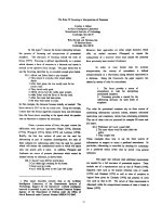
Fig. 1. DYRK1A distribution in DS. DYRK1A
immunolabeling with mAb 7F3 in the hippo-
campus of a 56-year-old DS subject illus-
trates sector- and layer-specific differences
in the distribution of DYRK1A in neurons
and neuronal processes in CA1-4 and the
dentate gyrus (DG) (A). The most intensive
reaction is observed in CA1 pyramidal neu-
rons in bodies and apical dendrites (B, C).
High magnification of a neuron shows
immunoreactivity in the nucleus, cytoplasm
and synapses in the CA4 sector (D). Immu-
noreactivity in the cortex is weaker than in
the hippocampus and is more prominent in
pyramidal than granule neurons (E, F).
In DS, regionally astrocytes show strong
diffuse cell body immunoreactivity (temporal
lobe; G). DYRK1A immunoreactivity in the
corpora amylacea in the dentate gyrus (H)
reflects DYRK1A contribution to astrocytes
and neuronal degeneration.
Role of DYRK1A in neurodegenerative diseases J. Wegiel et al.
238 FEBS Journal 278 (2011) 236–245 ª 2010 The Authors Journal compilation ª 2010 FEBS
further tau phosphorylation at Ser199, Ser202, Thr205
and Ser208 with glycogen synthase kinase-3b (GSK-
3b) [20,22]. Phosphorylation of tau by both DYRK1A
and GSK-3b enhances both self-aggregation and fibril
formation in vitro [20,23]. The link between overex-
pression of DYRK1A and tau phosphorylation is also
detected in Ts65Dn mice, a mouse model of DS tri-
somy that carries an additional copy of the distal seg-
ment of murine chromosome 16, including the
DYRK1A gene [24]. These mice reveal 1.5-fold greater
expression and activity of DYRK1A and increased tau
protein phosphorylation [20].
The direct evidence of the contribution of over-
expressed DYRK1A to neurofibrillary degeneration in
DS is a several-fold greater number of DYRK1A-posi-
tive neurofibrillary tangles (NFTs) in the brains of
people with DS ⁄ AD than in the brains of people with
sporadic AD (Fig. 2) [19]. DYRK1A phosphorylates
tau protein at the sites that are phosphorylated in AD.
In NFTs, tau protein is phosphorylated by several pro-
tein kinases, including GSK-3b, cyclin-dependent
kinase5 (CDK5), c-Jun N-terminal kinase, extracellular
signal-regulated kinases 1 ⁄ 2 and p38 mitogen-activated
protein kinases at more than 30 phosphorylation sites
[25–30]. Several kinases in their activated forms colo-
calize with NFTs in AD, including extracellular signal-
regulated kinase 2 [31], microtubule-affinity-regulating
kinase [32], GSK-3b [33], CDK5 [34], Cdc2-related
kinase [35] and casein kinase 1d [36]. DYRK1A not
only phosphorylates tau protein, but also colocalizes
with NFTs. The presence of DYRK1A-positive NFTs
in all subjects with DS ⁄ AD but in only 60% of people
diagnosed with sporadic AD suggests a link between
DYRK1A overexpression in DS and neurofibrillary
degeneration. The presence of DYRK1A-positive
NFTs in all NFT-positive subjects with DS who are
38–51 years of age indicates that DYRK1A contrib-
utes to the early onset of neurofibrillary degeneration.
The increase with age in the percentage of DYRK1A-
positive NFTs up to 100% in some subjects from 58
to 67 years of age reflects the increasing contribution
of DYRK1A with age to the progression of neurofi-
brillary degeneration in DS subjects. However, in spo-
radic AD, the percentage of DYRK1A-positive NFTs
does not change with age or disease duration. Striking
differences in the detection of intracellular NFTs with
antibody G-19 (Santa Cruz Biotechnology, Santa
Cruz, CA, USA) in the majority of NFTs, the reaction
with antisera X1079 (Exalpha Biologicals, Shirley,
MA) and 324446 (EMD4Bioscience, Gibbstown, NJ)
in only 10% of G-19-positive NFTs, and the lack of
reaction of NFTs with antibody 7F3 indicate that epi-
topes detected with these antibodies against DYRK1A
are masked in complexes of DYRK1A with tau and
potentially with other proteins [19].
Neuropathological and molecular studies indicate
that overexpressed nuclear DYRK1A contributes to
the modification of the alternative splicing of tau and
neurofibrillary degeneration. DYRK1A phosphorylates
the alternative splicing factor (ASF), mainly at Ser227,
Ser234 and Ser238. The phosphorylation of these three
sites is neither catalyzed by the three other known
ASF kinases (SRPK1, SRPK2 and Clk ⁄ Sty [21]) nor
modulated by DNA topoisomerase I [37].
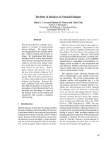
AB
CD
Fig. 2. Prevalence of 3R-tau-positive NFTs
in DS. The several-fold more DYRK1A-
positive NFTs in DS (A) than in AD (B), and
the several-fold more 3R-tau-positive NFTs
in DS (C) than in AD (D) are direct evidence
of the enhanced contribution of DYRK1A to
neurofibrillary degeneration in DS. The figure
illustrates changes in sector CA1 of a
54-year-old DS male (A, C) and an 84-year-
old male (B, D), both diagnosed with
severe AD.
J. Wegiel et al. Role of DYRK1A in neurodegenerative diseases
FEBS Journal 278 (2011) 236–245 ª 2010 The Authors Journal compilation ª 2010 FEBS 239
ASF binds to a polypurine enhancer of exonic splic-
ing enhancer located at tau exon 10 and promotes the
inclusion of exon 10 in the mRNA driving 4R-tau syn-
thesis [38,39]. Phosphorylation regulates the trafficking
and function of ASF. Phosphorylation of ASF by
DYRK1A drives this factor to nuclear speckles, the
site of storage of inactivated serine⁄ arginine-rich pro-
teins, including ASF. This mechanism prevents ASF
from facilitating tau exon 10 inclusion and upregulates
the expression of 3R-tau [21]. Equal levels of 3R- and
4R-tau are critical for optimal neuron function [40].
The predominance of 3R-tau results in the tauopathy
observed in Pick disease, whereas the predominance of
4R-tau causes tau pathology and neuronal loss in pro-
gressive supranuclear palsy and corticobasal degenera-
tion [41]. Phosphorylation of ASF by overexpressed
DYRK1A is considered the foundation for the approx-
imately four-fold increase in 3R-tau in DS. The
increase in the level of free 3R-tau available for abnor-
mal hyperphosphorylation contributes to alterations of
cell cytoskeleton and neurofibrillary degeneration in
DS [21]. Immunohistochemically, several-fold more
3R-tau-positive NFTs are seen in the DS brain than in
the AD brain (Fig. 2), further supporting the contribu-
tion of DYRK1A to neurofibrillary degeneration in
DS.
Application of 2D gel electrophoresis demonstrates
that the pattern of ASF phosphorylation is different in
subjects with DS ⁄ AD than in sporadic AD or control
subjects. The direct evidence of the prevalence of
3R-tau in DS ⁄ AD is a several-fold increase in the
number of 3R-tau-positive NFTs in the entorhinal
cortex, hippocampus, amygdala and neocortex of
DS ⁄ AD subjects in comparison with sporadic AD sub-
jects. Differences between neuron type-specific patterns
of DYRK1A nuclear expression and the rather uni-
form distribution of ASF suggest that the elevated
ratio of nuclear DYRK1A to ASF is a risk factor
determining neuron type susceptibility to neurofibril-
lary degeneration [42].
In DS, DYRK1A overexpression appears to be the
cause of gene dosage- dependent modifications of sev-
eral mechanisms that contribute to the early onset of
neurofibrillary degeneration, including DYRK1A
phosphorylation of tau protein at 11 sites [20,22,43];
the DYRK1A-stimulated, several-fold increase in the
rate of tau protein phosphorylation by GSK-3b
[20,43]; the several-fold increase in the number of
DYRK1A-positive NFTs in the brains of people with
DS ⁄ AD [17]; phosphorylation of ASF, leading to alter-
native splicing of exon 10; and the several-fold greater
number of 3R-tau-positive NFTs in the brains of peo-
ple with DS⁄ AD than in sporadic AD [21,42].
Neurofibrillary degeneration is the leading cause
of neuronal death and dementia in DS ⁄ AD and AD.
The multipathway involvement of DYRK1A in neuro-
fibrillary degeneration indicates that therapeutic inhibi-
tion of overexpressed DYRK1A activity to control
levels may delay the age of onset and inhibit the
progression of neurodegeneration in DS.
Contribution of overexpressed DYRK1A
to b-amyloidosis in DS
A broad spectrum of developmental and age-associated
changes in people with DS is considered a result of the
overexpression of genes localized on chromosome 21.
The extra copy of the gene encoding amyloid precursor
protein (APP) located on chromosome 21 appears to
be the main cause of the early onset of brain b-amyloi-
dosis in people with DS. Overexpression of APP has
been associated with an increase in Ab 42 levels in the
brains of fetuses with trisomy of chromosome 21 [44],
the development of diffuse Ab-positive plaques in
50% of individuals with DS younger than 30 years
of age [45,46], Alzheimer-type pathology in the major-
ity of DS subjects older than 40 years of age [46] and
an elevated risk of AD-associated dementia [47,48].
Experimental studies suggest that overexpression of
DYRK1A could be a primary risk factor contributing
to the enhancement of both b-amyloidosis and neuro-
fibrillary degeneration. Various kinases phosphorylate
the APP cytosolic domain, including GSK-3b [49],
Cdc2 kinase [50], CDK5 [51] and c-Jun N-terminal
kinase 3 [52]. Recent studies by Ryoo et al. [53]
revealed that DYRK1A phosphorylates APP at
Thr668 in vitro and in mammalian cells. The increase
in DYRK1A concentration is associated with increased
APP phosphorylation at Thr668 and colocalization of
DYRK1A and APP in cytosol [53]. Elevated levels of
phospho-APP are observed in AD, particularly in the
hippocampus [54]. The phosphorylation of APP at
Thr668 may facilitate the cleavage of APP by BACE1
[54] and c-secretase [54,55] and enhance the production
of Ab. Elevated Ab 40 and Ab 42 production by 160
and 17%, respectively, detected in the hippocampus of
DYRK1A transgenic mice, suggests that DYRK1A
overexpression promotes APP cleavage and Ab pro-
duction [53]. The accumulation of toxic, soluble Ab
oligomers inhibits many critical neuronal activities,
including long-term potentiation, leading to memory
deficit. Recent studies strongly support the hypothesis
that soluble Ab oligomers contribute to dementia in
AD [56]. Increased expression of DYRK1A mRNA in
the hippocampus of AD patients and in vitro stimula-
tion by Ab of DYRK1A mRNA expression in
Role of DYRK1A in neurodegenerative diseases J. Wegiel et al.
240 FEBS Journal 278 (2011) 236–245 ª 2010 The Authors Journal compilation ª 2010 FEBS
neuroblastoma cells [57] indicate that DYRK1A and Ab
may positively feedback and accelerate Ab production.
In DS, three copies of the APP and DYRK1A genes
result in increased APP and DYRK1A mRNAs [58,59]
and increased levels of DYRK1A and APP by 50 and
55%, respectively [53]. The increase in phospho-APP
in DS brains by 82 and 23% after normalization to
the levels of actin and APP, respectively, suggests that
elevations of DYRK1A and APP may give rise to
brain amyloidosis in DS through DYRK1A-mediated
phosphorylation of APP [53]. Elevated Ab levels could
subsequently increase expression of the DYRK1A
gene and enhance hyperphosphorylation of tau
[20,43,53,57]. These observations reveal a potential reg-
ulatory link between DYRK1A and APP proteolytic
cleavage, enhanced levels of A b upregulating
DYRK1A mRNA expression, and the cascades of
events associated with DYRK1A overexpression.
The role of DYRK1A in a-synucleino-
pathies and other forms of
neurodegeneration
Several reports have indicated that DYRK1A can con-
tribute to other forms of degeneration, including a-syn-
uclein aggregation and fibrillization in Lewy bodies,
granulovacuolar degeneration (GVD) in the hippocam-
pal pyramidal neurons, and neuronal and astrocyte
degeneration with DYRK1A-positive corpora amylacea
deposition in aging, AD, DS ⁄ AD and other diseases.
DYRK1A phosphorylates a-synuclein at Ser87,
enhances cytoplasmic aggregate formation and potenti-
ates a-synuclein proapoptotic effects [60]. a-synuclein-
positive Lewy bodies and neuritic processes frequently
occur in DS brains with AD phenotypes [61].
DYRK1A phosphorylates and binds a-synuclein [60]
and septin 4 (SEPT4) [62], and complexes of these
three proteins may contribute to the cytoplasmic
aggregation ⁄ fibrillization observed in Parkinson dis-
ease, Lewy body dementia and multiple-system atro-
phy [63,64]. SEPT4 has been detected in NFTs,
neuropil threads and dystrophic neurites in amyloid
plaques in AD [65]. Binding of DYRK1A to SEPT4
and the presence of SEPT4 and DYRK1A in NFTs
and Lewy bodies suggest that the DYRK1A ⁄ SEPT4
tandem may play a significant role in both tauopathies
and a-synucleinopathies.
GVD is observed in neurons in the majority of nor-
mal-aged subjects, and their number increases in persons
with AD [66] and DS [67]. The granular component of
vacuoles reacts with antibodies to tubulin [68], abnor-
mally phosphorylated tau [69] and GSK-3b [33,34], as
well as to ubiquitin [70]. The presence of DYRK1A
immunoreactivity in granules in neurons with GVD
detected with C-terminal antibodies (X1079 and 324446)
and the lack of reactivity with antibodies against the
N-terminus (7F3 and G-19) may indicate that only
N-terminally truncated products of DYRK1A contrib-
ute to GVD or are selectively accumulated in these
granules [19].
The strong immunoreactivity of the corpora amyla-
cea with antibodies detecting the amino and carboxyl
terminal portions of DYRK1A, including 7F3, G-19,
X1079 and 324446, suggests that DYRK1A is involved
in this common form of neuron and astrocyte degener-
ation and the early onset of these changes in DS [19].
Strong diffuse or granular immunoreactivity in the
cytoplasm of almost all astrocytes in areas with
massive astrocyte degeneration with corpora amylacea
formation suggests the link between the cytoplasmic
overexpression of DYRK1A and the risk of astrocyte
degeneration in aging, DS and AD.
Concluding remarks
For decades, the molecular mechanisms of DS develop-
mental abnormalities, mental retardation and early
onset of Alzheimer-type pathology remained elusive.
Recent studies indicate that the overexpression of
DYRK1A contributes to an early onset of neurofibril-
lary degeneration, b-amyloidosis, neuronal loss and
dementia in DS (Fig. 3). The progress that has been
made in the identification of overexpressed DYRK1A
as a factor involved in a broad spectrum of molecular,
functional and structural modifications underlying the
DS phenotypes offers a rationale for the design of new
preventive and therapeutic treatments of DS. One may
hypothesize that the inhibition of excessive activity of
DYRK1A may result in cytoplasmic and nuclear path-
ways of the prevention ⁄ delay of several forms of neu-
rodegeneration. A few potent DYRK1A inhibitors have
been described [12,71,72]. Among inhibitors, epigallo-
catechin 3-gallate (EGCG), the major polyphenolic fla-
vonoid in tea, has recently emerged as a candidate for
therapeutic or prophylactic applications. EGCG could
rescue the synaptic plasticity deficiency of Ts65Dn mice
[73]. EGCG and related catechins were also successfully
applied to treat the learning deficits associated with
DYRK1A transgenic mice [74]. Furthermore, EGCG
has been shown to modulate APP processing, which
subsequently leads to a reduction in Ab production
and cerebral amyloidosis in APP transgenic mice [75].
Potentially, selective inhibition of overexpressed
DYRK1A in DS could prevent ⁄ reduce some develop-
mental defects, including intellectual deficits, as well as
delay ⁄ reduce Alzheimer-type pathology and dementia.
J. Wegiel et al. Role of DYRK1A in neurodegenerative diseases
FEBS Journal 278 (2011) 236–245 ª 2010 The Authors Journal compilation ª 2010 FEBS 241
Acknowledgements
The authors are grateful for financial support from the
New York State Office of Mental Retardation and
Developmental Disabilities and grants from the
National Institutes of Health, National Institute of
Child Health and Human Development R01
HD043960 (JW), HD038295 (YWH); the National
Institute of Aging, AG08051 (JW) and R01 AG027429
(C-XG); the Alzheimer’s Association, IIRG-05-13095
(C-XG) and NIRG-08-91126; and the Jerome Lejeune
Foundation (YWH). The authors thank Ms Maureen
Marlow for editorial corrections.
References
1 Tejedor F, Zhu XR, Kaltenbach E, Ackermann A,
Baumann A, Canal I, Heisenberg M, Fischbach KF &
Pongs O (1995) Minibrain: a new protein kinase family
involved in postembryonic neurogenesis in Drosophila.
Neuron 14, 287–301.
2 Kentrup H, Becker W, Heukelbach J, Wilmes A, Schur-
mann A, Huppertz C, Kainulainen H & Joost H-G
(1996) Dyrk, a dual specificity protein kinase with
unique structural features whose activity is dependent
on tyrosine residues between subdomains VII and VIII.
J Biol Chem 271, 3488–3495.
3 Song W-J, Sternberg LR, Kasten-Sportes C, Van Keuren
ML, Chung S-H, Slack AC, Miller DM, Glover TW,
Chiang P-W, Lou L et al. (1996) Isolation of human and
murine homologues of the Drosophila minibrain gene:
human homolog maps to 21q22.2 in the Down syndrome
‘‘critical region’’. Genomics 38, 331–339.
4 Wisniewski KE (1990) Down syndrome children often
have brain with maturation delay, retardation of
growth, and cortical dysgenesis. Am J Med Genet Suppl
7, 274–281.
5 Wegiel J, Wisniewski HM, Dziewiatkowski J, Popovitch
ER & Tarnawski M (1996) Differential susceptibility to
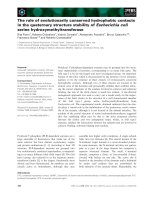
Fig. 3. Contribution of overexpressed DYRK1A to b-amyloidosis and neurofibrillary degeneration in DS. Gene-dose elevation of DYRK1A
expression associated with trisomy 21 could lead to the activation of two pathways contributing to neurofibrillary degeneration and one con-
tributing to brain b-amyloidosis. DYRK1A phosphorylates tau at 11 sites, including Thr212; primes tau phosphorylation by GSK-3b; promotes
tau aggregation into NFTs and the several-fold increase in the number of DYRK1A-positive NFTs. Phosphorylation of ASF by nuclear DYRK1A
increases the level of 3R-tau, leading to an imbalance in the 3R ⁄ 4R-tau ratio and triggering neurofibrillary degeneration with a several-fold
increase in 3R-tau-positive NFTs. Both cytoplasmic and nuclear pathways contribute to neurofibrillary degeneration, loss of neuron function
and neuronal death. DYRK1A phosphorylates APP at Thr688 and enhances APP amyloidogenic cleavage, resulting in an increased level of
Ab 40 ⁄ 42, the formation of toxic Ab oligomers and deposition of fibrillar amyloid in plaques. Brain amyloidosis contributes to a loss of neuro-
nal function and possibly also to neuronal loss. Moreover, elevated levels of Ab may upregulate DYRK1A expression and enhance the contri-
bution of overexpressed DYRK1A to neurofibrillary degeneration and b-amyloidosis.
Role of DYRK1A in neurodegenerative diseases J. Wegiel et al.
242 FEBS Journal 278 (2011) 236–245 ª 2010 The Authors Journal compilation ª 2010 FEBS
neurofibrillary pathology among patients with Down
syndrome. Dementia 7, 135–141.
6 Sadowski M, Wisniewski HM, Tarnawski M, Kozlow-
ski P, Lach B & Wegiel J (1999) Neuronal loss in the
entorhinal cortex of aged subjects with Down syn-
drome. Acta Neuropathol 97, 156–164.
7 Galceran J, de Graaf K, Tejedor FJ & Becker W (2003)
The MNB ⁄ DYRK1A protein kinase: genetic and
biochemical properties. J Neurol Transm Suppl 67,
139–148.
8 Becker W (2008) Dyrk1a. UCSD-Nature Molecule
Pages. doi:10.1038/mp.a000796.01.
9 Tejedor FJ & Ha
¨
mmerle B (2010) MNB ⁄ DYRK1A: a
multiple regulator of neuronal development. FEBS J
10 Lepagnol-Bestel A-M, Zvara A, Maussion G, Quignon
F, Ngimbous B, Ramoz N, Imbeaud S, Loe-Mie Y,
Benihoud K, Agier N et al. (2009) DYRK1A interacts
with the REST ⁄ NRSF-SWI ⁄ SNF chromatin remodeling
complex to deregulate gene clusters involved in the neu-
ronal phenotypic traits of Down syndrome. Hum Mol
Genet 18, 1405–1414.
11 Wegiel J, Kuchna I, Nowicki K, Frackowiak J, Dowjat
K, Silverman WP, Reisberg B, De Leon M, Wisniewski
T, Adayev T et al. (2004) Cell type- and brain struc-
ture-specific patterns of distribution of minibrain kinase
in human brain. Brain Res 1010, 69–80.
12 Fotaki V, Dierssen M, Alca
´
ntara S, Martı
´
nez S, Martı
´
E, Casas C, Visa J, Soriano E, Estivill X & Arbone
´
s
ML (2002) Dyrk1A haploinsufficiency affects viability
and cause developmental delay and abnormal brain
morphology in mice. Mol Cell Biol 22, 6636–6647.
13 Becker W & Sippl W (2010) DYRK1A: activation, reg-
ulation, and inhibition. FEBS J .
14 Dowjat WK, Adayev T, Kuchna I, Nowicki K,
Palminiello S, Hwang YW & Wegiel J (2007) Trisomy-
driven overexpression of DYRK1A kinase in the brain
of subjects with Down syndrome. Neurosci Lett 413,
77–81.
15 Becker W, Weber Y, Wetzel K, Eirmbter K, Tejedor FJ
& Joost HG (1998) Sequence characteristics, subcellular
localization, and substrate specificity of DYRK-related
kinases, a novel family of dual specificity protein
kinases. J Biol Chem 273, 25893–25902.
16 Murakami N, Bolton D & Hwang YW (2009) Dyrk1A
binds to multiple endocytic proteins required for forma-
tion of clathrin-coated vesicles. Biochemistry 48, 9297–
9305.
17 Marti E, Altafaj X, Dierssen M, de la Luna S, Fotaki
V, Alvarez M, Perez-Riba M, Ferrer I & Estivill X
(2003) Dyrk1A expression pattern supports specific
roles of this kinase in the adult central nervous system.
Brain Res 964, 250–263.
18 Ferrer I, Barrachina M, Puig B, Martinez de Lagran
M, Marti E, Avila J & Dierssen M (2005) Constitutive
Dyrk1A is abnormally expressed in Alzheimer disease,
Down syndrome, Pick disease, and related transgenic
models. Neurobiol Dis
20, 392–400.
19 Wegiel J, Dowjat K, Kaczmarski W, Kuchna I,
Nowicki K, Frackowiak J, Mazur Kolecka B, Wegiel J,
Silverman WP, Reisberg B et al. (2008) The role of
overexpressed DYRK1A protein in the early onset of
neurofibrillary degeneration in Down syndrome. Acta
Neuropathol 116, 391–407.
20 Liu F, Liang Z, Wegiel J, Hwang Y-W, Iqbal K, Grun-
dke-Iqubal I, Ramakrishna N & Gong C-X (2008)
Over-expression of Mnb ⁄ Dyrk1A contributes to neuro-
fibrillary degeneration in Down syndrome. FASEB J
22, 3224–3233.
21 Shi J, Zhang T, Zhou C, Chohan MO, Gu X, Wegiel
J, Zhou J, Hwang Y-W, Iqbal K, Grundke-Iqbal I
et al. (2008) Increased dosage of Dyrk1A alters alter-
native splicing factor (ASF)-regulated alternative splic-
ing of tau in Down syndrome. J Biol Chem 283,
28660–28669.
22 Woods YL, Cohen P, Becker W, Jakes R, Goedert M,
Wang X & Proud CG (2001) The kinase DYRK phos-
phorylates protein-synthesis initiation factor elF2Be at
Ser539 and the microtubule-associated protein tau at
Thr212: potential role for DYRK as a glycogen syn-
thase kinase 3-priming kinase. Biochem J 355, 609–615.
23 Liu F, Li B, Tung E-J, Grundke-Iqubal I, Iqbal K &
Gong C-X (2007) Site-specific effects of tau phosphory-
lation on its microtubule assembly activity and self-
aggregation. Eur J Neurosci 26, 3429–3436.
24 Reeves RH, Irving NG, Moran TH, Wohn A, Kitt C,
Sisodia SS, Schmidt C, Bronson RT & Davisson MT
(1995) A mouse model for Down syndrome exhibits
learning and behaviour deficits. Nat Genet 11, 177–184.
25 Singh TJ, Zaidi T, Grundke-Iqbal I & Iqbal K (1996)
Modulation of GSK-3-catalyzed phosphorylation of
microtubule-associated protein tau by non-proline-
dependent protein kinases. FEBS Lett 358, 4–8.
26 Lovestone S & Reynolds CH (1997) The phosphoryla-
tion of tau: a critical stage in neurodevelopment and
neurodegenerative processes. Neuroscience 78, 309–324.
27 Hanger DP, Betts JC, Loviny TLF, Blackstock WP &
Anderton BH (1998) New phosphorylation sites identi-
fied in hyperphosphorylated tau (paired helical filament-
tau) from Alzheimer’s disease brain using nanoelectro-
spray mass spectrometry. J Neurochem 71, 2465–2476.
28 Reynolds CH, Betts JC, Blackstock WP, Nebreda AR
& Anderton BH (2000) Phosphorylation sites on tau
identified by nanoelectrospray mass spectrometry:
differences in vitro between the mitogen activated
protein kinases ERK2, c-Jun N-terminal kinase and
P38, and glycogen synthase kinase-3b. J Neurochem 74,
1587–1595.
29 Lee VM-Y, Goedert M & Trojanowski JQ (2001) Neu-
rodegenerative tauopathies. Annu Rev Neurosci 24,
1121–1159.
J. Wegiel et al. Role of DYRK1A in neurodegenerative diseases
FEBS Journal 278 (2011) 236–245 ª 2010 The Authors Journal compilation ª 2010 FEBS 243
30 Gong C-X, Liu F & Grundke-Iqbal I (2005) Post-
translational modifications of tau protein in Alzheimer’s
disease. J Neurol Transm 112, 813–838.
31 Trojanowski JQ, Mawal-Dewan M, Schmidt ML,
Martin J & Lee VM (1993) Localization of the
mitogen-activated protein kinase ERK in Alzheimer’s
disease neurofibrillary tangles and senile plaque
neurites. Brain Res 618, 333–337.
32 Chin JY, Knowles RB, Schneider A, Drewes D, Man-
delkow E-M & Hyman BT (2000) Microtubule-affinity
regulating kinase (MARK) is tightly associated with
neurofibrillary tangles in Alzheimer brain: a fluores-
cence resonance energy transfer study. J Neuropathol
Exp Neurol 59, 966–971.
33 Leroy K, Boutajangout A, Authelet M, Woodgett JR,
Anderton BA & Brion J-P (2002) The active form of
glycogen synthase kinase-3b is associated with granulo-
vacuolar degeneration in neurons in Alzheimer’s
disease. Acta Neuropathol 103, 91–99.
34 Yamaguchi H, Ishiguro K, Uchida T, Takashima A,
Lemere CA & Imahori K (1996) Preferential labeling of
Alzheimer neurofibrillary tangles with antisera for tau
protein kinase (TPK) I ⁄ glycogen synthase kinase-3b
and cyclin-dependent kinase 5, a component of TPK II.
Acta Neuropathol 92, 232–241.
35 Liu W-K, Williams RT, Hall FL, Dickson DW & Yen
S-H (1995) Detection of a Cdc2-related kinase associ-
ated with Alzheimer paired helical filaments. Am J
Pathol 146, 228–238.
36 Schwab C, DeMaggio AJ, Ghoshal N, Binder LI, Kuret
J & McGeer PL (2000) Casein kinase 1 delta is associated
with pathological accumulation of tau in several neurode-
generative diseases. Neurobiol Aging 21, 503–510.
37 Rossi F, Labourier E, Forne T, Divita G, Derancourt
J, Riou JF, Antoine E, Cathala G, Brunel C & Tazi J
(1996) Specific phosphorylation of SR proteins by mam-
malian DNA topoisomerase I. Nature 381, 80–82.
38 Kondo S, Yamamoto N, Murakami T, Okumura M,
Mayeda A & Imaizumi K (2004) Tra2 beta, SF2 ⁄ ASF
and SRp30c modulate the function of an exonic splicing
enhancer in exon 10 of tau pre-mRNA. Genes Cells 9,
121–130.
39 D’Souza I & Schellenberg GD (2006) Arginine ⁄ serine-
rich protein interaction domain-dependent modulation
of a tau exon 10 splicing enhancer. J Biol Chem 281,
2460–2469.
40 Goedert M & Jakes R (2005) Mutations causing neuro-
degenerative tauopathies. Biochem Biophys Acta 1739,
240–250.
41 Liu F & Gong C-X (2008) Tau exon 10 alternative
splicing and tauopathies. Mol Neurodegener 3, 1–10.
42 Wegiel J, Kaczmarski W, Barua M, Kuchna I, Nowicki
K, Wang K-C, Wegiel J, Ma S-Y, Silverman WP, Reis-
berg B et al. The link between DYRK1A overexpres-
sion and several-fold enhancement of neurofibrillary
degeneration with 3-repeat tau protein in Down
syndrome. J Neuropathol Exp Neurol (in press).
43 Ryoo SR, Jeong HK, Radnaabazar C, Too JJ, Cho HJ,
Lee HW, Kim IS, Cheon YH, Ahn YS, Chung SH
et al. (2007) DYRK1A-mediated hyperphosphorylation
of Tau. A functional link between Down syndrome and
Alzheimer disease. J Biol Chem 2828, 34850–34857.
44 Teller JK, Russo C, DeBusk LM, Angelini G, Zaccheo
D, Dagna-Bricarelli F, Scartezzini P, Bertolini S, Mann
DMA, Tabaton M et al. (1996) Presence of soluble
amyloid ß-peptide precedes amyloid plaque formation
in Down’s syndrome. Nature Med 2, 93–95.
45 Lemere CA, Blusztajn JK, Yamaguchi H, Wisniewski
T, Saido TC & Selkoe DJ (1996) Sequence of deposition
of heterogenous amyloid b-peptides and APO E in
Down syndrome: implications for initial events in amy-
loid plaque formation. Neurobiol Dis 3, 16–32.
46 Wisniewski HM, Wegiel J & Popovitch ER (1994)
Age-associated development of diffuse and thioflavin-
S-positive plaques in Down syndrome. Dev Brain
Dysfunct 7 , 330–339.
47 Zigman WB, Schupf N, Sersen E & Silverman W (1995)
Prevalence of dementia in adults with and without
Down syndrome. Am J Ment Retard 100, 403–412.
48 Holland AJ, Hon J, Huppert FA, Stevens F & Watson
P (1998) Population-based study of the prevalence and
presentation of dementia in adults with Down’s syn-
drome. Br J Psychol 172, 493–498.
49 Aplin AE, Gibb GM, Jacobsen JS, Gallo JM & Anderton
BH (1996) In vitro phosphorylation of the cytoplasmic
domain of the amyloid precursor protein by glycogen
synthase kinase-3beta. J Neurochem 67, 699–707.
50 Suzuki T, Oishi M, Marshak DR, Czernik AJ, Nairn
AC & Greengard P (1994) Cell cycle-dependent
regulation of the phosphorylation and metabolism of
the Alzheimer amyloid precursor protein. EMBO J 13,
1114–1122.
51 Iijima K, Ando K, Takeda S, Satoh Y, Seki T, Itohara
S, Greengard P, Kirino Y, Nairn AC & Suzuki T
(2000) Neuron-specific phosphorylation of Alzheimer’s
beta-amyloid precursor protein by cyclin dependent
kinase 5. J Neurochem 75, 1085–1091.
52 Standen CL, Brownlees J, Grierson AJ, Kesavapany S,
Lau KF, McLoughlin DM & Miller CC (2001) Phos-
phorylation of thr(668) in the cytoplasmic domain of
the Alzheimer’s disease amyloid precursor protein by
stress-activated protein kinase 1b (Jun N-terminal
kinase-3). J Neurochem 76, 316–320.
53 Ryoo SR, Cho HJ, Le HW, Jeong HK, Radnaabazar
C, Kim YS, Kim MJ, Son MY, Seo H, Chung SH et al.
(2008) Dual specificity tyrosine (Y)-phosphorylation
regulated kinase 1A-mediated phosphorylation of amy-
loid precursor protein: evidence for a functional link
between Down syndrome and Alzheimer’s disease.
J Neurochem 104, 1333–1344.
Role of DYRK1A in neurodegenerative diseases J. Wegiel et al.
244 FEBS Journal 278 (2011) 236–245 ª 2010 The Authors Journal compilation ª 2010 FEBS
54 Lee MS, Kao SC, Lemere CA, Xia W, Tseng HC, Zhou
Y, Neve R, Ahlijanian MK & Tsai LH (2003) APP pro-
cessing is regulated by cytoplasmic phosphorylation.
J Cell Biol 163, 83–95.
55 Vingtdeux V, Hamdane M, Gompel M, Be
´
gard S, Dro-
becq H, Ghestem A, Grosjean ME, Kostanjevecki V,
Grognet P, Vanmechelen E et al. (2005) Phosphoryla-
tion of amyloid precursor carboxy-terminal fragments
enhances their processing by a gamma-secretase-depen-
dent mechanism. Neurobiol Dis 20 , 625–637.
56 Sakono M & Zako T (2010) Amyloid oligomers: forma-
tion and toxicity of Ab oligomers. FEBS J 277, 1348–
1358.
57 Kimura R, Kamino K, Yamamoto M, Nuripa A,
Kida T, Kazui H, Hashimoto R, Tanaka T, Kudo T,
Yamagata H et al. (2007) The DYRK1A gene,
encoded in chromosome 21 Down syndrome critical
region, bridges between beta-amyloid production and
tau phosphorylation in Alzheimer disease. Hum Mol
Genet 16, 15–23.
58 Guimera J, Casas C, Estivill X & Pritchard M (1999)
Human Minibrain homologue (MNBH ⁄ DYRK1): char-
acterization, alternative splicing, differential tissue
expression, and overexpression in Down syndrome.
Genomics 57, 407–418.
59 Engidawork E & Lubec G (2003) Molecular changes in
fetal Down syndrome brain. J Neurochem 84, 895–904.
60 Kim EJ, Sung Y, Lee HJ, Rhim H, Hasegawa M, Iwat-
subo T, Min do S, Kim J, Paik SR & Chung KC (2006)
Dyrk1A phosphorylates alpha-synuclein and enhances
intracellular inclusion formation. J Biol Chem 281,
33250–33257.
61 Lippa CF, Schmidt ML, Lee VM & Trojanowski J
(1999) Antibodies to a-synuclein detect Lewy bodies in
many Down’s syndrome brains with Alzheimer’s dis-
ease. Ann Neurol 45, 353–357.
62 Sitz JH, Baumga
¨
rtel K, Ha
¨
mmerle B, Papadopoulos C,
Hekerman P, Tejedor FJ, Becker W and Lutz B (2008)
The Down syndrome candidate dual-specificity tyrosine
phosphorylation-regulated kinase 1A phosphorylates
the neurodegeneration-related septin 4. Neuroscience
157, 596–605.
63 Ihara M, Tomimoto H, Kitayama H, Morioka Y, Akig-
uchi I, Shibasaki H, Noda M & Kinoshita M (2003)
Association of the cytoskeletal GTP-binding protein
Sept4 ⁄ H5 with cytoplasmic inclusions found in Parkin-
son’s disease and other synucleinopathies. J Biol Chem
278, 24095–24102.
64 Ihara M, Yamasaki N, Hagivara A, Tanigaki A, Kit-
ano A, Hikawa R, Tomimoto H, Noda M, Takanashi
M, Mori H et al. (2007) Sept4, a component of presyn-
aptic scaffold and Lewy bodies, is required for the
suppression of alpha-synuclein neurotoxicity. Neuron
53, 519–533.
65 Kinoshita A, Kinoshita M, Akiyama H, Tomimoto H,
Akiguchi I, Kumar S, Noda M & Kimura J (1998)
Identification of septins in neurofibrillary tangles in
Alzheimer’s disease. Am J Pathol 153, 1551–1560.
66 Tomlinson BE & Kitchener D (1972) Granulovacuolar
degeneration of hippocampal pyramidal cells. J Pathol
106
, 165–185.
67 Ball MJ & Nuttall K (1981) Topography of neurofibril-
lary tangles and granulovacuoles in hippocampi of
patients with Down’s syndrome: quantitative compari-
son with normal ageing and Alzheimer’s disease. Neuro-
pathol Appl Neurobiol 7, 13–20.
68 Price DL, Altschuler RJ, Struble RG, Casanova MF,
Cork LC & Murphy DB (1986) Sequestration of tubulin
in neurons in Alzheimer’s disease. Brain Res 385, 305–
310.
69 Dickson DW, Ksiezak-Reading H, Davies P & Yen SH
(1987) A monoclonal antibody that recognizes a phos-
phorylated epitope in Alzheimer neurofibrillary tangles,
neurofilaments and tau proteins immunostains
granulovacuolar degeneration. Acta Neuropathol 73,
254–258.
70 Lowe J, Blanchard A, Morrell K, Lennox G, Reynolds
L, Billett M, Landon M & Mayer RJ (1988) Ubiquitin
is a common factor in intermediate filament inclusion
bodies of diverse type in man, including those of
Parkinson’s disease, Pick’s disease, and Alzheimer’s
disease, as well as Rosenthal fibers in cerebellar astrocy-
tomas, cytoplasmic bodies in muscle, and Mallory
bodies in alcoholic liver disease. J Pathol 155, 9–15.
71 Bain J, McLauchlan H, Elliott M & Cohen P. (2003)
The specificities of protein kinase inhibitors: an update.
Biochem J 371, 199–204.
72 Bain J, Plater L, Elliott M, Shpiro N, Hastie CJ,
McLauchlan H, Klevernic I, Arthur JS, Alessi DR &
Cohen P (2007) The selectivity of protein kinase
inhibitors: a further update. Biochem J 408, 297–315.
73 Xie W, Ramakrishna N, Wieraszko A & Hwang YW
(2008) Promotion of neuronal plasticity by (-)-epigallo-
catechin-3-gallate. Neurochem Res 33, 776–783.
74 Guedj F, Sebrie C, Rivals I, Ledru A, Paly E, Bizot JC,
Smith D, Rubin E, Gillet B, Arbones M et al. (2009)
Green tea polyphenols rescue of brain defects induced
by overexpression of DYRK1A. PLoS ONE 4, e4606.
75 Rezai-Zadeh K, Shytle D, Sun N, Mori T, Hou H,
Jeanniton D, Ehrhart J, Townsend K, Zeng J, Morgan
D et al. (2005) Green tea epigallocatechin-3-gallate
(EGCG) modulates amyloid precursor protein cleavage
and reduces cerebral amyloidosis in Alzheimer trans-
genic mice. J Neurosci 25, 8807–8814.
J. Wegiel et al. Role of DYRK1A in neurodegenerative diseases
FEBS Journal 278 (2011) 236–245 ª 2010 The Authors Journal compilation ª 2010 FEBS 245