Báo cáo khoa học: Hematopoietic differentiation from human ESCs as a model for developmental studies and future clinical translations Invited review following the FEBS Anniversary Prize received on 5 July 2009 at the 34th FEBS Congress in Prague docx
Bạn đang xem bản rút gọn của tài liệu. Xem và tải ngay bản đầy đủ của tài liệu tại đây (208.01 KB, 12 trang )
REVIEW ARTICLE
Hematopoietic differentiation from human ESCs as a
model for developmental studies and future clinical
translations
Invited review following the FEBS Anniversary Prize received on
5 July 2009 at the 34th FEBS Congress in Prague
Inmaculada Moreno-Gimeno
1
, Maria H. Ledran
1,2
and Majlinda Lako
1,2
1 Centro de Investigacio
´
n Prı
´
ncipe Felipe, Valencia, Spain
2 Institute of Human Genetics, Newcastle University, International Centre for Life, UK
Why use human embryonic stem cells
or induced pluripotent stem cells for
hematopoiesis studies?
The first paper describing the derivation of human
embryonic stem cells (hESCs) was published in 1998 by
Thomson et al. [1]. Since then, many studies have been
performed using with hESCs in order to better under-
stand embryonic development and stem cell biology,
with the possibility of clinical application as well as
their use as tools for pharmaceutical research and drug
discovery being a major impetus for such investigations
[2]. The study of human hematopoiesis using hESCs
and induced pluripotent stem cells (hiPSCs) has been
Keywords
blood cells; engraftment; hematopoiesis;
hematopoietic differentiation; hematopoietic
ontogeny; hematopoietic progenitors; hESC
and iPSC therapeutic applications; human
embryonic stem cells; induced pluripotent
stem cells; SCID repopulating cells
Correspondence
I. Moreno-Gimeno, Centro de Investigacio
´
n
Prı
´
ncipe Felipe, Valencia 46012, Spain
Fax: 00 34 963289701
Tel: 00 34 963289680
E-mail:
(Received 22 June 2010, revised 10
September 2010, accepted 18 October 2010)
doi:10.1111/j.1742-4658.2010.07926.x
Human embryonic stem cells (hESCs) and induced pluripotent stem cells
are excellent models for the study of embryonic hematopoiesis in vitro,
aiding the design of new differentiation models that may be applicable to
cell-replacement therapies. Adult and fetal hematopoietic stem cells are cur-
rently being used in biomedical applications; however, the latest advances
in regenerative medicine and stem cell biology suggest that hESC-derived
hematopoietic stem cells are an outstanding tool for enhancing immuno-
therapy and treatments for blood disorders and cancer, for example. In this
review, we compare various methods used for inducing in vitro hematopoi-
etic differentiation from hESCs, based on co-culture with stromal cells or
formation of embryoid bodies, and analyse their ability to give rise to
hematopoietic precursors, with emphasis on their engraftment potential as
a measure of their functionality in vivo.
Abbreviations
AGM, aorta–gonad–mesonephros; BFU-E, burst forming unit-erythrocyte; BMP4, Bone morphogenetic protein 4; CFU-E, colony forming
unit-erythrocyte; FBS, fetal bovine serum; hESC, human embryonic stem cell; mESC, mouse embryonic stem cell; HSC, hematopoietic stem
cells; hiPSC, human induced pluripotent stem cells; NK, natural killer; NOD ⁄ SCID, non-obese diabetic ⁄ severe combined immuno-deficient;
VEGF, vascular endothelial growth factor.
5014 FEBS Journal 277 (2010) 5014–5025 ª 2010 The Authors Journal compilation ª 2010 FEBS
one of the most successful fields to date. It is known
that hESCs, characterized by their pluripotency and
theoretically unlimited proliferation ability, are capable
of producing all blood cell types when differentiated
under suitable conditions [3–8]. Various studies have
shown that hESC differentiation to hematopoietic lin-
eages closely mimics embryonic hematopoiesis [9,10],
making them an incomparable tool for the study of
hematopoiesis during embryonic development in states
of health or disease [11–13]. There is hope that the
insights gained by better understanding embryonic
hematopoiesis can meet clinical needs (in the fields of
AIDS, immunotherapy, blood disorders and cancer
treatment, for example) by helping to improve existing
therapies currently based on transfusions or allogeneic
hematopoietic stem cell transplantation [14].
HIV infections are known to affect the hematopoi-
etic system by specifically targeting white blood cells,
substantially weakening the immune system and even-
tually progressing to AIDs, and resulting in death,
usually as a result of secondary opportunistic infection
[15]. Therefore, cell-replacement therapy based on
reconstitution of the leukocytic hematopoietic com-
partment using a CD34
+
hESC-derived starting popu-
lation has been considered as a potential AIDS
therapy, and as a way to alleviate secondary effects
produced by anti-retroviral drugs [16]. Various studies
have now shown that functional B cells, natural killer
(NK) cells, dendritic cells and macrophages can be
derived from hESCs and used for immune therapy
[4,7,17–20]. Furthermore, by combining cellular and
gene therapy, anti-HIV-1 genes can be transduced into
CD34
+
hESC-derived macrophages and T cells, ren-
dering them insensitive to HIV infection, and therefore
giving hope that these types of cells could eventually
restore normal immune system function. [21]. Addi-
tionally, recent studies have highlighted the importance
of chemokine receptor 5 (CCR5) in HIV-1 infection;
silencing of CCR5 in HSCs (by RNA interference
technology) yields HIV-1-resistant hematopoietic pro-
genitors, potentially capable of restoring a healthy
hematopoietic compartment [22]. Moreover, Hu
¨
tter
et al. [23] showed the viability of this type of treatment
by transplanting CD34
+
stem cells expressing an inac-
tive form of CCR5 receptor (homozygous to the
CCR5 D32 deletion) into an individual suffering from
AIDS and acute myeloid leukemia. Following trans-
plantation and discontinuation of anti-retroviral ther-
apy, no HIV-1 virus was detected in the patient over a
20-month period [22,23]. A combination of gene and
cell therapy has also been developed to generate hESC
derived dendritic cells with enhanced in vitro antigen-
presenting function [19]. These hESC-derived dendritic
cells could potentially be used in clinical immune ther-
apy in order to induce immune responses in an anti-
gen-specific manner in patients suffering from cancer
or viral infections [20].
Cancer therapy is a principal goal of the current
clinical research on hESCs. Recently, regression of
metastatic melanoma tumors was achieved by trans-
plantation of adaptive T cells specific for tumor anti-
gens; however, this technique performed poorly in
human trials, as only 10% of the patients retained
the genetically engineered cells 1 year after infusion
[24,25]. A new anti-cancer therapeutic strategy involves
targeting of the innate immune system through the
cytolytic activity of NK cells. A recent study by Kauf-
man et al. showed that hESC-derived NK cells are
competent effector cells that can kill human tumor
cells in vivo. The authors compared hESC-derived NK
cells to those generated from umbilical cord blood,
and found that hESC-derived cells form a more mature
and homogeneous population with an increased ability
to kill tumor cells, and showed higher expression of
effector molecules as well as cytolytic competence, thus
representing a great advance in the use of hematopoi-
etic stem cells in anti-tumor therapy [26].
The latest studies in stem cell biology have shown
how somatic cells can be reprogrammed back to a
pluripotent state similar to that in hESCs [27,28].
These hiPSCs have opened new avenues for creating
in vitro disease models that can be used to help under-
stand the pathology of many genetic diseases and to
design new drugs. They are also considered a potential
source of allogenic cells for cell replacement therapy,
as hiPSCs could be derived specifically for each
patient. The clinical capacity of hiPSCs has been
recently described in mouse models. In 2007, Hanna
et al. reported the first proof that combined gene and
cell therapy could be used in the treatment of blood
disorders by transplanting modified cells into a
humanized mouse model of sickle cell anemia carrying
the human mutant variant of the b-globin gene (b
S
)
that is responsible for the disease. Fibroblasts from a
diseased animal were reprogrammed into iPSCs, and
the sickle cell anemia was corrected by homologous
recombination. These cells differentiated into hemato-
poietic progenitors, and, following irradiation of the
donor mouse resulting in the destruction of the hema-
topoietic compartment, were transplanted successfully
back into the mouse, resulting in recovery and correc-
tion of the disease phenotype [29]. Using similar meth-
odology, another study has shown phenotypic
correction of the clotting factor VIII disorder in a
hemophilia A mouse model [30]. Hemophilia A is a
genetic blood disorder characterized by mutations in
I. Moreno-Gimeno et al. New advances in human hematopoiesis from human ESCs
FEBS Journal 277 (2010) 5014–5025 ª 2010 The Authors Journal compilation ª 2010 FEBS 5015
the factor VIII gene that provoke impaired clotting
and spontaneous hemorrhages that may result in
death in severe cases. Tail-tip fibroblasts from healthy
mice were reprogrammed into pluripotent cells and
subsequently differentiated into endothelial cells pro-
ducing factor VIII, and were transplanted into mice
suffering from hemophilia A. The transplanted cells
engrafted and expressed endogenous factor VIII pro-
tein in vivo, resulting in phenotypic correction of the
disease [30]. Together, these two studies suggest that
iPSCs are a good cell source for correcting blood dis-
eases (especially monogenic disorders); however, more
work is required t o examine engraftment efficiency and
functionality in various animal models of diseases before
clinical medical application can become a reality.
Ontogeny of human hematopoiesis
Hematopoietic ontogeny has been the subject of inten-
sive investigation by several groups. As early as 1970,
Moore and Metcalf [31] demonstrated that primitive
hematopoiesis starts with the formation of yolk sac
blood islands in mouse embryos, and later studies by
Medvinsky and Dzierzak indicated that the aorta–
gonad–mesonephros (AGM) region of the embryo is a
primary source of definitive hematopoietic progenitors
[32,33]. These subsequently enter the circulation and
colonize the fetal liver (and other hematopoiesis-sup-
porting organs), where they mature before migrating
to the bone marrow [34,35]. Definitive hematopoiesis
is initiated in the aortic endothelium region of the
embryonic AGM, where blood cells emerge into
the aortic lumen, from CD31
+
endothelial cells lining
the lumen, in a process that could be understood as
hematopoietic transition. Both nascent cells express
CD31, but only cells budding into the aortic lumen spe-
cifically express CD34, c-kit and CD41, and, at a later
stage, CD45 [34] (Fig. 1). This process of budding has
been described recently in mice and zebrafish [36,37].
Because HSCs always emerge in close association
with endothelial cells, a common origin for these two
cell types has been hypothesized, a parent population
known as hemangioblasts [38,39]. Using the differenti-
ation of mouse ESC as an in vitro model, Keller’s
group isolated blast colony-forming cells that were
shown to be responsible for the generation of endothe-
lial and hematopoietic precursors, and hypothesized to
represent the in vitro equivalent of hemangioblasts [40].
Similarly, Bhatia’s group identified a population of
human cells derived from hESCs that were hypothe-
sized to be the in vitro human equivalent of the
hemangioblast, characterized by expression of the
endothelial markers PECAM-1 (CD31), Flk-1 (KDR)
and VE-cadherin (CD144), but not the hematopoietic
marker CD45 (termed CD45
neg
PFV cells), and hemo-
genic bi-phenotypic differentiation capacity [9].
Although the existence of the hemangioblast in vivo is
widely accepted and has been described in Drosophila,
zebrafish and mice [41–43], identification of a human he-
mangioblast in vivo has not yet been achieved, and is
hindered largely by the difficulty of obtaining early
human embryonic tissues. Nonetheless, insights gained
from work on human adult cells do suggest the existence
of a human hemangioblast. For example, the BCR ⁄ ABL
fusion gene has been found not only in bone marrow,
but also in the endothelial cells of patients suffering
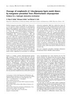
Fig. 1. Schematic representation of hematopoietic development in the embryo. The surface markers proving hematopoietic commitment at
each stage of the differentiation process are shown. Transcription factors required for differentiation are shown above the blue arrows
where appropriate. Runx1, runt-related transcription factor 1; Scl = Tal1, T-cell acute lymphocytic leukemia 1.
New advances in human hematopoiesis from human ESCs I. Moreno-Gimeno et al.
5016 FEBS Journal 277 (2010) 5014–5025 ª 2010 The Authors Journal compilation ª 2010 FEBS
from chronic myelogenous leukemia, suggesting that
translocation of these genes occurred in a progenitor
common to them both [44]. Further supporting this
hypothesis, both hematopoietic and endothelial cell lin-
eages can be derived from single CD34
+
KDR
+
cells
isolated from human bone marrow and cord blood [45].
Single-cell tracing and cell imaging of the aortic lumen
in vitro have shown that hemogenic endothelial cells,
that were initially defined by VE-cadherin and claudin
expression, acetylated low density lipoprotein uptake
and formation of tight junctions, start to co-express
CD41 and c-kit (early markers of definitive hematopoie-
sis) and change morphology over time. These cells con-
vert from adherent growth as endothelial colonies
towards a more ‘hematopoietic’ phenotype, character-
ized by loss of their characteristic ‘hemogenic’ expres-
sion profile whilst concurrently acquiring expression of
the pan-leukocyte marker CD45, and switching to
growth in suspension. Thus these studies firmly prove
the emergence of definitive blood cells from aortic endo-
thelium [35,46,47]. These findings have been confirmed
by many studies in mouse and human showing that
definitive human hematopoiesis originates from the aor-
tic embryonic endothelium [36,48].
Molecular studies have identified a number of tran-
scriptional factors that are key regulators of HSC
development. For instance, the stem cell leukemia
factor Scl, has been shown to play a pivotal role in
endothelial and hematopoietic differentiation. This
transcription factor is usually altered in patients suffer-
ing from T-cell leukemia, and it has been shown that
mESCs lacking Scl are unable to undergo hematopoie-
sis [49]. Other studies have shown that Scl is not
required for hemangioblast generation, but is necessary
for subsequent differentiation, as Scl mutants cannot
generate hemogenic endothelium from the hemangio-
blast and consequently no hematopoietic or endothelial
cells can be produced [50]. Another important tran-
scription factor required for the generation of blood
cells is Runx1. Alterations affecting Runx1 are
involved in acute myeloid leukemia and pediatric acute
lymphoblastic leukemia. In vitro studies using Runx1
null cells have shown that this transcription factor is
required for the production of definitive hematopoietic
cells, but not primitive hematopoietic or endothelial
cells, from hemogenic endothelium [51]. Despite this
progress, the complete developmental program that
facilitates the onset and progression of human embryo-
nic hematopoiesis remains to be further investigated.
Although the AGM region is considered the first site
of definitive hematopoiesis, recent studies have indi-
cated that the placenta acts as an additional extrame-
dullary hematopoietic organ during embryonic and
fetal development [52,53]. Hematopoietic precursors
found in the human placenta can give rise to erythro-
cytic and myelocytic lineages, and can also reconstitute
the hematopoietic system of myelo-ablated mice. These
findings suggest the possibility of using placenta as a
source of human hematopoietic progenitors for clinical
use. Following these investigations, Serikov et al. [54]
developed a protocol for cryopreservation and thawing
of the placenta to optimize the recovery of hematopoi-
etic precursors, demonstrating that placenta can yield
higher amounts of HSCs than umbilical cord blood,
and further suggesting that placenta could be banked
and used for clinical transplant in a similar way to
umbilical cord blood.
Which methods drive differentiation of
hESC to hematopoietic lineages?
In vitro differentiation of hESCs into HSCs has been
achieved through various methods, some based on cul-
turing hESCs in the presence of stromal cells that
mimic the embryonic hematopoietic developmental
environment, others using methods involving the for-
Table 1. Comparison of hematopoietic differentiation methods reported previously.
Method hESC lines
Differentiation
media Stages Cytokines
% CD34
+
in culture References
S17 co-culture H1, H1.1, H9.2 FBS added 1 No 1–2% at day 17 Kaufman et al. (2001) [3]
OP9 co-culture H1 and H9 FBS added 1 No 20% at day 7 Vodyanik et al. (2005) [4]
Hematopoietic
embryonic niches
H1, H9, hES-NCL1 FBS added 1 No 16% in AGM
co-culture
Ledran et al. (2008) [58]
Embryoid bodies H1, H9 FBS added 1 SCF, Flt-3, IL-3, IL-6,
G-CSF, BMP4
20% at day 15 Chadwick et al. (2003) [59]
Embryoid bodies H1, HES2 Serum-free 1 BMP4, bFGF – Kennedy et al. (2007) [61]
2 VEGF
Spin embryoid
bodies
hES2, hES3, hES4 Serum-free 1 BMP4, VEGF, SCF 23% at day 11 Ng et al. (2005, 2008)
[62,63]2 SCF, VEGF, IL-3, IL-6,
Tpo, Epo
I. Moreno-Gimeno et al. New advances in human hematopoiesis from human ESCs
FEBS Journal 277 (2010) 5014–5025 ª 2010 The Authors Journal compilation ª 2010 FEBS 5017
mation of three-dimensional cellular clumps termed
embryoid bodies. Various hematopoietic differentiation
methods are summarized in Table 1.
Co-culture with stromal cells
In 2001, work by Kaufman et al. [3] showed that co-
culture of hESCs with the cells lines S17 (derived from
murine bone marrow) or C166 (from murine yolk sac
endothelium) can induce differentiation towards blood
cells. After 17 days of co-culture, 2% of the hESCs
had formed hematopoietic progenitors as evaluated by
expression of CD34, an embryonic and adult hemato-
poietic stem cell marker. The kinetics of hematopoiesis
in co-culture with the S17 cell line showed increased
CD34 expression in two distinct stages of the differen-
tiation process. The first peak appeared in conjunction
with CD31 expression, and is proposed to mark a
hemogenic endothelium state, and the second started
at day 14, together with expression of the definitive
hematopoietic marker CD45, and was coincident with
observation of the first colonies in hematopoietic col-
ony assays [55]. The hematopoietic transcription fac-
tors SCL and GATA-2 were also expressed in these
cells between days 7 and 21 of differentiation. More-
over, when hESCs co-cultured with the S17 cell line or
CD34
+
cells derived from these co-cultures were trans-
ferred to semi-solid medium, they produced erythroid,
myeloid and megakaryocyte colonies, arising between
days 14 and 17, demonstrating the functionality of
hESC-derived CD34
+
precursors.
Using a similar approach, hESCs have also been
co-cultured with the mouse bone marrow stromal cell
line OP9, which is deficient in macrophage colony-
stimulating factor [56]. This method generates a higher
level of hematopoietic differentiation than co-culture
with the S17 cell line: almost 20% of initial hESCs dif-
ferentiated into CD34
+
progenitors after 7 days of co-
culture [4]. The kinetics of OP9 differentiation show
expression of CD34 from days 3 to 7, and the CD34
+
population showed enhanced CD43 and CD41 expres-
sion at approximately day 5, followed by CD45
expression at day 8. The expression of CD43 has been
found to mark the earliest hematopoietic cells arising
from hemato-endothelial commitment [57]. Using this
method, expression of hematopoietic transcription fac-
tors GATA-1 and GATA-2 was observed from days 2
and 3 of differentiation, coincident with the onset of
CD34 and SCL expression on days 3 and 4, respec-
tively. Additionally, colony-forming cells also arose
from CD34
+
cells from day 4, and, after subsequent
co-culture with MS-5 cells, showed production of both
lymphoid and myeloid lineages [4].
In our group, we used stromal cells derived from
embryonic hematopoietic niches to direct the differen-
tiation of hESCs towards hematopoietic progenitors
with greater efficiency. With this in mind, we com-
pared the ability of primary stromal cells from the
AGM and fetal liver regions, as well as three established
cell lines derived from the AGM region (AM20.1B4),
urogenital ridge (UG26.1B6) and fetal liver (EL08.1D2),
to induce hematopoietic differentiation of hESCs.
Expression of CD34 was found in all cases from day 6
and peaked at day 18, matching the peak of activity of
colony-forming cells. Co-culture of hESCs with pri-
mary AGM stromal cells produced the highest level of
CD34
+
and CD45
+
cells at day 18. Most importantly,
we found that some of these hESC-derived hemato-
poietic cells engrafted both primary and secondary
immunocompromised mice (treated with a sub lethal
dose of ionising radiation) at substantially higher levels
than described previously [58].
Formation of embryoid bodies
Differentiation of hESCs in the form of embryoid bodies
(EBs) is an easily modifiable alternative method that is
often used to obtain differentiated cell types in serum-
free and defined media, depending on the cytokines
and ⁄ or growth factors added. Many methods of EB-
mediated in vitro hematopoiesis have been described.
Chadwick et al. described the use of hematopoietic cyto-
kines in combination with BMP4 to induce hematopoie-
sis. Using this method, the proportion of CD34
+
cells at
day 15 of differentiation increased from 10% to 20%
when BMP4 and cytokines were added to the medium,
and 90% of the CD34
+
cells co-expressed CD45.
During this differentiation process, expression of hema-
topoietic transcription factors was detected, and hema-
topoietic colonies including macrophages, granulocytes,
megakaryocytes and erythrocytes emerged [59,60].
In 2007, Keller’s group developed a new two-step
approach for EB-mediated hematopoietic differentia-
tion, the first driven by BMP4 and basic fibroblast
growth factor, and the second by addition of VEGF
165
.
The kinetics of EB-derived hematopoiesis showed
expression of SCL, GATA-1 and RUNX1 from day 3,
consistent with up-regulation of CD34 expression,
detectable from day 4 of differentiation. The first
hematopoietic colony-forming cells colonies arose at
day 5, and consisted predominantly of primitive ery-
throid progenitors, followed by erythroid colonies and
macrophages that emerged at approximately day 8 of
differentiation. It is noteworthy that development of
hematopoietic progenitors from EBs in these conditions
was dependent on the presence of BMP4 [61].
New advances in human hematopoiesis from human ESCs I. Moreno-Gimeno et al.
5018 FEBS Journal 277 (2010) 5014–5025 ª 2010 The Authors Journal compilation ª 2010 FEBS
A further improvement in EB-mediated hematopoi-
etic differentiation was described in 2005 when spin
EBs were first described [62]. Spin EBs are embryoid
bodies produced from a defined number of cells that
are centrifuged to produce tight cell compaction, thus
improving experimental reproducibility compared to
standard EB derivation methods. The number of cells
used for EB formation and various augmenting factors
were found to be critical; optimum hematopoietic dif-
ferentiation was achieved when spin EBs were formed
from at least 1000 hESCs, and different cytokines were
required at each of two distinct differentiation stages
for efficient differentiation. Onset of CD34 expression
using this protocol was detected at day 6, concurrent
with expression of the RUNX1 transcription factor.
CD34 expression peaked at approximately day 10 and
was maintained at this level until day 26, when
approximately 30% of cells expressed CD45. More
primitive blast forming units erythroid colonies
(BFU-E) emerged on day 6, whereas more mature col-
ony forming unit (CFU-E) erythropoietic colonies and
myeloid cells arose later, probably from CD34
+
hema-
topoietic progenitors [62,63].
Successes and failures of
hematopoietic engraftment
The only definitive way to evaluate the full functional-
ity of hESC-derived hematopoietic cells is transplanta-
tion into immunocompromised animals in order to test
their ability to engraft and provide long-term multi-
lineage hematopoietic reconstitution. Various mouse
models have been used to study human hematopoiesis
in vivo; the most commonly used are genetically modi-
fied strains of the non-obese diabetic ⁄ severe combined
immuno-deficient (NOD ⁄ SCID) mouse.
To date, only a few studies have described the engraft-
ment potential of hESC-derived hematopoietic cells. In
2005, Bhatia’s group tested EB-differentiated cells by
transplanting these cells intravenously into the tail vein
of sub-lethally irradiated NOD ⁄ SCID mice. However,
more than 60% of the transplanted mice died after the
transplantation due to aggregation of human ESC-
derived hematopoietic cells in the presence of rodent
serum, resulting in the formation of pulmonary emboli.
To solve this problem, the authors performed intra-fem-
oral injections to deliver the cells directly into the bone
marrow. These animals showed hematopoietic reconsti-
tution 8 weeks after injection; however, the level of
human engraftment was low at 1% [64].
A second study used hematopoietic progenitors
derived from hESC co-culture with S17 cells for intra-
venous and intra-femoral transplantation into sub-leth-
ally irradiated NOD ⁄ SCID mice (treated with an
antibody against NK cells in order to avoid immune
rejection of the hESC-derived cells). Long-term
engraftment was achieved using both methods as
shown by expression of human CD45 3–6 months
post-transplantation. Additionally, secondary engraft-
ment was achieved following intravenous transplanta-
tion of bone marrow from primary recipients, but once
again the level of human engraftment was very low
(< 0.5% independently of intravenous or intra-femo-
ral injection) [65].
Our group has successfully used the NOD ⁄ LtSz-
SCID IL2rc
null
(NOG) mouse model [58] to test the
hematopoietic potential of hESCs differentiated in var-
ious embryonic hematopoietic environments by intra-
femoral injection. We found that hESC-derived hema-
topoietic cells engrafted primary and secondary recipi-
ents with multiple hematopoietic lineages for up to
12 weeks following transplantation. Most importantly,
engraftment levels were higher compared to previous
studies, reaching 16% when hESCs were co-cultured
with AM20.1B4, an established cell line derived from
the AGM embryonic region [58].
Future therapeutic applications for hESC-derived
hematopoietic cells will require a significant improve-
ment in the engraftment levels achieved to date. Low
engraftment may result from multiple factors, includ-
ing possible rejection by the host immune system, the
quantity administered, the developmental stage of the
cells derived from hESC differentiation, and the qual-
ity and ⁄ or viability of the transplanted cells. Moreover,
the variables affecting the study of human hematopoie-
sis in ‘humanized’ mice have been studied, and demon-
strate the importance of selecting the appropriate
mouse strain when designing experimental work. For
instance, truncation of IL2 receptor gamma chain
(IL2rc as in NSG mice) in the genetic background of
NOD ⁄ SCID mice has been shown to increase the
engraftment ability of NOD ⁄ SCID re-populating cells,
and consequently significantly improve human hemato-
poietic cell engraftment [66–68]. Host age is also
important, as it has been shown that newborn mice
support higher levels of engraftment than adults
because HSCs can better mature and develop in an
age-matched micro-environment [69]. This hypothesis
is in agreement with the results of a study of the long-
term engraftment of primary and secondary recipients
(22 months after transplantation) by Narayan et al.,
who studied HSC engraftment in fetal sheep by inject-
ing HSCs in utero into the fetal peritoneal cavity [70].
The site of transplantation (and therefore the differen-
tiation cues provided by the niche) is also very impor-
tant, as a recent study has shown than hESC-derived
I. Moreno-Gimeno et al. New advances in human hematopoiesis from human ESCs
FEBS Journal 277 (2010) 5014–5025 ª 2010 The Authors Journal compilation ª 2010 FEBS 5019
CD34
+
cells differentiate preferentially to endothelial
cells upon transplantation into the liver of immuno-
compromised recipients, suggesting that the environ-
ment surrounding the transplanted cells could
determine their final lineages [71].
Advantages and disadvantages of using
hESCs and hiPSCs for therapeutic
applications
The successful use of stem cells for clinical purposes is
one of the most appealing prospects in regenerative
medicine today. HSCs can be isolated from peripheral
blood, bone marrow and umbilical cord blood for exam-
ple, but problems arising from immunogenic matching,
resulting in rejection or graft-versus-host disease, as well
as the overall lack of donors, impair use of these cell
types for therapy. For these reasons, in vitro production
of hESC-derived HSCs represents a remarkable oppor-
tunity for regenerative medicine, as they represent not
only a theoretically unlimited source of more closely tis-
sue-matched donor cells, but can also reduce immune
responses following transplant of other cell types
derived from the same hESCs if hematopoietic chimer-
ism is first induced [60,72]. However, there are many
advantages and disadvantages regarding the use of
hESCs in medical practice, including some ethical objec-
tions regarding the use of human embryos for research,
even when discarded embryos are used.
Use of hiPSCs bypasses these ethical issues [27,28],
and several tissue-specific cells have been successfully
obtained using hiPSC differentiation, including hema-
topoietic and endothelial cells [73]. In theory, hiPSC
technology could allow the production of specific cell
types for the treatment of various patients or diseases
[74], but the suitability of these cells for therapeutic
use is still open to debate [75,76]. Experiments by Choi
et al. [73] suggest that HSCs derived from various
hiPSC lines differentiate following the same profile
than those derived from hESCs. However, recent work
by Lanza’s group showed that endothelial and hemato-
poietic cells obtained from hiPSC-derived hemangio-
blasts show decreased proliferation potential, an
apoptotic phenotype and early senescence, features
that were not observed in hESC-derived counterparts.
The same features were observed in hiPSC-derived reti-
nal pigment epithelial cells and cardiomyocytes [77,78],
evidence against a differentiated cell type-specific phe-
nomenon. The mechanism that results in such abnor-
malities is still unknown, but may be related to the
integration of viral vectors carrying the transcriptional
factors required for hiPSC reprogramming, or the
result of a perturbed epigenome.
A new method to reprogram hiPSCs based on epi-
somal vectors, which avoid the expression of transg-
enes in the iPSC lines generated, was described
recently [79]. However, it has been shown that the vec-
tors are not responsible for the differences in effective-
ness of hiPSC versus hESC differentiation, as neural
cells obtained from lentiviral, retroviral or episomal
vector iPSCs exhibit the same variability compared to
their hESC counterparts [80]. In order to clarify this,
comparative studies at the level of genomic DNA, cod-
ing RNA, microRNA and epigenetic events have
recently been performed [81–83]. These findings dem-
onstrated that, although very similar, hiPSCs and
hESCs are not identical in either their genomic DNA,
gene expresion pattern, epigenetic profile, teratoma
formation efficiency or latency [84]. In agreement with
this, two recently published studies using mouse mod-
els have shown that iPSCs retain a temporary tran-
scriptional and epigenetic memory of their original
somatic lineages, leading to preferential differentiation
of these cells towards their original tissue type [85,86].
This phenomenon was only observed when pluripotent
stem cells were obtained by exogenous expression of
reprogramming transcription factors, but not by
somatic cell nuclear transfer or embryonic stem cells
derived from fertilized embryos [85]. These findings
suggest that certain sets of somatic cells may be
selected prior to reprogramming with the aim of
enhancing the iPSC differentiation capacity using their
tissue-specific epigenetic memory. However, in many
cases, full pluripotency may be advisable or required
prior to re-differentiation, and therefore erasing the
epigenetic ⁄ transcriptional memory from iPSCs is an
important goal. Work towards achieving this by serial
reprogramming or by treating iPSCs with chromatin-
modifying chemicals has been described [85]. An alter-
native method for avoiding problems associated with
partially retained cellular epigenetic memory, by repro-
gramming human cord blood un-restricted somatic
stem cells, has recently been suggested [87]. It was
found that the iPSCs obtained from these cells are very
similar to hESCs in terms of morphology, molecular
signature and differentiation potential; nonetheless,
further studies on epigenetics and comparative analysis
using iPSCs obtained from other cells are required
[87].
Finally, some recent studies have suggested that hiP-
SCs might potentially be oncogenic. It is known that
some transcriptional factors used for hiPSC repro-
gramming, such as Lin28 and c-Myc, are tumorigenic,
and although their expression should be abolished in
hiPSCs, it is not known whether they can be activated
again at any time during differentiation [77,88].
New advances in human hematopoiesis from human ESCs I. Moreno-Gimeno et al.
5020 FEBS Journal 277 (2010) 5014–5025 ª 2010 The Authors Journal compilation ª 2010 FEBS
Another recent study showed that the p53 pathway
acts not only as a tumor suppressor, but also sup-
presses iPSC generation as determined using small
interfering RNA and p53 mutation [89]. These findings
indicate the need to improve hiPSC technology as well
as for a complete evaluation of the possible conse-
quences of use of hiPSCs for drug screening, as disease
models and for medical purposes.
Conclusions
The use of stem cells for the treatment of human
diseases is one of the principal goals in regenerative
medicine today. hESCs and hiPSCs, capable of direc-
ted differentiation into specific cell types with theoreti-
cally no limits on cell availability, are a promising
source of cells, but detailed investigation is required to
enable their application in clinical situations. In vitro
differentiation of hESCs to hematopoietic progenitors
has been successfully achieved by various methods and
groups, but the cells obtained using these approaches
are not completely functional in vivo, as demonstrated
by the low proportion of cells able to reconstitute the
hematopoietic system of immunodeficient animals. It is
worth highlighting that co-culture of hESCs with the
stromal cell line AM20.1B4, derived from the embry-
onic AGM region, yields the highest engraftment level
in NOD ⁄ LtSz-SCID ⁄ IL2rc
null
(NOG) mice, increasing
this proportion from < 1% to 16% [58]; however, this
is still not sufficient for any useful clinical application.
Human iPSCs are a promising alternative to the use
of hESCs for hematopoietic studies and in drug devel-
opment, but reasonable doubts regarding the suitabil-
ity of these cells for clinical therapies are starting to
emerge. The main objection to the potential clinical
use of hiPSC-derived cells is the use of viral vectors
required for initial reprogramming, which could confer
tumorigenic potential to these cells and their deriva-
tives. However, this is not the only problem: low dif-
ferentiation efficiencies and early senescence have also
been detected in hiPSC-derived cells.
For these reasons, future studies should strive to
improve differentiation protocols, in order to attain
higher engraftment levels. Additionally, the develop-
ment of safer and more efficient reprogramming meth-
ods for obtaining pluripotent stem cells may guarantee
the future safe application of hiPSC technologies for
clinical therapy.
Acknowledgements
The authors are grateful to financial support received
by from Leukemia and Lymphoma UK, Fanconi
Hope UK and the Fanconi Anemia Research Fund
USA, and funds for research in the field of regenera-
tive medicine through the collaboration agreement
between the Conselleria de Sanidad (Generalitat Valen-
ciana) and the Instituto de Salud Carlos III (Ministry
of Science and Innovation).
References
1 Thomson JA, Itskovitz-Eldor J, Shapiro SS, Waknitz
MA, Swiergiel JJ, Marshall VS & Jones JM (1998)
Embryonic stem cell lines derived from human blast-
ocysts. Science 282, 1145–1147.
2 Jensen J, Hyllner J & Bjo
¨
rquist P (2009) Human embry-
onic stem cell technologies and drug discovery. J Cell
Physiol 219, 513–519.
3 Kaufman DS, Hanson ET, Lewis RL, Auerbach R &
Thomson JA (2001) Hematopoietic colony-forming cells
derived from human embryonic stem cells. Proc Natl
Acad Sci USA 98, 10716–10721.
4 Vodyanik MA, Bork JA, Thomson JA & Slukvin II
(2005) Human embryonic stem cell-derived CD34
+
cells: efficient production in the coculture with OP9
stromal cells and analysis of lymphohematopoietic
potential. Blood 105, 617–626.
5 Qiu C, Olivier EN, Velho M & Bouhassira EE (2008)
Globin switches in yolk sac-like primitive and fetal-like
definitive red blood cells produced from human embry-
onic stem cells. Blood 111, 2400–2408.
6 Galic Z, Kitchen SG, Kacena A, Subramanian A,
Burke B, Cortado R & Zack JA (2006) T lineage differ-
entiation from human embryonic stem cells. Proc Natl
Acad Sci USA 103, 11742–11747.
7 Woll PS, Martin CH, Miller JS & Kaufman DS (2005)
Human embryonic stem cell-derived NK cells acquire
functional receptors and cytolytic activity. J Immunol
175, 5095–5103.
8 Menendez P, Wang L, Chadwick K, Li L & Bhatia M
(2004) Retroviral transduction of hematopoietic cells
differentiated from human embryonic stem cell-derived
CD45
(neg)
PFV hemogenic precursors. Mol Ther 10,
1109–1120.
9 Wang L, Li L, Shojaei F, Levac K, Cerdan C, Menen-
dez P, Martin T, Rouleau A & Bhatia M (2004) Endo-
thelial and hematopoietic cell fate of human embryonic
stem cells originates from primitive endothelium with
hemangioblastic properties. Immunity 21, 31–41.
10 Chang KH, Nelson AM, Cao H, Wang L, Nakamoto
B, Ware CB & Papayannopoulou T (2006) Definitive-
like erythroid cells derived from human embryonic stem
cells coexpress high levels of embryonic and fetal globins
with little or no adult globin. Blood 108, 1515–1523.
11 Lerou PH & Daley GQ (2005) Therapeutic potential of
embryonic stem cells. Blood Rev 19, 321–331.
I. Moreno-Gimeno et al. New advances in human hematopoiesis from human ESCs
FEBS Journal 277 (2010) 5014–5025 ª 2010 The Authors Journal compilation ª 2010 FEBS 5021
12 Menendez P, Bueno C & Wang L (2006) Human
embryonic stem cells: a journey beyond cell replacement
therapies. Cytotherapy 8, 530–541.
13 Lensch MW & Daley GQ (2006) Scientific and clinical
opportunities for modeling blood disorders with embry-
onic stem cells. Blood 107, 2605–2612.
14 Jenq RR & van den Brink MR (2010) Allogeneic
haematopoietic stem cell transplantation: individualized
stem cell and immune therapy of cancer. Nat Rev
Cancer 10, 213–221.
15 Kilareski EM, Shah S, Nonnemacher MR & Wigdahl B
(2009) Regulation of HIV-1 transcription in cells
of the monocyte–macrophage lineage. Retrovirology 6,
118.
16 Volberding PA & Deeks SG (2010) Antiretroviral therapy
and management of HIV infection. Lancet 376, 49–62.
17 Zhan X, Dravid G, Ye Z, Hammond H, Shamblott M,
Gearhart J & Cheng L (2004) Functional antigen-pre-
senting leucocytes derived from human embryonic stem
cells in vitro. Lancet 364, 163–171.
18 Anderson JS, Bandi S, Kaufman DS & Akkina R
(2006) Derivation of normal macrophages from human
embryonic stem (hES) cells for applications in HIV gene
therapy. Retrovirology 3, 24.
19 Su Z, Frye C, Bae KM, Kelley V & Vieweg J (2008)
Differentiation of human embryonic stem cells into
immunostimulatory dendritic cells under feeder-free
culture conditions. Clin Cancer Res 14, 6207–6217.
20 Senju S, Hirata S, Motomura Y, Fukuma D,
Matsunaga Y, Fukushima S & Matsuyoshi H (2010)
Pluripotent stem cells as source of dendritic cells for
immune therapy. Int J Hematol 91, 392–400.
21 Li MJ, Kim J, Li S, Zaia J, Yee JK, Anderson J,
Akkina R & Rossi JJ (2005) Long-term inhibition of
HIV-1 infection in primary hematopoietic cells by
lentiviral vector delivery of a triple combination of
anti-HIV shRNA, anti-CCR5 ribozyme, and a nucleolar-
localizing TAR decoy. Mol Ther 12, 900–909.
22 Liang M, Kamata M, Chen KN, Pariente N, An DS &
Chen IS (2010) Inhibition of HIV-1 infection by a
unique short hairpin RNA to chemokine receptor 5
delivered into macrophages through hematopoietic
progenitor cell transduction. J Gene Med 12, 255–265.
23 Hu
¨
tter G, Nowak D, Mossner M, Ganepola S, Mu
¨
ssig
A, Allers K, Schneider T, Hofmann J, Ku
¨
cherer C,
Blau O et al. (2009) Long-term control of HIV by
CCR5 Delta32 ⁄ Delta32 stem-cell transplantation.
N Engl J Med 360, 692–698.
24 Dudley ME, Wunderlich JR, Yang JC, Sherry RM,
Topalian SL, Restifo NP, Royal RE, Kammula U,
White DE, Mavroukakis SA et al. (2005) Adoptive cell
transfer therapy following non-myeloablative but
lymphodepleting chemotherapy for the treatment of
patients with refractory metastatic melanoma. J Clin
Oncol 23, 2346–2357.
25 Morgan RA, Dudley ME, Wunderlich JR, Hughes MS,
Yang JC, Sherry RM, Royal RE, Topalian SL,
Kammula US, Restifo NP et al. (2006) Cancer
regression in patients after transfer of genetically
engineered lymphocytes. Science 314, 126–129.
26 Woll PS, Grzywacz B, Tian X, Marcus RK, Knorr DA,
Verneris MR & Kaufman DS (2009) Human embryonic
stem cells differentiate into a homogeneous population
of natural killer cells with potent in vivo antitumor
activity.
Blood 113, 6094–6101.
27 Takahashi K, Tanabe K, Ohnuki M, Narita M,
Ichisaka T, Tomoda K & Yamanaka S (2007) Induction
of pluripotent stem cells from adult human fibroblasts
by defined factors. Cell 131, 861–872.
28 Yu J, Vodyanik MA, Smuga-Otto K, Antosiewicz-
Bourget J, Frane JL, Tian S, Nie J, Jonsdottir GA,
Ruotti V, Stewart R et al. (2007) Induced pluripotent
stem cell lines derived from human somatic cells.
Science 318, 1917–1920.
29 Hanna J, Wernig M, Markoulaki S, Sun CW, Meissner
A, Cassady JP, Beard C, Brambrink T, Wu LC,
Townes TM et al. (2007) Treatment of sickle cell
anemia mouse model with iPS cells generated from
autologous skin. Science 318, 1920–1923.
30 Xu D, Alipio Z, Fink LM, Adcock DM, Yang J, Ward
DC & Ma Y (2009) Phenotypic correction of murine
hemophilia A using an iPS cell-based therapy. Proc Natl
Acad Sci USA 106, 808–813.
31 Moore MAS & Metcalf D (1970) Ontogeny of the
hematopoietic system: yolk sac origin of in vivo and
in vitro colony forming cells in the developing mouse
embryo. Br J Haematol 18, 279–296.
32 Medvinsky A & Dzierzak E (1996) Definitive hemato-
poiesis is autonomously initiated by the AGM region.
Cell 86, 897–906.
33 Taoudi S, Gonneau C, Moore K, Sheridan JM, Black-
burn CC, Taylor E & Medvinsky A (2008) Extensive
hematopoietic stem cell generation in the AGM region
via maturation of VE-cadherin
+
CD45
+
pre-definitive
HSCs. Cell Stem Cell 3, 99–108.
34 Kumaravelu P, Hook L, Morrison AM, Ure J, Zhao S,
Zuyev S, Ansell J & Medvinsky A (2002) Quantitative
developmental anatomy of definitive haematopoietic
stem cells ⁄ long-term epopulating units (HSC ⁄ RUs): role
of the aorta–gonad–mesonephros (AGM) region and
the yolk sac in colonisation of the mouse embryonic
liver. Development 129, 4891–4899. Erratum in Develop-
ment (2003) 130, 425.
35 Zovein AC, Hofmann JJ, Lynch M, French WJ,
Turlo KA, Yang Y, Becker MS, Zanetta L, Dejana E,
Gasson JC et al. (2008) Fate tracing reveals the
endothelial origin of hematopoietic stem cells. Cell Stem
Cell 3 , 625–636.
36 Boisset JC, van Cappellen W, Andrieu-Soler C,
Galjart N, Dzierzak E & Robin C (2010) In vivo
New advances in human hematopoiesis from human ESCs I. Moreno-Gimeno et al.
5022 FEBS Journal 277 (2010) 5014–5025 ª 2010 The Authors Journal compilation ª 2010 FEBS
imaging of haematopoietic cells emerging from the
mouse aortic endothelium. Nature 464, 116–120.
37 Kissa K & Herbomel P (2010) Blood stem cells emerge
from aortic endothelium by a novel type of cell transi-
tion. Nature 464, 112–115.
38 Sabin FR (1920) Studies on the origin of blood vessels
and of red blood corpuscles as seen in the living blasto-
derm of chicks during the second day of incubation.
Contrib Embryol 9, 214.
39 Murray PDF (1932) The development in vitro of blood of
the early chick embryo. Proc R Soc London 111, 497–521.
40 Choi K, Kennedy M, Kazarov A, Papadimitriou JC &
Keller G (1998) A common precursor for hematopoietic
and endothelial cells. Development 125, 725–732.
41 Mandal L, Banerjee U & Hartenstein V (2004) Evidence
for a fruit fly hemangioblast and similarities between
lymph-gland hematopoiesis in fruit fly and mammal
aorta–gonadal–mesonephros mesoderm. Nat Genet 36,
1019–1023.
42 Vogeli KM, Jin SW, Martin GR & Stainier DY (2006) A
common progenitor for haematopoietic and endothelial
lineages in the zebrafish gastrula. Nature 443, 337–339.
43 Huber TL, Kouskoff V, Fehling HJ, Palis J & Keller G
(2004) Haemangioblast commitment is initiated in the
primitive streak of the mouse embryo. Nature 432,
625–630.
44 Gunsilius E, Duba HC, Petzer AL, Ka
¨
hler CM, Gru
¨
ne-
wald K, Stockhammer G, Gabl C, Dirnhofer S, Clausen
J & Gastl G (2001) Evidence from a leukaemia model
for maintenance of vascular endothelium by bone-mar-
row-derived endothelial cells. Lancet 355, 1688–1691.
45 Pelosi E, Valtieri M, Coppola S, Botta R, Gabbianelli
M, Lulli V, Marziali G, Masella B, Mu
¨
ller R, Sgadari
C et al. (2002) Identification of the hemangioblast in
postnatal life. Blood 100, 3203–3208.
46 Lancrin C, Sroczynska P, Stephenson C, Allen T,
Kouskoff V & Lacaud G (2009) The haemangioblast
generates haematopoietic cells through a haemogenic
endothelium stage. Nature 457, 892–895.
47 Eilken HM, Nishikawa SI & Schroeder T (2009) Con-
tinuous single-cell imaging of blood generation from
haemogenic endothelium. Nature 457, 896–901.
48 Oberlin E, El Hafny B, Petit-Cocault L & Souyri M
(2010) Definitive human and mouse hematopoiesis orig-
inates from the embryonic endothelium: a new class of
HSCs based on VE-cadherin expression. Int J Dev Biol
54, 1165–1173.
49 Porcher C, Swat W, Rockwell K, Fujiwara Y, Alt FW
& Orkin SH (1996) The T cell leukemia oncoprotein
SCL ⁄ tal-1 is essential for development of all hematopoi-
etic lineages. Cell 86, 47–57.
50 D’Souza SL, Elefanty AG & Keller G (2005) SCL ⁄
Tal-1 is essential for hematopoietic commitment of the
hemangioblast but not for its development. Blood 105,
3862–3870.
51 Chen MJ, Yokomizo T, Zeigler BM, Dzierzak E &
Speck NA (2009) RUNX1
is required for the endothelial
to haematopoietic cell transition but not thereafter.
Nature 457, 887–891.
52 Ba
´
rcena A, Kapidzic M, Muench MO, Gormley M,
Scott MA, Weier JF, Ferlatte C & Fisher SJ (2009) The
human placenta is a hematopoietic organ during the
embryonic and fetal periods of development. Dev Biol
327, 24–33.
53 Robin C, Bollerot K, Mendes S, Haak E, Crisan M,
Cerisoli F, Lauw I, Kaimakis P, Jorna R, Vermeulen M
et al. (2009) Human placenta is a potent hematopoietic
niche containing hematopoietic stem and progenitor cells
throughout development. Cell Stem Cell 5, 385–395.
54 Serikov V, Hounshell C, Larkin S, Green W, Ikeda H,
Walters MC & Kuypers FA (2009) Human term pla-
centa as a source of hematopoietic cells. Exp Biol Med
234, 813–823.
55 Woll PS, Morris JK, Painschab MS, Marcus RK, Kohn
AD, Biechele TL, Moon RT & Kaufman DS (2008)
Wnt signaling promotes hematoendothelial cell
development from human embryonic stem cells. Blood
111, 122–131.
56 Ji J, Vijayaragavan K, Bosse M, Menendez P, Weisel K
& Bhatia M (2008) OP9 stroma augments survival of
hematopoietic precursors and progenitors during hema-
topoietic differentiation from human embryonic stem
cells. Stem Cells 26, 2485–2495.
57 Vodyanik MA, Thomson JA & Slukvin II (2006) Leuk-
osialin (CD43) defines hematopoietic progenitors in
human embryonic stem cell differentiation cultures.
Blood 108, 2095–2105.
58 Ledran MH, Krassowska A, Armstrong L, Dimmick I,
Renstro
¨
m J, Lang R, Yung S, Santibanez-Coref M,
Dzierzak E, Stojkovic M et al. (2008) Efficient hemato-
poietic differentiation of human embryonic stem cells
on stromal cells derived from hematopoietic niches. Cell
Stem Cell 3, 85–98.
59 Chadwick K, Wang L, Li L, Menendez P, Murdoch B,
Rouleau A & Bhatia M (2003) Cytokines and BMP-4
promote hematopoietic differentiation of human embry-
onic stem cells. Blood 102, 906–915.
60 Wang L, Menendez P, Cerdan C & Bhatia M (2005)
Hematopoietic development from human embryonic
stem cell lines. Exp Hematol 33, 987–996.
61 Kennedy M, D’Souza SL, Lynch-Kattman M,
Schwantz S & Keller G (2007) Development of the
hemangioblast defines the onset of hematopoiesis in
human ES cell differentiation cultures. Blood 109,
2679–2687.
62 Ng ES, Davis RP, Azzola L, Stanley EG & Elefanty
AG (2005) Forced aggregation of defined numbers of
human embryonic stem cells into embryoid bodies fos-
ters robust, reproducible hematopoietic differentiation.
Blood 106, 1601–1603.
I. Moreno-Gimeno et al. New advances in human hematopoiesis from human ESCs
FEBS Journal 277 (2010) 5014–5025 ª 2010 The Authors Journal compilation ª 2010 FEBS 5023
63 Ng ES, Davis RP, Hatzistavrou T, Stanley EG &
Elefanty AG (2008) Directed differentiation of human
embryonic stem cells as spin embryoid bodies and a
description of the hematopoietic blast colony forming
assay. Curr Protoc Stem Cell Biol. doi:10.1002/
9780470151808.sc01d03s4.
64 Wang L, Menendez P, Shojaei F, Li L, Mazurier F,
Dick JE, Cerdan C, Levac K & Bhatia M (2005) Gener-
ation of hematopoietic repopulating cells from human
embryonic stem cells independent of ectopic HOXB4
expression. J Exp Med 201 , 1603–1614.
65 Tian X, Woll PS, Morris JK, Linehan JL & Kaufman
DS (2006) Hematopoietic engraftment of human
embryonic stem cell-derived cells is regulated by recipi-
ent innate immunity. Stem Cells 24, 1370–1380.
66 McDermott SP, Eppert K, Lechman E, Doedens M &
Dick JE (2010) Comparison of human cord blood
engraftment between immunocompromised mouse
strains. Blood 116, 193–200.
67 Levac K, Menendez P & Bhatia M (2005) Intra-bone
marrow transplantation facilitates pauci-clonal human
hematopoietic repopulation of NOD ⁄ SCID ⁄ b2m
) ⁄ )
mice. Exp Hematol 33, 1417–1426.
68 Bueno C, Montes R, de la Cueva T, Gutierrez-Ara
´
nda
I & Menendez P (2010) Intra-bone marrow transplanta-
tion of human CD34
+
cells into NOD ⁄ LtSz-scid IL-
2rc
null
mice permits multilineage engraftment without
previous irradiation. Cytotherapy 12, 45–49.
69 Brehm MA, Cuthbert A, Yang C, Miller DM,
DiIorio P, Laning J, Burzenski L, Gott B, Foreman
O, Kavirayani A et al. (2010) Parameters for
establishing humanized mouse models to study human
immunity: analysis of human hematopoietic stem cell
engraftment in three immunodeficient strains of mice
bearing the IL2rc
null
mutation. Clin Immunol 135,
84–98.
70 Narayan AD, Chase JL, Lewis RL, Tian X, Kaufman
DS, Thomson JA & Zanjani ED (2006) Human embry-
onic stem cell-derived hematopoietic cells are capable of
engrafting primary as well as secondary fetal sheep
recipients. Blood 107, 2180–2183.
71 Tian X, Hexum MK, Penchev VR, Taylor RJ, Shultz
LD & Kaufman DS (2009) Bioluminescent imaging
demonstrates that transplanted human embryonic stem
cell-derived CD34
+
cells preferentially develop into
endothelial cells. Stem Cells 27, 2675–2685.
72 Menendez P, Bueno C, Wang L & Bhatia M (2005)
Human embryonic stem cells: potential tool
for achieving immunotolerance? Stem Cell Rev 1, 151–
158.
73 Choi KD, Yu J, Smuga-Otto K, Salvagiotto G,
Rehrauer W, Vodyanik M, Thomson J & Slukvin I
(2009) Hematopoietic and endothelial differentiation of
human induced pluripotent stem cells. Stem Cells 27,
559–567.
74 Park IH, Arora N, Huo H, Maherali N, Ahfeldt T,
Shimamura A, Lensch MW, Cowan C, Hochedlinger K
& Daley GQ (2008) Disease-specific induced pluripotent
stem cells. Cell 134, 877–886.
75 Lako M, Armstrong L & Stojkovic M (2010) Induced
pluripotent stem cells: it looks simple but can looks
deceive? Stem Cells 28, 845–850.
76 Teo AK & Vallier L (2010) Emerging use of stem cells
in regenerative medicine. Biochem J 428, 11–23.
77 Feng Q, Lu SJ, Klimanskaya I, Gomes I, Kim D,
Chung Y, Honig GR, Kim KS & Lanza R (2010)
Hemangioblastic derivatives from human induced
pluripotent stem cells exhibit limited expansion and
early senescence. Stem Cells 28, 704–712.
78 Zhang J, Wilson GF, Soerens AG, Koonce CH, Yu J,
Palecek SP, Thomson JA & Kamp TJ (2009)
Functional cardiomyocytes derived from human
induced pluripotent stem cells. Circ Res 104, e30–e41.
79 Yu J, Hu K, Smuga-Otto K, Tian S, Stewart R, Slukvin
II & Thomson JA (2009) Human induced pluripotent
stem cells free of vector and transgene sequences. Sci-
ence 324, 797–801.
80 Hu BY, Weick JP, Yu J, Ma LX, Zhang XQ, Thomson
JA & Zhang SC (2010) Neural differentiation of human
induced pluripotent stem cells follows developmental
principles but with variable potency. Proc Natl Acad
Sci USA 107, 4335–4340.
81 Chin MH, Mason MJ, Xie W, Volinia S, Singer M,
Peterson C, Ambartsumyan G, Aimiuwu O, Richter L,
Zhang J et al. (2009) Induced pluripotent stem cells and
embryonic stem cells are distinguished by gene expres-
sion signatures. Cell Stem Cell 5, 111–123.
82 Pick M, Stelzer Y, Bar-Nur O, Mayshar Y, Eden A &
Benvenisty N (2009) Clone- and gene-specific aberra-
tions of parental imprinting in human induced pluripo-
tent stem cells. Stem Cells 27, 2686–2690.
83 Doi A, Park IH, Wen B, Murakami P, Aryee MJ,
Irizarry R, Herb B, Ladd-Acosta C, Rho J, Loewer S
et al. (2009) Differential methylation of tissue- and
cancer-specific CpG island shores distinguishes human
induced pluripotent stem cells, embryonic stem cells
and fibroblasts. Nat Genet 41, 1350–1353.
84 Gutierrez-Aranda I, Ramos-Mejı
´
a V, Bueno C, Mun
˜
oz-
Lo
´
pez M, Real PJ, Macı
´
aA,Sa
´
nchez L, Ligero G,
Garcı
´
a-Pe
´
rez JL & Menendez P (2010) Human induced
pluripotent stem cells develop teratoma more efficiently
and faster than human embryonic stem cells regardless
the site of injection. Stem Cells 28, 1568–1570.
85 Kim K, Doi A, Wen B, Ng K, Zhao R, Cahan P, Kim
J, Aryee MJ, Ji H, Ehrlich LI et al. (2010) Epigenetic
memory in induced pluripotent stem cells. Nature 467,
285–290.
86 Polo JM, Liu S, Figueroa ME, Kulalert W, Eminli S,
Tan KY, Apostolou E, Stadtfeld M, Li Y, Shioda T
et al. (2010) Cell type of origin influences the molecular
New advances in human hematopoiesis from human ESCs I. Moreno-Gimeno et al.
5024 FEBS Journal 277 (2010) 5014–5025 ª 2010 The Authors Journal compilation ª 2010 FEBS
and functional properties of mouse induced pluripotent
stem cells. Nat Biotechnol 28, 848–855.
87 Zaehres H, Ko
¨
gler G, Arauzo-Bravo MJ, Bleidissel M,
Santourlidis S, Weinhold S, Greber B, Kim JB, Buchhe-
iser A, Liedtke S et al. (2010) Induction of pluripotency
in human cord blood unrestricted somatic stem cells.
Exp Hematol 38, 809–818.
88 West JA, Viswanathan SR, Yabuuchi A, Cunniff K,
Takeuchi A, Park IH, Sero JE, Zhu H, Perez-Atayde
A, Frazier AL et al. (2009) A role for Lin28 in primor-
dial germ-cell development and germ-cell malignancy.
Nature 460, 909–913.
89 Hong H, Takahashi K, Ichisaka T, Aoi T, Kanagawa
O, Nakagawa M, Okita K & Yamanaka S (2009)
Suppression of induced pluripotent stem cell generation
by the p53–p21 pathway. Nature 460, 1132–1135.
I. Moreno-Gimeno et al. New advances in human hematopoiesis from human ESCs
FEBS Journal 277 (2010) 5014–5025 ª 2010 The Authors Journal compilation ª 2010 FEBS 5025