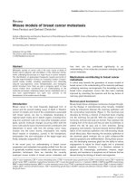
Mouse models of breast cancer metastasis: Anna Fantozzi and Gerhard Christofori pdf
Bạn đang xem bản rút gọn của tài liệu. Xem và tải ngay bản đầy đủ của tài liệu tại đây (332.13 KB, 11 trang )
Available online />
Review
Mouse models of breast cancer metastasis
Anna Fantozzi and Gerhard Christofori
Institute of Biochemistry and Genetics, Department of Clinical-Biological Sciences (DKBW), Center of Biomedicine, University of Basel, Mattenstrasse
28, CH-4058 Basel, Switzerland
Corresponding author: Gerhard Christofori,
Published: 26 July 2006
This article is online at />© 2006 BioMed Central Ltd
Breast Cancer Research 2006, 8:212 (doi:10.1186/bcr1530)
Abstract
how their use has contributed significantly to our
understanding of the molecular processes underlying breast
cancer metastasis.
Metastatic spread of cancer cells is the main cause of death of
breast cancer patients, and elucidation of the molecular mechanisms underlying this process is a major focus in cancer research.
The identification of appropriate therapeutic targets and proof-ofconcept experimentation involves an increasing number of experimental mouse models, including spontaneous and chemically
induced carcinogenesis, tumor transplantation, and transgenic
and/or knockout mice. Here we give a progress report on how
mouse models have contributed to our understanding of the
molecular processes underlying breast cancer metastasis and on
how such experimentation can open new avenues to the
development of innovative cancer therapy.
Introduction
Breast cancer is the most frequently diagnosed form of
cancer and the second leading cause of death in Western
women [1]. Death, and most of the complications associated
with breast cancer, are due to metastasis developing in
regional lymph nodes and in distant organs, including bone,
lung, liver, and brain [1,2]. As in many other metastatic cancer
types, specific molecular changes occurring within both the
tumor cells and the tumor microenvironment contribute to the
detachment of tumor cells from the primary tumor mass,
invasion into the tumor stroma, intravasation into nearby
blood vessels or lymphatics, survival in the bloodstream,
extravasation into and colonization of the target organ and,
finally, metastatic outgrowth [3,4].
In the recent past, our understanding of breast cancer
progression and metastasis has greatly profited from the use
of genetically modified mouse models and advanced transplantation techniques. Here we describe the currently
employed mouse models of breast cancer metastasis and
Mechanisms contributing to breast cancer
metastasis
A critical step towards the generation of mouse models of
breast cancer is the understanding of the molecular pathways
underlying mammary carcinogenesis. Our knowledge on how
breast tumor progression occurs has also been markedly
improved by unraveling the dynamics and the key factors of
mammary gland development.
Mammary gland development
Mouse breast tissue undergoes continuous changes throughout the lifespan of reproductively active females, mediated
mainly by interactions between the mammary epithelium and
the surrounding mesenchyme (Figure 1). The mammary bud
develops by forming a network of branched ducts invading
into the mammary fat pad [5]. With the release of ovarian
hormones, terminal end buds are formed. They represent the
invading front of the ducts and they are able to proliferate, to
extend into the fat pad, and to form branches. During
pregnancy and lactation, hormone-induced terminal differentiation of the mammary epithelium into milk-secreting lobular
alveoli takes place. After weaning, the secretory epithelium of
the mammary gland involutes into an adult nulliparous-like
state by apoptosis and redifferentiation. During these processes, the developing mammary gland has the ability to induce
angiogenesis to adjust for blood supply and is protected
against premature involution; it is therefore resistant to
apoptosis [6]. Interestingly, proliferation, invasion, angiogenesis, and resistance to apoptosis are all features that are
abused during the etiology of breast carcinogenesis.
COX = cyclo-oxygenase; CSF = colony-stimulating factor; CTGF = connective tissue growth factor; ECM = extracellular matrix; EGF = epidermal
growth factor; EMT = epithelial–mesenchymal transition; IGF = insulin-like growth factor; IL = interleukin; MEKK = MAP kinase/ERK kinase kinase;
MMP = matrix metalloproteinase; MMTV = murine mammary tumour virus; PTHrP = parathyroid hormone-related protein; PyMT = polyoma middle T
antigen; SDF = stromal cell-derived factor; TGF = transforming growth factor; VCAM = vascular cell adhesion molecule; VEGF = vascular endothelial growth factor.
Page 1 of 11
(page number not for citation purposes)
Breast Cancer Research
Vol 8 No 4
Fantozzi and Christofori
Figure 1
Schematic representation of epithelial–stromal interactions during mammary gland development. The mammary bud originates at the embryonic
level and starts proliferating after birth. Pubertal hormones drive the invasion of the fat pad by the generation of epithelial ducts and terminal end
buds (TEB). Proliferation and side branching continues until epithelial ducts fill the adult mammary gland. Pregnancy hormones induce the full
development and proliferation of the mammary gland and the transformation of the lobular alveoli into milk-secreting ducts. After lactation the
mammary gland involutes to return to a nulliparous-like state via apoptosis, redifferentiation and remodeling processes. C/EBP, CCAAT-enhancerbinding protein; CSF, colony-stimulating factor; DDR, discoidin domain receptor; ECM, extracellular matrix; HSPG, heparan sulfate proteoglycan;
GH, growth hormone; IGF, insulin-like growth factor; IRF, interferon regulatory factor; MMP, matrix metalloproteinase; NFκB, nuclear factor-κB;
Ptc-1, patched-1; TGF, transforming growth factor; TIMP, tissue inhibitor of metalloproteinases.
Transformation and metastasis
Mammary gland morphogenesis and branching involve the
regulatory function of several signaling pathways, including
signaling by Wnt family members [7], transforming growth
factor-β (TGF-β) [8], insulin-like growth factor-I (IGF-I) [9],
and epidermal growth factor (EGF) and others [10]. These
pathways are frequently activated during the tumorigenic
process by mutation or gene amplification, thus allowing the
mammary epithelium to expand, proliferate, and invade neighboring tissue. The cross-talk and interactions between tumor
cells and the surrounding stroma, the extracellular matrix
(ECM), and infiltrating cells of the immune system are constantly modulating tumor development. The mammary stroma,
composed of pre-adipocytes, adipocytes, fibroblasts, endothelial cells, and inflammatory cells, contributes functionally to
mammary gland development [6]. In a similar manner,
tumor–stroma interactions, occurring via soluble growth
factors, cytokines and chemokines, remodeling of the extracellular matrix, or direct cell–cell adhesion, are critical for
tumor growth, migration, and metastasis. Alteration of the
expression or function of adhesion molecules responsible for
Page 2 of 11
(page number not for citation purposes)
the adhesion of breast cancer cells to themselves, to stromal
cells, or to tumor matrix, including integrin family members,
immunoglobulin-domain cell adhesion molecules (such as L1
and NCAM), cadherin family members, or other cell surface
receptors (such as CD44), contributes predominantly to latestage tumor progression and metastatic dissemination of
cancer cells [11,12].
The formation of new blood vessels (angiogenesis) is crucial
for the growth and persistence of primary solid tumors and
their metastases, and it has been assumed that angiogenesis
is also required for metastatic dissemination, because an
increase in vascular density will allow easier access of tumor
cells to the circulation. Induction of angiogenesis precedes
the formation of malignant tumors, and increased vascularization seems to correlate with the invasive properties of
tumors and thus with the malignant tumor phenotype [13]. In
fact, angiogenesis indicates poor prognosis and increased
risk of metastasis in many cancer types, including breast
cancer [14]. With the recent identification of lymphangiogenic factors and their receptors it has also been possible to
Available online />
investigate the causal role of lymphangiogenesis in the
metastatic process (reviewed in [15]). It is therefore not surprising that molecules essential for mammary gland development, many of them stromal factors, are also critical
participants in breast carcinogenesis.
The knowledge gained on the several mechanisms contributing to tumor progression can be used to design and
generate better mouse models. At the same time, such
models allow a thorough investigation of all different aspects
of multistage breast carcinogenesis, including the genetic
alterations leading to tumor onset, neovascularization, tumor
progression, and formation of metastasis in secondary
organs.
Breast cancer metastasis models
Tumor transplantation
There are various ways to mimic breast cancer growth and
metastasis in tumor transplantation experiments. The site of
injection, together with the specific tropism of the chosen
breast cancer cell line used, largely defines primary and
secondary metastatic growth. Orthotopic or ectotopic
implantation of cancer cells in the skin or mammary fat pad,
with the formation of primary tumors and the subsequent
formation of metastasis, in part resembles the multiple stages
involved in malignant breast cancer development in patients
[16]. In contrast, tail vein injection results mainly in lung
metastasis, whereas portal vein injection provokes colonization of the liver, and intracardiac infusion gives rise to a
broader target organ spectrum, including bone. Notably, the
direct introduction of cancer cells into the blood circulation
should be considered an assay of organ colonization and not
a true metastatic process.
Depending on the species or genetic background of donor
and host, syngeneic or xenograft tumor transplantations need
to be distinguished. Transplantation of cancer cells from one
mouse into another mouse with identical genetic backgrounds (syngeneic transplantation) bypasses the immunologic host-versus-graft reaction and concomitantly allows the
investigation of the contribution of an intact immune system
to malignant tumor progression [17,18]. Syngeneic mouse
models have been employed to establish organ-specific
metastasis models by several rounds of transplantation/
metastasis formation and the selection of metastatic cell lines
in vivo [19]. For example, 4T1 cells, which originally derive
from a spontaneous mouse mammary tumor of a BALB/C
mouse, grow rapidly when injected into the fat pad of a
syngeneic animal and metastasize to lungs, liver, bone, and
brain [19,20]. Sublines of 4T1 cells, which exhibit various
degrees of metastatic dissemination, have been employed
recently to generate distinct gene expression signatures for
each stage of tumor progression, namely primary tumor
formation, lymph node colonization, metastatic outgrowth in
the lymph node, and distant organ metastasis. These experiments led to the identification of the transcriptional repressor
Twist, some members of the cadherin family of cell–cell
adhesion molecules, and various chemokines as critical
factors in the distinct stages of metastatic tumor progression
[20]. This and other syngeneic mouse models have also been
successfully employed for the testing of experimental drugs
designed to interfere with tumor malignancy [18,21].
To investigate the growth and metastasis of human breast
cancer cell lines in vivo, xenograft transplantation experiments are performed in immunocompromised mice [22].
Human breast cancer cells can be injected subcutaneously,
intravenously, intracardially, or orthotopically into the fat pad
of the mouse [23]. For example, MDA-MB-231 cells, an
estrogen-independent breast cancer cell line derived from the
pleural effusion of a cancer patient, is able to colonize bone,
liver, lung, adrenal glands, ovary, and brain after intravenous
injection [24]. This cell line and organ-specific metastatic
variants thereof have recently been used to identify and
functionally implicate a number of genes in organ-specific
metastasis, including IL-11, osteopontin and the connective
tissue growth factor (CTGF) in osteolytic metastasis [25,26],
and epiregulin, CXCL1, matrix metalloproteinase-1 (MMP-1),
cyclo-oxygenase-2 (COX-2), inhibitor of differentiation-1 (Id1)
and others in lung metastasis [27] (see below).
The implantation of established cell lines derived from human
breast cancer is relatively simple and allows the genetic or
pharmacological manipulation of the implanted cells. However, there are clear limitations to xenograft models. First,
immune responses, which have a key role during tumor
development, are impaired in immunocompromised mice.
Second, stromal components are not of tumor origin. For
example, carcinoma-associated fibroblasts derived from a
breast cancer patient support the growth of a breast carcinoma
cell line better than the normal tissue in a xenograft mouse
co-implantation model. Carcinoma-associated fibroblasts
seem to activate and sustain CXCR4/stromal cell-derived
factor (SDF-1)-mediated chemokine signaling and to recruit
endothelial progenitors to the growing tumor, thereby
promoting angiogenesis [28,29]. Last, human cells are
apparently not fully adapted to grow in a murine environment.
For example, breast cancer metastasis to bone has recently
been investigated in an experimental mouse system in which
both the breast cancer cells and the metastatic target organ,
the bone, are of human origin [30]. After orthotopic injection,
cancer cells predominantly colonize the bone of human origin,
thus exhibiting a species-specific osteotropism.
Genetically modified mice
Several promoters can be used to drive the expression of
transgenes in the mammary epithelium (Table 1), and many
known oncogenes have been expressed under their control
to initiate or modulate breast carcinogenesis in mice, including ErbB2/Neu, polyoma middle T antigen (PyMT), simian
virus 40 (SV40) T antigen, Ha-Ras, Wnt-1, TGF-α, and
c-Myc. MMTV-Neu and MMTV-PyMT transgenic mice (in
Page 3 of 11
(page number not for citation purposes)
Breast Cancer Research
Vol 8 No 4
Fantozzi and Christofori
Table 1
Mammary gland-specific promoters
Promoter
Origin
Expression
Activation
MMTV-LTR
Mouse mammary tumor virus
Breast epithelial cells, several
other tissues
Steroid hormones
WAP
Whey acidic protein
Secretory mammary epithelium
Lactogenic hormones
C3(1)
Rat prostate steroid-binding
protein (PSBP)
Epithelial cells of prostate and
mammary gland
Estrogen (ductal and alveolar
mammary epithelium)
B-LG
Bovine β-lactoglobulin
Mammary gland
Pregnancy and lactation
MT
Metallothionein
Most mammary cells
Zn2+
References
[42]
[96,97]
[36]
[98,99]
[100]
which the expression of the oncogene is driven by the Mouse
Mammary Tumor Virus promoter) develop metastasis in lung
and lymph nodes, mainly after their first pregnancy, while
other transgenic mice have to be combined to generate
double-transgenic mice that efficiently develop malignant
cancers [31-35]. C3(1)-SV40 T-antigen transgenic mice
develop invasive mammary carcinomas independently of
hormone supplementation or pregnancy, with a 15%
incidence of lung metastasis. This model recapitulates the
loss of estrogen receptor-α expression that is frequently
observed in human breast cancer [36]. The most commonly
used transgenic mouse models that develop metastatic
mammary cancer are summarized in Table 2.
MMTV-Neu
Amplification of the gene encoding ErbB2, a member of the
EGF receptor gene family, is associated with 15 to 20% of
human breast cancers, and in about 30% of cases the
increased expression of an activated form of ErbB2 is
detected. Consistent with this notion is the observation that
transgenic expression of an activated form of the rat homolog
of ErbB2 (Neu) in MMTV-Neu transgenic mice results in the
development of multifocal adenocarcinomas with lung metastases at about 15 weeks after pregnancy [42]. Transgenic
expression of wild-type ErbB2 in mammary gland also
provokes tumor formation and metastatic dissemination, yet
with longer latency.
Investigating the functional role of distinct genes during the
multiple stages of breast carcinogenesis requires the ability
to modulate their function in time and space [37]. Inducible
transgene expression can be obtained by the use of the
bacteria-derived tetracycline-inducible system permitting the
switching on or off (Tet-On/Tet-Off system) of a gene of
interest in a tissue- and time-specific manner [38]. In contrast,
mice are modified by the genetic ablation of a gene of interest
in an inducible manner to generate conditional knockouts
with the use of the Cre/loxP phage recombinase system, for
example [39]. To ablate a gene at a certain time point in
mammary epithelial cells, recombinase activity can be
controlled by the expression of a tamoxifen-inducible version
of Cre (MMTV-ERTM-Cre) or by using the tetracyclineinducible system to drive Cre expression [40].
Doxycycline-inducible expression of ErbB2 in mammary
epithelial cells of transgenic mice also results in invasive
mammary carcinoma and extensive metastasis, yet the tumors
regress with the loss of ErbB2 expression upon the
withdrawal of doxycycline. However, most mice exhibit
recurrences of the tumors [43]. These recurrent tumors
exhibit epithelial–mesenchymal transition (EMT), which seems
to be mediated by the upregulated expression of the
transcriptional repressor Snail, a molecular process that
seems to have a high prognostic value in predicting human
breast cancer recurrence. Expression of oncogenic versions
of ErbB2 that bind either Grb-2 or Shc demonstrate that
focal mammary tumors with a high rate of lung metastasis
require Grb-2-mediated signaling, whereas low metastatic
multifocal mammary tumors rely on Shc function [44].
First comparisons of gene expression profiles obtained from
mammary gland tumor models initiated by different oncogenes have revealed several common and oncogene-specific
targets and similarities with human molecular breast cancer
pathology [41]. The challenge now is to test whether genes
identified in gene expression profiling experiments with
patient samples are able to modulate breast carcinogenesis
in transgenic mouse models, for example in the wellcharacterized MMTV-Neu and MMTV-PyMT mouse models of
breast carcinogenesis or in improved versions of these.
MMTV-PyMT
Mammary gland-specific expression of PyMT under the
control of the MMTV promoter/enhancer in transgenic mice
(MMTV-PyMT) results in widespread transformation of the
mammary epithelium and in the development of multifocal
mammary adenocarcinomas and metastatic lesions in the
lymph nodes and in the lungs [45]. Tumor formation and
progression in these mice is characterized by four stages:
hyperplasia, adenoma/mammary intra-epithelial neoplasia,
and early and late carcinoma [46]. The close similarity of this
Page 4 of 11
(page number not for citation purposes)
Available online />
Table 2
Transgenic mouse models of breast cancer metastasis
TG mouse model
Expression
Tumor
incidence
(%)
Tumor
latency
(months)
Metastasis
incidence
(%)
Metastatic
site
Metastasis
latency
(months)
>85
7a
b
LN
[101]
60
8
b
Lung, LN
[7,16,77]
References
Single-transgenic mice
MMTV-Cox2
Mammary gland
MMTV-Wnt1
Mammary gland
MMTV-Neu
Mammary gland
100
6.8a
72
Lung
8
[16,102]
MMTV-Neu activated
Mammary gland
100
3a–5
20
Lung
3.5
[42,44]
MMTV-Neu (YB)
Mammary gland
100
6a
65
Lung
2
[44,67]
44
Lung
2
3.5
MMTV-Neu (YD)
Mammary gland
100
3.6a
MMTV-PyMT
Mammary gland
100
1–6
>85; 51
Lung; LN
MTB-TAN
Mammary gland
100
–
92
Lung
[16,103]
MT-Met
Mammary gland
b
10
b
Lung; LN; kidney;
heart; cecum
[16,104]
C3(1)-Tag
Mammary gland
100
3–6
b
Lung
[16,105]
Wap-Notch4
Mammary gland
100
6.2
High
Lung
[106]
Lung, LN
[107]
[16,108]
[16,45,51]
Wap-T-NP
Mammary gland
12–83
11
b
Wap-Ras
Mammary gland,
salivary gland
100
6
14
Lung
Wap-HGF
Mammary gland
89
1–2
22
Lung
H19-IGF2
Mammary gland
50–100
>9
38
Lung, spleen; liver
[16,110]
10–18a
50
Lung, liver
[111]
100
9.8a
39
Lung
[112]
100
3.5a
66
Lung
86.8
8a
12
Lung
1–2
[109]
Composite-transgenic mice
p53fp/fp MMTV-Cre Wap-Cre
Mammary gland deletion 100
p53+/– MMTV-∆N-β-catenin
Mammary gland
CD44–/–MMTV-PyMT
MMTV-Neu;SR2F
Mammary gland
Mammary gland
3.5
[12]
[70]
MMTV-NeuYB;TβRI(AAD)
Mammary gland
8.9a
65
Lung
MMTV-NeuYD;TβRI(AAD)
Mammary gland
4.4a
44
>Extravascular
MMTV-NeuYB;TβRII(∆Cyt)
Mammary gland
6a
65
MMTV-rtTA/TetOp-TGFβ1S223/225; MMTV-PyMT
Mammary gland
1.8a
>10-fold
Lung
MMTV-Neu; S100A4
Mammary gland
12
50
Lung
[16,113]
Mammary gland
90
4a
b
Lung, LN
[7,77]
MMTV-Wnt1; int2
[67]
3.2
[68]
Mammary gland
100
1.5a
31
Lung, LN
3.5
MMTV-PyMT; Plg–/–
Mammary gland
100
1.5a
25c
Lung
3
MMTV-PyMT; VEGF
Mammary gland
100
1–2
100
Lung
2
[91]
Mammary gland
100
3
25
Lung
4
[53]
MMTV-PyMT;
MMTV-PyMT;
uPA–/–
2
MEKK1–/–
[45,51,52]
aTumor
t50 was reported; bmetastasis/tumor appearance but not incidence was reported; clung metastasis in all Plg–/– mice analyzed versus 56% in
control mice; metastasis was dependent on tumor burden. HGF, hepatocyte growth factor; LN, lymph nodes.
model to human breast cancer is also exemplified by the fact
that in these mice a gradual loss of steroid hormone
receptors (estrogen and progesterone) and β1-integrin is
associated with overexpression of ErbB2 and cyclin D1 in
late-stage metastatic cancer [47]. The MMTV-PyMT mouse
model of breast cancer is furthermore characterized by short
Page 5 of 11
(page number not for citation purposes)
Breast Cancer Research
Vol 8 No 4
Fantozzi and Christofori
latency, high penetrance, and a high incidence of lung
metastasis occurring independently of pregnancy and with a
reproducible kinetics of progression.
In MMTV-PyMT transgenic mice, increased metastatic
potential has been shown to depend on the presence of
macrophages in primary tumors and on the establishment of a
chemoattractant paracrine loop of colony-stimulating factor-1
(CSF-1) and EGF ligands between macrophages and tumor
cells [48,49]. In MMTV-PyMT/CSF-1–/– mice, tumor progression and metastasis are significantly delayed but restored on
the overexpression of CSF-1 in the mammary gland [48,50].
The crucial role of macrophages in sustaining tumor progression was further shown by depletion of plasminogen, a downstream effector of CSF-1, either by its genetic ablation or by
affecting the expression of its activator uPA, resulting in
significantly reduced metastasis in the MMTV-PyMT mouse
model without affecting primary tumor growth [51,52]. The
uPA/plasminogen system may contribute to metastasis mainly
by ECM degradation. The relevance of this mechanism is
further supported by experiments with MEKK1-deficient
MMTV-PyMT mice, which show a significant delay in lung
metastasis, whereas no differences are observed in the
primary tumor growth. MEKK1 signaling is involved in cell
adhesion and controls uPA induction. Accordingly, MEKK1deficient mice display decreased levels of uPA, which result
in reduced levels of activated plasminogen and impaired
tumor cell migration and invasiveness [53].
The role of adhesion molecules during mammary gland tumor
progression has also been addressed with the use of MMTVPyMT mice. Specifically, loss of CD44 promotes lung metastasis in these mice, highlighting the role of tumor–stroma
interaction for adhesion and invasion [12]. CD44 expression
on tumor cells mediates their interaction with hyaluronanexpressing stromal cells and results in increased cancer
progression. Loss of another adhesion molecule, Muc-1, in the
MMTV-Wnt1 tumor model results in a delayed onset of
tumorigenesis as well as delayed metastasis to lungs. Muc-1
seems to form complexes with β-catenin at the cell membrane
and in the cytoplasm of cells at the tumor’s invading front [54].
Recent results indicate that changes in cell adhesion have a
critical function in tumor progression [11]. For example, the
epithelial adherens junction molecule E-cadherin is
considered a tumor and invasion suppressor. Forced expression of E-cadherin prevents tumor cell migration and invasion,
whereas inhibition of E-cadherin function enhances tumor cell
invasion and metastatic dissemination. E-cadherin is irreversibly lost in more than 85% of invasive lobular breast cancer
associated with an invasive phenotype, and in the remaining
15% the retention of E-cadherin is associated with dysfunctional adhesion. Interestingly, a transgenic mouse model
of epithelial loss of both E-cadherin and p53 develops
metastatic mammary carcinoma resembling human invasive
lobular breast cancer (J. Jonkers, personal communication).
Page 6 of 11
(page number not for citation purposes)
Taken together, these examples indicate that transgenic
mouse models of breast cancer metastasis are essential to
understanding the role of several molecules in modulating key
steps during malignant progression.
In vivo imaging
Non-invasive in vivo imaging techniques have been developed
to reveal metastatic mammary tumors in experimental
systems. Cell lines and transgenic mice can be engineered to
express luminescent or fluorescent markers, permitting the
visualization of primary tumor growth and the formation of
metastatic nodes in live animals over time. MMTV-enhanced
green fluorescent protein (eGFP) mice or mice in which
expression of eGFP or luciferase marker genes is ‘switched
on’ in the mammary gland in a Cre-dependent way upon
crossing with either WAP-Cre or MMTV-Cre mice have been
generated [55-57]. Tumor growth and metastasis formation
can be easily monitored in composite transgenic animals after
crossing of these mice with breast cancer mouse models
[58]. Moreover, tumor progression and the actual metastatic
mobility of tumor cells can be detected in live animals by
multiphoton microscopy, positron-enhanced tomography
scans, and magnetic resonance analysis [59-61].
Furthermore, the newest technologies, including intravital
microscopy [62,63], in vivo flow cytometry [64], and
multicolor fluorescent-based approaches, provide the
possibility of quantitatively detecting individual tumor cells in
living animals and documenting their clearance, motility, and
migration to or retention in target organs.
Molecular pathways dissected using breast
cancer mouse models
β
Transforming growth factor-β
TGF-β exerts a dual role during tumor progression: by
inducing the expression of cell cycle inhibitors, it acts as a
tumor suppressor during the initial phases of tumor
progression. Yet it promotes metastasis and invasion in the
later stages by inducing EMT [8]. The role of TGF-β in breast
cancer metastasis is still under investigation. One of its major
functions, beside the induction of EMT, is inducing the
migration and intravasation of breast cancer cells into the
circulation, thereby promoting osteolytic metastasis [65].
Expression of TGF-β1 in double-transgenic MMTV-Neu/
MMTV-TGF-β1 mice increased the number of cancer cells
circulating in the blood as well as the lung metastases,
whereas primary tumors developed at unchanged frequency
[66,67]. Inducible expression of TGF-β1 in mammary glands
of MMTV-PyMT transgenic mice also demonstrated the prometastatic function of TGF-β1 [68]. Transgenic mice
expressing TGF-βRI or a dominant-negative version of
TGF-βRII under the control of the MMTV promoter crossed
with MMTV-Neu mice promoted and repressed, respectively,
tumor metastasis [44]. Surprisingly, conditional knockout of
TGF-βRII in the mammary epithelium of the MMTV-PyMT
mouse resulted in increased metastasis formation [69].
Together, these experiments in mouse models demonstrate
Available online />
the pivotal role of TGF-β signaling in breast carcinogenesis.
These observations have implications for the development of
anti-metastatic therapies. For example, long-term treatment of
MMTV-Neu mice with a soluble version of TGF-βRII protects
MMTV-Neu mice from metastasis without increasing primary
tumor growth, hence selectively blocking the metastatic
effects of TGF-β while not affecting its functions in early
tumor stages [70]. Chronic exposure to the soluble TGF-βRII
in these mice did not cause any unwanted side effects,
suggesting a potential avenue for the development of therapy.
Small inhibitors of the TGF-β receptor kinase activity and
agents specifically blocking TGF-β-mediated signaling
pathways are currently in clinical trials [71].
EGF family members
The importance of TGF-α, an EGF family member, in mammary tumor onset has been demonstrated by the transgenic
expression of TGF-α under the control of several mammary
epithelium-specific promoters. Such tissue-specific expression
has led to distorted mammary gland development. However,
primary tumors and pulmonary metastasis formed only after
the combination of several additional tumor-promoting
factors, such as crossing TGF-α transgenic mice with MMTVMyc transgenic mice or treating MMTV-TGF-α mice with
chemical carcinogens. In double-transgenic MMTV-TGF-α;
MMTV-TGF-β mice, tumor development is, however,
suppressed [72].
We have already introduced the importance of ErbB2 in
breast carcinogenesis. In addition, amplification of the gene
encoding EGFR correlates with increased metastasis and is a
bad prognosis factor in breast cancer [73]. MMTV-Neu mice
have also been extensively employed to investigate the
functional contribution of EGFR to mammary carcinogenesis.
EGFR-mediated signaling contributes to invasion, intravasation and metastasis, along with the mitogenic signaling in
this model [49,74,75]. Moreover, EGFR contribution to
metastasis was shown by using MTLn3 rat mammary adenocarcinoma cells injected into the fat pad of mice. By
quantifying the number of tumor cells in the blood as a direct
measure of cell intravasation it was possible to show that
EGFR acts via increased cell motility and intravasation rather
then by affecting cell proliferation [76]. A neutralizing antibody against ErbB2 (Herceptin) has been developed to
repress the tumorigenic stimuli of ErB2 and has been
approved for clinical use (reviewed in [10]). Together with
newly developed inhibitors of EGFR signaling, combinatorial
repression of EGFR and ErbB2 activity may therefore be an
efficient way to combat breast cancer.
Wnt signaling
Wnt family members were the first proto-oncogenes to be
discovered by an MMTV-mediated insertion–activation
mechanism. Transgenic expression of Wnt-1 in the mammary
gland of transgenic mice results in mammary adenocarcinomas with metastasis to lymph nodes and lungs [7].
Moreover, Wnt-1 collaborates with fibroblast growth factor-3,
another MMTV-insertion-activated gene, in tumor onset.
Surprisingly, in double-transgenic MMTV-Wnt-1;MMTV-TGF-β
animals, tumor cell proliferation is not repressed by TGF-β
expression, showing an opposite effect to that observed for
MMTV-TGF-α;MMTV-TGF-β mice (see above) [77].
Genes involved in organ-specific metastasis
Cancers developing in a certain organ usually exhibit particular
patterns of organ-specific metastasis. Breast cancer predominantly colonizes bone, followed by axillary and other
lymph nodes, lung, liver, brain, and (rarely) adrenal glands. A
combination of physical factors, such as lymphatic and blood
vessel capillary networks encountered by disseminating
tumor cells, and environmental factors, such as chemoattractive cytokines or chemokines and the presence of
‘vasculature addresses’, contribute to the specific dissemination of metastastic cancer cells [78,79]. One possible
underlying mechanism is that breast cancer cells follow a
cytokine gradient by co-opting immune cells’ strategies to
arrive at target organs [80].
Xenograft transplantation experiments using the MDA-MB231 cell line have been instrumental in demonstrating the
functional role of certain genes in organ-specific breast
cancer metastasis. For instance, prevention of CXCR4
expression by using short interfering RNA technology or
blocking its function with specific antibodies or synthetic
peptides repressed the formation of lung metastasis,
indicating that the CXCR4 ligand, SDF-1, expressed by
metastatic target organs, is recruiting tumor cells via CXCR4,
which is expressed on breast cancer cells [80-82].
Orthotopic, intracardiac, and tail vein injections of MDA-MB231 cells have also been performed to identify genes
modulating organ-specific metastasis, for example to bone or
lung [25,27]. Gene expression profiling experiments with
sublines of MDA-MB-231 selected for organ-specific metastasis have identified specific gene expression signatures for
different organ-specific metastases. The functional involvement of these genes and factors in directing organ-specific
metastasis was demonstrated subsequently. Genes involved
in lung metastasis include those encoding the EGF-like factor
epiregulin, CXCL1, MMP-1 and MMP-2, SPARC, vascular
cell adhesion molecule-1 (VCAM-1), Id1, and COX-2, and
genes promoting bone metastasis include those encoding
IL-11, osteopontin, CTGF, CXCR4, and MMP-1 [25,27].
Overexpression of osteopontin induces metastasis of poorly
metastatic MDA-MB-231 cells, whereas its downregulation is
correlated with reduced osteolytic metastasis [26]. Osteopontin upregulates uPA plasminogen activator, which, upon
binding to integrins and surface receptors, provokes the
activation of both the hepatocyte growth factor (HGF) and
EGF pathways [83]. Xenograft transplantation of MT2994
primary breast cancer cells has shown that the expression of
osteopontin was associated with a constitutive activation of
Page 7 of 11
(page number not for citation purposes)
Breast Cancer Research
Vol 8 No 4
Fantozzi and Christofori
the phosphoinositide 3-kinase pathway and a metastatic
phenotype of tumor cells [74]. Moreover, osteopontin can
induce the expression of alternatively spliced isoforms v6 and
v9 of CD44 in breast cancer cells, leading to an increase in
cell migration [84].
In a similar approach, sublines of the breast cancer cell line
MDA-MB-435 have been selected for specific colonization of
lung, lymph node, and thorax. Several adhesion and matrix
molecules are correlated with lymph node metastasis,
including CD73, a cell surface protein previously implicated
in lymphocyte homing to lymph nodes [85]. Moreover,
MDA-MB-468 variant metastatic cells with tropism to lymph
nodes may use differential expression of adhesion molecules
and may mimic angiogenesis pathways to reach lymph nodes
[86]. Notably, these cells express α9β1 integrin, an integrin
that is specifically expressed on lymphatic endothelial cells
and can bind many ligands previously implicated in
metastasis, including osteopontin, tenascin C, VCAM-1 and
the lymphangiogenic factors vascular endothelial growth
factor (VEGF)-C and VEGF-D.
Recent work has documented a role for RANK/RANK ligand
(RANKL) signaling together with parathyroid hormone-related
protein (PTHrP) and osteoprotegerin in bone metastasis.
Treatment with a humanized antibody against PTHrP significantly suppressed osteolytic metastasis in mice injected with
a subline of MDA-MB-231 that showed high metastatic ability
to bone and expressed high levels of PTHrP, IL-8, IL-6, and
MMP-1 [87]. The importance of the role of RANK/RANKL
signaling in the regulation of tumor cell migration has also
been reported for melanoma cells in vivo [88], whereas
experiments performed with MDA-MB-231 breast cancer
cells have shown that the RANK soluble receptor,
osteoprotegerin, is effective in specifically decreasing bone
metastasis by preventing the signaling that mediates the
differentiation and activation of osteoclasts [89]. However, in
an intra-tibial ectotopic injection model of osteoprotegerin
and PTHrP overexpression by MCF-7 breast cancer cells it
was revealed that overexpression of osteoprotegerin by tumor
cells actually supports tumor growth [90].
Upregulated expression of VEGF-C, and to a smaller extent
that of VEGF-D, is highly correlated with lymphangiogenesis
and lymph node metastasis in cancer patients. Moreover,
forced expression of VEGF-C or VEGF-D in tumor cell lines
or in transgenic mouse models of tumorigenesis results in
upregulated lymphangiogenesis and in the formation of lymph
node metastasis [15]. The role of lymphangiogenesis and
angiogenesis in breast cancer metastasis is a major focus of
current research. Mammary overexpression of the blood
vessel angiogenic factor VEGF-A markedly accelerates the
formation of lung metastasis in MMTV-PyMT mice, not only by
promoting tumor angiogenesis but also by sustaining tumor
proliferation and survival [91]. In a xenograft tumor transplantation model using MDA-MB-231 breast cancer cell line
Page 8 of 11
(page number not for citation purposes)
variants with brain tropism, the formation of brain metastases
seems highly dependent on the presence of VEGF-A [92].
Moreover, in orthotopic xenograft transplantation of human
breast cancer cells with high or low metastatic ability
(MDA-MB-435 and MCF-7, respectively), overexpression of
VEGF-C induces intra-tumoral lymphoangiogenesis and the
subsequent formation of lymph node and lung metastasis
[93,94]. Blockade of VEGF receptor-3 signaling by specific
antibodies inhibits regional and distant lymph node
metastasis in these models, whereas VEGF receptor-2
inhibition results in a suppression of angiogenesis and tumor
growth. Notably, a combination of the two treatments
suppresses the formation of metastases better than single
treatments [95]. These results indicate that angiogenic and
lymphangiogenic factors may have central roles in defining
organ-specific breast cancer metastasis.
Conclusion
Elucidation of the molecular mechanisms underlying breast
cancer progression and metastasis has gained tremendously
from mouse models in which the multiple stages of tumor
progression are recapitulated. However, despite their obvious
convenience in basic cancer research and in the testing of
experimental therapies, the use of mouse models carries
several limitations. There are obvious differences between
human and mouse tumorigenesis, among which are the
kinetics of carcinogenesis and the final size of tumors, differences in cell intrinsic features such as the requirements to
transform cells, and differences in organ-specific gene
expression, in physiology, metabolism, pathology, and in the
immune system. Moreover, metastatic dissemination occurs
mainly via hematogenous spreading to lungs and lymph
nodes in MMTV-PyMT and MMTV-Neu mice, as opposed to
the initial spreading of cancer cells to local lymph nodes via
the lymphatics in human breast cancer.
Another important aspect to the understanding of breast
cancer metastasis is the role of different subpopulations of
breast cells, including cancer stem cells. A great effort is put
into their isolation by means of molecular markers or
functional assays. The use of transplanted breast cancer
stem cells isolated from mice harboring different genetic
modifications thereby offers a valuable tool not only in the
unraveling of breast cancer development but also in
designing effective therapeutic strategies.
Recent technological advances have greatly improved the
use of animal models in breast cancer research, such as the
use of bioluminescence and fluorescence systems, magnetic
resonance, positron-enhanced tomography scans or in vivo
confocal analysis to image tumor development in live animals,
also allowing observation for long periods. Moreover,
extended time-lapse observation of labeled tumor cells in vivo
provides new insights into the actual dynamics of tumor
growth, extravasation, cell migration, and organ colonization,
as well as the contribution of the tumor stroma and subsets of
Available online />
immune cells. Finally, gene expression analysis of tumor
samples matched with normal tissue from patients will
provide gene signatures that will have to be tested in vivo by
proof-of-concept experiments in reliable mouse models of
breast cancer metastasis.
In the future it will be necessary to generate mouse models
that more accurately recapitulate human breast carcinogenesis, while offering the advantages of model systems,
such as easy genetic or pharmacological manipulation and
imaging. The quest for such improved models has just begun.
Competing interests
The authors declare that they have no competing interests.
Acknowledgements
We are grateful to Dr Miguel Cabrita and Dr Franỗois Lehembre for
critical comments on the manuscript, and to Dr Jos Jonkers for sharing
unpublished results. Research in the laboratory of the authors is supported by the Krebsliga Beider Basel, Novartis Pharma Inc., NCCR
Molecular Oncology, the Swiss National Science Foundation and the
EU-FP6 framework programs LYMPHANGIOGENOMICS LSHG-CT2004-503573 and BRECOSM LSHC-CT-2004-503224.
References
1.
2.
3.
4.
5.
6.
7.
8.
9.
10.
11.
12.
13.
14.
15.
16.
17.
Weigelt B, Peterse JL, van ’t Veer LJ: Breast cancer metastasis:
markers and models. Nat Rev Cancer 2005, 5:591-602.
Fisher B, Bauer M, Wickerham DL, Redmond CK, Fisher ER, Cruz
AB, Foster R, Gardner B, Lerner H, Margolese R, et al.: Relation
of number of positive axillary nodes to the prognosis of
patients with primary breast cancer. An NSABP update.
Cancer 1983, 52:1551-1557.
Chambers AF, Groom AC, MacDonald IC: Dissemination and
growth of cancer cells in metastatic sites. Nat Rev Cancer
2002, 2:563-572.
Hanahan D, Weinberg RA: The hallmarks of cancer. Cell 2000,
100:57-70.
Hennighausen L, Robinson GW: Think globally, act locally: the
making of a mouse mammary gland. Genes Dev 1998, 12:
449-455.
Wiseman BS, Werb Z: Stromal effects on mammary gland
development and breast cancer. Science 2002, 296:1046-1049.
Li Y, Hively WP, Varmus HE: Use of MMTV-Wnt-1 transgenic
mice for studying the genetic basis of breast cancer. Oncogene 2000, 19:1002-1009.
Siegel PM, Massagué J: Cytostatic and apoptotic actions of
TGF-beta in homeostasis and cancer. Nat Rev Cancer 2003, 3:
807-821.
Pollak MN, Schernhammer ES, Hankinson SE: Insulin-like growth
factors and neoplasia. Nat Rev Cancer 2004, 4:505-518.
Hynes NE, Lane HA: ERBB receptors and cancer: the complexity of targeted inhibitors. Nat Rev Cancer 2005, 5:341-354.
Cavallaro U, Christofori G: Cell adhesion and signalling by cadherins and Ig-CAMs in cancer. Nat Rev Cancer 2004, 4:118-132.
Lopez JI, Camenisch TD, Stevens MV, Sands BJ, McDonald J,
Schroeder JA: CD44 attenuates metastatic invasion during
breast cancer progression. Cancer Res 2005, 65:6755-6763.
Ferrara N, Kerbel RS: Angiogenesis as a therapeutic target.
Nature 2005, 438:967-974.
Weidner N: Tumoural vascularity as a prognostic factor in
cancer patients: the evidence continues to grow. J Pathol
1998, 184:119-122.
Alitalo K, Tammela T, Petrova TV: Lymphangiogenesis in development and human disease. Nature 2005, 438:946-953.
Khanna C, Hunter K: Modeling metastasis in vivo. Carcinogenesis 2005, 26:513-523.
Gravekamp C, Sypniewska R, Gauntt S, Tarango M, Price P,
Reddick R: Behavior of metastatic and nonmetastatic breast
tumors in old mice. Exp Biol Med (Maywood) 2004, 229:665675.
18. Ottewell PD, Coleman RE, Holen I: From genetic abnormality to
metastases: murine models of breast cancer and their use in
the development of anticancer therapies. Breast Cancer Res
Treat 2005:1-13.
19. Aslakson CJ, Miller FR: Selective events in the metastatic
process defined by analysis of the sequential dissemination
of subpopulations of a mouse mammary tumor. Cancer Res
1992, 52:1399-1405.
20. Yang J, Mani SA, Donaher JL, Ramaswamy S, Itzykson RA, Come
C, Savagner P, Gitelman I, Richardson A, Weinberg RA: Twist, a
master regulator of morphogenesis, plays an essential role in
tumor metastasis. Cell 2004, 117:927-939.
21. Torrero MN, Henk WG, Li S: Regression of high-grade malignancy in mice by bleomycin and interleukin-12 electrochemogenetherapy. Clin Cancer Res 2006, 12:257-263.
22. Hurst J, Maniar N, Tombarkiewicz J, Lucas F, Roberson C,
Steplewski Z, James W, Perras J: A novel model of a metastatic
human breast tumour xenograft line. Br J Cancer 1993, 68:
274-276.
23. Kim JB, O’Hare MJ, Stein R: Models of breast cancer: is
merging human and animal models the future? Breast Cancer
Res 2004, 6:22-30.
24. Yoneda T, Michigami T, Yi B, Williams PJ, Niewolna M, Hiraga T:
Actions of bisphosphonate on bone metastasis in animal
models of breast carcinoma. Cancer 2000, 88:2979-2988.
25. Kang Y, Siegel PM, Shu W, Drobnjak M, Kakonen SM, CordonCardo C, Guise TA, Massagué J: A multigenic program mediating breast cancer metastasis to bone. Cancer Cell 2003, 3:
537-549.
26. Adwan H, Bauerle TJ, Berger MR: Downregulation of osteopontin and bone sialoprotein II is related to reduced colony formation and metastasis formation of MDA-MB-231 human
breast cancer cells. Cancer Gene Ther 2004, 11:109-120.
27. Minn AJ, Gupta GP, Siegel PM, Bos PD, Shu W, Giri DD, Viale A,
Olshen AB, Gerald WL, Massagué J: Genes that mediate breast
cancer metastasis to lung. Nature 2005, 436:518-524.
28. Orimo A, Gupta PB, Sgroi DC, Arenzana-Seisdedos F, Delaunay
T, Naeem R, Carey VJ, Richardson AL, Weinberg RA: Stromal
fibroblasts present in invasive human breast carcinomas
promote tumor growth and angiogenesis through elevated
SDF-1/CXCL12 secretion. Cell 2005, 121:335-348.
29. Cabioglu N, Summy J, Miller C, Parikh NU, Sahin AA, Tuzlali S,
Pumiglia K, Gallick GE, Price JE: CXCL-12/stromal cell-derived
factor-1alpha transactivates HER2-neu in breast cancer cells
by a novel pathway involving Src kinase activation. Cancer
Res 2005, 65:6493-6497.
30. Kuperwasser C, Dessain S, Bierbaum BE, Garnet D, Sperandio K,
Gauvin GP, Naber SP, Weinberg RA, Rosenblatt M: A mouse
model of human breast cancer metastasis to human bone.
Cancer Res 2005, 65:6130-6138.
31. Stewart TA, Pattengale PK, Leder P: Spontaneous mammary
adenocarcinomas in transgenic mice that carry and express
MTV/myc fusion genes. Cell 1984, 38:627-637.
32. Schoenenberger CA, Andres AC, Groner B, van der Valk M,
LeMeur M, Gerlinger P: Targeted c-myc gene expression in
mammary glands of transgenic mice induces mammary
tumours with constitutive milk protein gene transcription.
EMBO J 1988, 7:169-175.
33. Sandgren EP, Schroeder JA, Qui TH, Palmiter RD, Brinster RL,
Lee DC: Inhibition of mammary gland involution is associated
with transforming growth factor alpha but not c-myc-induced
tumorigenesis in transgenic mice. Cancer Res 1995, 55:39153927.
34. Cardiff RD, Anver MR, Gusterson BA, Hennighausen L, Jensen
RA, Merino MJ, Rehm S, Russo J, Tavassoli FA, Wakefield LM, et
al.: The mammary pathology of genetically engineered mice:
the consensus report and recommendations from the
Annapolis meeting. Oncogene 2000, 19:968-988.
35. Sinn E, Muller W, Pattengale P, Tepler I, Wallace R, Leder P:
Coexpression of MMTV/v-Ha-ras and MMTV/c-myc genes in
transgenic mice: synergistic action of oncogenes in vivo. Cell
1987, 49:465-475.
36. Green JE, Shibata MA, Yoshidome K, Liu ML, Jorcyk C, Anver MR,
Wigginton J, Wiltrout R, Shibata E, Kaczmarczyk S, et al.: The
C3(1)/SV40 T-antigen transgenic mouse model of mammary
cancer: ductal epithelial cell targeting with multistage progression to carcinoma. Oncogene 2000, 19:1020-1027.
Page 9 of 11
(page number not for citation purposes)
Breast Cancer Research
Vol 8 No 4
Fantozzi and Christofori
37. Gossen M, Bujard H: Tight control of gene expression in mammalian cells by tetracycline-responsive promoters. Proc Natl
Acad Sci USA 1992, 89:5547-5551.
38. Gunther EJ, Belka GK, Wertheim GB, Wang J, Hartman JL, Boxer
RB, Chodosh LA: A novel doxycycline-inducible system for the
transgenic analysis of mammary gland biology. FASEB J
2002, 16:283-292.
39. Sauer B, Henderson N: Cre-stimulated recombination at loxPcontaining DNA sequences placed into the mammalian
genome. Nucleic Acids Res 1989, 17:147-161.
40. Wagner KU, Wall RJ, St-Onge L, Gruss P, Wynshaw-Boris A,
Garrett L, Li M, Furth PA, Hennighausen L: Cre-mediated gene
deletion in the mammary gland. Nucleic Acids Res 1997, 25:
4323-4330.
41. Desai KV, Xiao N, Wang W, Gangi L, Greene J, Powell JI, Dickson
R, Furth P, Hunter K, Kucherlapati R, et al.: Initiating oncogenic
event determines gene-expression patterns of human breast
cancer models. Proc Natl Acad Sci USA 2002, 99:6967-6972.
42. Muller WJ, Sinn E, Pattengale PK, Wallace R, Leder P: Singlestep induction of mammary adenocarcinoma in transgenic
mice bearing the activated c-neu oncogene. Cell 1988, 54:
105-115.
43. Moody SE, Perez D, Pan TC, Sarkisian CJ, Portocarrero CP,
Sterner CJ, Notorfrancesco KL, Cardiff RD, Chodosh LA: The
transcriptional repressor Snail promotes mammary tumor
recurrence. Cancer Cell 2005, 8:197-209.
44. Dankort D, Maslikowski B, Warner N, Kanno N, Kim H, Wang Z,
Moran MF, Oshima RG, Cardiff RD, Muller WJ: Grb2 and Shc
adapter proteins play distinct roles in Neu (ErbB-2)-induced
mammary tumorigenesis: implications for human breast
cancer. Mol Cell Biol 2001, 21:1540-1551.
45. Guy CT, Cardiff RD, Muller WJ: Induction of mammary tumors
by expression of polyomavirus middle T oncogene: a transgenic mouse model for metastatic disease. Mol Cell Biol
1992, 12:954-961.
46. Lin EY, Jones JG, Li P, Zhu L, Whitney KD, Muller WJ, Pollard JW:
Progression to malignancy in the polyoma middle T oncoprotein mouse breast cancer model provides a reliable model for
human diseases. Am J Pathol 2003, 163:2113-2126.
47. Maglione JE, Moghanaki D, Young LJ, Manner CK, Ellies LG,
Joseph SO, Nicholson B, Cardiff RD, MacLeod CL: Transgenic
Polyoma middle-T mice model premalignant mammary
disease. Cancer Res 2001, 61:8298-8305.
48. Lin EY, Nguyen AV, Russell RG, Pollard JW: Colony-stimulating
factor 1 promotes progression of mammary tumors to malignancy. J Exp Med 2001, 193:727-740.
49. Wyckoff J, Wang W, Lin EY, Wang Y, Pixley F, Stanley ER, Graf T,
Pollard JW, Segall J, Condeelis J: A paracrine loop between
tumor cells and macrophages is required for tumor cell
migration in mammary tumors. Cancer Res 2004, 64:70227029.
50. Gouon-Evans V, Rothenberg ME, Pollard JW: Postnatal
mammary gland development requires macrophages and
eosinophils. Development 2000, 127:2269-2282.
51. Almholt K, Lund LR, Rygaard J, Nielsen BS, Dano K, Romer J,
Johnsen M: Reduced metastasis of transgenic mammary
cancer in urokinase-deficient mice. Int J Cancer 2005, 113:
525-532.
52. Bugge TH, Lund LR, Kombrinck KK, Nielsen BS, Holmback K,
Drew AF, Flick MJ, Witte DP, Dano K, Degen JL: Reduced
metastasis of Polyoma virus middle T antigen-induced
mammary cancer in plasminogen-deficient mice. Oncogene
1998, 16:3097-3104.
53. Cuevas BD, Winter-Vann AM, Johnson NL, Johnson GL: MEKK1
controls matrix degradation and tumor cell dissemination
during metastasis of polyoma middle-T driven mammary
cancer. Oncogene 2006.
54. Schroeder JA, Adriance MC, Thompson MC, Camenisch TD,
Gendler SJ: MUC1 alters beta-catenin-dependent tumor formation and promotes cellular invasion. Oncogene 2003, 22:
1324-1332.
55. Hoffman RM: Green fluorescent protein imaging of tumor cells
in mice. Lab Anim (NY) 2002, 31:34-41.
56. Ahmed F, Wyckoff J, Lin EY, Wang W, Wang Y, Hennighausen L,
Miyazaki J, Jones J, Pollard JW, Condeelis JS, et al.: GFP expression in the mammary gland for imaging of mammary tumor
cells in transgenic mice. Cancer Res 2002, 62:7166-7169.
Page 10 of 11
(page number not for citation purposes)
57. Lyons SK, Meuwissen R, Krimpenfort P, Berns A: The generation
of a conditional reporter that enables bioluminescence
imaging of Cre/loxP-dependent tumorigenesis in mice.
Cancer Res 2003, 63:7042-7046.
58. Kawamoto S, Niwa H, Tashiro F, Sano S, Kondoh G, Takeda J,
Tabayashi K, Miyazaki J: A novel reporter mouse strain that
expresses enhanced green fluorescent protein upon Cremediated recombination. FEBS Lett 2000, 470:263-268.
59. Dubey P, Su H, Adonai N, Du S, Rosato A, Braun J, Gambhir SS,
Witte ON: Quantitative imaging of the T cell antitumor
response by positron-emission tomography. Proc Natl Acad
Sci USA 2003, 100:1232-1237.
60. Naumov GN, Wilson SM, MacDonald IC, Schmidt EE, Morris VL,
Groom AC, Hoffman RM, Chambers AF: Cellular expression of
green fluorescent protein, coupled with high-resolution in vivo
videomicroscopy, to monitor steps in tumor metastasis. J Cell
Sci 1999, 112:1835-1842.
61. Sweeney TJ, Mailander V, Tucker AA, Olomu AB, Zhang W, Cao
Y, Negrin RS, Contag CH: Visualizing the kinetics of tumor-cell
clearance in living animals. Proc Natl Acad Sci USA 1999, 96:
12044-12049.
62. Condeelis J, Segall JE: Intravital imaging of cell movement in
tumours. Nat Rev Cancer 2003, 3:921-930.
63. Chambers AF, MacDonald IC, Schmidt EE, Koop S, Morris VL,
Khokha R, Groom AC: Steps in tumor metastasis: new concepts from intravital videomicroscopy. Cancer Metastasis Rev
1995, 14:279-301.
64. Georgakoudi I, Solban N, Novak J, Rice WL, Wei X, Hasan T, Lin
CP: In vivo flow cytometry: a new method for enumerating circulating cancer cells. Cancer Res 2004, 64:5044-5047.
65. Yin JJ, Selander K, Chirgwin JM, Dallas M, Grubbs BG, Wieser R,
Massagué J, Mundy GR, Guise TA: TGF-beta signaling blockade inhibits PTHrP secretion by breast cancer cells and bone
metastases development. J Clin Invest 1999, 103:197-206.
66. Muraoka RS, Koh Y, Roebuck LR, Sanders ME, Brantley-Sieders
D, Gorska AE, Moses HL, Arteaga CL: Increased malignancy of
Neu-induced mammary tumors overexpressing active transforming growth factor beta1. Mol Cell Biol 2003, 23:86918703.
67. Siegel PM, Shu W, Cardiff RD, Muller WJ, Massagué J: Transforming growth factor beta signaling impairs Neu-induced
mammary tumorigenesis while promoting pulmonary metastasis. Proc Natl Acad Sci USA 2003, 100:8430-8435.
68. Muraoka-Cook RS, Kurokawa H, Koh Y, Forbes JT, Roebuck LR,
Barcellos-Hoff MH, Moody SE, Chodosh LA, Arteaga CL: Conditional overexpression of active transforming growth factor
beta1 in vivo accelerates metastases of transgenic mammary
tumors. Cancer Res 2004, 64:9002-9011.
69. Forrester E, Chytil A, Bierie B, Aakre M, Gorska AE, Sharif-Afshar
AR, Muller WJ, Moses HL: Effect of conditional knockout of the
type II TGF-beta receptor gene in mammary epithelia on
mammary gland development and polyomavirus middle T
antigen induced tumor formation and metastasis. Cancer Res
2005, 65:2296-2302.
70. Yang YA, Dukhanina O, Tang B, Mamura M, Letterio JJ, MacGregor J, Patel SC, Khozin S, Liu ZY, Green J, et al.: Lifetime exposure to a soluble TGF-beta antagonist protects mice against
metastasis without adverse side effects. J Clin Invest 2002,
109:1607-1615.
71. Dumont N, Arteaga CL: Targeting the TGF beta signaling
network in human neoplasia. Cancer Cell 2003, 3:531-536.
72. Pierce DF Jr, Gorska AE, Chytil A, Meise KS, Page DL, Coffey RJ
Jr, Moses HL: Mammary tumor suppression by transforming
growth factor beta 1 transgene expression. Proc Natl Acad Sci
USA 1995, 92:4254-4258.
73. Ross JS, Fletcher JA: The HER-2/neu oncogene in breast
cancer: prognostic factor, predictive factor, and target for
therapy. Stem Cells 1998, 16:413-428.
74. Zhang G, He B, Weber GF: Growth factor signaling induces
metastasis genes in transformed cells: molecular connection
between Akt kinase and osteopontin in breast cancer. Mol
Cell Biol 2003, 23:6507-6519.
75. Xue C, Liang F, Mahmood R, Vuolo M, Wyckoff J, Qian H, Tsai KL,
Kim M, Locker J, Zhang ZY, Segall JE: ErbB3-dependent motility
and intravasation in breast cancer metastasis. Cancer Res
2006, 66:1418-1426.
76. Xue C, Wyckoff J, Liang F, Sidani M, Violini S, Tsai KL, Zhang ZY,
Available online />
77.
78.
79.
80.
81.
82.
83.
84.
85.
86.
87.
88.
89.
90.
91.
92.
93.
94.
Sahai E, Condeelis J, Segall JE: Epidermal growth factor receptor overexpression results in increased tumor cell motility in
vivo coordinately with enhanced intravasation and metastasis.
Cancer Res 2006, 66:192-197.
Kwan H, Pecenka V, Tsukamoto A, Parslow TG, Guzman R, Lin
TP, Muller WJ, Lee FS, Leder P, Varmus HE: Transgenes
expressing the Wnt-1 and int-2 proto-oncogenes cooperate
during mammary carcinogenesis in doubly transgenic mice.
Mol Cell Biol 1992, 12:147-154.
Kolonin M, Pasqualini R, Arap W: Molecular addresses in blood
vessels as targets for therapy. Curr Opin Chem Biol 2001, 5:
308-313.
Fidler IJ: The organ microenvironment and cancer metastasis.
Differentiation 2002, 70:498-505.
Muller A, Homey B, Soto H, Ge N, Catron D, Buchanan ME,
McClanahan T, Murphy E, Yuan W, Wagner SN, et al.: Involvement of chemokine receptors in breast cancer metastasis.
Nature 2001, 410:50-56.
Liang Z, Yoon Y, Votaw J, Goodman MM, Williams L, Shim H:
Silencing of CXCR4 blocks breast cancer metastasis. Cancer
Res 2005, 65:967-971.
Liang Z, Wu T, Lou H, Yu X, Taichman RS, Lau SK, Nie S,
Umbreit J, Shim H: Inhibition of breast cancer metastasis by
selective synthetic polypeptide against CXCR4. Cancer Res
2004, 64:4302-4308.
Tuck AB, Arsenault DM, O’Malley FP, Hota C, Ling MC, Wilson
SM, Chambers AF: Osteopontin induces increased invasiveness and plasminogen activator expression of human
mammary epithelial cells. Oncogene 1999, 18:4237-4246.
Khan SA, Cook AC, Kappil M, Gunthert U, Chambers AF, Tuck
AB, Denhardt DT: Enhanced cell surface CD44 variant (v6, v9)
expression by osteopontin in breast cancer epithelial cells
facilitates tumor cell migration: novel post-transcriptional,
post-translational regulation. Clin Exp Metastasis 2006.
Lee H, Lin EC, Liu L, Smith JW: Gene expression profiling of
tumor xenografts: in vivo analysis of organ-specific metastasis. Int J Cancer 2003, 107:528-534.
Vantyghem SA, Allan AL, Postenka CO, Al-Katib W, Keeney M,
Tuck AB, Chambers AF: A new model for lymphatic metastasis: development of a variant of the MDA-MB-468 human
breast cancer cell line that aggressively metastasizes to
lymph nodes. Clin Exp Metastasis 2005, 22:351-361.
Saito H, Tsunenari T, Onuma E, Sato K, Ogata E, Yamada-Okabe
H: Humanized monoclonal antibody against parathyroid
hormone-related protein suppresses osteolytic bone metastasis of human breast cancer cells derived from MDA-MB-231.
Anticancer Res 2005, 25:3817-3823.
Jones DH, Nakashima T, Sanchez OH, Kozieradzki I, Komarova
SV, Sarosi I, Morony S, Rubin E, Sarao R, Hojilla CV, et al.: Regulation of cancer cell migration and bone metastasis by
RANKL. Nature 2006, 440:692-696.
Morony S, Capparelli C, Sarosi I, Lacey DL, Dunstan CR,
Kostenuik PJ: Osteoprotegerin inhibits osteolysis and
decreases skeletal tumor burden in syngeneic and nude
mouse models of experimental bone metastasis. Cancer Res
2001, 61:4432-4436.
Fisher JL, Thomas-Mudge RJ, Elliott J, Hards DK, Sims NA, Slavin
J, Martin TJ, Gillespie MT: Osteoprotegerin overexpression by
breast cancer cells enhances orthotopic and osseous tumor
growth and contrasts with that delivered therapeutically.
Cancer Res 2006, 66:3620-3628.
Schoeffner DJ, Matheny SL, Akahane T, Factor V, Berry A, Merlino
G, Thorgeirsson UP: VEGF contributes to mammary tumor
growth in transgenic mice through paracrine and autocrine
mechanisms. Lab Invest 2005, 85:608-623.
Kim LS, Huang S, Lu W, Lev DC, Price JE: Vascular endothelial
growth factor expression promotes the growth of breast
cancer brain metastases in nude mice. Clin Exp Metastasis
2004, 21:107-118.
Skobe M, Hawighorst T, Jackson DG, Prevo R, Janes L, Velasco
P, Riccardi L, Alitalo K, Claffey K, Detmar M: Induction of tumor
lymphangiogenesis by VEGF-C promotes breast cancer
metastasis. Nat Med 2001, 7:192-198.
Mattila MM, Ruohola JK, Karpanen T, Jackson DG, Alitalo K,
Harkonen PL: VEGF-C induced lymphangiogenesis is associated with lymph node metastasis in orthotopic MCF-7 tumors.
Int J Cancer 2002, 98:946-951.
95. Roberts N, Kloos B, Cassella M, Podgrabinska S, Persaud K, Wu
Y, Pytowski B, Skobe M: Inhibition of VEGFR-3 activation with
the antagonistic antibody more potently suppresses lymph
node and distant metastases than inactivation of VEGFR-2.
Cancer Res 2006, 66:2650-2657.
96. Hennighausen L: The mammary gland as a bioreactor: production of foreign proteins in milk. Protein Expr Purif 1990, 1:3-8.
97. Lipnik K, Petznek H, Renner-Muller I, Egerbacher M, Url A,
Salmons B, Gunzburg WH, Hohenadl C: A 470 bp WAP-promoter fragment confers lactation independent, progesterone
regulated mammary-specific gene expression in transgenic
mice. Transgenic Res 2005, 14:145-158.
98. Ali S, Clark AJ: Characterization of the gene encoding ovine
beta-lactoglobulin. Similarity to the genes for retinol binding
protein and other secretory proteins. J Mol Biol 1988, 199:
415-426.
99. Bortner DM, Rosenberg MP: Induction of mammary gland
hyperplasia and carcinomas in transgenic mice expressing
human cyclin E. Mol Cell Biol 1997, 17:453-459.
100. Palmiter RD, Sandgren EP, Koeller DM, Brinster RL: Distal regulatory elements from the mouse metallothionein locus stimulate gene expression in transgenic mice. Mol Cell Biol 1993,
13:5266-5275.
101. Liu CH, Chang SH, Narko K, Trifan OC, Wu MT, Smith E, Haudenschild C, Lane TF, Hla T: Overexpression of cyclooxygenase-2 is sufficient to induce tumorigenesis in transgenic
mice. J Biol Chem 2001, 276:18563-18569.
102. Guy CT, Webster MA, Schaller M, Parsons TJ, Cardiff RD, Muller
WJ: Expression of the neu protooncogene in the mammary
epithelium of transgenic mice induces metastatic disease.
Proc Natl Acad Sci USA 1992, 89:10578-10582.
103. Moody SE, Sarkisian CJ, Hahn KT, Gunther EJ, Pickup S, Dugan
KD, Innocent N, Cardiff RD, Schnall MD, Chodosh LA: Conditional activation of Neu in the mammary epithelium of transgenic mice results in reversible pulmonary metastasis. Cancer
Cell 2002, 2:451-461.
104. Jeffers M, Fiscella M, Webb CP, Anver M, Koochekpour S, Vande
Woude GF: The mutationally activated Met receptor mediates
motility and metastasis. Proc Natl Acad Sci USA 1998, 95:
14417-14422.
105. Maroulakou IG, Anver M, Garrett L, Green JE: Prostate and
mammary adenocarcinoma in transgenic mice carrying a rat
C3(1) simian virus 40 large tumor antigen fusion gene. Proc
Natl Acad Sci USA 1994, 91:11236-11240.
106. Gallahan D, Jhappan C, Robinson G, Hennighausen L, Sharp R,
Kordon E, Callahan R, Merlino G, Smith GH: Expression of a
truncated Int3 gene in developing secretory mammary epithelium specifically retards lobular differentiation resulting in
tumorigenesis. Cancer Res 1996, 56:1775-1785.
107. Schulze-Garg C, Lohler J, Gocht A, Deppert W: A transgenic
mouse model for the ductal carcinoma in situ (DCIS) of the
mammary gland. Oncogene 2000, 19:1028-1037.
108. Nielsen LL, Discafani CM, Gurnani M, Tyler RD: Histopathology
of salivary and mammary gland tumors in transgenic mice
expressing a human Ha-ras oncogene. Cancer Res 1991, 51:
3762-3767.
109. Gallego MI, Bierie B, Hennighausen L: Targeted expression of
HGF/SF in mouse mammary epithelium leads to metastatic
adenosquamous carcinomas through the activation of multiple signal transduction pathways. Oncogene 2003, 22:84988508.
110. Pravtcheva DD, Wise TL: Metastasizing mammary carcinomas
in H19 enhancers-Igf2 transgenic mice. J Exp Zool 1998, 281:
43-57.
111. Lin SC, Lee KF, Nikitin AY, Hilsenbeck SG, Cardiff RD, Li A, Kang
KW, Frank SA, Lee WH, Lee EY: Somatic mutation of p53
leads to estrogen receptor alpha-positive and -negative
mouse mammary tumors with high frequency of metastasis.
Cancer Res 2004, 64:3525-3532.
112. Ridgeway AG, McMenamin J, Leder P: P53 levels determine
outcome during beta-catenin tumor initiation and metastasis
in the mammary gland and male germ cells. Oncogene 2006.
113. Davies MP, Rudland PS, Robertson L, Parry EW, Jolicoeur P, Barraclough R: Expression of the calcium-binding protein S100A4
(p9Ka) in MMTV-neu transgenic mice induces metastasis of
mammary tumours. Oncogene 1996, 13:1631-1637.
Page 11 of 11
(page number not for citation purposes)