Chest Radiographic Findings in Primary Pulmonary Tuberculosis: Observations from High School Outbreaks pdf
Bạn đang xem bản rút gọn của tài liệu. Xem và tải ngay bản đầy đủ của tài liệu tại đây (195.59 KB, 6 trang )
612
Korean J Radiol 11(6), Nov/Dec 2010
Chest Radiographic Findings in Primary
Pulmonary Tuberculosis: Observations
from High School Outbreaks
Objective: To describe the radiographic findings of primary pulmonary tubercu-
losis (TB) in previously healthy adolescent patients.
Materials and Methods: The Institutional Review Board approved this retro-
spective study, with a waiver of informed consent from the patients. TB outbreaks
occurred in 15 senior high schools and chest radiographs from 58 students with
identical strains of TB were analyzed by restriction fragment length polymorphism
analysis by two independent observers. Lesions of nodule(s), consolidation, or
cavitation in the upper lung zones were classified as typical TB. Mediastinal
lymph node enlargement; lesions of nodule(s), consolidation, or cavitation in
lower lung zones; or pleural effusion were classified as atypical TB. Inter-observ-
er agreement for the presence of each radiographic finding was examined by
kappa statistics.
Results: Of 58 patients, three (5%) had normal chest radiographs. Cavitary
lesions were present in 25 (45%) of 55 students. Lesions with upper lung zone
predominance were observed in 27 (49%) patients, whereas lower lung zone pre-
dominance was noted in 18 (33%) patients. The remaining 10 (18%) patients had
lesions in both upper and lower lung zones. Pleural effusion was not observed in
any patient, nor was the mediastinal lymph node enlargement. Hilar lymph node
enlargement was seen in only one (2%) patient. Overall, 37 (67%) students had
the typical form of TB, whereas 18 (33%) had TB lesions of the atypical form.
Conclusion: The most common radiographic findings in primary pulmonary TB
by recent infection in previously healthy adolescents are upper lung lesions,
which were thought to be radiographic findings of reactivation pulmonary TB by
remote infection.
ulmonary tuberculosis (TB) has been classified into primary and reactiva-
tion (post-primary) forms (1). In about 5% of individuals infected by
Mycobacterium tuberculosis (M. tuberculosis), the infection progresses to
active disease within two years after infection. This progressive primary TB is consid-
ered to occur typically in childhood. An additional 5% develop active disease at some
later point in their lives, and this reactivation TB is considered to occur typically in
adults (2).
Traditionally, it has been thought that the radiographic manifestations of primary TB
infection are distinct from those of reactivation TB (1). Mediastinal lymph node
enlargement, lower lobe lesions, and pleural effusions are considered to be characteris-
tics of primary TB infection, whereas upper lobe lesions, cavitation, and fibrosis are
considered to be typical of reactivation TB (3-5). However, recent studies using
genotyping methods for M. tuberculosis isolates have shown that the radiographic
Won-Jung Koh, MD
1
Yeon Joo Jeong, MD
2
O Jung Kwon, MD
1
Hee Jin Kim, MD
3
En Hi Cho, MD
4
Woo Jin Lew, MD
3
Kyung Soo Lee, MD
5
Index terms:
Adolescent
Mycobacterium tuberculosis
Pulmonary tuberculosis
Thoracic radiography
DOI:10.3348/kjr.2010.11.6.612
Korean J Radiol 2010;11:612-617
Received June 25, 2010; accepted
after revision July 1, 2010.
1
Division of Pulmonary and Critical Care
Medicine, Department of Medicine,
Samsung Medical Center, Sungkyunkwan
University School of Medicine, Seoul 135-
710, Korea;
2
Department of Radiology,
Pusan National University Hospital,
Pusan National University School of
Medicine and Medical Research Institute,
Busan 612-617, Korea;
3
Korean Institute
of Tuberculosis, Seoul 121-150, Korea;
4
Korea Centers for Disease Control and
Prevention, Seoul 122-701, Korea;
5
Department of Radiology, Samsung
Medical Center, Sungkyunkwan
University School of Medicine, Seoul 135-
710, Korea
Corresponding author:
Kyung Soo Lee, MD, Department of
Radiology, Samsung Medical Center,
Sungkyunkwan University School of
Medicine, 50 Ilwon-dong, Gangnam-gu,
Seoul 135-710, Korea.
Tel. (822) 3410-2511
Fax. (822) 3410-2559
e-mail:
P
features are often similar in patients who apparently have
primary disease by recent infection and those who have
reactivation TB by remote infection (6, 7).
To confirm that TB in an adult is due to recent infection,
we document recent tuberculin skin test conversions or
utilize restriction fragment length polymorphism (RFLP)
analysis (DNA fingerprinting with the IS6110 insertion
sequence) of M. tuberculosis isolates (8-10). Isolates from
patients infected with epidemiologically unrelated strains
of TB have different RFLP patterns, whereas those from
patients with epidemiologically linked strains generally
have identical RFLP patterns. Therefore, clustered cases of
TB, defined as those in which the isolates have identical or
closely related genotypes, have usually recently been
transmitted. To evaluate the radiographic findings of
primary pulmonary TB in previously healthy adolescents,
we reviewed the chest radiographs of a large number of
patients with TB whose isolates had been subjected to
RFLP analysis.
MATERIALS AND METHODS
The Institutional Review Board approved this retrospec-
tive study, with a waiver of informed consent from the
patients.
Study Subjects
From January 2007 to December 2009, TB outbreaks
occurred in 15 senior high schools in South Korea. By
reviewing the medical records of the Korean Institute of
Tuberculosis, we identified all 90 students in whom
culture-proved TB demonstrated identical strains of TB by
RFLP analysis with the IS6110 insertion sequence. All
isolates from the same school appeared to be the same M.
tuberculosis strain.
Ministry of Education, Science and Technology of Korea
performs student medical check-ups when students are in
the first and fourth grades of elementary school and in the
first grade of middle and high school. The students’
medical check-up includes chest radiographic examination
for the evaluation of pulmonary TB. All 90 students in our
study also underwent chest radiographic examination in
the first grade of middle or high school. Because all these
students were previously healthy and had normal chest
radiographs in their previous student medical check-ups,
we considered this recent infection proven by RFLP
analysis as primary TB. The mean interval between the
time of the last normal chest radiographs and the time of
TB diagnosis for each patient was 1.25 years (range; 0.5-
2.5 years). The mean age of these 90 students was 17 ±
1.2 years, and 64 students (71%) were male. Underlying
chronic disease was not reported in any student. Moreover,
none of the students with active pulmonary TB had a
previous history of TB treatment.
All these students were referred to public health centers,
where they received formal chest radiographic examina-
tions with a regular-sized (14 × 17-inch) film (n = 32) or
digital radiographs (n = 58). Of the 90 students, initial
chest radiographs were available in 58 students who
underwent chest radiographic examinations with a digital
radiographic (radiographic units from various vendor
companies) technique. Imaging parameters for digital
radiography were as follows: image size, 14 × 17-inch or
17 × 17-inch; maximum tube currents, 650 mA; usual
exposure amount, 1 or 2 mAs; tube voltage, 100-120 kVp;
focal spot size, 1.2 mm; detector-focus distance, 183 cm.
Chest radiographic examinations were performed by the
postero-anterior view only. Thus, these 58 students consti-
tuted the study population for the analysis of chest
radiographic characteristics.
Image Analysis
All chest radiographic image data of the 58 patients were
directly interfaced to a picture archiving and communica-
tions system (M-view; Marotec Medical System, Seoul,
Korea) which allowed to display all image data on monitors
(four monitors, 2048 × 2560 image matrices, 10-bit
viewable gray scale, and 145.9-ft-lambert luminescence).
The initial chest radiographs of the students with newly
diagnosed TB were reviewed independently by two chest
radiologists who had 21 and eight years of experience, in
chest radiology respectively, and differences in observed
findings were resolved by consensus. The observers
assessed the presence of lung parenchymal abnormalities
including nodule(s), consolidation, and cavities. The
presence or absence of pleural effusion and lymph node
enlargement of the mediastinum or hilum was also
recorded. Nodule(s) (≤ 3 cm in diameter; large nodules, ≥
10 mm in diameter, small nodules, < 10 mm in diameter)
were considered present when there was a rounded
opacity, either well or poorly defined. Consolidation was
defined as a homogeneous increase in pulmonary
parenchymal opacity that obscured the margins of vessels
and airway walls. A cavity was diagnosed when an air-
filled space was noticed within the pulmonary consolida-
tion, mass, or nodule (11).
The distributions (upper or lower zone) and the laterality
(unilateral or bilateral) of lung lesions were also analyzed.
Lesions were considered to be in the upper lung zone if
cephalad to the pulmonary hila and in the lower lung zone
if caudad to the hila.
After the analysis of chest radiographic findings, the
Chest Radiographic Findings of Primary Pulmonary Tuberculosis in High School Outbreaks
Korean J Radiol 11(6), Nov/Dec 2010
613
findings were considered typical of the previous definition
of reactivation pulmonary TB by remote infection if lesions
of consolidation, nodule(s), or cavities were present in the
upper lung zone(s). The presence of concurrent hilar
lymphadenopathy, a lower lung lesion, or pleural effusion
did not change the characterization of typical TB. The
findings were regarded to be atypical if mediastinal lymph
node enlargement, lower lung zone abnormalities, or a
pleural effusion was present. Radiographs with a cavitary
lesion or segmental or lobar consolidation in the lower
lung zones were also considered atypical (5-7).
Statistical Analyses
Statistical analyses were performed using commercially
available software (SPSS 15.0; SPSS, Chicago, IL). The
agreement between the two radiologists for the presence
or absence of each radiographic finding was examined by
using the k statistic. A k-value of 0-0.20 indicates slight
agreement; 0.21-0.40, fair agreement; 0.41-0.60,
moderate agreement; 0.61-0.80, substantial agreement;
and 0.81-1.00, almost perfect agreement.
RESULTS
Two observers had almost perfect agreement for the
identification of mediastinal lymph node enlargement (k =
1.00), hilar lymph node enlargement (k = 1.00), pleural
effusion (k = 1.00), large nodule (k = 0.965), cavity (k =
0.894), and consolidation (k = 0.813). There was substan-
tial agreement between the two radiologists for the identi-
fication of small nodules (k = 0.742).
Of the 58 patients that underwent chest radiographs,
three had normal chest radiographs. Table 1 demonstrates
summarized abnormal chest radiographic findings in
remaining 55 patients. Cavitary lesions were present in 25
(45%) students. Pleural effusion was not observed in any
patient, nor was mediastinal lymph node enlargement.
Hilar lymph node enlargement was seen in only one
patient (2%).
Lesions with upper lung zone predominance were
observed in 27 (49%) patients and lesions with lower lung
zone predominance were observed in 18 (33%) patients.
Remaining 10 (18%) patients had lesions in both upper
and lower lung zones. Bilateral involvement of lung lesions
Koh et al.
614
Korean J Radiol 11(6), Nov/Dec 2010
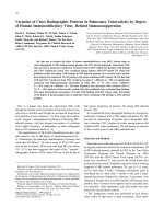
Fig. 1. Primary pulmonary tuberculosis in 18-year-old boy with
typical radiographic findings. Chest radiograph shows patchy
consolidation, nodules, and cavities (arrows) in bilateral upper
lung zones.
Table 1. Abnormal Radiographic Findings in Primary
Pulmonary Tuberculosis in Previously Healthy
Adolescent Patients (n = 55)
Variables Number
Small nodules (D < 10 mm) 53 (96%)
Large nodules (10 mm ≤ D < 30 mm) 28 (51%)
Cavity 25 (45%)
Consolidation 14 (25%)
Hilar lymph node enlargement 1 (2%)
Mediastinal lymph node enlargement 0
Pleural effusion 0
Note.─ D = diameter
Fig. 2. Pulmonary tuberculosis in 18-year-old boy with typical
radiographic findings. Chest radiograph shows cavitary nodule
(arrow) with multiple small nodules (arrowheads) in left upper
lung zone.
was observed in 13 (24%) patients. Overall, 37 (67%)
students had the typical form of reactivation TB (Figs. 1,
2), and 18 (33%) had TB lesions of the atypical form,
based on chest radiograph findings (Fig. 3).
DISCUSSION
The aim of this study was to describe the radiographic
findings of primary pulmonary TB in previously healthy
adolescent patients with recent infection. We found that
primary pulmonary TB in our teenage high-school students
typically present with upper lobe nodule(s), consolidation,
or cavitary lesion(s) on chest radiographs. Mediastinal
lymph node enlargement or pleural effusion was not seen
in our patients. These findings have been traditionally
considered as typical chest radiographic findings of reacti-
vation TB with remote infection. In reactivation TB, the
chest radiographs have been regarded to show patchy
consolidation and poorly-defined nodules involving the
upper lobes. In one-third of patients, cavities are present
within lung abnormalities (12, 13).
Primary TB has been considered to be mainly a disease
of infancy and childhood. The most common radiographic
abnormalities of primary TB in infancy and childhood are
intra-thoracic lymph node enlargement, pleural effusion,
and lower lobe lung lesions (14-17). Primary TB can also
occur in adults and hence a shift toward delayed presenta-
tion in adults may be related to a decrease in childhood
exposure and an increasing number of immunocompro-
mised hosts (14). Primary tuberculosis in adolescents and
adults tends to manifest itself as lung parenchymal lesions
in the upper lobes or superior segments of the lower lobes
(14, 17). In addition, pleural effusion or mediastinal lymph
node enlargement is occasional. Cavitation, usually within
area of consolidation, can also occur in adolescent or adult
primary TB as in our cases. Early cavitation in primary TB
is more common and occurs more quickly in adults than in
any other age group (14). Therefore, primary TB in adoles-
cents and adults can manifest upper lobe cavitary consoli-
dation without mediastinal or hilar lymph node enlarge-
ment or pleural effusion, and thus show traditionally-
regarded typical chest radiographic findings of reactivation
TB with remote infection.
The radiographic findings observed in our study concur
with those examined in the study of Sant’Anna et al. (18),
who evaluated radiographic findings of pulmonary TB
observed in the adolescent age group. In their study,
although mode (primary, endogenous reactivation or
exogenous reinfection) of infection was not clearly
mentioned, lung parenchymal lesions were located in the
upper lobes in 57% of patients, whereas cavitary lesions
occurred in 183 (32%) of 564 patients (28% [67 of 243
patients] consisting of 10 to15 year old adolescents and
36% [116 of 321] consisting of 16 to 19 adolescents) (18).
Recent studies based on DNA fingerprinting suggest that
chest radiographic features are similar in patients who
apparently have primary disease and those who have
reactivation TB (6, 7). Additionally, more than 70% of
adult patients with TB pleurisy (which had been regarded
as a primary TB manifestation rather than reactivation TB)
had features of reactivation TB in the lung parenchyma
(19). Moreover, cavitary lung lesions do occur within six
months of initial infection; in other words, cavitary lesions
manifest as radiographic findings of primary TB pulmonary
infection (20). These observations suggest that typical
reactivation-type pulmonary TB can result from primary
infection, endogenous reactivation, or exogenous reinfec-
tion (21, 22).
Impaired host immunity has been regarded as a predis-
posing factor for TB disease. Human immunodeficiency
virus (HIV)-seropositive pulmonary TB patients with
crucial immunodeficiency (CD4 T lymphocyte count, <
200/mm
3
) have a higher prevalence of mediastinal
lymphadenopathy and a lower prevalence of cavitation
than do HIV-seronegative patients (23, 24). Previous
studies demonstrated that these radiologic findings of TB in
HIV-infected patients reflect impaired cell-mediated
immunity (6, 7). Thus, the traditional concept of differ-
ences in chest radiographic findings between children and
Chest Radiographic Findings of Primary Pulmonary Tuberculosis in High School Outbreaks
Korean J Radiol 11(6), Nov/Dec 2010
615
Fig. 3. Pulmonary tuberculosis in 18-year-old boy with atypical
radiographic findings. Chest radiograph shows cavitary consoli-
dation (arrow) and nodules in right lower lung zone. Lesions were
classified as atypical because they were located in lower lung
zone without involvement of upper lung zone.
adults with TB disease may reflect differential efficacy of
the immune response, rather than differences in the timing
of infection (6, 7). An important predictor of radiographic
appearance may be the integrity of the host immune
response, as determined by patient age and immunodefi-
ciency (25). Neonate, young children, or HIV-infected
persons who have impaired cell-mediated immune
responses show a tendency to have the atypical form of
TB, whereas immunocompetent patients tend to have the
typical form of previously known reactivation reactivation
TB (6, 7).
Several characteristics of our study population were
unique; all were previously healthy senior high school
students, with a mean age of 17 years, and no patient had
any underlying chronic illness. All students were
demonstrated to be infected with an identical strain of M.
tuberculosis at each school, which was proven by DNA
fingerprint testing. These findings suggest that our adoles-
cent patients were recently infected and they had recently
developed primary pulmonary TB.
Our study has several limitations. First, our study
subjects were senior high school students (adolescents).
Thus, our results may not be generalized to children or
adults. Second, chest radiographs of all patients were not
available; thus, a selection bias may be present. Third, we
evaluated radiographic findings only, even in the postero-
anterior direction only; thus, we might not have found
mediastinal or hilar lymph node enlargement or minimal
pleural effusion. In addition, three students in our study
had normal chest radiographs, despite having culture-
confirmed active TB. It has been reported that the
radiographs may be normal or show only mild or nonspe-
cific findings in patients with active disease (12). Common
causes of a missed diagnosis of TB are failure to recognize
hilar and mediastinal lymphadenopathy and the oversight
of mild parenchymal abnormalities such as small centrilob-
ular nodules. However, inter-observer agreement in the
identification of hilar or mediastinal lymph node enlarge-
ment and pleural effusion were almost perfect in our study.
Fourth, because we did not have enough data on serial
tuberculin skin test results, students with previously
normal chest radiographs and no history of tuberculosis
were regarded to have primary TB infection. Thus, we
used a broad definition of primary TB infection (14).
Finally, we did not evaluate the effect of BCG vaccination
on the host immune response and radiologic manifestation
of TB infection. Our national policy for preventing
tuberculosis recommends BCG vaccination in the neonatal
period. BCG vaccination may affect host immune response
and radiologic manifestations of TB infection.
In conclusion, the most common radiographic findings of
primary pulmonary TB by recent infection in previously
healthy adolescents are upper lung lesions, including
nodule(s), consolidation, and cavitation, which were
previously thought to be typical radiographic findings of
reactivation pulmonary TB by remote infection.
References
1. Diagnostic Standards and Classification of Tuberculosis in
Adults and Children. This official statement of the American
Thoracic Society and the Centers for Disease Control and
Prevention was adopted by the ATS Board of Directors, July
1999. This statement was endorsed by the Council of the
Infectious Disease Society of America, September 1999. Am J
Respir Crit Care Med 2000;161:1376-1395
2. Small PM, Fujiwara PI. Management of tuberculosis in the
United States. N Engl J Med 2001;345:189-200
3. Lee KS, Song KS, Lim TH, Kim PN, Kim IY, Lee BH. Adult-
onset pulmonary tuberculosis: findings on chest radiographs and
CT scans. AJR Am J Roentgenol 1993;160:753-758
4. Lee JY, Lee KS, Jung KJ, Han J, Kwon OJ, Kim J, et al.
Pulmonary tuberculosis: CT and pathologic correlation. J
Comput Assist Tomogr 2000;24:691-698
5. Jeong YJ, Lee KS. Pulmonary tuberculosis: up-to-date imaging
and management. AJR Am J Roentgenol 2008;191:834-844
6. Jones BE, Ryu R, Yang Z, Cave MD, Pogoda JM, Otaya M, et
al. Chest radiographic findings in patients with tuberculosis with
recent or remote infection. Am J Respir Crit Care Med
1997;156:1270-1273
7. Geng E, Kreiswirth B, Burzynski J, Schluger NW. Clinical and
radiographic correlates of primary and reactivation tuberculosis:
a molecular epidemiology study. JAMA 2005;293:2740-2745
8. Tabet SR, Goldbaum GM, Hooton TM, Eisenach KD, Cave MD,
Nolan CM. Restriction fragment length polymorphism analysis
detecting a community-based tuberculosis outbreak among
persons infected with human immunodeficiency virus. J Infect
Dis 1994;169:189-192
9. Small PM, Hopewell PC, Singh SP, Paz A, Parsonnet J, Ruston
DC, et al. The epidemiology of tuberculosis in San Francisco. A
population-based study using conventional and molecular
methods. N Engl J Med 1994;330:1703-1709
10. Alland D, Kalkut GE, Moss AR, McAdam RA, Hahn JA,
Bosworth W, et al. Transmission of tuberculosis in New York
City. An analysis by DNA fingerprinting and conventional
epidemiologic methods. N Engl J Med 1994;330:1710-1716
11. Hansell DM, Bankier AA, MacMahon H, McLoud TC, Mu¨ller
NL, Remy J. Fleischner Society: glossary of terms for thoracic
imaging. Radiology 2008;246:697-722
12. Woodring JH, Vandiviere HM, Fried AM, Dillon ML, Williams
TD, Melvin IG. Update: the radiographic features of pulmonary
tuberculosis. AJR Am J Roentgenol 1986;146:497-506
13. Krysl J, Korzeniewska-Kosela M, Mu¨ller NL, FitzGerald JM.
Radiologic features of pulmonary tuberculosis: an assessment of
188 cases. Can Assoc Radiol J 1994;45:101-107
14. Choyke PL, Sostman HD, Curtis AM, Ravin CE, Chen JT,
Godwin JD, et al. Adult-onset pulmonary tuberculosis.
Radiology 1983;148:357-362
15. Khan MA, Kovnat DM, Bachus B, Whitcomb ME, Brody JS,
Snider GL. Clinical and roentgenographic spectrum of
pulmonary tuberculosis in the adult. Am J Med 1977;62:31-38
16. Kim WS, Choi JI, Cheon JE, Kim IO, Yeon KM, Lee HJ.
Koh et al.
616
Korean J Radiol 11(6), Nov/Dec 2010
Chest Radiographic Findings of Primary Pulmonary Tuberculosis in High School Outbreaks
Korean J Radiol 11(6), Nov/Dec 2010
617
Primary tuberculosis in infants: radiographic and CT findings.
AJR Am J Roentgenol 2006;187:1024-1033
17. Leung AN, Mu¨ller NL, Pineda PR, FitzGerald JM. Primary
tuberculosis in childhood: radiographic manifestations.
Radiology 1992;182:87-91
18. Sant’Anna C, March MF, Barreto M, Pereira S, Schmidt C.
Pulmonary tuberculosis in adolescents: radiographic features.
Int J Tuberc Lung Dis 2009;13:1566-1568
19. Kim HJ, Lee HJ, Kwon SY, Yoon HI, Chung HS, Lee CT, et al.
The prevalence of pulmonary parenchymal tuberculosis in
patients with tuberculous pleuritis. Chest 2006;129:1253-1258
20. Marais BJ, Parker SK, Verver S, van Rie A, Warren RM.
Primary and postprimary or reactivation tuberculosis: time to
revise confusing terminology. AJR Am J Roentgenol
2009;192:W198
21. Marais BJ, Gie RP, Schaaf HS, Hesseling AC, Obihara CC,
Starke JJ, et al. The natural history of childhood intra-thoracic
tuberculosis: a critical review of literature from the pre-
chemotherapy era. Int J Tuberc Lung Dis 2004;8:392-402
22. Andronikou S, Vanhoenacker FM, De Backer AI. Advances in
imaging chest tuberculosis: blurring of differences between
children and adults. Clin Chest Med 2009;30:717-744
23. Barnes PF, Bloch AB, Davidson PT, Snider DE Jr. Tuberculosis
in patients with human immunodeficiency virus infection. N
Engl J Med 1991;324:1644-1650
24. Leung AN, Brauner MW, Gamsu G, Mlika-Cabanne N, Ben
Romdhane H, Carette MF, et al. Pulmonary tuberculosis:
comparison of CT findings in HIV-seropositive and HIV-
seronegative patients. Radiology 1996;198:687-691
25. Newton SM, Brent AJ, Anderson S, Whittaker E, Kampmann B.
Paediatric tuberculosis. Lancet Infect Dis 2008;8:498-510