The Nature of Disease: Pathology for the Health Professions_2 pot
Bạn đang xem bản rút gọn của tài liệu. Xem và tải ngay bản đầy đủ của tài liệu tại đây (20.74 MB, 351 trang )
16
CHAPTER
389
Diseases of the Liver and Biliary Tract
This chapter begins with a review of the normal anatomy and physiology of the liver and biliary tract.
Diseases and disorders discussed include cirrhosis, hepatitis, alcoholic and metabolic liver disease, gallstones
and other gallbladder disease, and cancers of the liver and biliary tract.
BACK TO BASICS
• Liver Anatomy
• Liver Function
THE LIVER RESPONSE TO INJURY
• Anatomic Patterns of Liver Injury
• Functional Patterns of Liver Injury
CIRRHOSIS
• Anatomic Types of Cirrhosis
• The Pathophysiology of Cirrhosis
• Clinical Features of Cirrhosis
VIRAL HEPATITIS
• Clinicopathologic Syndromes
• Hepatitis A Virus (HAV) Infection
• Hepatitis B Virus (HBV) Infection
• Hepatitis C Virus (HCV) Infection
• Hepatitis D Virus (HDV) Infection
• Hepatitis E Virus (HEV) Infection
• The Anatomic Pathology of Hepatitis
AUTOIMMUNE HEPATITIS
LIVER ABSCESS
TOXIC LIVER INJURY
ALCOHOLIC LIVER DISEASE
• Fatty Liver
• Alcoholic Hepatitis
• Alcoholic Cirrhosis
INHERITED METABOLIC AND PEDIATRIC LIVER DISEASE
• Hemochromatosis
• Wilson Disease
• Hereditary Alpha-1 Antitrypsin Deficiency
• Neonatal Cholestasis, Biliary Atresia, and Hepatitis
• Reye Syndrome
DISEASE OF INTRAHEPATIC BILE DUCTS
• Primary Biliary Cirrhosis
• Primary Sclerosing Cholangitis
CIRCULATORY DISORDERS
TUMORS OF THE LIVER
• Primary Carcinomas of the Liver
• Cholangiocarcinoma
DISEASES OF THE GALLBLADDER AND EXTRAHEPATIC
BILE DUCTS
• Diseases of the Gallbladder
• Diseases of Extrahepatic Bile Ducts
After studying this chapter you should be able to:
1. Trace the flow of blood through the liver
2. Name the major functions of the liver
3. Explain the enterohepatic circulation
4. Name the major functional reactions of the liver to injury
5. Explain the difference between conjugated and unconjugated bilirubin
6. Name one cause of unconjugated hyperbilirubinemia and one of conjugated hyperbilirubinemia
7. Define cirrhosis, and name the two most common causes
8. Explain why cirrhosis causes portal hypertension
9. Name two hemodynamic consequences of portal hypertension
10. Outline the main clinical manifestations of cirrhosis
11. Name several clinicopathologic syndromes associated with viral hepatitis
12. Contrast the mode of transmission and clinical course of hepatitis A and hepatitis B infection
Learning Objectives
90981 ch 16.qxd 10/9/06 10:14 AM Page 389
Key Terms and Concepts
The liver regulates the composition of blood by disposing
of waste products (bilirubin, for example), convert-
ing substances from one form into another (glycogen
into glucose and vice versa), secreting substances into the
intestines (bile and cholesterol), and producing plasma
proteins (albumin, coagulation factors, and others).
The anatomy of the liver, portal venous system, and
bile ducts is illustrated in Figure 16-1. Nutrients ab-
sorbed by the intestine do not enter the general circula-
tion directly; instead they flow first to the liver via the
portal vein. The vein gains its name from the fact that
the liver is a gate (a portal or doorway) through which
blood must pass before entering the general circulation.
This “gate” effect is unique in human anatomy: venous
blood goes from one capillary system (the intestine), is
collected into a large vein (portal), and passes through
a second capillary system (the liver) before entering the
general circulation. The liver also receives arterial blood
from the hepatic artery, which merges with portal blood
before flowing through the hepatic vein into the inferior
vena cava.
Two of every three deaths are premature; they are related to the loafer’s heart,
smoker’s lung and drinker’s liver.
DR. THOMAS J. BASSLER, PATHOLOGIST; QUOTED BY JAMES FIXX (1932–1984),
IN
THE COMPLETE BOOK OF RUNNING
(Random House, 1977)
390
Part 2 • Diseases of Organ Systems
13. Name the most important epidemiologic fact about hepatitis C, and name the most common serious conse-
quences of hepatitis C infection
14. Name one common acute and one common chronic change induced in the liver by alcohol abuse
15. Explain the difference between primary biliary cirrhosis and sclerosing cholangitis
16. Name the liver condition most commonly associated with hepatocellular carcinoma
17. Name the two major categories of gallstones, and know which is the most common
18. Name several risk factors that favor the formation of gallstones
19. Discuss some of the most common causes of extrahepatic bile duct obstruction
BACK TO BASICS
• portal vein
• hepatocytes
• bile duct
• bile
• bile acids
• bilirubin
• unconjugated bilirubin
• conjugated bilirubin
• jaundice
• cholestasis
THE LIVER RESPONSE TO INJURY
• hepatitis
• cirrhosis
• hepatic failure
CIRRHOSIS
• portal cirrhosis
• biliary cirrhosis
• portal hypertension
VIRAL HEPATITIS
• viral hepatitis
• carrier state
• chronic viral hepatitis
• hepatitis A virus (HAV)
• hepatitis B virus (HBV)
• hepatitis C virus (HCV)
ALCOHOLIC LIVER DISEASE
• fatty liver
INHERITED METABOLIC AND PEDIATRIC LIVER DISEASE
• neonatal cholestasis
DISEASE OF INTRAHEPATIC BILE DUCTS
• primary biliary cirrhosis
TUMORS OF THE LIVER
• hepatocellular carcinoma
DISEASES OF THE GALLBLADDER AND EXTRAHEPATIC
BILE DUCTS
• cholelithiasis
• cholecystitis
BACK TO BASICS
90981 ch 16.qxd 10/9/06 10:14 AM Page 390
391
Chapter 16 • Diseases of the Liver and Biliary Tract
A
A
B
Inferior vena cava
Left hepatic vein
Hepatic
duct
Common
bile duct
Hepatic artery
Portal vein
Cystic duct
Liver
Right hepatic vein
Central vein
To hepatic vein,
then to inferior
vena cava
Portal
triad
Plates of
hepatocytes
Hepatic artery
from aorta
Blood mixes
in sinusoids
Bile duct
Portal vein
from bowel
Bile canaliculi
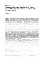
Figure 16-1 The liver, portal venous system, and bile ducts.
AA,,
The liver and biliary system.
BB,,
The hepatic lobule. Blood flows into the central
vein from branches of the portal vein and hepatic artery clustered in portal triads at the edge of the lobule. Bile flows in the opposite direction—
from the interior of the lobule to bile ducts in the portal triads.
90981 ch 16.qxd 10/9/06 10:14 AM Page 391
392
Part 2 • Diseases of Organ Systems
LIVER ANATOMY
Upon entering the portal circulation, blood and freshly
absorbed nutrients are brought into close contact with
the main functional cells (parenchyma) of the liver (he-
patocytes). Liver cells are formed into plates one or two
cells thick that are sandwiched between large venous
capillaries (hepatic sinusoids). Hepatic sinusoids have
no basement membrane and are therefore much more
permeable than other capillaries are, so that even large
protein and lipid molecules cross freely. Venous sinu-
soid walls contain fixed macrophages known as Kupffer
cells, which, unlike other macrophages, do not move
about but remain in place to filter portal blood.
The plates of hepatic cells are arranged into a hepatic
lobule. In the middle of each lobule is a central vein sur-
rounded by hepatocytes. The corners of each lobule are
defined by several portal triads consisting of 1) a
branch of the hepatic artery bringing blood from the
aorta, 2) a portal vein carrying blood from the GI tract,
and 3) a small bile duct that carries bile out of the liver.
Blood entering the lobule from the hepatic artery and
portal vein flows into venous sinusoids, percolates
through hepatic plates, and is collected in the central
vein for delivery to the general circulation.
The liver is a large gland that secretes bile into the in-
testines. Bile is a mixture of metabolic waste and bile
acids, which emulsify (make water soluble) dietary fat
so it can be absorbed by the intestinal mucosa. Bile is
excreted by hepatocytes into a network of small intra-
hepatic bile ducts that carry it out of the liver and into
the common bile duct (hepatic duct), which connects to
the intestine at the ampulla of Vater in the duodenum. A
reserve of bile is held in the gallbladder and discharged
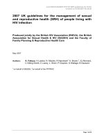
after meals. Figure 16-2 illustrates that much of the bile
acid excreted into the intestine is reabsorbed by the
small bowel and sent back to the liver for reuse, a
process known as the enterohepatic circulation. Only a
small amount of bile acid finds its way into feces.
LIVER FUNCTION
As is outlined in Table 16-1, the liver has five main
functions: 1) detoxification and excretion of metabolic
waste, drugs, and hormones; 2) lipid and carbohydrate
metabolism; 3) protein synthesis; 4) conjugation and
excretion of bilirubin; and 5) synthesis and excretion of
bile acids.
The liver clears blood of endogenous metabolic waste
(especially bilirubin and ammonia), chemicals and toxins
(especially drugs), and hormones (especially estrogen). It
does so by excreting them into bile, as it does with
bilirubin, or converting them into something not harm-
ful, as it does by converting ammonia to urea.
The liver modulates blood glucose concentration by
storing glucose as glycogen and reconverts and excretes it
on demand. Additionally, the liver synthesizes triglyc-
erides and cholesterol and, when necessary, burns fat
and excretes the ketones produced by the process.
Ketones are an acidic by-product of fat metabolism that
accounts for the acidosis (ketosis) that occurs when di-
abetic patients are without insulin and cannot burn glu-
cose, and the liver must burn fat to produce energy
(Chapter 17).
The liver produces virtually all plasma proteins except
antibodies (immunoglobulins), which are made by B
lymphocytes of the immune system. Plasma protein
consists of albumin, alpha and beta globulins—all made
by the liver—and gamma globulins (immunoglobulins,
or antibodies, made by lymphocytes of the immune sys-
tem, Chapter 8). The most abundant liver protein is al-
bumin, which forms about three fourths of plasma pro-
tein and provides most of the osmotic pressure of
plasma (Chapter 5) that holds water in blood. Other
proteins produced by the liver include clotting factors
(Chapter 11) and specialized proteins for fat (and trans-
port of fat, hormones, iron, and other substances.
The liver excretes bilirubin into bile ducts, which
carry it to the bowel. Bilirubin is an intensely yellow
pigment, most of which is produced in the spleen from
the hemoglobin of old red blood cells the spleen has re-
moved from the circulation. This new bilirubin is not
water soluble and must be attached to a protein for
transport to the liver, where it is made water soluble
and excreted in bile. In the liver, bilirubin is joined
(conjugated) to glucuronide to make conjugated biliru-
bin, which is water soluble and can be excreted in bile.
Before reaching the liver, the water-insoluble bilirubin
is called unconjugated bilirubin. Increased blood
bilirubin (of either type) causes jaundice (icterus), a
yellow discoloration of skin and sclera that is a hallmark
of increased blood bilirubin.
Bacteria in the bowel convert bilirubin into uro-
bilinogen, a compound that gives feces its brown color.
Patients with jaundice usually have pale stools owing to
a lack of urobilinogen. However, some urobilinogen is
produced directly by the liver, secreted into blood, and
The circulation of blood through the intestines
and liver is unique in human anatomy: blood
from one capillary system, the intestinal, flows
into another capillary system, the hepatic, be-
fore returning to the heart.
90981 ch 16.qxd 10/9/06 10:14 AM Page 392
393
Chapter 16 • Diseases of the Liver and Biliary Tract
Liver
Gallbladder
Common bile duct
Reabsorption
and reexcretion
Bile
Portal vein
Small intestine
Feces
Figure 16-2 The enterohepatic circulation. Bile acids
are secreted into bile, absorbed back into blood by the in-
testine, and recirculated via the portal vein for remetabo-
lism by the liver.
MMeettaabboolliicc TTaasskk HHeeppaattooccyyttee AAccttiioonnss
Detoxification and drug metabolism • Chemical alteration and excretion of hormones and drugs
• Production and excretion of urea and other compounds that are less toxic
than the parent compound
Lipid and carbohydrate metabolism • Conversion of glucose to glycogen and fat
• Production of glucose from glycogen and other compounds
• Secretion of glucose into blood
• Synthesis and secretion of triglyceride and cholesterol into blood
• Excretion of cholesterol into bile
• Production of ketones from fatty acid
Protein synthesis • Synthesis and secretion into blood of albumin, transport proteins and blood
coagulation (clotting) factors
Conjugation and excretion of bilirubin • Conjugation of bilirubin with glucuronide and excretion of it into bile ducts
Synthesis and excretion of bile acids • Synthesis of bile acids by liver cells and secretion of them into bile ducts
Liver Functions
Table 16-1
90981 ch 16.qxd 10/9/06 10:14 AM Page 393
394
Part 2 • Diseases of Organ Systems
excreted into urine, giving urine its faint amber color.
Jaundiced patients may have dark urine because of in-
creased urobilinogen.
The liver produces and excretes bile acids. Bile acids,
the main constituent of bile, emulsify fat like soap does
grease, solubilizing it for absorption. Bile acids are pro-
duced by the liver from cholesterol, and bile acid excre-
tion is the main way by which the body rids itself of
cholesterol. However, most bile acids and the choles-
terol they contain are reabsorbed by the intestinal mu-
cosa and returned to blood (enterohepatic circulation).
The enterohepatic circulation is, therefore, very impor-
tant in cholesterol metabolism. Most cholesterol ab-
sorbed from the intestine is not from diet but from re-
absorption of bile acids. New drugs lower blood
cholesterol by blocking intestinal absorption of choles-
terol. Obstruction of bile excretion is referred to as
cholestasis.
The Liver Response to Injury
The liver responds remarkably well to injury—it has
enormous functional reserve and must suffer a marked
decline of capacity before becoming symptomatic.
However, because normal liver function is so crucial in
metabolism, liver disease looms large in human illness.
Laboratory evaluation of blood is critical in the diagno-
sis and management of liver disease. The nearby Lab
Tools box lists and describes the most common lab
tests.
ANATOMIC PATTERNS OF LIVER INJURY
The injury to liver prompts one of four consequences:
inflammation, degeneration, necrosis, and fibrosis.
• Inflammation: Inflammation of the liver is termed
hepatitis, most of which is caused by virus infec-
tions.
• Degeneration: Hepatocytes may undergo hydropic
(watery) or fatty degeneration (Chapter 2) in re-
sponse to toxic or autoimmune injury. For example,
alcohol abuse typically causes fatty degeneration,
which can proceed to injury that is more serious if al-
cohol abuse continues.
• Necrosis: Liver cells may die as blocks of tissue (in-
farcts, Chapter 2) or as single cells. In some types of
diffuse hepatic injuries, such as viral hepatitis and
chronic alcohol injury, individual liver cells die one
by one, becoming shrunken, dead cells known as
Councilman bodies.
• Fibrosis: Scarring can occur as a result of severe he-
patic injury. Cirrhosis (discussed in detail below) is
a patterned, permanent scarring (fibrosis) of the en-
tire liver from long-standing, severe injury that de-
stroys the normal architecture and replaces it with
scar tissue.
FUNCTIONAL PATTERNS OF LIVER INJURY
The direct metabolic consequences of hepatic injury are:
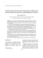
• Jaundice: a yellow discoloration of skin and sclerae
(Fig. 16-3) caused by an excess of blood bilirubin
• Cholestasis: an accumulation of bile acids and cho-
lesterol in blood when there is obstruction of bile
flow inside or outside of the liver
• Hepatic failure: the loss of hepatic metabolic func-
tion severe enough to cause clinical symptoms.
Each of these consequences may be associated with
widespread ill effect on the body.
MAJOR DETERMINANTS OF DISEASE
• The metabolic consequences of liver disease
are serious and include:
– Toxic accumulations of:
metabolic waste (especially ammonia
and bilirubin)
drugs and toxins
endogenous hormones (especially estro-
gen)
– Bleeding, associated with a deficiency of
coagulation factors
– Edema, associated with a deficiency of
plasma albumin
– Failure to absorb intestinal fat because of a
deficiency of bile acids
• Viral hepatitis is a common contagious dis-
ease
• Cirrhosis is the final endpoint for many liver
diseases
• Portal hypertension is the most important
consequence of cirrhosis and can be associ-
ated with liver failure and severe hemorrhage
• Stones often form in the gallbladder and may
pass into and obstruct the bile duct
90981 ch 16.qxd 10/9/06 10:14 AM Page 394
395
Chapter 16 • Diseases of the Liver and Biliary Tract
Jaundice and Cholestasis
Jaundice usually becomes visible when blood bilirubin
level is Ͼ2 mg/dL (normal Ͻ1.2 mg/dL). As is illus-
trated in Figure 16-4, jaundice can be caused by three
conditions:
• the presence of excessive amounts of bilirubin (pre-
hepatic jaundice), such as accompanies red cell de-
struction in hemolytic anemia
• defective liver functioning (hepatic jaundice), such
as occurs with viral hepatitis, drug interference with
liver function, or cirrhosis
• biliary obstruction (posthepatic jaundice), such as
occurs when pancreatic cancer occludes the common
bile duct
Prehepatic jaundice causes an increase in blood of wa-
ter-insoluble unconjugated bilirubin. The most com-
mon serious cause of increased amounts of unconju-
gated bilirubin in blood is hemolytic anemia, such as
sickle cell disease (Chapter 11).
Increased unconjugated bilirubin can also occur
when there is hepatic malfunction. For newborns un-
conjugated hyperbilirubinemia is especially dangerous,
because it is toxic to the underdeveloped brain. Because
the immature livers of premature infants are unable to
conjugate bilirubin effectively, during the first several
weeks of life a marked increase of unconjugated biliru-
bin may cause kernicterus (from German: kern, nucleus,
and icterus, jaundice), a severe neurologic condition re-
sulting from toxic deposits of water-insoluble, uncon-
jugated bilirubin in the brain (Chapter 7).
However, the most common cause of increased un-
conjugated bilirubin in blood is Gilbert syndrome
(pronounced jeel-bear), a very common and harmless
condition associated with a mild increase of unconju-
gated bilirubin that is the result of a genetic enzyme defi-
Figure 16-3 Scleral icterus of jaundice.
Liver Function Tests
The most useful laboratory tests for liver function are:
•
Enzymes
: The liver is packed with enzymes. In liver dis-
ease these enzymes are washed into blood, where they
are easily measured. Even mild liver-cell injury can cause
minor increases in levels of liver enzymes. Elevation of
lactic dehydrogenase (LDH), aspartate aminotransferase
(AST), and alanine aminotransferase (ALT) suggests he-
patic cellular damage. Alkaline phosphatase levels also
may be increased in liver disease, but they tend to rise
highest in bile duct diseases. Red blood cells contain
some of these enzymes, and in vitro damage (hemolysis)
to red cells during specimen collection or handling can
cause misleading enzyme level increases.
•
Bilirubin
: The liver metabolizes and excretes bilirubin into
the bile ducts. If hemolytic disease has been excluded, in-
creased levels of blood bilirubin usually indicate at least
moderate liver disease or bile duct disease.
•
Proteins
: The liver makes albumin and many other plasma
proteins. Low levels of plasma albumin and blood coag-
ulation factors are characteristic of moderate to serious
liver disease.
•
Coagulation tests
: The liver makes most of the coagula-
tion proteins (factors), and liver disease can cause abnor-
mal (prolonged) prothrombin time and partial thrombo-
plastin time.
•
Hepatitis virus antigens and antibodies
: Each type of hep-
atitis virus is distinguished by characteristic patterns of
virus antigens and antibodies in blood.
•
Autoimmune antibodies
: Antimitochondrial antibodies in
blood are characteristic of primary biliary cirrhosis; anti-
smooth muscle antibodies are characteristic of chronic
autoimmune hepatitis.
LAB TOOLS
90981 ch 16.qxd 10/9/06 10:14 AM Page 395
396
Part 2 • Diseases of Organ Systems
ciency, which is usually detected accidentally while the
patient is being seen for an unrelated illness or when the
patient has been fasting for 12 hours or more. A similar
genetic syndrome causing increased conjugated biliru-
bin is the Dubin-Johnson syndrome.
Jaundice associated with hepatic disorders (hepatic
jaundice) may cause an increase in blood of either un-
conjugated or conjugated bilirubin, because interference
can occur either before or after conjugation in the liver.
Jaundice resulting from bile duct obstruction (pos-
thepatic jaundice) is characterized by increased water-
soluble, conjugated bilirubin in blood because by the
time bilirubin enters hepatic ducts all of it is conjugated,
and obstruction forces conjugated bilirubin out of the
ducts and into blood. Gallstones and pancreatic cancer
are the two most common causes of duct obstruction.
Cholestasis is usually accompanied by jaundice and
sometimes by severe pruritus (itching) because of the
deposition of bile acids in the skin. Cholestasis may re-
sult from primary liver disease, drug interference with
bile secretion, pregnancy, and a variety of other condi-
tions. Because bile is the means by which the body rids
itself of excess cholesterol, blood cholesterol levels may
become markedly elevated and associated with yellow
deposits of cholesterol in skin (xanthomas). Because bile
duct epithelium is rich in alkaline phosphatase, a char-
acteristic laboratory finding is marked increase of blood
alkaline phosphatase levels, whereas levels of other liver
enzymes are usually normal or only mildly increased.
Hepatic Failure
Hepatic failure is the most severe consequence of liver
disease—most patients die within a few weeks or
months. It may follow sudden injury, as in sudden, se-
vere viral hepatitis (fulminant hepatitis), or it may be
the result of chronic injury, as with chronic hepatitis or
chronic alcoholism. About 90% of hepatic function
must be destroyed before failure occurs.
The clinical features of hepatic failure are:
• Jaundice because of failure to excrete bilirubin
• Ascites because of increased portal pressure and low
blood osmotic pressure
• Fetor hepaticus, literally “liver breath,” because of an
accumulation of volatile waste products such as am-
monia
A Prehepatic jaundice (unconjugated hyperbilirubinemia)
B Hepatic jaundice (unconjugated or conjugated
hyperbilirubinemia)
C Posthepatic jaundice (conjugated hyperbilirubinemia)
1
1
2
2
Red blood cell
Major cause:
• Hemolysis
Major causes:
• Viral hepatitis
• Toxins
• Therapeutic drugs
• Cirrhosis
• Neoplasms
Gallstones in
bile ducts
Macrophage
Hemoglobin
Bilirubin
Circulating bilirubin
Metastatic
cancer
Stones
Gallbladder
Bile duct
Cirrhosis
Pancreatic cancer
Major causes:
Figure 16-4 Three types of jaundice.
AA,,
Prehepatic jaundice causes
increased levels of unconjugated bilirubin in blood.
BB,,
Hepatic jaundice
may be caused by interference with the liver’s ability to conjugate biliru-
bin or to secrete it after conjugation; increased levels of blood bilirubin
may be of either conjugated or unconjugated type.
CC,,
Posthepatic jaun-
dice is caused by obstruction of bile flow inside or outside of the liver;
increased levels of blood bilirubin are of conjugated type.
90981 ch 16.qxd 10/9/06 10:14 AM Page 396
397
Chapter 16 • Diseases of the Liver and Biliary Tract
• Hypoalbuminemia because of diminished hepatic
production of protein
• Hypoglycemia because of lack of liver glycogen stores
• Hyperammonemia because of failure of the liver to
convert ammonia to urea
• Palmar erythema (redness of the palms of the hands),
spider angiomata (small skin hemangiomas), testicular
atrophy, balding, and gynecomastia (enlarged male
breasts) because of increases in blood estrogen from
impaired liver metabolism of estrogen
• Bleeding disorders because of deficiency of blood clot-
ting factors made by the liver
Moreover, kidney and brain function can be seri-
ously impaired by liver failure. (A summary of the clin-
ical features of hepatic failure can also be found later in
the chapter in Figure 16-10, which appears with the dis-
cussion of cirrhosis.)
Hepatorenal syndrome is renal failure owing to
acute hepatic failure. The cause is not completely
clear—it appears to result from renal vasoconstriction
and low renal blood flow, but the kidney is pathologi-
cally normal. Hepatic encephalopathy, an especially se-
rious complication of hepatic failure, occurs when ac-
cumulated ammonia and other unmetabolized waste
products exert a toxic effect on the brain. Neurologic
signs include rigidity, hyperreflexia, and, rarely,
seizures. Fatal coma may occur. A particularly charac-
teristic sign is asterixis, a rapid extension-flexion mo-
tion of the head and extremities that can be demon-
strated by testing for “hepatic flap”—the arms are held
extended and the hands dorsiflexed. A pulsating, flap-
ping, or hand waving motion constitutes a positive test.
Cirrhosis
Cirrhosis is the final, common end-stage for a variety of
chronic liver diseases. As is illustrated in Figures 16-5
and 16-6, cirrhosis is a patterned fibrosis of the entire
liver characterized pathologically by a three-dimen-
sional web of interconnecting bands of scar tissue, di-
viding the liver into small nodules separated from one
another by dense fibrous tissue, an architecture that
makes cirrhotic livers tense and hard. Cirrhosis is pro-
gressive, irreversible, and incurable.
Cirrhosis can be classified by cause, such as alcoholic
or hepatitic, but regardless of cause there are only two
anatomic types of cirrhosis:
• portal cirrhosis, caused by diffuse liver cell injury
• biliary cirrhosis, caused by chronic disease of the bil-
iary tree
A
B
A
B
Nodule of
hepatocytes
Fibrous
tissue
Figure 16-5 Cirrhosis.
AA,,
Normal liver.
BB,,
Cirrhosis. Note the small size
of the cirrhotic liver.
Figure 16-6 Cirrhosis.
AA,,
Gross section.
BB,,
Microscopic study. Note
the nodular pattern in both specimens. The liver is divided into nodules
by a web of fibrous (scar) tissue. Dark discoloration of some nodules is
caused by the accumulation of bile pigment in lobules. The cause of cir-
rhosis in this patient is unknown.
90981 ch 16.qxd 10/9/06 10:14 AM Page 397
398
Part 2 • Diseases of Organ Systems
In early cases, portal and biliary cirrhosis are easy to
distinguish by microscopic examination, but as disease
progresses the distinctions disappear. In the end the
cause of most cases of cirrhosis cannot be determined
by study of the liver: clinical findings and history are of
paramount importance. However, a few rare types of
cirrhosis have highly characteristic microscopic find-
ings. For example, in hemochromatosis (discussed be-
low), the body is overloaded with iron, much of which
is deposited in the liver. A second example is hereditary
alpha-1 antitrypsin deficiency, which is associated with
early emphysema (Chapter 14) and distinctive micro-
scopic findings in the liver.
Table 16-2 lists the causes of cirrhosis and the mi-
croscopic pattern associated with each. Cirrhosis is
among the top 10 causes of death in the western hemi-
sphere, two thirds of it resulting from alcoholism and
chronic viral hepatitis. Less common causes are genetic
hemochromatosis, and diseases of bile ducts. In about
one third of cases, the cause is unknown (cryptogenic
cirrhosis).
ANATOMIC TYPES OF CIRRHOSIS
The term portal cirrhosis is assigned to cirrhosis oc-
curring with repeated episodes of liver cell necrosis that
are followed by hepatocyte regeneration and growth of
fibrous tissue from the area of the portal triad. Portal
cirrhosis is by far the most common type of cirrhosis
and includes all forms of cirrhosis other than those de-
scribed immediately below as biliary cirrhosis. The ma-
jority of portal cirrhosis results from alcoholic liver dis-
ease and chronic viral hepatitis.
Far less common is biliary cirrhosis, which results
from chronic inflammation of bile ducts. Primary bil-
iary cirrhosis is an autoimmune disease (Chapter 8) of
intrahepatic bile ducts. Secondary biliary cirrhosis de-
velops as a consequence of prolonged inflammation of
bile ducts, usually associated with obstruction of bile
flow because of gallstones lodged in the common bile
duct. Another cause of bile duct inflammation and fi-
brosis that causes biliary cirrhosis is sclerosing cholangi-
tis, which is associated with chronic ulcerative colitis
(Chapter 15).
THE PATHOPHYSIOLOGY OF CIRRHOSIS
Cirrhosis obstructs free flow of portal blood through
the liver and causes portal hypertension; that is, high
blood pressure in the portal venous system. This ob-
struction of portal blood flow through the liver diverts
(shunts) blood around the liver through alternative
(collateral) vessels in the GI tract, spleen, and skin, as is
depicted in Figure 16-7.
The hemodynamic consequences of portal hyperten-
sion are ascites, congestive splenomegaly (Fig. 16-8),
and various types of prominent veins that result from
the shunting of blood around the liver: esophageal
varices (see Fig. 15-8, Chapter 15), hemorrhoids, and
prominent veins radiating outward from the umbilicus,
known as caput medusa—literally snake-head—so
named because of its likeness to the female serpent-
haired monster, Medusa, from Greek mythology.
Ascites (from Greek, askos, for bag) is an intraperi-
toneal accumulation of watery (serous) fluid. This fluid
seeps from portal venules as a result of high portal
blood pressure and low blood osmotic pressure caused
by low blood albumin owing to low output of albumin
by the liver. Ascites becomes clinically evident when
about 500 ml of intraperitoneal fluid have accumulated;
however, as is depicted in Figure 16-9, fluid accumula-
tion may be massive.
CLINICAL FEATURES OF CIRRHOSIS
The clinical features of cirrhosis are summarized in
Figure 16-10 and result from four phenomena:
• Failure to metabolize estrogen and ammonia. Failing
hepatic metabolism of estrogen results in high levels
CCaauussee MMiiccrroossccooppiicc TTyyppee
Alcohol abuse Portal
Chronic viral hepatitis B or C Portal
Biliary obstruction Biliary
Gallstones
Cystic fibrosis
Autoimmune disease
Primary biliary cirrhosis Biliary
Sclerosing cholangitis Biliary
Autoimmune hepatitis Portal
Inherited metabolic disease Portal
Hemochromatosis
Alpha-1 antitrypsin deficiency
Wilson disease
Cryptogenic (unknown) Portal
Causes and Microscopic Types of
Cirrhosis
Table 16-2
Cirrhosis is always associated with portal hy-
pertension.
90981 ch 16.qxd 10/9/06 10:14 AM Page 398
399
Chapter 16 • Diseases of the Liver and Biliary Tract
of blood estrogen, which in men accounts for thin-
ning scalp and genital hair, enlarged breasts (gy-
necomastia), red palms (palmar erythema), atrophic
testes, and spider angiomas—small vascular malfor-
mations of skin that feature a tiny central vessel from
which spider-like vessels radiate outward. Women
may have abnormal menstrual bleeding. Failing he-
patic metabolism of ammonia and other waste prod-
ucts accounts for hepatic coma and fetor hepaticus
(liver breath).
• Protein synthesis failure. Failing hepatic protein syn-
thesis causes decreased plasma albumin, which con-
tributes to ascites and peripheral edema (Chapter 5),
and decreased production of blood coagulation fac-
tors, which accounts for bleeding tendencies, usually
evident as easy bruising (skin purpura).
Portal circulation
Systemic circulation
Diaphragm
Liver
Stomach
Cirrhosis obstructs
portal blood flow
Superior
epigastric
vein
Umbilical vein
Inferior
epigastric
vein
Superior mesenteric vein
Inferior mesenteric vein
Anus
Inferior vena cava
Splenic vein
Spleen
Veins of
abdominal
wall
Portal vein
Esophageal and gastric varices
Hemorrhoids
Caput medusa
(periumbilical
varices)
Splenomegaly
Figure 16-7 Portal hypertension. The hemodynamic consequences of obstructed portal blood flow.
Figure 16-8 Congestive splenomegaly in cirrhosis. Both specimens
are from the same patient. Note that the spleen
(Top)
is much larger
than the liver is; normally the opposite is true.
90981 ch 16.qxd 10/9/06 10:14 AM Page 399
400
Part 2 • Diseases of Organ Systems
• Excretory failure. Failing hepatic excretion of biliru-
bin causes jaundice.
• Portal hypertension. Portal hypertension causes hem-
orrhoids, esophageal varices, splenomegaly, and ca-
put medusa veins radiating from the umbilicus, and
contributes to the formation of ascites.
Patients with cirrhosis also lose muscle mass (muscle
wasting); however, the cause is not clear. In early cir-
rhosis the liver may be large, but as scarring progresses
it always shrinks to less than normal size.
Cirrhosis may go undetected for years. Sometimes
the delay is so great that when symptoms appear the
original cause may not be apparent. Initial symptoms
may be nothing more than anorexia, fatigue, and weight
loss. But hepatic symptoms, jaundice especially, are
usually present and may appear quite suddenly, mim-
Figure 16-9 Ascites.
7
Muscle
wasting
Liver large
or small
Hepatocellular
carcinoma
1
Hair thin
Coma
Fetor hepaticus
2
3
1
1
2
3
4
1
2
3
8
5
6
4
2
1
3
Failure to metabolize estrogen and ammonia
Spider nevi
4
Ascites (low albumin)
Purpura (low clotting factors)
1
2
Edema (low albumin)
3
Protein synthesis failure
2
Collateral veins (caput medusae)
Ascites
3
Edema
4
1
Jaundice
Excretory failure
1
Splenomegaly
Portal hypertension
Gynecomastia
5
Palmar erythema
6
Absent or reduced pubic hair
7
Small testes
8
Figure 16-10 Clinical features of cirrhosis.
90981 ch 16.qxd 10/9/06 10:14 AM Page 400
icking acute liver disease. The most common causes of
death from cirrhosis are hepatic failure, gastrointestinal
hemorrhage from esophageal varices, and hepatocellu-
lar carcinoma (discussed below).
Viral Hepatitis
Viral hepatitis is infection by one of the several viruses
that preferentially infect the liver: hepatitis viruses A, B,
C, D, and E, which are designated HAV (for hepatitis A
virus), HBV, and so on. Other viruses can incidentally
infect the liver and cause hepatitis, most notably the cy-
tomegalovirus or the Epstein-Barr virus of infectious
mononucleosis.
Hepatitis viruses are distinguished from one another
according to the following clinical characteristics (sum-
marized in Table 16-3):
• Mode of transmission: Is the virus transmitted by oral-
fecal contamination, close personal contact, contam-
inated water, or blood contamination (needlestick or
transfusion)?
• Length of incubation period: What is the length of time
from infection to symptomatic disease?
• Carrier state: After recovery from acute infection,
does the virus linger, so that an apparently healthy
person continues to infect others?
• Chronic hepatitis: Can the virus cause chronic hepa-
titis?
• Fulminant hepatitis: Can the virus cause a sudden,
catastrophic hepatitis?
• Hepatocellular carcinoma: Is the virus associated with
increased risk of hepatocellular carcinoma?
401
Chapter 16 • Diseases of the Liver and Biliary Tract
MMaajjoorr HHeeppaattiittiiss VViirruusseess
CChhaarraacctteerriissttiicc HHeeppaattiittiiss AA HHeeppaattiittiiss BB HHeeppaattiittiiss CC
New cases per annum United 11,000 8,000 28,000
States (2001)
Transmission
Route Fecal-oral Parenteral or close contact Parenteral or close contact
Mother to child No Yes ?
Incubation period 3–6 weeks 2 weeks to 6 months 2 weeks to 6 months
Viremia Very short Long Long
Carrier state No Yes, uncommon Yes, common
Chronic hepatitis No 5–10% Ͼ50%
Increased risk for hepatocellular No Yes Yes
carcinoma
Blood markers* anti-HAV (IgM) HBsAg HCV-RNA
anti-HAV (IgG) HBeAg anti-HCV
anti-HBs
anti-HBc (IgM)
anti-HBc (IgG)
anti-HBe
Vaccine available Yes Yes No
*Marker key: HBsAg, hepatitis B surface antigen; anti-HBc (IgM), anti-hepatitis B core antibody, acute phase (IgM) type
Characteristics of Major Hepatitis Viruses
Table 16-3
• Hepatitis A is a mild, epidemic disease spread
by contaminated food and water; it does not
cause chronic hepatitis or cirrhosis.
• Hepatitis B and hepatitis C are spread from
individual to individual by needles or sexual
contact; both can cause chronic hepatitis and
cirrhosis.
90981 ch 16.qxd 10/9/06 10:14 AM Page 401
402
Part 2 • Diseases of Organ Systems
CLINICOPATHOLOGIC SYNDROMES
Viral hepatitis can cause several clinical syndromes or
diseases:
• asymptomatic hepatitis
• carrier state
• acute viral hepatitis
• chronic viral hepatitis
• fulminant hepatic failure
• hepatocellular carcinoma
However, not every virus can produce each of these;
and some of these disorders can be caused by diseases
other than viral hepatitis.
Asymptomatic hepatitis may produce no lasting
liver injury and be detected only by accident. The usual
circumstance is unexpected abnormal blood tests—ab-
normally elevated liver enzymes, for example—ob-
tained as part of an annual physical exam or in the
course of attention to an unrelated condition.
The carrier state exists in a patient who despite be-
ing asymptomatic harbors the virus and is therefore ca-
pable of transmitting the virus to others. The percent-
age of infected people who become carriers varies
greatly from one type of viral hepatitis to another. For
example, few patients infected with hepatitis B virus
(HBV) become carriers; conversely, many of those with
hepatitis C virus (HCV) develop asymptomatic chronic
infection and, therefore, fit the definition of carrier.
Acute viral hepatitis is an acute illness that typically
progresses through four clinical phases:
• Incubation usually lasts a few weeks. Peak infectivity
occurs about the time symptoms appear.
• The symptomatic prejaundice phase is usually marked
by constitutional symptoms, including malaise, fati-
gability, nausea, and anorexia. However, right upper-
quadrant pain, low-grade fever, headache, skin rash,
vomiting, diarrhea, or muscle and joint aches may
occur.
• The symptomatic jaundice phase begins as jaundice
(icterus) appears and other symptoms fade. The jaun-
dice reflects a rise in conjugated bilirubin. Because
conjugated bilirubin is water soluble, it is excreted in
urine, causing a brown discoloration. Because less
bilirubin is getting into the gut, stools may be pale.
With HAV infection, most adults become jaundiced,
but most children do not. About half of patients with
HBV infection become jaundiced, but patients with
HCV infection are rarely jaundiced.
• Convalescence begins as jaundice fades, infectivity
disappears, and antibodies appear in blood to confer
immunity.
Chronic viral hepatitis is defined as viral hepatitis
proven by liver biopsy, with six months or more of lab-
oratory or clinical evidence of disease activity. Not all
patients with chronic hepatitis have chronic viral hepa-
titis—autoimmune hepatitis and alcoholic hepatitis,
discussed below, are examples. About 10% of patients
with hepatitis B infections develop chronic hepatitis,
whereas more than 50% of patients with hepatitis C do
so. Most patients with chronic hepatitis show few spe-
cific clinical signs and symptoms, and the extent of dis-
ease is revealed only by laboratory tests.
On the other hand, some patients may be very ill and
exhibit a variety of laboratory and clinical abnormali-
ties. Laboratory coagulation tests are often abnormal
because liver impairment affects the production of co-
agulation factors. Increased levels of enzymes by
necrotic liver cells are detected in blood. Impaired
bilirubin excretion causes increased levels of bilirubin.
The immune system (Chapter 8) reacts to the virus in-
fection by producing large amounts of immunoglobu-
lin, appearing in blood as hypergammaglobulinemia.
Moreover, about 10% of patients with hepatitis B or
hepatitis C infection develop autoimmune disease, usu-
ally kidney disease (glomerulonephritis, Chapter 19) or
vasculitis (Chapter 12).
Patients with chronic hepatitis may have an en-
larged, tender liver because of liver inflammation, and
they may have an enlarged spleen (splenomegaly) re-
sulting from portal hypertension and reaction of the im-
mune system to the infection. Appearance of palmar
erythema or spider angiomas (owing to failure of the
liver to metabolize estrogen) or other signs of liver fail-
ure is evidence of severe chronic viral hepatitis.
Fulminant hepatic failure denotes explosively acute
liver disease that progresses to hepatic failure and en-
cephalopathy in a very short time, usually a few weeks.
Fulminant hepatitis (Fig. 16-11) accounts for more than
half of cases, most of which are associated with hepati-
tis A or B infections. Other causes include suicidal doses
of acetaminophen, heat stroke, acute fatty liver of preg-
nancy, wild mushroom poisoning, and adverse reac-
tions to drugs.
Finally, patients with hepatitis B and C infections are
at substantially increased risk for development of hepa-
tocellular carcinoma.
HEPATITIS A VIRUS (HAV) INFECTION
HAV is the cause of epidemic hepatitis, which is prima-
rily spread by oral-fecal contamination of water or food,
for instance, contaminated shellfish (oysters, shrimp).
Shared food utensils, kissing, handshaking, and sexual
activity are less common modes of transmission. HAV
90981 ch 16.qxd 10/9/06 10:14 AM Page 402
403
Chapter 16 • Diseases of the Liver and Biliary Tract
infection is benign and self-limited (it resolves sponta-
neously), with an incubation period of 2–6 weeks. It is
the most common type of hepatitis infection in the
world. Infection is much more common than actual dis-
ease is; by mid adult life about half of people in devel-
oped countries have blood anti-HAV antibodies as evi-
dence of infection, but few recall being ill. Rates are
higher in developing nations, where most children have
evidence of infection (anti-hepatitis antibodies in
blood) by age ten. About 10,000 new clinical cases are
reported each year in the United States; however, be-
cause the great majority of cases are asymptomatic, it is
likely that hundreds of thousands of unreported infec-
tions occur each year. Fatalities are very rare. HAV in-
fection has no carrier state, does not cause chronic hep-
atitis, and very rarely causes fulminant hepatitis or
death. It is most common in poor countries without
modern sanitation and hygiene. A vaccine is available.
As is depicted in Figure 16-12A, the virus infects the
liver and quickly begins to be shed in feces. It appears
transiently in blood (viremia), but the viremia is so
short that risk of blood-borne transmission—by trans-
fusion of infected donor blood or accidental needle-
stick by health care personnel—is very low.
As is illustrated in Figure 16-12B, jaundice, in-
creased liver enzymes in blood, and the appearance of
IgM type anti-HAV antibodies are clinical markers of
disease progress. Recall from Chapter 8 that IgM anti-
bodies are acute phase antibodies, and IgG antibodies
Figure 16-11 Hepatitis with massive hepatic necrosis. Dark spots
are hemorrhagic necrosis around central veins; red areas are nonhem-
orrhagic necrosis.
Weeks
IgM anti-HAV
Jaundice
2 4 6 8 10 12
A Presence of hepatitis A
virus in liver, feces,
and blood
B Markers of hepatitis A
virus in blood
Relative levelRelative level
Incubation period Acute disease Recovery
IgG anti-HAV
Jaundice
Feces
Liver
Blood
• Clinical symptoms
• Increased liver enzymes
in blood
• Increased bilirubin in blood
Exposure
Exposure
Figure 16-12 Hepatitis A: clinical phases and blood markers of infection.
AA,,
Infection of the liver is followed quickly by the appearance of
virus in blood and feces.
BB,,
Jaundice or other symptoms of acute infection are accompanied by the appearance of IgM-type acute phase antibodies
in blood. The appearance of IgG-type antibodies signals recovery and immunity against reinfection.
90981 ch 16.qxd 10/9/06 10:14 AM Page 403
404
Part 2 • Diseases of Organ Systems
appear later and confer immunity. Anti-HAV (IgM) an-
tibodies are a valuable diagnostic tool to confirm the di-
agnosis of HAV infection. Vaccination produces immu-
nity by stimulating the production of IgG anti-HAV
antibodies.
HEPATITIS B VIRUS (HBV) INFECTION
HBV is a much more serious disease than hepatitis A is.
Unlike hepatitis A, it is not spread by food, water, or ca-
sual contact—it spreads by needlestick or sexual con-
tact, and it infects hundreds of millions of people world-
wide. The incubation period varies greatly: from a few
weeks to six months. Outcomes of infection are illus-
trated in Figure 16-13. In the United States new cases
have fallen dramatically to near 10,000 annually be-
cause of improved public awareness and vaccination.
However, about two thirds of infections are asympto-
matic, so the number of actual infections is about three
times the number of reported cases. Most symptomatic
infections appear as a syndrome of acute hepatitis that
resolves quickly with supportive care. A carrier state
evolves in less than 10% of infections, most likely in
neonates and people with impaired immunity.
As is depicted in Figure 16-14A, the viremia of acute
infection is indicated by detection in blood of a particu-
lar hepatitis B antigen, hepatitis B surface antigen, des-
ignated HBsAg. Hepatitis B viremia may last for many
weeks in acute infection, or for years in chronic infection
(Fig. 16-14B) or in asymptomatic carriers (Fig. 16-14C),
a critical fact in its infectivity. It is transmitted in blood,
saliva, and semen and can be spread by heterosexual or
homosexual contact, blood transfusion, renal dialysis,
and needlestick accidents among health care workers
and intravenous drug users. Some infected mothers in-
fect the fetus in utero or during vaginal delivery. In one
third of cases, the method of infection is not known.
Laboratory tests for hepatitis markers (antigens and
antibodies) are critical in the diagnosis and manage-
ment of hepatitis B. HBV antigens usually appear first.
HBsAg is the first marker to appear and is an indicator
of viremia and, therefore, of infectivity.
Anti-hepatitis B antibodies are also important markers
of disease and the state of patient immunity to reinfec-
tion. Significant variation occurs in the type of antibody
(acute phase IgM or late phase IgG, Chapter 8), the time
at which they appear, and whether or not they disappear
or persist, features that reveal much about the state of the
infection and patient immunity. Antibody to hepatitis B
core antigen (anti-HBc) is the first to appear and is use-
ful as an early indicator of HBV infection (Fig. 16-14A).
The appearance of antibodies to hepatitis B surface anti-
gen—anti-HBs—marks the beginning of recovery and is
not usually detectable until viremia (HBsAg in blood)
has disappeared. Anti-HBs confers immunity and is the
antibody created by vaccination. Anti-HBs does not ap-
pear in patients who develop chronic hepatitis B (Fig.
16-14B) or hepatitis B carrier state (Fig. 16-14C).
Hepatitis B infection
65%
Subclinical
infection
90%
Recovery
Symptomatic acute hepatitis
Chronic hepatitis
10% Chronic
hepatitis
35%
Symptomatic
acute hepatitis
70-90%
Asymptomatic
carrier state
1%
1%
10-30%
2-6% annually
90%
ACUTE LIVER
FAILURE AND DEATH
HEPATOCELLULAR
CARCINOMA
CHRONIC HEPATITIS
AND CIRRHOSIS
RECOVERY
100%
Figure 16-13 Outcomes of hepatitis B infection.
90981 ch 16.qxd 10/9/06 10:14 AM Page 404
405
Chapter 16 • Diseases of the Liver and Biliary Tract
A Acute hepatitis B with recovery
C He
p
atitis B carrier state
Relative level
YearsMonths
123456
Antibody in blood
Anti-HBs does
not appear
B Chronic hepatitis B
Relative level
YearsExposure Months
123456
Virus in
blood
Anti-HBs does
not appear
Relative level
Incubation period Acute disease Recovery
Virus in
blood
Virus in
blood
Antibody in blood
Antibodies in blood
Anti-HBc
Anti-HBc
Anti-HBc
HBsAg
HBsAg
HBsAg
Anti-HBs
Exposure
YearsExposure Months
123456
• Clinical symptoms
• Increased liver enzymes
in blood
• Increased bilirubin in blood
• Clinical symptoms
• Increased liver enzymes
in blood
• Increased bilirubin in blood
• Clinical symptoms
• Increased liver enzymes
in blood
• Increased bilirubin in blood
Figure 16-14 Hepatitis B: clinical phases and blood markers of infection.
AA,,
Acute infection is characterized by rapid appearance of the virus
in blood before symptoms appear, disappearance of the virus from blood, and the appearance in blood of antibodies to hepatitis B surface antigen
(HBsAg).
BB,,
Chronic hepatitis is signaled by continuing jaundice or clinical symptoms, or the continued presence of virus in blood (as is indicated by
the detection in blood of HBsAg).
CC,,
The carrier state is indicated by disappearance of clinical symptoms and the persistence of virus in blood (as is
indicated by the detection in blood of HBsAg).
90981 ch 16.qxd 10/9/06 10:14 AM Page 405
406
Part 2 • Diseases of Organ Systems
HEPATITIS C VIRUS (HCV) INFECTION
HCV is a major cause of chronic liver disease (chronic
hepatitis, cirrhosis, and hepatocellular carcinoma). The
incubation period varies from a few weeks to six
months, similar to hepatitis B. About 40,000 new cases
are reported each year. Nearly 2% of the United States
population has blood antibodies, indicating previous
infection. In more half of these cases, virus is detectable
in the blood, indicating a chronic carrier state. HCV is
responsible for half of all chronic liver disease in the
United States.
Over half of new HCV infections are a consequence
of intravenous drug abuse—the great majority of IV
drug users are infected. About 15% of cases can be ac-
counted for by transmission through sexual activity and
by infected health care workers, and by neonatal trans-
mission. However, in about one third of cases the man-
ner of infection cannot be determined because the ini-
tial infection is usually asymptomatic, and years later,
when chronic infection is detected, the patient has no
recollection of having had hepatitis. Outcomes of infec-
tion are depicted in Figure 16-15. About 3 million peo-
ple in the United States are chronically infected.
Viremia is marked by detection in blood of virus
RNA (HCV-RNA). In acute infection (Fig. 16-16A),
anti-HCV appears promptly as a marker of acute im-
mune response but does not confer immunity. Over half
of patients with HCV progress to chronic infection (Fig.
16-16B). Many of these patients remain asymptomatic,
but others have relapsing symptoms marked by reap-
pearance of detectable HCV-RNA and elevated levels of
liver enzymes and bilirubin in blood. After 20 years
about 25%–35% of those with chronic hepatitis develop
cirrhosis. Of those developing cirrhosis a few percent
develop hepatocellular carcinoma each year. HCV is a
mutating RNA virus with dozens of subtypes, which has
frustrated hope for a vaccine. Case Study 16.1 at the end
of this chapter focuses on a patient with hepatitis C.
HEPATITIS D VIRUS (HDV) INFECTION
HDV (delta virus) is peculiar—it cannot exist without
HBV. It can co-infect at the same time HBV is acquired,
in which case the infection takes on characteristics of
the usual HBV infection. In most instances of co-infec-
tion, the immune system successfully overcomes both
of the viruses, and the patient recovers. On the other
hand, HDV can infect someone who is already a carrier
of HBV, in which case the asymptomatic HBV carrier
develops acute hepatitis syndrome, or chronic hepatitis
evolves. Most such infections occur among patients
who inject illegal drugs, and in patients with hemo-
philia. Patients in both of these groups have multiple
opportunities for infection—first by HBV infection,
then by HDV infection on a subsequent injection or
transfusion.
HEPATITIS E VIRUS (HEV) INFECTION
HEV infection is rare in the United States, but it is the
most common form of epidemic hepatitis in India,
where it is more common even than hepatitis A. Like
hepatitis A it is transmitted by food and water and
causes epidemics from time to time in Asia and Africa.
The disease usually is mild and self-limiting, but it is ex-
ceptionally dangerous in pregnant women—20% of
cases are fatal. It does not appear to have a carrier state
and does not cause chronic hepatitis.
Hepatitis C infection
25-35%
Cirrhosis
2-5%
per year
30-50%
Recovery
50-70%
Chronic
hepatitis
65-75%
Chronic hepatitis
without cirrhosis
Chronic hepatitis
HEPATOCELLULAR
CARCINOMA
Figure 16-15 Outcomes of hepatitis C infection.
90981 ch 16.qxd 10/9/06 10:14 AM Page 406
407
Chapter 16 • Diseases of the Liver and Biliary Tract
A Acute hepatitis C with recovery
Anti-HCV
antibody in blood
Exposure
Months
123456
Virus in blood
(HCV RNA)
B Chronic hepatitis C with chronic hepatitis or carrier state
Symptomatic
relapses
Months
123456
Virus in blood
(HCV RNA)
Anti-HCV
antibody
in blood
Incubation period Acute disease Recovery
Exposure
• Clinical symptoms
• Increased liver
enzymes in blood
• Increased bilirubin
in blood
• Clinical symptoms
• Increased liver
enzymes in blood
• Increased bilirubin
in blood
THE ANATOMIC PATHOLOGY OF HEPATITIS
A liver biopsy from a patient in the carrier state of hep-
atitis is usually normal, with two exceptions: hepatitis C
carriers usually have microscopic evidence of low-grade
inflammation, and in hepatitis B carriers the virus par-
ticles may be seen as a “ground glass” appearance of he-
patocyte cytoplasm.
The microscopic changes seen in acute hepatitis are
similar among the various types. Hepatocytes show hy-
dropic (watery) degeneration (Chapter 2), and chronic
inflammation is present. Necrosis occurs, most often af-
fecting scattered individual cells rather than entire
blocks of tissue. Tiny plugs of bile appear between liver
cells, indicating that bile flow is obstructed in the small-
est bile ducts. In fulminant hepatitis or hepatic failure
the inflammatory reaction is overshadowed by exten-
sive blocks of cell necrosis.
In chronic hepatitis all of the above changes of hepa-
titis are present, but the damage is more severe and liver
architecture shows considerable disorganization. The
inflammatory reaction is more intense, and necrosis is
more extensive. A network of scar tissue appears that
can evolve into cirrhosis if inflammation persists.
Autoimmune Hepatitis
Autoimmune hepatitis, accounting for about 20% of all
cases of chronic hepatitis, is a syndrome of chronic hep-
atitis not associated with a viral infection, although its
microscopic features are indistinguishable from those
of chronic viral hepatitis. The clinical picture varies
from mild to severe hepatitis, and most patients are
young women. As a rule no blood markers of viral hep-
atitis are present, but a few patients may have false-pos-
Figure 16-16 Hepatitis C: clinical phases and blood markers of infection.
AA,,
Acute infection with recovery is characterized by permanent dis-
appearance of clinical symptoms and disappearance of the virus from blood (as is indicated by inability to detect hepatitis C virus RNA—HCV RNA—
in blood).
BB,,
Chronic hepatitis is characterized by reappearance of jaundice or clinical symptoms and persistent evidence of the virus in blood, as is
indicated by detection in blood of hepatitis C virus RNA (HCV RNA). The carrier state is indicated by asymptomatic persistent evidence of the virus
in blood.
90981 ch 16.qxd 10/9/06 10:14 AM Page 407
408
Part 2 • Diseases of Organ Systems
itive anti-HCV antibody test results. Most patients have
high titers of autoantibodies—such as antinuclear, anti-
smooth muscle, or anti-mitochondrial antibodies—and
in more than half of patients some other autoimmune
disease is present, such as ulcerative colitis, Sjögren
syndrome, thyroiditis, systemic lupus erythematosus,
or rheumatoid arthritis. As with many autoimmune dis-
eases (Chapter 8), there is an increased frequency of as-
sociation with certain HLA genotypes. Most patients re-
spond well to immunosuppressive therapy, but a few
patients progress to cirrhosis.
Liver Abscess
Anatomically, a liver abscess is similar to any other ab-
scess (Chapter 3)—it is a focal collection of necrotic
and acute inflammatory debris and fluid. Liver abscess
is rare in industrialized countries, but when it occurs it
is most often caused by bacteria or fungi that reach the
liver by direct ascent up the biliary tree, as in ascending
cholangitis (discussed below), hematogenous spread
from another infected site, or penetrating injury. Liver
abscess most often occurs in patients who are immu-
nodeficient or on cancer chemotherapy or who are very
old or severely debilitated from chronic disease.
Diagnosis is often missed or delayed because symptoms
of the abscess are obscured by other serious clinical
problems or the patient is too dulled by disability or de-
mentia to respond. Antibiotic therapy may control
smaller lesions, but surgical drainage usually is required
for larger ones.
On the other hand, in nations with poor sanitary sys-
tems, most liver abscesses are caused by infection with
Entamoeba histolytica, a protozoan parasite that is
spread by fecal contamination of unwashed food.
Organisms burrow into the intestinal wall and spread
up the portal vein to infect the liver.
Toxic Liver Injury
Injury from toxins or drugs should always be suspected
in liver disease because the liver metabolizes and ex-
cretes most drugs and other exogenous compounds, al-
most any of which in sufficiently large amounts can
cause liver damage. Symptoms of acute toxic injury
span the continuum of liver injury from almost imper-
ceptible to fatal, and onset ranges from instantaneous to
weeks after exposure. Mild injury may be asymptomatic
and detectable only by modest elevations of liver en-
zymes in blood, whereas severe injury can cause hepatic
failure or hepatic coma and death. Patients with drug-
induced or toxin-induced liver disease usually recover
upon withdrawal of the agent.
There are two types of liver reactions to drugs and
toxins. First are those that are dose related; that is, liver
damage is certain if enough chemical is present.
(Historically most cases of liver toxicity were industrial,
but improved occupational safety regulations have
nearly eliminated the problem.) Today acute, dose-
related liver injury is uncommon. When it happens, it is
usually the result of large doses of chemotherapy agents
or of suicidal doses of drugs such as acetaminophen
(Tylenol ©).
Second, and much more common, is unpredictable
toxic injury, where the damage is out of proportion to the
dose. These reactions, called “idiosyncratic,” occur
when people cannot metabolize a chemical as well as
others can. The chemical may initiate autoimmune hep-
atitis. Although microscopically, idiosyncratic reactions
are indistinguishable from chronic viral hepatitis, labo-
ratory markers of virus infection are present in patients
with viral hepatitis. Idiosyncratic reactions have been
attributed to a very long list of drugs, among them sul-
fonamide antibiotics, isoniazid (an antituberculosis
drug), halothane (a gas anesthetic), and chlorpro-
mazine (a tranquilizer).
Drugs or toxins may incite neoplastic growth. For in-
stance, oral contraceptives can stimulate the develop-
ment of large benign liver tumors (adenomas), and
chronic industrial exposure to vinyl chloride can cause
hepatocellular carcinoma. Reye syndrome (discussed in
detail below) is a potentially fatal liver and brain syn-
drome caused by aspirin use in children in some situa-
tions.
However, the most important drug affecting the liver
is one not mentioned above—alcohol (ethanol).
Alcoholic Liver Disease
They never taste who always drink;
They always talk who never think.
MATTHEW PRIOR (1664–1721), ENGLISH POET AND DIPLOMAT
Alcohol abuse is a fact of antiquity. It is no less true
today: alcoholism is the leading cause of liver disease in
industrialized countries. About 20 million Americans
abuse alcohol (about 10% of adults) and about 25% of
hospitalized patients have some alcohol-related prob-
lem.
The best evidence linking alcohol to liver disease is
epidemiologic: 1) Evidence shows a direct relationship
between the amount of alcohol consumed and the de-
velopment of cirrhosis; and 2) during prolonged short-
90981 ch 16.qxd 10/9/06 10:14 AM Page 408
409
Chapter 16 • Diseases of the Liver and Biliary Tract
ages of alcohol there is less cirrhosis—in the United
States during Prohibition (1919–1933) and in France
during World War II (1939–1945) deaths from cirrho-
sis declined. As a rule, the amount of alcohol necessary
to produce cirrhosis is about 200 grams of ethanol per
day—the approximate amount in one pint (near 500
ml) of whiskey, gin, or vodka or two bottles of wine—
and consumed regularly for 10–16 years. Even so, only
about 16% of alcoholics develop cirrhosis. Women are
more prone to develop alcoholic cirrhosis than men are.
Alcohol directly damages hepatocytes, producing
three distinct lesions: fatty liver, alcoholic hepatitis, and
cirrhosis, which usually occur in sequence.
FATTY LIVER
All alcoholics develop fatty livers. The first sign of alco-
hol injury is fatty degeneration (Chapter 2) of hepato-
cytes, also known as steatosis, or fatty liver, as is de-
picted in Figure 10-10 (Chapter 10) and Figure 16-17.
In severe cases the liver is large (sometimes two or three
times normal), yellow, and greasy. Exactly how alcohol
causes fatty liver is unclear, but there is no doubt that
alcohol itself is the cause: withdrawal produces com-
plete reversal. For example, a week at the beach down-
ing 8–10 beers a day is likely to produce a fatty liver to
go along with the sunburn. But the fat disappears with
return to normal habits at home.
Patients with fatty liver are usually asymptomatic,
though they may have mild elevations of liver enzyme
levels in blood. Elevations of liver enzyme levels indi-
cate that even though fatty liver is fully reversible, he-
patocytes are being damaged. Continued damage may
lead to increasingly severe liver disease. Evidence of
damaged hepatocytes is present in Figure 16-17: Dying
hepatocytes appear as small, round, dark cells
(Councilman bodies), and clumps of damaged protein
appear as irregular reddish deposits (Mallory’s alcoholic
hyaline, or Mallory bodies).
Not to be forgotten in this discussion is the other
damage done by alcohol abuse—social disruption, can-
cers of the oral cavity and esophagus, pancreatitis, car-
diomyopathy, fetal alcohol syndrome, brain damage,
and accidents of every kind.
Historical people who were alcoholic include literary titans
Edgar Allen Poe (1809–1849), F. Scott Fitzgerald
(1896–1940), Dylan Thomas (1914–1953), and athlete Jim
Thorpe (1888–1953). Alcoholism takes nothing from their
accomplishments; if anything it makes them all the more re-
markable that they triumphed despite the burden.
Poe, most famous for his haunting poem,
The Raven
,
died at age 40. Fitzgerald, author of the classic American
novel
The Great Gatsby
, died at age 44. Thomas, the Welsh
poet most famous for his poem
Do Not Go Gentle into That
Good Night
, may have been anticipating his own death at
age 39 when he wrote these immortal lines:
Jim Thorpe, however, lived to age 64. At the 2000
Olympics in Sydney, Australia, Thorpe, a Native American,
was voted the greatest athlete of the 20
th
century, a fact
that astonished legions of sports-crazed Americans who
had never heard of him. In the 1912 Olympics Thorpe won
Gold Medals in the decathlon and pentathlon (which to-
gether encompass 16 sports); he played professional base-
ball, hitting for a lifetime average of .252 in six seasons
with the Giants, Braves, and Reds; he played professional
football, playing on both offense and defense and scoring
25 touchdowns in one season for the Canton Bulldogs, af-
ter which he became president of what would later be-
come the National Football League; in 1950 he was named
by the Associated Press as the greatest football profes-
sional ever. He was formidable at every sport he tried: bas-
ketball, lacrosse, hockey, archery, handball, tennis, boxing,
wrestling, bowling, billiards, darts, shooting, golf, gymnas-
tics, and swimming. He even won first place in a school
dance contest while a student at Carlisle (Pennsylvania) In-
dian School. In 1941 on a return trip to Carlisle, Thorpe
stood at midfield and drop-kicked a football over the goal.
He then turned and placekicked a second ball for a suc-
cessful field goal at the other end of the field—both at the
age of 52 and wearing street shoes.
In a triumph of legalism over justice, Thorpe’s Olympic
Medals were stripped from him in 1913 because it was
found he had played semi-professional baseball, something
he did not hide like others, who played under false names.
He battled alcoholism the last twenty years of his life and
died penniless in 1953.
Thorpe’s Olympic medals were restored by the Interna-
tional Olympic Committee in 1983.
History of Medicine
FAMOUS PEOPLE WITH ALCOHOLISM
90981 ch 16.qxd 10/9/06 10:14 AM Page 409
410
Part 2 • Diseases of Organ Systems
ALCOHOLIC HEPATITIS
Alcoholic hepatitis is a subacute or chronic form of al-
cohol liver injury characterized by inflammation, hepa-
tocyte necrosis, and early fibrosis, which can progress
to cirrhosis if alcohol abuse continues. Why some pa-
tients progress from fatty liver to alcoholic hepatitis is a
mystery—a patient may have fatty liver for many years
without a change in drinking habits but suddenly de-
velop alcoholic hepatitis that eventually progresses to
cirrhosis.
Clinical features depend on the degree of liver injury.
Occasionally alcoholic hepatitis may appear so sud-
denly that bile duct obstruction or viral hepatitis is sus-
pected. Malaise, anorexia, right upper quadrant pain,
and jaundice are common. Leukocytosis and fever may
be present, depending upon the extent of liver cell
necrosis. Blood enzyme levels are moderately elevated.
Each bout of alcoholic hepatitis carries a 10–20%
chance of death, not merely from liver disease, but also
from intestinal hemorrhage, pancreatitis, and other al-
cohol-related problems. Abnormal clotting tests, low
blood albumin, or clinical signs of hepatic failure are a
bad prognostic sign because they do not become abnor-
mal until the liver is severely damaged.
Established alcoholic hepatitis may not be reversible
even with complete abstention from alcohol. Among
those who quit drinking completely, one in five will nev-
ertheless progress to cirrhosis. Patients who continue to
drink usually develop cirrhosis within a few years.
ALCOHOLIC CIRRHOSIS
Alcoholic cirrhosis is the final and irreversible stage of
alcoholic liver disease and is similar clinically and
anatomically to other forms of cirrhosis discussed ear-
lier (Fig. 16-5 through Fig.16-10). It is worth repeating
that despite monumental alcohol intake, most alco-
holics do not develop cirrhosis—only about 15% do.
Alcoholics suffer from the usual consequences of he-
patic failure and portal hypertension upon which is
piled the train wreck of social ruin, gastric ulcers, can-
cers of the mouth, throat, and esophagus, encephalopa-
thy, accidents, and pancreatitis. It’s enough to kill you.
It should be no surprise that alcoholic cirrhosis is one of
the leading causes (with chronic viral hepatitis B and C)
for adult liver transplantation in the United States.
Inherited Metabolic and Pediatric
Liver Disease
Discussed here are diverse and rather uncommon in-
herited and sporadic liver diseases that primarily affect
children.
HEMOCHROMATOSIS
The small intestine avidly absorbs iron, but there is no
excretory pathway, especially in men (women regularly
shed iron with the blood they lose with each menstrual
period). Virtually all body iron reserves are stored in the
liver, which therefore is directly affected by iron over-
load. Hemochromatosis is the toxic accumulation of an
excessive amount of iron in cells, especially in liver,
heart, and pancreas.
Primary (inherited) hemochromatosis is an autoso-
mal recessive disorder caused by abnormally high iron
absorption from the intestine. It is surprisingly com-
mon: among people of northern European ancestry—
about 1 in 10 persons are heterozygous carriers of the
faulty gene, and about 1 in 200 persons is diseased (ho-
mozygous), making hemochromatosis one of the most
Dying hepatocyte
(Councilman body)
Damaged hepatocyte
protein (Mallory body)
Fat
Fibrous tissue
of early cirrhosis
Figure 16-17 Alcoholic liver. Fatty liver (steatosis) and alcoholic hep-
atitis. Large clear areas are hepatocytes filled with fat. Hepatitis is indi-
cated by necrotic liver cells (Councilman bodies), intracellular degener-
ative inclusions (Mallory alcoholic hyaline), and fibrosis.
90981 ch 16.qxd 10/9/06 10:14 AM Page 410
411
Chapter 16 • Diseases of the Liver and Biliary Tract
common inborn errors of metabolism in the United
States. Secondary (acquired) hemochromatosis is usu-
ally the result of repeated blood transfusions given as
treatment for sickle cell anemia, thalassemia, or aplastic
anemia. In such cases, iron can be converted to an ex-
cretable form by intravenous infusion of chemicals that
bind (chelate) iron.
Hemochromatosis usually does not become sympto-
matic until adulthood because it takes many years to ac-
cumulate enough iron to cause damage. Men are af-
fected 10 times more often than women are. As is
depicted in Figure 16-18, in fully developed cases iron
deposits in the liver cause cirrhosis; in the pancreas, di-
abetes; in cardiac muscle, heart failure; in skin, brown
pigmentation; in joints, arthritis; and in the pituitary,
pituitary failure (Chapter 18).
The pathology features excessive amounts of iron de-
posited in the liver, pancreas, heart (Fig. 16-19), en-
docrine glands, joints, and skin. Cirrhosis is universal
in untreated cases. In some patients the first complaint
is hypogonadism, the origins of which, in these pa-
tients, is not well understood. Presumably, it is related
to iron deposition in the endocrine glands, including in
the pituitary (Chapter 17).
Diagnosis depends on finding clinical features or
markedly increased levels of blood iron, ferritin, and
transferrin (Chapter 11) in the absence of another
known cause. Diagnosis is confirmed by liver biopsy
showing marked iron overload. Screening family mem-
bers of the patient is very important. If the diagnosis of
primary hemochromatosis is made early, most patients
can expect to live normal lives if the excessive accumu-
lation of iron is removed by periodic bleeding (phle-
botomy). Patients with transfusion hemochromatosis
who continue to need blood because of their underlying
anemia can be treated effectively with injectable chem-
icals that bind (chelate) iron in a form that allows iron
excretion in urine.
Hypopituitarism
Skin
pigmentation
Cardiac
failure
Hepatocellular
carcinoma
Cirrhosis
Diabetes
Arthritis
Testicular
atrophy
Figure 16-18 Complications of hemochromatosis.
Figure 16-19 Hemochromatosis. Iron stain of myocardium.
90981 ch 16.qxd 10/9/06 10:14 AM Page 411
412
Part 2 • Diseases of Organ Systems
WILSON DISEASE
Wilson disease is a rare autosomal recessive inherited
disorder of copper metabolism that results in toxic ac-
cumulation of copper, mainly in the brain and liver.
Copper is absorbed in the gut and excreted in bile.
Absorbed copper is carried by albumin to the liver,
where it is bound to a copper-bearing protein, cerulo-
plasmin, and secreted back into blood for metabolic dis-
tribution. In Wilson disease biliary excretion of copper
is decreased, and copper accumulates, with toxic effect,
primarily in the liver and brain. Blood levels of copper
and ceruloplasmin are low in Wilson disease.
First symptoms may occur at any age but are usually
manifest by young adult years. Earliest signs are neuro-
logic—behavioral oddities, psychosis, or tremors and
abnormal gait. The liver may show fatty change and
chronic hepatitis or cirrhosis, changes that combined
with the neurologic symptoms may result in misdiag-
nosis of alcoholic liver disease. The diagnosis of Wilson
disease can be confirmed by liver biopsy slides stained
for copper. Copper deposits in the eye also occur in
Wilson disease, demonstrated as a brown-green arc
(Kayser-Fleischer ring) where the sclera meets the
cornea. Early diagnosis is critical. Although many pa-
tients given long-term copper chelation therapy do
well, in some liver disease is relentless, and liver trans-
plantation offers the only hope.
HEREDITARY ALPHA-1 ANTITRYPSIN
DEFICIENCY
Alpha-1 antitrypsin (AAT), a protein made by the liver,
inhibits the action of trypsin and other protein-digest-
ing enzymes that are released by neutrophils in acute
inflammatory reactions. AAT was discussed in Chapter
14 because of its importance in the pathogenesis of em-
physema caused by smoking. AAT deficiency is an in-
herited disorder that results in low levels of AAT activ-
ity in the lungs and accumulation of excessive amounts
of defective AAT in the liver, which appear in liver cells
as distinctive protein globules and damage liver cells by
an unknown mechanism.
Only about 10% of patients with AAT deficiency de-
velop clinically significant liver disease. In a minority of
patients AAT deficiency appears initially as neonatal
cholestasis (discussed below). However, in most pa-
tients liver disease does not appear until hepatitis or cir-
rhosis is diagnosed in adolescence or adult life. The
lungs are not severely affected unless the patient
smokes, in which case early and severe emphysema oc-
curs. Smoking causes inflammation, which results in re-
lease of protein-digesting enzymes that are not opposed
by AAT, and the diminished AAT activity in the lungs
allows the inflammatory enzymes to dissolve alveolar
septa to produce emphysema.
In patients with liver or lung disease, the diagnosis is
made by finding low levels of blood AAT activity and
characteristic microscopic evidence of AAT accumula-
tion in hepatocytes.
NEONATAL CHOLESTASIS, BILIARY ATRESIA,
AND HEPATITIS
Almost all newborns, premature infants in particular,
have physiologic jaundice because of increased unconju-
gated bilirubin for a week or two after birth, until the
liver matures enough to produce the glucuronide nec-
essary to conjugate bilirubin. Jaundice associated with
other causes usually demonstrates an increase of conju-
gated bilirubin and is termed neonatal cholestasis until
a definitive cause can be determined. Neonatal cholesta-
sis results from one of two main types of defects: bile
duct obstruction, usually termed primary biliary atre-
sia, and neonatal hepatitis, a collective term for every-
thing else. It is important to realize that these are clini-
cal terms of convenience and not specific diagnoses, nor
are the diseases necessarily inflammatory. The finding
of increased neonatal conjugated bilirubin should stim-
ulate a search for identifiable infectious, toxic, or meta-
bolic disease (of which alpha-1 antitrypsin deficiency is
the most common). Affected neonates typically have
jaundice, dark urine, light stools, and an enlarged liver.
Differential diagnosis is difficult but important because
extrahepatic bile duct obstruction is sometimes surgi-
cally correctible, and prolonged obstruction can cause
severe liver disease or death.
Most cases prove to be idiopathic hepatitis or biliary
stenosis, which together probably represent a contin-
uum of diseases from diffuse hepatic inflammation on
the one hand to fibrous scarring of bile ducts on the
other. This suggests that neonatal hepatitis and biliary
atresia result from a common inflammatory process that
might better be termed “infantile obstructive cholan-
giopathy.”
REYE SYNDROME
Reye syndrome is a combination of fatty liver and acute
brain dysfunction (encephalopathy) in children that
most often develops a few days after an acute viral ill-
ness treated with aspirin. Onset is heralded by severe
vomiting, lethargy, irritability, and hepatomegaly.
Jaundice is usually absent initially. About 25% of these
youngsters progress to coma. Death may be attributable
to liver or to neurologic disease. The cause is unknown,
90981 ch 16.qxd 10/9/06 10:14 AM Page 412
413
Chapter 16 • Diseases of the Liver and Biliary Tract
but epidemiologic evidence links it to aspirin adminis-
tration for viral illness in persons less than 18 years of
age. The disease is more complex than simple aspirin
toxicity because the doses consumed are far too small to
be toxic. Aspirin is now rarely used in the United States
to treat childhood fevers, and Reye syndrome is there-
fore uncommon in the United States.
Disease of Intrahepatic Bile Ducts
Prolonged obstruction in the extrahepatic biliary tree
results in severe intrahepatic damage. The most com-
mon cause of obstruction is cholelithiasis (gallstones),
discussed below. Other causes of obstruction include
biliary stenosis, malignancy in the head of the pancreas,
and fibrous strictures (scars) from surgical procedures.
Extrahepatic obstruction leads to nonbacterial inflam-
mation in small, intrahepatic bile ducts, which if pro-
longed leads to a special type of cirrhosis, secondary bil-
iary cirrhosis. Obstruction also encourages ascending
bacterial infection from the GI tract (ascending cholan-
gitis).
PRIMARY BILIARY CIRRHOSIS
Primary biliary cirrhosis is an autoimmune disease that
evolves from inflammatory destruction of intrahepatic
bile ducts. Nearly 90% of patients also have another au-
toimmune disease (Chapter 8) such as systemic lupus
or Sjögren syndrome, and virtually all have high titers
of antimitochondrial antibody. As is illustrated in Figure
16-20, biopsy in early disease reveals accumulations of
lymphocytes surrounding bile ducts. Later, bile duct
scarring occurs and progresses to cirrhosis.
Nearly all patients are middle-aged women who pre-
sent with cholestasis. They have itchy skin (pruritus) and
increased levels of blood cholesterol because of retained
bile acids, and their blood alkaline phosphatase level is
markedly elevated because of damage to the bile duct ep-
ithelium. Jaundice appears late and usually signals that
hepatic failure is near. Impaired bile excretion may cause
malabsorption syndrome (Chapter 15). Death results
from hepatic failure and portal hypertension. The only
effective treatment is liver transplantation.
PRIMARY SCLEROSING CHOLANGITIS
Primary sclerosing cholangitis is a chronic liver disease
caused by inflammation and fibrosis of intrahepatic and
extrahepatic bile ducts. Two thirds of patients have ul-
cerative colitis (Chapter 15). Less often, other inflam-
matory bowel disease is present; however, in most of
the other third no underlying disease can be found. The
pathology of sclerosing cholangitis is characterized by
scarring of bile ducts inside and outside of the liver.
Microscopically, findings in the liver are distinctive—
onionskin fibrosis that encircles and eventually de-
stroys the ducts.
Although most patients with ulcerative colitis are
women, most patients with primary sclerosing cholan-
gitis are men under age 40. The typical patient is an
adult male with long-standing ulcerative colitis who
slowly develops signs and symptoms of liver disease.
Few patients have blood autoimmune antibodies.
Biliary cirrhosis is the end point. Liver transplantation
is the definitive treatment.
Circulatory Disorders
Blood flow may be obstructed in the portal vein as it
flows into the liver, as it flows through the liver, or in
the hepatic vein as it flows out of the liver.
The portal vein can become obstructed and produce
changes similar to the portal hypertension caused by
cirrhosis. Obstruction is usually attributable to throm-
bosis (Chapter 5) associated with intra-abdominal in-
Normal liver
Chronic
inflammation
Bile duct
Figure 16-20 Primary biliary cirrhosis. Microscopic study of early
stage showing a collar of chronic inflammatory cells (lymphocytes)
around small bile duct.
90981 ch 16.qxd 10/9/06 10:14 AM Page 413