Báo cáo khoa học: Multidrug efflux pumps: The structures of prokaryotic ATP-binding cassette transporter efflux pumps and implications for our understanding of eukaryotic P-glycoproteins and homologues ppt
Bạn đang xem bản rút gọn của tài liệu. Xem và tải ngay bản đầy đủ của tài liệu tại đây (527.26 KB, 14 trang )
MINIREVIEW
Multidrug efflux pumps: The structures of prokaryotic
ATP-binding cassette transporter efflux pumps and
implications for our understanding of eukaryotic
P-glycoproteins and homologues
Ian D. Kerr
1
, Peter M. Jones
2
and Anthony M. George
2
1 School of Biomedical Sciences, University of Nottingham, UK
2 Department of Medical and Molecular Biosciences, Institute for the Biotechnology of Infectious Diseases, University of Technology
Sydney, Australia
Introduction
The spectre of multidrug resistance (MDR) haunts
many a clinical intervention. Most pertinent to this
minireview is the resistance of leukaemias and many
solid tumours to anticancer chemotherapy [1]. Encoded
within the human genome are three ATP-binding
cassette (ABC) transporters which have been shown to
be able to contribute towards the MDR phenotype.
These three proteins, ABCB1 (P-glycoprotein), ABCC1
(multidrug resistance protein 1; MRP1) and ABCG2
(breast cancer resistance protein; BCRP) have been
the focus of numerous biochemical studies since their
original cloning and identification [2–4]. Among the
advances have been the demonstration of multiple
pharmacologically distinct binding sites for transported
drugs, [5–7], the determination of kinetic parameters
for ATPase activity [8–10], and the establishment of
high-level expression systems amenable to purification
and structural work [11–13].
In spite of this, the structure at high resolution of
any of these three proteins remains unknown, although
all three have been imaged using low- to medium-reso-
lution electron microscopy (EM) [12,14,15]. Recently,
the crystal structure of a murine ABCB1 homologue
has been published [16], but it still leaves us some way
Keywords
ABC transporter; ABCC1; ABCG2; homology
modelling; MsbA; multidrug pump;
P-glycoprotein; Sav1866; structure; transport
mechanism
Correspondence
I. D. Kerr, School of Biomedical Sciences,
University of Nottingham, Queen’s Medical
Centre, Nottingham NG7 2UH, UK
Tel: +44 115 8230122
E-mail:
(Received 22 July 2009, revised
1 October 2009, accepted 22 October 2009)
doi:10.1111/j.1742-4658.2009.07486.x
One of the Holy Grails of ATP-binding cassette transporter research is a
structural understanding of drug binding and transport in a eukaryotic
multidrug resistance pump. These transporters are front-line mediators of
drug resistance in cancers and represent an important therapeutic target in
future chemotherapy. Although there has been intensive biochemical
research into the human multidrug pumps, their 3D structure at atomic
resolution remains unknown. The recent determination of the structure of
a mouse P-glycoprotein at subatomic resolution is complemented by struc-
tures for a number of prokaryotic homologues. These structures have pro-
vided advances into our knowledge of the ATP-binding cassette exporter
structure and mechanism, and have provided the template data for a num-
ber of homology modelling studies designed to reconcile biochemical data
on these clinically important proteins.
Abbreviations
ABC, ATP-binding cassette; CFTR, cystic fibrosis transmembrane conductance regulator; EM, electron microscopy; ICL, intracellular loop;
MDR, multidrug resistance; NBD, nucleotide-binding domain; TM, transmembrane; TMD, transmembrane domain.
550 FEBS Journal 277 (2010) 550–563 ª 2009 The Authors Journal compilation ª 2009 FEBS
short of the goal of an atomic resolution (3 A
˚
or
better) structure that could allow rational interpreta-
tion of the MDR phenomenon in ABC transporters.
Although the secondary structure and membrane-span-
ning topology of the three proteins differ, a common
‘functional core’ may well exist because all three com-
prise a pair of nucleotide-binding domains (NBDs),
which are well conserved across the ABC transporter
family [17], and two transmembrane domains (TMDs)
with six membrane-spanning a helices (although as dis-
cussed later, ABCC1 contains an additional N-terminal
domain).
This fu nctional core is represented i n a nu mber of pro-
karyotic ABC proteins including Sav1866, MsbA and
LmrA [18–20]. Sav1866 and MsbA are now available at
medium to high resolu tion [21, 22], a nd the t hird (LmrA)
may be a functional ho mologue of ABCB1 (see below)
[18]. This prompts the mai n questions that this minireview
seeks to address: to what extent can the structural data
for Sav1866 and MsbA be used as templates for MDR-
type ABC exporters in general? Do these data map the
conformational states that a dynamic ABC transporter
adopts during its catalytic cycle? In this minireview, there-
fore, we use the high-res olution templates o f prokaryo tic
MDR exporters to look over the horizon to the eukary-
otic MDR pumps, as typified by ABCB1, ABCC1 and
ABCG2. In the final part of t he minireview we ask to
what extent the r ecent s tructure of murine ABCB1A [16]
will replace the current homology models of ABCB1.
Throughout our discussion it is worth recalling that the
prokaryotic ABC exporters mentioned are homodimers,
as is the eukaryotic MDR pump ABCG2, whereas
ABCB1 and ABCC1 c omprise a single polypeptid e.
A crystallization of the recent history
of ABC exporters: when seeing is
believing
A brief account of the complete structures of ABC
exporters can be summarized as belonging to two
phases, pre-Sav1866 (2001-2006) and post-Sav1866
(2006-present). The first phase commenced with the
publication of the first complete ABC crystal structure
in 2001, namely the MsbA lipid A half-transporter
from Escherichia coli. The bacterial homodimeric
MsbA is a close homologue of human ABCB1, the
eukaryotic MDR pump that continues to attract the
most interest and study. The first MsbA structure was
followed by two further MsbAs reported by the same
group in 2003 and 2005, and these were at higher reso-
lution and in different orientations. During the pre-
Sav1866 stage, these MsbA exporter structures were
called into question [23,24] because there were discrep-
ancies between them and other structural and bio-
chemical data on complete ABC transporters and
isolated dimeric NBDs [25–30].
These anomalies were not fully understood until late
in 2006 when Kaspar Locher’s group published the
structure of the Sav1866 protein from Staphylococ-
cus aureus, solved as a homodimer at 3.0 A
˚
resolution,
with outward-facing TMDs and a canonical NBD
dimer with ADP sandwiched between Walker-A and
Signature motifs [21,31]. To date, this new structure has
proved to be ‘bullet-proof’, with a seemingly convincing
tertiary scaffold. The Chang group has since realized
that his crystallographic data-processing package had
led him to interpret the MsbA data incorrectly, result-
ing in the retraction of these papers. The reinterpreted
data (summarized in Table 1) has been republished [22]
and this is discussed below. Much has been written sub-
sequent to these retractions and the most pertinent
comments appear in Petsko [32], with its cautionary
reminder that structural data should be consistent with
the majority of the available biochemical data.
The six structures published for prokaryotic ABC
exporters are summarized in Table 1. Resolution is
highest for the ADP-bound form of Sav1866, and
decreases to < 5 A
˚
for the MsbA structures in the
unliganded state (unliganded with respect to nucleotide
substrate). For the majority of the MsbA structures,
Table 1. Structural data for bacterial homologues of eukaryotic ABCB multidrug pumps.
ABC Organism Ligand Resolution (A
˚
) NBDs Reference
Sav1866 Staphylococcus aureus ADP 3.0 Closed [21]
S. aureus AMP–PNP
a
3.4 Closed [37]
MsbA Escherichia coli – 5.3 Open [22]
Vibrio cholerae – 5.5 Closed
c
[22]
Salmonella typhi AMP–PNP 4.5; 3.7
b
Closed [22]
Salmonella typhi ADP.Vi 4.2 Closed [22]
ABC, ATP-binding cassette; NBD, nucleotide-binding domain.
a
Soaked into ADP-containing crystals.
b
Contains side chain atoms. All other MsbA structures listed are Ca trace only.
c
Although the NBDs
are closed in this conformation, there is no apposition of Walker-A motif with Signature motif.
I. D. Kerr et al. The structure of eukaryotic ABC multidrug pumps
FEBS Journal 277 (2010) 550–563 ª 2009 The Authors Journal compilation ª 2009 FEBS 551
the level of resolution is not so great as to allow
unambiguous determination of side chain orientation.
Simulated annealing approaches to ‘extend’ the resolu-
tion of MsbA structural data have been described pre-
viously [33] and such efforts may also be applied to
the revised data. However, the important factors perti-
nent to comparisons with eukaryotic ABC MDR
pumps are: (a) the way in which the two half trans-
porters come together to form a dimeric unit, because
this reveals possible domain organizations for eukary-
otic MDR pumps; and (b) the assignment of secondary
structure and transmembrane topology.
In the second of these factors the structures show
agreement. They each describe six transmembrane
(TM) helices per TMD, followed a classical ABC
transporter NBD structure (Fig. 1). Of the six TM
helices in each monomer, five are extended signifi-
cantly compared with the presumed membrane thick-
ness of 35 A
˚
(only helix 1, which is preceded by a
perpendicular ‘elbow’ helix demarking the membrane
surface is not extended in this way). The other five
helices have an average length of 43–44 amino acids,
a span of almost 70 A
˚
, and thus make a substantial
contribution to the cytoplasmic structure of the
protein. The five long helices provide three main con-
tact points to the NBD. The extension to TM6 has a
direct covalent linkage into the NBD itself, whereas
the linker regions between TM helices 2 and 3 and
between TM helices 4 and 5 provide noncovalent
interactions with sites on the NBD. These latter two
linker regions, called intracellular loops 1 and 2
(ICL1 and ICL2) can both be represented structurally
as a pair of antiparallel a helices, connected by an 8–
12 amino acid stretch (also a-helical in secondary
structure and called ‘coupling helices’ 1 and 2) form-
ing the bottom of the loops and making significant
contacts with residues in the NBD [21].
The unexpected finding of the Sav1866 structure,
seen later in the revised MsbA structures, is that the
TMD of one ABC protomer contacts the NBD of
the second protomer and vice versa (Fig. 1). Specifi-
cally, the second coupling helix, between TM helices
4 and 5, exclusively contacts the NBD of the other
Sav1866 molecule in the dimeric arrangement. The
contact surfaces on the NBDs include residues C-ter-
minal to the Walker-A motif, and to a conserved
motif (‘X-loop’) immediately N-terminal to the ABC
transporter Signature sequence [21]. Also of note, TM
helices 4 and 5 are splayed away from one of the
TMDs and form the majority of their interhelical
contacts with TM helices from the opposite protomer.
This cross-protomer interaction clearly suggests a
mechanism for co-operativity as exemplified by ABC
transporters, and the direct contacts of the TMDs
with the NBDs also hint at how transported sub-
strate–TMD interactions could be communicated to
the ATP-binding pockets in the NBDs. Thus far, pro-
karyotic ABC import systems do not show this cross-
protomer interaction, and thus we should be cautious
about the extent to which we interpret bacterial
MDR structural data and apply it to eukaryotic ABC
exporters.
First, we need to understand the degree to which the
prokaryotic ABC proteins can function as multidrug
pumps (discussed below), and second, we need to vali-
date some of the key structural findings. The latter has
been provided in a cross-linking study of cysteine-free
human ABCB1 [34] in which it has been demonstrated
that a cysteine residue introduced into the loop
between TM helices 8 and 9 (equivalent to TM2 and
TM3 in either Sav1866 protomer) can be cross-linked
to a cysteine residue introduced just C-terminal to the
Walker-A motif of the opposite NBD. This is convinc-
ing supporting evidence that ABCB1, and possibly
other eukaryotic ABCB proteins, deploy a similar
‘domain swapping’ and TMD–NBD interface to that
observed in the Sav1866 structure.
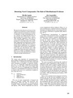
Fig. 1. The cross-protomer interaction of Sav1866. The intracellular
portion of a Sav1866 homodimer is represented in cartoon fashion;
the two Sav1866 molecules are coloured yellow and red, and blue
and green. Bound nucleotide is rendered in grey space-filling repre-
sentation. The cross-protomer (‘domain swapping’) interaction is
illustrated by the intracellular loops of one TMD (blue) interacting
primarily with the NBD of the opposite protomer (yellow).
The structure of eukaryotic ABC multidrug pumps I. D. Kerr et al.
552 FEBS Journal 277 (2010) 550–563 ª 2009 The Authors Journal compilation ª 2009 FEBS
Function of the prokaryotic ABC
exporters
For the bacterial exporters to be used as structural
templates for understanding eukaryotic MDR pumps
also requires demonstration that they are sufficiently
similar in terms of function. This is pertinent because
the physiological relevance of Sav1866 is not clear,
and the function of MsbA is in the transport of lipid
A (a component of the lipolysaccharide outer mem-
brane) [20,35]. Detailed characterization of their func-
tion has been undertaken and provides some evidence
that MsbA and Sav1866 can function as multidrug
pumps. For Sav1866, the protein was characterized in
a Lactococcus lactis expression system [19]. Inside-out
vesicles, intact cells and proteoliposomes containing
purified, reconstituted protein were used to determine
that the transport substrate specificity of Sav1866
includes the dye Hoescht33342 and ethidium bromide,
and ATPase activities further added verapamil and
vinblastine to the list of compounds with which
Sav1866 interacts [19]. For MsbA, similar studies
argue that the protein is a functional homologue of
multidrug pumps – the protein can confer resistance to
ethidium bromide and transport this cation, as well as
another DNA-intercalating agent Hoescht 33342.
Furthermore, membranes containing MsbA have an
ATPase activity that is stimulated by daunomycin, and
can interact with a further typical MDR substrate azi-
dopine [36]. Intriguingly, not only can MsbA substitute
for LmrA in conferring multidrug resistance on E. coli,
but LmrA can restore growth of a MsbA temperature-
sensitive mutant, suggesting functional complementar-
ity [36]. Moreover, LmrA can substitute for human
ABCB1 in transfected tissue culture cells as a MDR
determinant [18]. Thus, LmrA, MsbA and Sav1866,
irrespective of their physiological roles, all interact
with multiple substrates, many of which are also sub-
strates ⁄ modulators of the human multidrug pumps
(Table 2). The ability to function across species barri-
ers also suggests that the study of other eukaryotic
ABC proteins might be advanced by the identification
and characterization of bacterial homologues.
Conformational transitions observed in
prokaryotic MDR pumps
The structural data for MsbA (Table 1 and Fig. 2)
describe three different configurations of the trans-
porter [22]. An open, nucleotide-free state was
observed for the E. coli structure, in which the two
NBDs are a significant distance apart (50 A
˚
; Fig. 2A).
A second nucleotide-free state was observed for the
Vibrio cholera MsbA in which the NBDs are now
closed (Fig. 2B, but not in the classical sandwich
dimer, i.e. there is no Walker-A motif ⁄ Signature motif
interaction) [22,27,30,31]. This ‘closed apo’ structure
can be arrived at from the ‘open apo’ structure by a
rigid body closure, centred on a hinge in the extracellu-
lar loops [22]. Formation of the closed, nucleotide-
bound structure (as observed in Salmonella typhi
MsbA; Fig. 2C) requires a further pair of motions to
align the NBDs, thus forming the nucleotide sandwich
dimer, and a concomitant retraction of TM1 and TM2
from TM3 and TM6, generating an outward facing
configuration of the TMDs similar to that observed in
Sav1866 [21,22,37] (Fig. 2D). These three conforma-
tional states are postulated to be intermediates in the
functional cycle of MsbA – but their magnitude calls
this into question.
EPR spectroscopy has the power to give dynamic
structural data on membrane proteins, determining
inter-residue distances, residue accessibility and confor-
mational transitions [38]. For MsbA, several studies
have attempted to determine the likelihood of the
extreme conformations observed, and to verify the
structures themselves [39–42]. One potential limitation
here is the low resolution of the MsbA data which
means that accessibilities of residues have to be
inferred from Ca positions, an imperfect science. Resi-
due accessibility studies of the Signature and His-loop
regions [42] are only partially consistent with the wide
Table 2. Substrate interaction with the prokaryotic and eukaryotic MDR pumps. n ⁄ r, not recorded.
Ethidium bromide Hoescht 33342 Verapamil Vinblastine Daunomycin Azidopine
Sav1866 [19] Low l
M Low lM 10–50 lM 5 lM n ⁄ rn⁄ r
MsbA [35,36] Low l
M Low lM n ⁄ rn⁄ r 10–50 lM < lM
ABCB1 n ⁄ r Low lM [95] Low lM [6,95] < lM [6] [6] [96]
ABCC1 [97,98] n ⁄ rn⁄ r Yes Yes Yes n ⁄ r
ABCG2 n ⁄ r Low l
M [99] No [99] No [99] < lM [5] Yes, [100]
LmrA [18,101] Low l
M Low lM 10–20 lM 10-20 lM 2–5 lM < lM
I. D. Kerr et al. The structure of eukaryotic ABC multidrug pumps
FEBS Journal 277 (2010) 550–563 ª 2009 The Authors Journal compilation ª 2009 FEBS 553
‘open apo’ structure because two of the five Signature
sequence residues and one of the His-loop residues are
buried according to EPR quenching data [42], but
rather exposed in the E. coli structure [22]. The ‘closed
apo’ structure only partially remedies this conflict.
Moreover, EPR data in multiple configurations of
MsbA argue that the major conformational changes
occur upon nucleotide hydrolysis, suggesting that the
difference seen between the two ‘apo’ structures is a
reflection of crystallographic conditions rather than
being physiological.
Furthermore, Dong et al. [43] investigated the struc-
ture of MsbA in liposomes and mapped conformational
changes during the ATPase cycle by EPR analysis of
112 spin-labelled mutants trapped in four intermediate
states, including apo and AMP–PNP bound. Notably,
this study found that residues in the N-terminal half of
TM helix 6, (residues 284-296), show very low accessibil-
ity to the aqueous phase in all stages of the transport
cycle examined. The accessibility data are in excellent
agreement with cysteine mutagenesis studies of the
equivalent region in ABCB1 [44] (residues 331-343), but
are harder to reconcile with EM images of ABCB1
showing a 5–6 nm diameter, 5 nm deep aqueous cham-
ber within the membrane open to the cell exterior [45]
(although this conformation was obtained under nucleo-
tide-free conditions for EM). The inaccessibility of the
N-terminal half of TM6 to the solvent is even more
puzzling given that in the MsbA accessibility studies
[43], C-terminal regions of TM helix 6 (residues 300 and
303) located near the middle of the membrane, are
accessible to the bulk solvent in all phases of the trans-
port cycle. Clearly, the accessibility data are at odds
with the MsbA crystal structures (and even the Sav1866
structure), and full reconciliation to experimental data
might only be explained by the trapping of other TMD
A
B
D
C
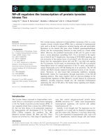
Fig. 2. Conformational states of MDR type
ABC exporters. The structures of ‘open apo’
MsbA (A), ‘closed apo’ MsbA (B), nucleo-
tide-bound MsbA (C) and nucleotide-bound
Sav1866 (D) are shown as Ca traces with
the two monomers in red and blue, respec-
tively. The bound nucleotide is rendered as
green space-filling in the lower two panels.
The structure of eukaryotic ABC multidrug pumps I. D. Kerr et al.
554 FEBS Journal 277 (2010) 550–563 ª 2009 The Authors Journal compilation ª 2009 FEBS
configurations that MsbA ⁄ Sav1866 adopt during the
translocation cycle.
Lastly, spin–spin distance data obtained on deter-
gent or liposome-embedded MsbA [39] identified both
the magnitude and sign of interdomain distance
changes occurring in the transition from the nucleo-
tide-free to the ADP ⁄ Vi-trapped states of MsbA. With
all five pairs of residues (located along the axis of the
protein perpendicular to the membrane) the sign of
the distance change was the same as that observed in
the three structural states of Chang, and the magni-
tudes of distance changes for three of the four pairs of
residues for which data were obtained in liposomes
correlates well with the change in distance observed
going from the ‘closed apo’ V. cholerae structure to
the vanadate-trapped Salmonella typhi structure
[22,39]. Perhaps most pertinently among these data,
the distance between residues within the NBDs
changes by 30 A
˚
according to EPR data, this is incom-
patible with a transition from the ‘open apo’ structure
which would be accompanied by a 50 A
˚
distance
change. In summary, it remains unclear whether either
of the nucleotide-free states of MsbA is physiologically
relevant, and the extent of the conformational transi-
tions seen remains questionable. Indeed, recent com-
mentaries have addressed whether the Sav1866
structures could be compatible with these elaborate
TMD and NBD movements [21,46].
To what extent can the structures of
prokaryotic ABC exporter proteins be
used as homology models for
eukaryotic members of the family?
Homology modelling is a process that generates a 3D
map of a target protein, built against a template of a
Table 3. Sequence identity comparisons of human and prokaryotic multidrug pump nucleotide-binding domains (NBD). The NBDs
are defined for this purpose as encompassing residues from 10 N-terminal to the conserved aromatic reside of the A-loop to 10-residues
C-terminal to the conserved histidine of the His-loop [17].
ABCB1
NBD1
ABCB1
NBD2
ABCC1
NBD1
ABCC1
NBD2
ABCG2
NBD MsbA LmrA Sav1866
ABCB1
NBD1
100
ABCB1
NBD2
61 100
ABCC1
NBD1
28 29 100
ABCC1
NBD2
32 32 26 100
ABCG2 18 21 12 14 100
MsbA 53 48 32 35 13 100
LmrA 44 40 24 35 24 40 100
Sav1866 49 47 33 38 23 55 50 100
Table 4. Sequence identity comparisons of human and prokaryotic multidrug pump transmembrane domains (TMD). The TMDs are defined
for this purpose as being from the first amino acid of the first predicted transmembrane (TM) helix to the final residue of the last predicted
helix (although the additional five TM helices at the N-terminus of ABCC1 are ignored for this exercise).
ABCB1
TMD1
ABCB1
TMD2
ABCC1
TMD1
ABCC1
TMD2
ABCG2
TMD MsbA LmrA Sav1866
ABCB1
TMD1
100
ABCB1
TMD2
26 100
ABCC1
TMD1
8 7 100
ABCC1
TMD2
10 9 11 100
ABCG2 4 3 8 8 100
MsbA 17 16 8 14 8 100
LmrA 16 25 11 8 6 16 100
Sav1866 13 15 13 13 6 18 20 100
I. D. Kerr et al. The structure of eukaryotic ABC multidrug pumps
FEBS Journal 277 (2010) 550–563 ª 2009 The Authors Journal compilation ª 2009 FEBS 555
close homologue, whose X-ray structure is known and
which has mutual sequence similarity [47]. Homology
modelling, in essence ‘structural mimicry’, is particu-
larly appropriate for membrane proteins for which
there is a scarcity of high-resolution structures. Several
algorithms are available to generate a homology
model, including modeller [48], insight ii [49], and
internet-based servers such as swiss-model [50] and
what if [51]. In general, homology modelling
approaches follow a four-step process involving: tem-
plate selection, sequence alignment, model building,
and model optimization and validation.
Template selection is made using similarity search
algorithms such as blast or psi-blast from the RCSB
Protein Data Bank (PDB). An accurate alignment
using a program such as clustalw requires a degree
of manual adjustment of the two sequences in order to
accommodate unmatched gaps and insertions for
which there is no equivalent template sequence. Many
ABC exporters have the conserved architectural scaf-
fold of two TMDs in 6 + 6 helical bundles, and two
NBDs in a ‘head-to-tail’ configuration. However, when
selecting templates for eukaryotic MDR homology
modelling a cautionary tale emerges from sequence
comparisons (Tables 3 and 4) providing the percentage
amino acid identity across the NBD and the TMD to
prokaryotic ABC exporters. The data demonstrate that
ABCB1 is much more similar to the prokaryotic ABC
exporters than ABCC1 and ABCG2. In particular,
ABCG2 shows barely any homology to the bacterial
species, particularly in the TMDs, but also in the
NBDs the percentage sequence identity is in the low
20s. This is discussed further below.
Model building is the easiest of the four stages, rely-
ing as it does essentially on a default task of the soft-
ware, provided that the target–template alignment is
matched accurately. modeller, for example, extracts
the distance and dihedral angle restraints from the
alignment then combines these restraints with
CHARMM energy terms to generate the target 3D
model with proper stereochemistry [47]. Multiple struc-
tures are usually generated and one of these is sub-
jected to validation of the restraints and backbone
angles using programs such as what if; or the best
‘raw’ structure is optimised by short energy minimiza-
tion runs of the order of 2 ns, using a molecular
dynamics package such as gromacs [52].
Homology models of ABC exporters began appear-
ing during the pre-Sav1866 period and, because of the
apparent congruence of the MsbA structure with the
‘generic’ ABC transporter architecture, MsbA was
used as the template for all homology models, whose
target ABC proteins were: MsbA itself [33], ABCB1
[53–56], ABCC1 [57], BmrA [58] and LmrA [59,59a].
Unfortunately, much of this early work amounted to
very little following the retraction of all three MsbA
structures. In retrospect, the first MsbA structure was
a poor template choice because its resolution was
low at only 4.8 A
˚
with the Ca backbone electron
density map lacking side chain definition; and the
NBDs were nondimeric at 50 A
˚
apart with an incor-
rect tail-to-tail orientation. Nevertheless, who could
blame those with the requisite skills from building
homology models of their favourite ABC transport-
ers? Of the pre-Sav1866 homology models, only one
was rendered correctly [56]; and this was achieved by
rotating the NBDs of MsbA ⁄ ABCB1 150° relative
to the cognate TMDs, generating a P-glycoprotein
homology model with a consensus NBD–NBD inter-
face and outward-facing TMD helical bundles that
bears some resemblance to the Sav1866 TMD tertiary
structure. This model was also broadly consistent
with cross-linking data [60] and low-resolution EM
images of ABCB1 [14,61].
The appearance in late 2006 of the S. aureus
Sav1866 half-transporter ushered in new homology
models of other ABC transporters, namely ABCB1
[62,63], LmrA [64], ABCG2 [65,66], ABCC1 [67],
ABCC4 and C5 [68,69], as well as the possibly bidirec-
tional plant auxin transporter ABCB4 [70]. Among
this Sav1866-based group, the two ABCG2 models
presented problems during their construction and sub-
sequent interpretive analyses of cross-linking data and
ligand docking, chiefly because of the NBD–TMD
reverse domain order, the lack of conserved structural
motifs ICD2 and ICD3, and shorter TMD helices and
low sequence identity with Sav1866 (Tables 3 and 4;
6–23%). Despite these limitations, one of these
ABCG2 studies [65] reported blind docking calcula-
tions on their homology model to discern clearly dis-
tinct but neighbouring TMD-binding sites for
rhodamine, doxorubicin and prazosin; although it
should be stressed that the bound ligands, if correctly
docked, would be positioned at or near low-affinity
binding sites because the ABCG2 homology model
was constructed with the TMDs in the outward-facing
conformation. The authors of both ABCG2 studies
acknowledged the limitations of the models and that
further refinement and authentication were required
[65,66]. The ABCB1 and ABCC1 models were much
better matched to the Sav1866 template because their
primary sequences align more closely. In the case of
ABCC1, the model was built without including the
ABCC subfamily-specific N-terminal five-helix TMD0
domain [67]. ABCB1 was rendered as three homology
models representing the three catalytic states of closed
The structure of eukaryotic ABC multidrug pumps I. D. Kerr et al.
556 FEBS Journal 277 (2010) 550–563 ª 2009 The Authors Journal compilation ª 2009 FEBS
(ATP bound), semi-open and open apo or ADP
bound. These models were generated by a ‘cut and
paste’ approach, using the Sav1866 nucleotide-bound
NBD-ICLs or the MalK nucleotide-free NBDs, and
Sav1866 for the ABCB1 TMDs, which were subse-
quently refined and energy minimized. The authors of
all of these post-Sav1866 models contend that they
that are generally consistent with a raft of cysteine
cross-linking studies and spin-labelling and EPR stud-
ies [62,63]. With respect to the ABCB1 cross-linking
data for residues within the TM segments, if the crite-
rion for correlation is that the length of any successful
cross-linker (plus 6 A
˚
for the cysteine side chains) falls
within 4 A
˚
of any distance for the residue pair from
the three different modelled conformations in O’Mara
& Tieleman [62], then 28 ⁄ 52 results fit (see Supplemen-
tary Table 2 in O’Mara & Tieleman [62]). Given the
latitude of the correlation criterion, this fit is probably
better described as ‘fair’ rather than ‘good’.
Plainly, homology modelling uses a crystal structure
template to generate a ‘look-alike’ ABC transporter.
For the NBDs, the model building can be very accu-
rate because the sequences are highly conserved (in the
order of 50%) and the NBD tertiary folds of tem-
plate and target superimpose very closely with small
root mean square deviations. However, there is only
low sequence similarity in the TMDs ( 15%). A sec-
ond caution is that no matter how accurate a homol-
ogy model can be rendered using a seemingly reliable
template such as Sav1866, homology models suffer
from the same interpretive limitations as static crystal
structures in representing ‘snapshots’ of a multistep
transport mechanism. In general, homology models are
comparable with medium-resolution structures and
would not usually be of sufficient quality to be used
for structure-based design directly, although there is
scope for using X-ray scattering, cross-linking data
and MD simulations to improve the models [13,71,72].
Homology modelling of ABCB1 is
consistent with regions showing
correlated evolution
The homology models of ABCB1, as discussed above,
can be validated against biochemical data. We have
attempted a validation against bioinformatics data
using the principle of residue co-evolution, i.e. the
extent to which evolution of a residue i in a given
protein is coupled to evolution of a residue j. Although
i and j may be close together in the 3D structure (and
thus their co-evolution would be expected on structural
grounds), it has also been determined that co-evolution
of pairs of residues at distant sites is indicative of an
allosteric communication between the two sites, as
recently explored for the cystic fibrosis transmembrane
conductance regulator (CFTR) [73]. Many methods
are available to determine which regions of a protein
are subject to co-evolutionary constraint, and descrip-
tion of these is beyond the current review (but see refs
[74–78]). For ABCB1, blast analysis [79] and muscle
sequence alignment [80] enabled the generation of a
multiple sequence alignment of over 150 ABCB type
sequences from eukaryotic organisms. Analysis of this
alignment using several algorithms [74–78,81] enabled
identification of regions in the primary sequence of
ABCB1 that are co-evolving with other regions. When
the highest scoring (i.e. most likely to be co-evolving)
regions are mapped onto the nucleotide-bound model
of O’Mara & Tieleman [62] (Fig. 3), it is striking to
observe that the majority of these map to key domain–
domain interfaces. For example, ICL2, which is in
direct contact with NBD2 [21], is co-evolving with at
least three other regions of ABCB1. Two of these are
located in the a-helical region of NBD2, explaining
mutagenesis data for ABCB1, as previously described
[82], whereas the third is located in TM helix 12 pro-
viding an intriguing co-evolutionary perspective on the
allosteric communication between NBD1 and TMD2.
Further analysis of the data provides many stimulating
opportunities for functional analysis of other ABCB1
mutant isoforms.
Is the structural basis for interdomain
communication observed for several
ABC proteins likely to be preserved
across the whole family?
The TMD–NBD ‘transmission interface’ features the
structurally conserved two short coupling helices that
nevertheless share little or no sequence similarity
among the different transporters [83]. The coupling
helices are deployed roughly parallel to the membrane
and ‘fit’ into grooves in the tops of the NBDs, in the
manner of a ball and socket joint. Despite this con-
served interface, Sav1866 is the only structure in which
the coupling helices are domain swapped – that is, the
coupling helix from TMD1–NBD1 interacts with
NBD2 and vice versa; and this effect is supported by
experimental cross-linking and genetic data for the
eukaryotic drug exporters ABCB1 [34], Yor1p [84] and
the chloride channel CFTR [85,86]. Domain swapping
of the coupling helices does not occur in any of the
ABC importer structures and, if this clear distinction
between importers and exporters is maintained in
future solved ABC transporter structures, it could
inform about the mechanics of translocation for which
I. D. Kerr et al. The structure of eukaryotic ABC multidrug pumps
FEBS Journal 277 (2010) 550–563 ª 2009 The Authors Journal compilation ª 2009 FEBS 557
the TMDs need to adopt alternately inward- and out-
ward-facing conformations for the import or export of
allocrites.
Intriguingly, only ABC exporters contain the con-
served short X-loop motif (consensus TEVGERG) that
is located just N-terminal to the Signature sequence
and that appears to be involved in cross-linking the
ICLs to one another. Thus the X-loop’s chief function
could be to enable the mechanical domain swapping of
the ICL helices for ABC exporters. An increasing
number of recent studies of naturally occurring or
artificially swapped domains has widened the range of
functions of domain swapping to include mechanistic
considerations. For example, interdomain contacts
between the coupling helices and NBDs of CFTR
comprise aromatic clusters important for stabilization
of the interfaces and also involve the Q-loops and
X-loops that are in close proximity to the ATP-binding
sites [85,86]. The aromatic clusters within the ICLs of
CFTR are almost certainly involved in effecting inter-
domain communication between the NBDs and
TMDs, and such a cluster is found in Sav1866 at the
interface of ICL2 and the NBD, but whether this holds
true for ABC transporters generally remains to be
seen. These examples of differences between the
TMD–NBD interface might therefore only pertain to
the mechanistic coupling involved in import versus
export among ABC transporters, that is, between sub-
families, and it is likely that the structural basis for
interdomain communication is preserved across the
prokarya and eukarya kingdoms within the ABC fam-
ily, but has evolved to meet the needs of specific
functions.
An allosteric model of ABC exporter
function
A simple, modified allosteric model for membrane
pumps was proposed by Jardetsky in 1966 [87]. To
function as a pump, a membrane protein need only
meet three structural conditions, it must: (a) contain a
cavity in the interior large enough to admit the solute;
(b) be able to assume inward- and outward-facing con-
figurations such that the cavity is alternately open to
one side of the membrane; and (c) contain a binding
site for the transported species within the cavity, the
affinity of which is different in the two configurations.
In this model, pumps for different molecules need dif-
fer only in the specificity of binding sites, and the same
pump molecule could be adapted to translocate more
than one molecular species. For prokaryotic ABC
importers we are already seeing these multiple confor-
mations at higher resolution [25,88–90]. From such
structures, and despite structurally unrelated TMD
folds, a unified alternating access model for ABC
importers and exporters, based on the Jardetsky allo-
steric model, has been proposed [91a] and developed
further by comparative analysis of several full-length
ABC structures [83].
Despite the obvious appeal of this model, there
remain several unanswered questions regarding sub-
strate transport through MDR-type ABC exporters. If
the NBDs are directly coupled mechanically to open-
ing of the TMDs then what is the magnitude of
domain separation required to enable access of trans-
port substrate (drug)? Does this vary according to the
size of the substrate, and whether it is strongly parti-
tioned into the inner leaflet of the membrane (as is
likely for many MDR transporter substrates) [91]?
Fig. 3. Co-evolving residues of ABCB family members map to
domain interfaces in ABCB1 homology models. Co-evolution analy-
sis of ABCB sequences was performed using tools at http://coevo-
lution.gersteinlab.org/coevolution/ and residues identified by
multiple analyses are superimposed onto a structural model of
ABCB1 [62]. Regions are coloured as follows: red,177–186 (C-termi-
nal to the coupling helix of ICL1, TMD1); blue, 145–155 (N-terminal
to the coupling helix of ICL1, TMD1); purple, 807–819 (C-terminal
to the coupling helix ICL1, TMD2); orange, 895–915 (ICL2, TMD2);
green, 255–268 (ICL2, TMD1); yellow, 465–475 (Gln loop, NBD1);
cyan, 540–545 (Signature–Walker-B, NBD1); pink, 1220–1229 (His
loop, NBD2 in the lower foreground); salmon, 1120–1140 (Gln loop
and C-terminal helix, NBD2 in the background).
The structure of eukaryotic ABC multidrug pumps I. D. Kerr et al.
558 FEBS Journal 277 (2010) 550–563 ª 2009 The Authors Journal compilation ª 2009 FEBS
How does the interior cavity differ for different sub-
strates? What are the repulsive forces that drive the
TMDs and ⁄ or NBDs apart and how is the extent of
domain separation controlled? What are the attractive
forces that bring the domains back into contact – is it
possible that electrostatic attraction across a solvent-
filled gap is sufficient to enable NBD re-association in
a timely and specific manner? As discussed above, the
Sav1866 structure is conformationally constrained by
the intertwined TMD ‘wings’ and domain-swapped
ICL–NBDs, prompting the authors to suggest that the
two subunits are unlikely to move independently and
their maximum separation during the transport cycle is
therefore limited [21]. A detailed mechanistic descrip-
tion of substrate translocation through the TMDs of
MDR-type ABC exporters and its allosteric linkage to
ATP binding and hydrolysis within the NBDs will
require their structural characterization in multiple
states, including bound nucleotides and drug substrate.
The power of molecular dynamics will also be central
to this challenge.
The Holy Grail? A structure for a
eukaryotic MDR pump
Recently, the structure of mouse ABCB1a has been
described by Aller et al., [16] resolved to a resolution of
3.8 A
˚
. At first glance the structure seems to tick all the
boxes with regard to a structural understanding of mul-
tidrug binding. The structure is comparable in terms of
the fold and the domain–domain interactions to the
structures of MsbA and Sav1866. Furthermore, a cavity
is contributed by both TMDs, and is sufficiently large
to accommodate a cyclic peptide drug molecule, with
stereospecificity. However, a number of concerns arise
from close inspection of the structure. Of most rele-
vance to the current discussion are the resolution, the
completeness of the structure, the spatial separation of
the NBDs and the drug-bound state. First, the resolu-
tion is at best 3.8 A
˚
, which is considerably lower than
the Sav1866 structure. The exact orientation of many
side chain residues will be difficult to determine at this
resolution and the very high B-factors in the structure
are a reflection of this uncertainty. Second, the struc-
ture does not address one of the major topological dis-
tinctions between a prokaryotic MDR homologue and
eukaryotic ABCB MDR pumps, namely the presence
of a linker domain between the two halves of the
transporter. The mouse ABCB1a structure is missing
the 56 amino acids between the end of the first NBD
and the start of the second TMD (the first 32 residues
are also unresolved). The missing linker region means
that no light can be shed on this important region –
phosphorylation of which influences the potency of
several transported substrates to increase the ATPase
activity implying a role in TMD–NBD communication
[92], and that the spatial separation of the NBDs may
not reflect the separation(s) observed physiologically.
Finally, with relevance to this minireview series, the
drug-bound state has been determined with stereoi-
somers of a cyclic peptide (related to MDR reversal
agents from blue–green algae; dendromamides) [93].
These are poorly characterized in terms of their inter-
action with any of the eukaryotic MDR pumps, unlike
the compounds listed in Table 2. Until the structure of
ABCB1 with drug bound reaches the quality of the
bacterial resistance nodulation division multidrug
pumps [94], it seems likely that we will continue to
rely on computational approaches (homology model-
ling and drug docking) in order to elucidate aspects of
both the structure of eukaryotic MDR pumps, and
their interaction with a multitude of chemically dis-
tinct compounds.
Acknowledgements
P. M. Jones is supported by Cure Cancer Australia
and UTS IBID fellowships.
References
1 Bates SE (2003) Solving the problem of multidrug
resistance: ABC transporters in clinical oncology. In
ABC Proteins: From Bacteria to Man (Holland IB,
Cole SPC, Kuchler K & Higgins CF eds), pp. 359–391.
Academic Press, New York, NY.
2 Chen CJ, Chin JE, Ueda K, Clark DP, Pastan I, Got-
tesman MM & Roninson IB (1986) Internal duplica-
tion and homology with bacterial transport proteins in
the mdr1 (P-glycoprotein) gene from multidrug-resis-
tant human cells. Cell 47, 381–389.
3 Cole SP, Bhardwaj G, Gerlach JH, Mackie JE, Grant
CE, Almquist KC, Stewart AJ, Kurz EU, Duncan AM
& Deeley RG (1992) Overexpression of a transporter
gene in a multidrug-resistant human lung cancer cell
line. Science 258, 1650–1654.
4 Doyle LA, Yang W, Abruzzo LV, Krogmann T, Gao
Y, Rishi AK & Ross DD (1998) A multidrug resistance
transporter from human MCF-7 breast cancer cells.
Proc Natl Acad Sci USA 95, 15665–15670.
5 Clark R, Kerr ID & Callaghan R (2006) Multiple drug
binding sites on the R482G isoform of the ABCG2
transporter. Br J Pharmacol 149, 506–515.
6 Martin C, Berridge G, Higgins CF, Mistry P, Charlton
P & Callaghan R (2000) Communication between mul-
tiple drug binding sites on P-glycoprotein. Mol Phar-
macol 58, 624–632.
I. D. Kerr et al. The structure of eukaryotic ABC multidrug pumps
FEBS Journal 277 (2010) 550–563 ª 2009 The Authors Journal compilation ª 2009 FEBS 559
7 Rothnie A, Callaghan R, Deeley RG & Cole SP (2006)
Role of GSH in estrone sulfate binding and transloca-
tion by the multidrug resistance protein 1
(MRP1 ⁄ ABCC1). J Biol Chem 281, 13906–13914.
8 Callaghan R, Berridge G, Ferry DR & Higgins CF
(1997) The functional purification of P-glycoprotein is
dependent on maintenance of a lipid–protein interface.
Biochem Biophys Acta 1328 , 109–124.
9 Mao Q, Leslie EM, Deeley RG & Cole SP (1999)
ATPase activity of purified and reconstituted
multidrug resistance protein MRP1 from drug-
selected H69AR cells. Biochim Biophys Acta 1461,
69–82.
10 Ozvegy C, Varadi A & Sarkadi B (2002) Characteriza-
tion of drug transport, ATP hydrolysis, and nucleotide
trapping by the human ABCG2 multidrug transporter.
Modulation of substrate specificity by a point muta-
tion. J Biol Chem 277, 47980–47990.
11 Mao Q, Deeley RG & Cole SP (2000) Functional
reconstitution of substrate transport by purified multi-
drug resistance protein MRP1 (ABCC1) in phospho-
lipid vesicles. J Biol Chem 275, 34166–34172.
12 McDevitt CA, Collins RF, Conway M, Modok S,
Storm J, Kerr ID, Ford RC & Callaghan R (2006)
Purification and 3D structural analysis of oligomeric
human multidrug transporter ABCG2. Structure 14,
1623–1632.
13 McDevitt CA, Shintre CA, Grossmann JG, Pollock
NL, Prince SM, Callaghan R & Ford RC (2008) Struc-
tural insights into P-glycoprotein (ABCB1) by small
angle X-ray scattering and electron crystallography.
FEBS Lett 582, 2950–2956.
14 Rosenberg MF, Callaghan R, Modok S, Higgins CF &
Ford RC (2005) Three-dimensional structure of P-gly-
coprotein: the transmembrane regions adopt an asym-
metric configuration in the nucleotide-bound state.
J Biol Chem 280, 2857–2862.
15 Rosenberg MF, Mao Q, Holzenburg A, Ford RC,
Deeley RG & Cole SP (2001) The structure of the mul-
tidrug resistance protein 1 (MRP1 ⁄ ABCC1). Crystalli-
zation and single-particle analysis. J Biol Chem 276,
16076–16082.
16 Aller SG, Yu J, Ward A, Weng Y, Chittaboina S,
Zhuo R, Harrell PM, Trinh YT, Zhang Q, Urbatsch
IL et al. (2009) Structure of P-glycoprotein reveals a
molecular basis for poly-specific drug binding. Science
323, 1718–1722.
17 Kerr ID (2002) Structure and association of ATP bind-
ing cassette transporter nucleotide-binding domains.
Biochem Biophys Acta 1561 , 47–64.
18 van Veen HW, Venema K, Bolhuis H, Oussenko I,
Kok J, Poolman B, Driessen AJ & Konings WN
(1996) Multidrug resistance mediated by a bacterial
homolog of the human multidrug transporter MDR1.
Proc Natl Acad Sci USA 93, 10668–10672.
19 Velamakanni S, Yao Y, Gutmann DA & van Veen
HW (2008) Multidrug transport by the ABC trans-
porter Sav1866 from Staphylococcus aureus. Biochemis-
try 47, 9300–9308.
20 Zhou Z, White KA, Polissi A, Georgopoulos C &
Raetz CR (1998) Function of Escherichia coli MsbA,
an essential ABC family transporter, in lipid A and
phospholipid biosynthesis. J Biol Chem 273, 12466–
12475.
21 Dawson RJ & Locher KP (2006) Structure of a bacte-
rial multidrug ABC transporter. Nature 443, 180–185.
22 Ward A, Reyes CL, Yu J, Roth CB & Chang G (2007)
Flexibility in the ABC transporter MsbA: alternating
access with a twist. Proc Natl Acad Sci USA 104,
19005–19010.
23 Davidson AL & Chen J (2005) Structural biology.
Flipping lipids: is the third time the charm?
Science
308, 963–965.
24 Higgins CF & Linton KJ (2001) Structural biology.
The xyz of ABC transporters. Science 293 , 1782–1784.
25 Locher KP, Lee AT & Rees DC (2002) The E. coli
BtuCD structure: a framework for ABC transporter
architecture and mechanism. Science 296, 1091–1098.
26 Chen J, Lu G, Lin J, Davidson AL & Quiocho FA
(2003) A tweezers-like motion of the ATP-binding cas-
sette dimer in an ABC transport cycle. Mol Cell 12,
651–661.
27 Hopfner KP, Karcher A, Shin DS, Craig L, Arthur
LM, Carney JP & Tainer JA (2000) Structural biology
of Rad50 ATPase: ATP-driven conformational control
in DNA double-strand break repair and the ABC–AT-
Pase superfamily. Cell 101, 789–800.
28 Lamers MH, Perrakis A, Enzlin JH, Winterwerp
HHK, de Wind N & Sixma TK (2000) The crystal
structures of DNA mismatch repair protein MutS
binding to a G.T mismatch. Nature 407, 711–717.
29 Oblomova G, Ban C, Hsieh P & Yang W (2000)
Crystal structures of mismatch repair protein MutS
and its complex with a substrate DNA. Nature 407,
703–710.
30 Smith PC, Karpowich N, Millen L, Moody JE, Rosen J,
Thomas PJ & Hunt JF (2002) ATP binding to the motor
domain from an ABC transporter drives formation of a
nucleotide sandwich dimer. Mol Cell 10, 139–149.
31 Jones PM & George AM (1999) Subunit interactions
in ABC transporters: towards a functional architecture.
FEMS Microbiol Lett 179, 187–202.
32 Petsko GA (2007) And the second shall be first. Gen-
ome Biol 8, 103.
33 Campbell JD, Biggin PC, Baaden M & Sansom MSP
(2003) Extending the structure of an ABC transporter
to atomic resolution: modelling and simulation studies
of MsbA. Biochemistry 42, 3666–3673.
34 Zolnerciks JK, Wooding C & Linton KJ (2007) Evi-
dence for a Sav1866-like architecture for the human
The structure of eukaryotic ABC multidrug pumps I. D. Kerr et al.
560 FEBS Journal 277 (2010) 550–563 ª 2009 The Authors Journal compilation ª 2009 FEBS
multidrug transporter P-glycoprotein. FASEB J 21,
3937–3948.
35 Doerrler WT & Raetz CR (2002) ATPase activity of
the MsbA lipid flippase of Escherichia coli. J Biol
Chem 277, 36697–36705.
36 Reuter G, Janvilisri T, Venter H, Shahi S, Balakrish-
nan L & van Veen HW (2003) The ATP binding cas-
sette multidrug transporter LmrA and lipid transporter
MsbA have overlapping substrate specificities. J Biol
Chem 278, 35193–35198.
37 Dawson RJ & Locher KP (2007) Structure of the mul-
tidrug ABC transporter Sav1866 from Staphylococ-
cus aureus in complex with AMP–PNP. FEBS Lett
581, 935–938.
38 Altenbach C, Marti T, Khorana HG & Hubbell WL
(1990) Transmembrane protein structure: spin labeling
of bacteriorhodopsin mutants. Science 248, 1088–
1092.
39 Borbat PP, Surendhran K, Bortolus M, Zou P, Freed
JH & McHaourab HS (2007) Conformational motion
of the ABC transporter MsbA induced by ATP hydro-
lysis. PLoS Biol 5, e271.
40 Buchaklian AH, Funk AL & Klug CS (2004) Resting
state conformation of the MsbA homodimer as studied
by site-directed spin labeling. Biochemistry 43, 8600–
8606.
41 Buchaklian AH & Klug CS (2005) Characterization of
the Walker A motif of MsbA using site-directed spin
labeling electron paramagnetic resonance spectroscopy.
Biochemistry 44, 5503–5509.
42 Buchaklian AH & Klug CS (2006) Characterization of
the LSGGQ and H motifs from the Escherichia coli
lipid A transporter MsbA. Biochemistry 45, 12539–
12546.
43 Dong J, Yang G & McHaourab HS (2005) Structural
basis of energy transduction in the transport cycle of
MsbA. Science 308, 1023–1028.
44 Rothnie A, Storm J, McMahon R, Taylor A, Kerr ID
& Callaghan R (2005) The coupling mechanism of
P-glycoprotein involves residue L339 in the sixth mem-
brane spanning segment. FEBS Lett 579, 3984–3990.
45 Rosenberg MF, Kamis AB, Callaghan R, Higgins CF
& Ford RC (2003) Three-dimensional structures of the
mammalian multidrug resistance P-glycoprotein dem-
onstrate major conformational changes in the trans-
membrane domains upon nucleotide binding. J Biol
Chem 278, 8294–8299.
46 Schuldiner S (2006) Structural biology: the ins and outs
of drug transport. Nature 443, 156–157.
47 Chang C & Swaan PW (2006) Computational appro-
aches to modeling drug transporters. Eur J Pharm Sci
27, 411–424.
48 Sali A & Blundell TL (1993) Comparative protein
modelling by satisfaction of spatial restraints. J Mol
Biol 234, 779–815.
49 Greer J (1990) Comparative modeling methods: appli-
cation to the family of the mammalian serine proteases.
Proteins 7, 317–334.
50 Schwede T, Kopp J, Guex N & Peitsch MC (2003)
SWISS-MODEL: an automated protein homology-
modeling server. Nucleic Acids Res
31, 3381–3385.
51 Vriend G (1990) WHAT IF: a molecular modeling and
drug design program. J Mol Graph 8, 52–56.
52 Lindahl E, Hess B & van der Spoel D (2001) GRO-
MACS 3.0: a package for molecular simulation and
trajectory analysis. J Mol Model 7, 306–317.
53 Omote H & Al-Shawi MK (2006) Interaction of trans-
ported drugs with the lipid bilayer and P-glycoprotein
through a solvation exchange mechanism. Biophys J
90, 4046–4059.
54 Pleban K, Kopp S, Csaszar E, Peer M, Hrebicek T,
Rizzi A, Ecker GF & Chiba P (2005) P-glycoprotein
substrate binding domains are located at the trans-
membrane domain ⁄ transmembrane domain interfaces:
a combined photoaffinity labelling–protein homology
modeling approach. Mol Pharmacol 67, 365–374.
55 Seigneuret M & Garnier-Suillerot A (2003) A structural
model for the open conformation of the mdr1 P-glyco-
protein based on the MsbA crystal structure. J Biol
Chem 278, 30115–30124.
56 Stenham DR, Campbell JD, Sansom MSP, Higgins
CF, Kerr ID & Linton KJ (2003) An atomic detail
model for the human ATP binding cassette trans-
porter, P-glycoprotein, derived from disulphide
cross-linking and homology modelling. FASEB J 15,
2287–2289.
57 Campbell JD, Koike K, Moreau C, Sansom MS,
Deeley RG & Cole SP (2004) Molecular modeling
correctly predicts the functional importance of
Phe594 in transmembrane helix 11 of the multidrug
resistance protein, MRP1 (ABCC1). J Biol Chem
279, 463–468.
58 Dalmas O, Orelle C, Foucher AE, Geourjon C, Crouzy
S, Di Pietro A & Jault JM (2005) The Q-loop disen-
gages from the first intracellular loop during the
catalytic cycle of the multidrug ABC transporter
BmrA. J Biol Chem 280, 36857–36864.
59 Shilling R, Federici L, Walas F, Venter H, Velama-
kanni S, Woebking B, Balakrishnan L, Luisi B & van
Veen HW (2005) A critical role of a carboxylate in
proton conduction by the ATP-binding cassette
multidrug transporter LmrA. FASEB J 19, 1698–
1700.
59a Ecker GF, Pleban K, Kopp S, Csaszar E, Poelarends
GJ, Putman M, Kaiser D, Konings WN & Chiba P
(2004) A three-dimensional model for the substrate
binding domain of the multidrug ATP binding cassette
transporter LmrA. Mol Pharmacol 66, 1169–1179.
60 Loo TW & Clarke DM (2000) The packing of the
transmembrane segments of human multidrug
I. D. Kerr et al. The structure of eukaryotic ABC multidrug pumps
FEBS Journal 277 (2010) 550–563 ª 2009 The Authors Journal compilation ª 2009 FEBS 561
resistance P-glycoprotein is revealed by disulfide cross-
linking analysis. J Biol Chem 275, 5253–5256.
61 Lee JY, Urbatsch IL, Senior AE & Wilkens S (2002)
Projection structure of P-glycoprotein by electron
microscopy. Evidence for a closed conformation of the
nucleotide-binding domains. J Biol Chem 277, 40125–
40131.
62 O’Mara M & Tieleman DP (2007) P-glycoprotein
models of the apo and ATP-bound states based on
homology with Sav1866 and MalK. FEBS Lett 581,
4217–4222.
63 Ravna AW, Sylte I & Sager G (2007) Molecular model
of the outward facing state of the human P-glycopro-
tein (ABCB1), and comparison to a model of the
human MRP5 (ABCC5). Theor Biol Med Model 4, 33.
64 Federici L, Woebking B, Velamakanni S, Shilling RA,
Luisi B & van Veen HW (2007) New structure model
for the ATP-binding cassette multidrug transporter
LmrA. Biochem Pharmacol 74, 672–678.
65 Hazai E & Bikadi Z (2008) Homology modeling of
breast cancer resistance protein (ABCG2). J Struct Biol
162, 63–74.
66 Li YF, Polgar O, Okada M, Esser L, Bates SE & Xia
D (2007) Towards understanding the mechanism of
action of the multidrug resistance-linked half-ABC
transporter ABCG2: a molecular modeling study.
J Mol Graph Model 25, 837–851.
67 DeGorter MK, Conseil G, Deeley RG, Campbell RL
& Cole SP (2008) Molecular modeling of the human
multidrug resistance protein 1 (MRP1 ⁄ ABCC1).
Biochem Biophys Res Commun 365, 29–34.
68 Ravna AW & Sager G (2008) Molecular model of the
outward facing state of the human multidrug resistance
protein 4 (MRP4 ⁄ ABCC4). Bioorg Med Chem Lett 18,
3481–3483.
69 Ravna AW, Sylte I & Sager G (2008) A molecular
model of a putative substrate releasing conformation
of multidrug resistance protein 5 (MRP5). Eur J Med
Chem 43, 2557–2567.
70 Yang H & Murphy AS (2009) Functional expression
and characterization of Arabidopsis ABCB, AUX 1
and PIN auxin transporters in Schizosaccharomy-
ces pombe. Plant J 59, 179–191.
71 Ecker GF, Stockner T & Chiba P (2008) Computa-
tional models for prediction of interactions with ABC-
transporters. Drug Discov Today 13, 311–317.
72 Fan H & Mark AE (2004) Refinement of homology-
based protein structures by molecular dynamics simula-
tion techniques. Protein Sci 13, 211–220.
73 Vergani P, Lockless SW, Nairn AC & Gadsby DC
(2005) CFTR channel opening by ATP-driven tight
dimerization of its nucleotide-binding domains. Nature
433, 876–880.
74 Clarke ND (1995) Covariation of residues in the home-
odomain sequence family. Protein Sci 4, 2269–2278.
75 Dekker JP, Fodor A, Aldrich RW & Yellen G (2004)
A perturbation-based method for calculating explicit
likelihood of evolutionary co-variance in multiple
sequence alignments. Bioinformatics 20, 1565–1572.
76 Gobel U, Sander C, Schneider R & Valencia A (1994)
Correlated mutations and residue contacts in proteins.
Proteins 18
, 309–317.
77 Larson SM, Di Nardo AA & Davidson AR (2000)
Analysis of covariation in an SH3 domain sequence
alignment: applications in tertiary contact prediction
and the design of compensating hydrophobic core sub-
stitutions. J Mol Biol 303, 433–446.
78 Lockless SW & Ranganathan R (1999) Evolutionarily
conserved pathways of energetic connectivity in protein
families. Science 286, 295–299.
79 Altschul SF, Madden TL, Schaffer AA, Zhang J,
Zhang Z, Miller W & Lipman DJ (1997) Gapped
BLAST and PSI-BLAST: a new generation of protein
database search programs. Nucleic Acids Res 25, 3389–
3402.
80 Edgar RC (2004) MUSCLE: multiple sequence align-
ment with high accuracy and high throughput. Nucleic
Acids Res 32, 1792–1797.
81 Yip KY, Patel P, Kim PM, Engelman DM, McDer-
mott D & Gerstein M (2008) An integrated system for
studying residue coevolution in proteins. Bioinformatics
24, 290–292.
82 Lawson J, O’Mara ML & Kerr ID (2008) Structure-
based interpretation of the mutagenesis database for
the nucleotide binding domains of P-glycoprotein. Bio-
chim Biophys Acta 1778, 376–391.
83 Hollenstein K, Dawson RJ & Locher KP (2007) Struc-
ture and mechanism of ABC transporter proteins. Curr
Opin Struct Biol 17, 412–418.
84 Pagant S, Brovman EY, Halliday JJ & Miller EA
(2008) Mapping of interdomain interfaces required for
the functional architecture of Yor1p, a eukaryotic
ATP-binding cassette (ABC) transporter. J Biol Chem
283, 26444–26451.
85 He L, Aleksandrov AA, Serohijos AW, Hegedus T,
Aleksandrov LA, Cui L, Dokholyan NV & Riordan
JR (2008) Multiple membrane–cytoplasmic domain
contacts in the cystic fibrosis transmembrane conduc-
tance regulator (CFTR) mediate regulation of channel
gating. J Biol Chem 283, 26383–26390.
86 Serohijos AW, Hegedus T, Aleksandrov AA, He L, Cui
L, Dokholyan NV & Riordan JR (2008) Phenylalanine-
508 mediates a cytoplasmic–membrane domain contact
in the CFTR 3D structure crucial to assembly and
channel function. Proc Natl Acad Sci USA 105, 3256–
3261.
87 Jardetzky O (1966) Simple allosteric model for mem-
brane pumps. Nature 211, 969–970.
88 Hvorup RN, Goetz BA, Niederer M, Hollenstein K,
Perozo E & Locher KP (2007) Asymmetry in
The structure of eukaryotic ABC multidrug pumps I. D. Kerr et al.
562 FEBS Journal 277 (2010) 550–563 ª 2009 The Authors Journal compilation ª 2009 FEBS
the structure of the ABC transporter-binding
protein complex BtuCD–BtuF. Science 317, 1387–
1390.
89 Pinkett HW, Lee AT, Lum P, Locher KP & Rees DC
(2007) An inward-facing conformation of a putative
metal-chelate-type ABC transporter. Science 315,
373–377.
90 Oldham ML, Khare D, Quiocho FA, Davidson AL
& Chen J (2007) Crystal structure of a catalytic
intermediate of the maltose transporter. Nature 450,
515–521.
91 Gatlik-Landwojtowicz E, Aanismaa P & Seelig A
(2006) Quantification and characterization of P-glyco-
protein–substrate interactions. Biochemistry 45, 3020–
3032.
91a van der Does C & Tampe
´
R (2004) How do ABC
transporters drive transport? Biol Chem 385, 927–933.
92 Szabo K, Bakos E, Welker E, Muller M, Goodfellow
HR, Higgins CF, Varadi A & Sarkadi B (1997) Phos-
phorylation site mutations in the human multidrug
transporter modulate its drug-stimulated ATPase
activity. J Biol Chem 272, 23165–23171.
93 Ogino J, Moore RE, Patterson GM & Smith CD
(1996) Dendroamides, new cyclic hexapeptides from a
blue-green alga. Multidrug-resistance reversing activity
of dendroamide A. J Nat Prod 59, 581–586.
94 Nikaido H & Takatsuka Y (2009) Mechanisms of
RND multidrug efflux pumps. Biochim Biophys Acta
1794, 769–781.
95 Qu Q & Sharom FJ (2002) Proximity of bound
Hoechst 33342 to the ATPase catalytic sites places the
drug binding site of P-glycoprotein within the
cytoplasmic membrane leaflet. Biochemistry 41, 4744–
4752.
96 Storm J, O’Mara ML, Crowley EH, Peall J, Tieleman
DP, Kerr ID & Callaghan R (2007) Residue G346 in
transmembrane segment six is involved in inter-domain
communication in P-glycoprotein. Biochemistry 46,
9899–9910.
97 Bakos E, Evers R, Szakacs G, Tusnady GE, Welker E,
Szabo K, de Haas M, van Deemter L, Borst P, Varadi
A et al. (1998) Functional multidrug resistance protein
(MRP1) lacking the N-terminal transmembrane
domain. J Biol Chem 273, 32167–32175.
98 Huang Z, Chang X, Riordan JR & Huang Y (2004)
Fluorescent modified phosphatidylcholine floppase
activity of reconstituted multidrug resistance-associated
protein MRP1. Biochim Biophys Acta 1660, 155–
163.
99 Robey RW, Honjo Y, van de Laar A, Miyake K, Regis
JT, Litman T & Bates SE (2001) A functional assay for
detection of the mitoxantrone resistance protein, MXR
(ABCG2). Biochim Biophys Acta 1512, 171–182.
100 Shukla S, Robey RW, Bates SE & Ambudkar SV
(2006) The calcium channel blockers, 1,4-dihydropyri-
dines, are substrates of the multidrug resistance-linked
ABC drug transporter, ABCG2. Biochemistry 45,
8940–8951.
101 Woebking B, Reuter G, Shilling RA, Velamakanni S,
Shahi S, Venter H, Balakrishnan L & van Veen HW
(2005) Drug–lipid A interactions on the Escherichia coli
ABC transporter MsbA. J Bacteriol 187, 6363–6369.
I. D. Kerr et al. The structure of eukaryotic ABC multidrug pumps
FEBS Journal 277 (2010) 550–563 ª 2009 The Authors Journal compilation ª 2009 FEBS 563