Báo cáo khoa học: Okadaic acid induces DNA fragmentation via caspase-3-dependent and caspase-3-independent pathways in Chinese hamster ovary (CHO)-K1 cells pot
Bạn đang xem bản rút gọn của tài liệu. Xem và tải ngay bản đầy đủ của tài liệu tại đây (269.77 KB, 9 trang )
Okadaic acid induces DNA fragmentation via
caspase-3-dependent and caspase-3-independent
pathways in Chinese hamster ovary (CHO)-K1 cells
Ikuko Kitazumi, Yoko Maseki, Yoshiko Nomura, Akiko Shimanuki, Yumi Sugita and Masayoshi
Tsukahara
Bio Process Research and Development Laboratories, Kyowa Hakko Kirin Co., Ltd, Hagiwara, Takasaki, Gunma, Japan
Introduction
Apoptosis is a crucial cellular mechanism that is
involved in inflammation, cell differentiation, and cell
proliferation. Apoptotic cells are characterized by dis-
tinctive morphological and biochemical changes,
including plasma membrane blebbing, nuclear conden-
sation, DNA fragmentation, and phosphatidylserine
(PS) exposure. These changes are largely mediated
by the activation of caspases, a family of cysteinyl
aspartate-specific proteases whose target proteins are
particularly important indicators of the apoptotic sig-
nal pathway. The activation of other proteases also
plays a key role in apoptotic cell death. They consti-
tute alternative signal pathways that frequently overlap
the caspase-dependent pathway. Among them, caspase-3
is involved in nuclear changes by cleaving substrates
such as poly(ADP-ribose) polymerase (PARP), an
Keywords
apoptosis; caspase-3; caspase inhibitor;
DNA fragmentation; okadaic acid
Correspondence
M. Tsukahara, Bio Process Research and
Development Laboratories, Kyowa Hakko
Kirin Co., Ltd, 100-1 Hagiwara, Takasaki,
Gunma 370-0013, Japan
Fax: +81 27 353 7400
Tel: +81 27 353 7382
E-mail: masayoshi.tsukahara@
kyowa-kirin.co.jp
Database
The sequences for CHO-K1 caspase-3 cDNA
reported in this article have been submitted
to the GenBank database under the
accession number FJ940732
(Received 10 June 2009, revised 23 October
2009, accepted 11 November 2009)
doi:10.1111/j.1742-4658.2009.07493.x
DNA fragmentation is a hallmark of apoptosis that occurs in a variety of
cell types; however, it remains unclear whether caspase-3 is required for its
induction. To investigate this, we produced caspase-3 knockout Chinese
hamster ovary (CHO)-K1 cells and examined the effects of gene knockout
and treatment with caspase-3 inhibitors. Okadaic acid (OA) is a potent
inhibitor of the serine⁄ threonine protein phosphatases (PPs) PP1 and
PP2A, which induce apoptotic cellular reactions. Treatment of caspase-
3
) ⁄ )
cells with OA induced DNA fragmentation, indicating that caspase-3
is not an essential requirement. However, in the presence of benzyloxycar-
bonyl-Asp-Glu-Val-Asp (OMe) fluoromethylketone (z-DEVD-fmk), DNA
fragmentation occurred in CHO-K1 cells but not in caspase-3
) ⁄ )
cells, sug-
gesting that caspase-3 is involved in OA-induced DNA fragmentation that
does not utilize DEVDase activity. In the absence of caspase-3, DEVDase
activity may play an important role. In addition, OA-induced DNA frag-
mentation was reduced but not blocked in CHO-K1 cells, suggesting that
caspase-3 is involved in caspase-independent OA-induced DNA fragmenta-
tion. Furthermore, OA-induced cleavage of caspase-3 and DNA fragmenta-
tion were blocked by pretreatment with the wide-ranging serine protease
inhibitor 4-(2-aminoethyl)-benzenesulfonyl fluoride hydrochloride. These
results suggest that serine proteases regulate DNA fragmentation upstream
of caspase-3.
Abbreviations
AAD, aminoactinomycin; AEBSF, 4-(2-aminoethyl)-benzenesulfonyl fluoride hydrochloride; CHO, Chinese hamster ovary; DIG, digoxigenin;
NaCl ⁄ P
i
(–), calcium and magnesium-free phosphate buffered saline; OA, okadaic acid; PARP, poly(ADP-ribose) polymerase; PP, protein
phosphatase;p PS, phosphatidylserine; TLCK, N-a-tosyl-
L-lysine chloromethyl ketone; TPCK, N-tosyl-L-phenylalanine chloromethyl ketone;
z-DEVD-fmk, benzyloxycarbonyl-Asp-Glu-Val-Asp (OMe) fluoromethylketone; z-VAD-fmk, benzyloxycarbonyl-Val-Ala-Asp fluoromethylketone.
404 FEBS Journal 277 (2010) 404–412 ª 2009 The Authors Journal compilation ª 2009 FEBS
inhibitor of caspase-activated DNase ⁄ DNA fragmenta-
tion factor 45 (ICAD ⁄ DFF45) [1–3]. Cells have multiple
cleavage mechanisms, as shown by cleavage induction of
these substrates in caspase-3-deficient cells [4,5].
Various methods are used for blocking caspase activa-
tion. One example is the addition of caspase inhibitors,
which are frequently used to define caspase-independent
events. However, it is unclear whether caspase inhibitors
actually prevent caspase activation or whether they
effectively block caspase-dependent apoptotic events.
Caspase inhibition is ineffective [6], although some
reports suggest that the caspase-3 ⁄ 7 inhibitor is effective
at blocking DNA fragmentation [7–9]. Furthermore,
caspase inhibitors have been found to inhibit more
than just the targeted caspase. This uncertainty over
the requirement of caspase-3 for DNA fragmentation
led us to target it for gene knockout in the pres-
ent study. We determined the genome sequence of
Chinese hamster ovary (CHO)-K1 cells, and produced
caspase-3-deficient CHO-K1 (caspase-3
) ⁄ )
) cells to
examine the differences between treatment with inhibi-
tors and gene knockout on DNA fragmentation, and
to investigate whether caspase-3 is required for DNA
fragmentation.
We used okadaic acid (OA), a potent inhibitor of
the serine ⁄ threonine protein phosphatase (PP) type 1
(PP1) and type 2 (PP2A), to induce caspase-3 activa-
tion and apoptotic cellular reactions, including DNA
fragmentation. Inhibition of PP2A (low OA concen-
tration) reduces apoptosis by decreasing mitochon-
drial cytochrome c release, whereas inhibition of PP1
(high OA concentration) induces apoptosis through
p53-dependent cell death pathways [10–12]. It is
unclear whether OA-induced caspase-3 activation is
necessary for apoptosis; one study demonstrated that
caspase-3 is important in the process of OA-induced
apoptosis [13], whereas another showed that OA
induces apoptosis in the human breast carcinoma cell
line MCF-7, which lacks expression of caspase-3 [14].
Furthermore, a caspase inhibitor was reported to
block OA-induced caspase-3 activity but not apopto-
sis [15].
Here, OA induced DNA fragmentation in both
CHO-K1 caspase-3
) ⁄ )
cells and CHO-K1 cells treated
with the caspase-3 ⁄ 7 inhibitor benzyloxycarbonyl-Asp-
Glu-Val-Asp (OMe) fluoromethylketone (z-DEVD-
fmk), indicating that caspase-3 is not essential for
DNA fragmentation. However, z-DEVD-fmk blocked
OA-induced DNA fragmentation in caspase-3
) ⁄ )
cells,
but not in CHO-K1 cells, suggesting that caspase-3 is
involved in OA-induced DNA fragmentation indepen-
dently from DEVDase activity, and that DEVDase or
DEVDase-like activity is involved in DNA fragmenta-
tion in caspase-3
) ⁄ )
cells. Furthermore, the wide-
ranging serine protease inhibitor 4-(2-aminoethyl)-
benzenesulfonyl fluoride hydrochloride (AEBSF)
blocked caspase-3 activation and DNA fragmentation,
suggesting that serine proteases are involved in
DNA fragmentation, probably upstream of caspase-3
activation.
Results
Sequencing of CHO-K1 full-length caspase-3
cDNA and genomic structure
Full-length CHO-K1 cell caspase-3 cDNA was isolated
using 5¢-RACE and 3¢-RACE, and used to determine
the genomic sequence of caspase-3. We confirmed that
the cDNA sequence is coincident with the genomic
sequence. Our caspase-3 cDNA sequence differed from
the GenBank database sequence (accession no.
AY479976; see Fig. S1). A caspase-3 gene-targeting
vector was constructed according to our sequence, and
used to knock out exons 4–6 of the CHO-K1 cell cas-
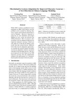
pase-3 gene (Fig. 1A). Exon 6 contains the cleavage
site that is required for its activation [16]. Gene knock-
out was confirmed by Southern blot analysis, which
revealed additional diagnostic bands from the targeted
alleles, as expected (Fig. 1B). Western blotting with a
caspase-3 antibody did not detect pro-caspase-3
expression after gene knockout (Fig. 1C).
The caspase-3/7 inhibitor z-DEVD-fmk causes
differential effects on OA-induced DNA
fragmentation in CHO-K1 and caspase-3
)
/
)
cells
To establish whether caspase-3 is required for
OA-induced DNA fragmentation in CHO-K1 cells, we
investigated the differences in OA response between
CHO-K1 and caspase-3
) ⁄ )
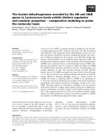
cells. As shown in
Fig. 2A,B, both cell types similarly showed PS expo-
sure and DNA fragmentation, which are characteristic
of apoptosis. OA induced DNA fragmentation both
with and without caspase-3, indicating that caspase-3
is not essential for the induction of DNA fragmenta-
tion. Next, CHO-K1 cells were stimulated with OA in
the presence of z-DEVD-fmk. DNA fragmentation still
occurred (Fig. 2A), as well as PS exposure and cleav-
age of the caspase-3 active form, although caspase-3-
specific DEVDase activity was completely blocked
(Fig. 2B,C). These results indicate that z-DEVD-fmk is
unable to prevent apoptotic cell reactions, including
DNA fragmentation, supporting the above finding that
caspase-3 is not essential for OA-induced DNA frag-
mentation in CHO-K1 cells.
I. Kitazumi et al. Involvement of caspase-3 in DNA fragmentation
FEBS Journal 277 (2010) 404–412 ª 2009 The Authors Journal compilation ª 2009 FEBS 405
Although caspase-3 gene knockout or inhibition
of caspase-3 activity is ineffective in preventing
OA-induced DNA fragmentation in CHO-K1 cells,
z-DEVD-fmk was successful at inhibiting OA-induced
DNA fragmentation in caspase-3
) ⁄ )
cells (Fig. 2A). The
difference between CHO-K1 cells and caspase-3
) ⁄ )
cells
is the presence of caspase-3, and it is hypothesized that
caspase-3 activity, and not DEVDase activity of cas-
pase-3, is involved in the mechanism by which OA
induces DNA fragmentation in CHO-K1 cells. In the
case of caspase-3
) ⁄ )
cells, DEVDase or DEVDase-like
activity may play an important role in inducing DNA
fragmentation.
OA induces caspase-dependent and
caspase-independent DNA fragmentation in the
presence of caspase-3
We next examined whether the activation of other
caspases is required for DNA fragmentation involving
caspase-3. In the presence of the pan-caspase inhibitor
benzyloxycarbonyl-Val-Ala-Asp fluoromethylketone
(z-VAD-fmk), OA-induced DNA fragmentation was
reduced but not completely inhibited in CHO-K1 cells
(Fig. 2A). Therefore, OA induced DNA fragmentation
in both a caspase activation-independent and caspase
activation-dependent manner. By contrast, z-VAD-fmk
completely blocked OA-induced DNA fragmentation
in caspase-3
) ⁄ )
cells, indicating that only caspase
Probe
Cleavage cite
Pro
-caspase-3
n.s.
A
ScaI
pCasp3-Hygro
pCasp3-Zeo
Wild-type allele
Hygro-target allele
Zeo-target allele
C
Wild type (5 kb)
Zeo
Hygro
Zeo-targeted
Hygro-targeted
(3.1 kb)
NS (4.5 kb)
CHO-K1
B
Caspase-3
–/–
CHO-K1
Caspase-3
–/–
Hygro
Zeo
Fig. 1. Establishment of caspase-3
) ⁄ )
cells. (A) Targeted disruption
of the caspase-3 gene. Targeting vector constructs, the wild-type
allele and targeted mutant alleles are shown. The open box indi-
cates hygromycin (Hygro)-resistance or zeocin (Zeo)-resistance
gene; the shaded box in the wild-type allele indicates targeted ex-
ons. The arrow indicates the caspase-3 cleavage site in the target
exon. (B) Strategy to differentiate homologous recombinants from
the wild-type allele by Southern blot analysis, using a 3¢ external
probe (black box). Hybridization of ScaI-digested CHO-K1 cell geno-
mic DNA to the probe gives a 5 kb DNA band. Targeted integration
is revealed by the appearance of an additional 3.1 kb diagnostic
band from the target allele with the pCasp3–Hygro vector and a
4.5 kb band from the targeted allele with the pCasp3–Zeo vector.
(C) Lack of pro-caspase-3 expression in untreated cells, detected
by western blot analysis using antibody against caspase-3.
A
B
C
CHO-K1 Caspase-3
–/–
OA
z-DEVD-fmk
z-VAD-fmk
DNA
fragmentation
–
–
–
+
–
–
+
+
–
+
–
+
+
+
+
–
–
–
+
–
–
+
+
–
+
–
+
+
+
+
CHO-K1
OA
z-DEVD-fmk
z-VAD-fmk
Cleaved caspase-3
/inhibitor
Cleaved cas
p
ase-3
Pro-caspase-3
–
–
–
+
–
–
+
+
–
+
–
+
+
+
+
CHO-K1 Caspase-3
–/–
PS (+) cells
20
-
40
-
60
-
80
-
0 -
100 -
Caspase3 activity
150 -
100
-
50
-
0 -
OA
z-DEVD-fmk
z-VAD-fmk
–
–
–
+
–
–
+
+
–
+
–
+
+
+
+
–
–
–
+
–
–
+
+
–
+
–
+
+
+
+
Fig. 2. Effects of caspase-3 gene knockout and caspase inhibitors
in apoptotic cell reactions. CHO-K1 and caspase-3
) ⁄ )
cells were
preincubated with 50 l
M caspase-3 ⁄ 7 inhibitor z-DEVD-fmk
and pan-caspase inhibitor z-VAD-fmk for 1 h, and then stimulated
with 300 n
M OA for 24 h. (A) DNA fragmentation was analyzed
on 2% agarose gels. (B) Caspase-3-specific DEVDase activity and
percentages of PS-exposing cells. Upper panel: DEVDase
enzyme activities were measured as pmol pNA liberated ⁄ h ⁄ lg total
protein. Lower panel: percentage of PS-exposing cells (annexin
V
+
⁄ 7-AAD
)
and annexin V
+
⁄ 7-AAD
+
) was determined by double
staining with annexin–phycoerythrin to detect PS-exposing cells,
and 7-AAD to detect dead cells. Values are expressed as
means ± standard deviations from four separate experiments. (C)
No inhibitory effect of caspase inhibitors in caspase-3 processing.
Cell extracts were subjected to western blot analysis using anti-
body against caspase-3.
Involvement of caspase-3 in DNA fragmentation I. Kitazumi et al.
406 FEBS Journal 277 (2010) 404–412 ª 2009 The Authors Journal compilation ª 2009 FEBS
activation-dependent DNA fragmentation occurred in
the case of caspase-3 deficiency. In addition, z-VAD-
fmk blocked caspase-3-specific DEVDase activity but
did not inhibit caspase-3 cleavage and PS exposure like
z-DEVD-fmk (Fig. 2B,C). There was no change when
z-DEVD-fmk and z-VAD-fmk pretreatments were
applied together, suggesting that caspase-3, with the
exception of DEVDase activity, contributes to both
OA-induced caspase activity-independent and caspase
activity-dependent DNA fragmentation.
Serine proteases are involved in OA-induced
DNA fragmentation upstream of caspase-3
It was previously reported that the serine protease
inhibitors N-tosyl-l-phenylalanine chloromethyl ketone
(TPCK) and N-a-tosyl-l-lysine chloromethyl ketone
(TLCK) did not block caspase-3 cleavage, although
they substantially inhibited DEVDase activity [17]. On
the other hand, it was also reported that the general
serine protease inhibitor AEBSF indirectly inhibits cas-
pase-3 cleavage and DEVDase activity [18]. To inhibit
caspase-3 activity in an alternative way, CHO-K1 cells
were pretreated with TPCK, TLCK, and AEBSF. Like
the caspase inhibitors, TPCK and TLCK were unable
to inhibit DNA fragmentation and cleavage of
caspase-3, but caspase-3-specific DEVDase activity was
reduced (Fig. 3A–C). AEBSF blocked caspase-3-spe-
cific DEVDase activity and cleavage of caspase-3
(Fig. 3B,C), and also inhibited OA-induced DNA frag-
mentation (Fig. 3A), suggesting that AEBSF-sensitive,
but TPCK-insensitive or TLCK-insensitive serine pro-
teases, are involved in OA-induced DNA fragmenta-
tion upstream of caspase-3 cleavage. In the case of
caspase-3
) ⁄ )
cells, OA-induced DNA fragmentation
was also blocked by AEBSF but not by TPCK or
TLCK (Fig. 3A), indicating that AEBSF-sensitive but
TPCK-insensitive or TLCK-insensitive serine proteases
mediate OA-induced DNA fragmentation indepen-
dently of caspase-3. Additionally, AEBSF inhibited
OA-induced PS exposure in both CHO-K1 cells and
caspase-3
) ⁄ )
cells (Fig. 3B), suggesting that AEBSF-
sensitive, but TPCK-insensitive or TLCK-insensitive,
serine proteases mediate apoptotic reactions, inducing
DNA fragmentation, upstream of caspase-3 cleavage
or caspase-3-independently.
Caspase inhibitors decrease PARP cleavage only
in the absence of caspase-3
The nuclear repair enzyme PARP is activated in
response to DNA damage. PARP degradation is used
as an indicator of caspase-3 activity because it is one
of the main caspase-3 substrates involved in genomic
processes [19]. Knockout of caspase-3 and treatment
with z-DEVD-fmk had no effect on OA-induced
PARP degradation (Fig. 4), so caspase-3 is not essen-
tial for PARP degradation. Treatment with z-VAD-
fmk had little effect on PARP degradation in CHO-K1
cells; by contrast, z-DEVD-fmk slightly inhibited and
z-VAD-fmk partially inhibited OA-induced PARP deg-
radation in caspase-3
) ⁄ )
cells (Fig. 4). These results
indicate that caspase-3 is involved in caspase-indepen-
dent intranuclear reactions and that OA-induced
PARP degradation is highly dependent on caspase
activation in the absence of caspase-3. In addition,
TPCK and TLCK did not inhibit PARP degradation,
but it was completely blocked by AEBSF in both cell
types, supporting the idea that AEBSF-sensitive, but
A
B
C
OA
TPCK
TLCK
AEBSF
CHO-K1
Cleaved
caspase-3
Pro-caspase-3
–
–
–
–
+
–
–
–
+
+
–
–
+
–
+
–
+
–
–
+
CHO-K1 Caspase-3
–/–
OA
TPCK
TLCK
AEBSF
–
–
–
–
DNA
fragmentation
+
–
–
–
+
+
–
–
+
–
+
–
+
–
–
+
–
–
–
–
+
–
–
–
+
+
–
–
+
–
+
–
+
–
–
+
CHO-K1 Caspase-3
–/–
OA
TPCK
TLCK
AEBSF
–
–
–
–
+
–
–
–
+
+
–
–
+
–
+
–
+
–
–
+
–
–
–
–
+
–
–
–
+
+
–
–
+
–
+
–
+
–
–
+
-
-
-
-
-
-
-
-
-
-
150
100
50
0
Caspase3 activity
20
40
60
80
0
100
PS (+) cells
Fig. 3. Effect of serine protease inhibition on OA-induced apoptotic
cell reactions. CHO-K1 and caspase-3
) ⁄ )
cells were stimulated with
300 n
M OA for 24 h in the presence of 10 lM TPCK, 100 lM TLCK,
or 400 l
M AEBSF. Inhibitors were added 1 h before OA addition.
(A) DNA fragmentation. (B) Caspase-3-specific DEVDase activity
(upper panel) and percentages of PS-exposing cells (lower panel).
(C) Cleavage of caspase-3.
I. Kitazumi et al. Involvement of caspase-3 in DNA fragmentation
FEBS Journal 277 (2010) 404–412 ª 2009 The Authors Journal compilation ª 2009 FEBS 407
TPCK-insensitive or TLCK-insensitive, serine prote-
ases mediate apoptotic reactions upstream of caspase-3
cleavage or caspase-3-independently.
Discussion
Capase-3 is a key mediator of apoptosis, and most
apoptotic pathways lead to its activation, resulting in
the cleavage of a wide range of cytoplasmic and
nuclear proteins. However, it is widely reported that
inactivation or an absence of caspase-3 does not inhi-
bit apoptosis [20,21]. DNA fragmentation, one of the
hallmarks of apoptotic cells, is also evident in caspase-
3-deficient cells [22]. By contrast, it has been shown
that caspase-3-deficient cells fail to exhibit DNA frag-
mentation [23,24]. It does not necessarily appear that
the requirement for caspase-3 for DNA fragmentation
may depend on the combination of cell type and level
of stimulation.
In this study, we found that OA induced DNA frag-
mentation in both caspase-3
) ⁄ )
cells and z-DEVD-
treated CHO-K1 cells (Fig. 2A), indicating that
caspase-3 is not essential for OA-induced DNA frag-
mentation in CHO-K1 cells. It is unlikely that the
discrepancy between previously reported caspase-3-
deficient cells and our own caspase-3
) ⁄ )
cells in the
induction of DNA fragmentation is caused by different
mechanisms of caspase-3 deletion or different stimuli.
For instance, the caspase-3-deficient cell line MCF-7 is
frequently used to investigate the role of caspase-3 in
apoptosis. MCF-7 caspase-3 deficiency is due to exon
3 skipping during splicing of the caspase-3 pre-mRNA
[23], but although our caspase-3
) ⁄ )
cells lacked exons
4–6, both cell types underwent apoptosis in response
to OA. We also confirmed that the protein kinase C
inhibitor staurosporine, which has been shown to
induce caspase activation and DNA fragmentation [6],
induced DNA fragmentation in caspase-3
) ⁄ )
cells
(data not shown). However, even if gene knockout and
treatment with inhibitors do not affect a reaction, the
factor being blocked may nevertheless still be involved
in the reaction. For example, this study showed that z-
DEVD-fmk blocked DNA fragmentation in caspase-3-
deficient cells but not in caspase-3-expressing cells
(Fig. 2A). Although caspase-3 is therefore not essential
for DNA fragmentation, it still maintains its function
to induce DNA fragmentation in the absence of
DEVDase activity.
Apoptosis is induced not only by the caspase-depen-
dent process, but also by other processes that do not
require caspase activation. As shown in Fig. 2A,C,
OA-induced DNA fragmentation was not completely
blocked by z-VAD-fmk in CHO-K1 cells. In the pres-
ence of caspase-3, therefore, OA can induce DNA
fragmentation without caspase activation. Addition-
ally, z-VAD-fmk partially blocked DNA fragmenta-
tion, as compared with only slight inhibition by
z-DEVD-fmk. Although OA can induce caspase-inde-
pendent DNA fragmentation, it is highly dependent on
caspase activation. By contrast, OA-induced DNA
fragmentation in caspase-3-deficient cells was depen-
dent on caspase activity. In support of these findings,
it was previously shown that DNA fragmentation
occurs through at least two redundant parallel path-
ways: caspase-dependent and caspase-independent
[22,25]. In the case of CHO-K1 cells, OA appears to
induce DNA fragmentation by at least three different
pathways: caspase-3 and other caspase activation-
dependent pathways not involving DEVDase activity;
caspase-3-dependent and other caspase activation-inde-
pendent pathways not involving DEVDase activity;
and other caspase activation-dependent pathways
involving DEVDase activity. It therefore appears that
the induction pathway of apoptosis varies according to
the type of caspase involved.
Caspase-3 is initially synthesized as a zymogen.
During apoptosis, pro-caspase-3 is cleaved sequentially
to generate the active p17 and p12 subunits that form
the active heterotetramer [26]. Here, treatment of
CHO-K1 cells with z-DEVD-fmk and z-VAD-fmk
caused cleavage of caspase-3 despite inhibition of cas-
pase-3 DEVDase activity (Fig. 2B,C). It is unlikely
that the failure to inhibit caspase-3 cleavage in CHO-
K1 cells was due to insufficient amounts of caspase
inhibitors, because OA-induced caspase-3 cleavage was
not inhibited when CHO-K1 cells were treated with
high concentrations of caspase inhibitors (data not
shown). The caspase inhibitors used in this study irre-
versibly bind to activated caspases. Following inhibitor
treatment, we detected caspase-3 fragments with a
slightly higher molecular mass than fragments that had
not been treated (Fig. 2C). This molecular mass
kDa
CHO-K1 Caspase-3
–/–
OA
z-DEVD-fmk
z-VAD-fmk
TPCK
TLCK
AEBSF
–
–
–
–
–
–
+
–
–
–
–
–
+
+
–
–
–
–
+
–
+
–
–
–
+
+
+
–
–
–
+
–
–
+
–
–
+
–
–
–
+
–
+
–
–
–
–
+
–
–
–
–
–
–
+
–
–
–
–
–
+
+
–
–
–
–
+
–
+
–
–
–
+
+
+
–
–
–
+
–
–
+
–
–
+
–
–
–
+
–
+
–
–
–
–
+
150 -
100 -
PA RP
Cleaved
PARP
Fig. 4. Differential effect of caspase inhibitors and serine protease
inhibitors on PARP degradation between CHO-K1 and caspase-3
) ⁄ )
cells. CHO-K1 and caspase-3
) ⁄ )
cells were preincubated with each
inhibitor for 1 h, and then stimulated with 300 n
M OA for 24 h. Cell
extracts were subjected to western blot analysis using antibody
against PARP.
Involvement of caspase-3 in DNA fragmentation I. Kitazumi et al.
408 FEBS Journal 277 (2010) 404–412 ª 2009 The Authors Journal compilation ª 2009 FEBS
change may be caused by binding of caspase inhibi-
tors. It was previously shown that processing of cas-
pase-3 still occurred in the presence of z-VAD-fmk,
but that fragments were inactive because they bound
caspase inhibitors [18]. Although caspase inhibitors
bind to activated caspase-3 and inhibit DEVDase
activity, proteolytic activity of caspase-3, including
substrate cleavage and DNA fragmentation, still
occurs in CHO-K1 cells. Activities inhibited by caspase
inhibitors (e.g. DEVDase activity) do not appear to be
involved in inducing apoptotic responses.
It was previously reported that serine proteases were
involved in the cascade to affect the cleavage step of
caspase-3 and PARP degradation [27]. As an example,
the proapoptotic mitochondrial serine protease
Omi ⁄ HtrA2 is released into the cytosol during apopto-
sis, and enhances caspase activation by inactivating
inhibitor of apoptosis proteins. The serine protease
activity of Omi ⁄ HtrA2 is also able to induce direct
degradation of inhibitor of apoptosis proteins and cas-
pase-independent cell death [28,29]. In this study,
TPCK and TLCK could not inhibit caspase-3 cleav-
age, and thus subsequently induce PARP degradation
and DNA fragmentation. On the other hand, AEBSF
completely blocked caspase-3 cleavage, PARP degrada-
tion, and DNA fragmentation. It is possible that
TPCK-insensitive or TLCK-insensitive but AEBSF-
sensitive serine proteases are involved in PARP degra-
dation and DNA fragmentation via caspase-3 process-
ing. However, AEBSF also blocked PARP degradation
and DNA fragmentation in the absence of caspase-3,
indicating that serine proteases are involved in nuclear
changes via caspase-3-independent pathways.
As DEVDase activity plays an important role in
OA-induced DNA fragmentation in caspase-3-deficient
cells, what induces DNA fragmentation in caspase-3-
deficient CHO-K1 cells remains to be clarified. Cas-
pase-7 also demonstrates DEVDase activity, and it has
been reported that caspase-3 and caspase-7 have some
overlapping substrate specificities, because they share a
common DXXD motif, although they are functionally
distinct. In the absence of caspase-3, caspase-7 is a
potential substitute and also cleaves PARP but at a
different level of activity [30]. It has also been shown
that caspase-7 is as efficient as caspase-3 for inducing
DNA fragmentation in caspase-3 knockout mice [31],
and has a much higher affinity for PARP than cas-
pase-3 in vitro [19]. We confirmed that OA also
induced caspase-7 cleavage in CHO-K1 cells and cas-
pase-3
) ⁄ )
cells, but caspase-7 cleavage still occurred
even though DNA fragmentation was inhibited in
caspase inhibitor-treated caspase-3
) ⁄ )
cells (Fig. S2).
If caspase-7 is involved in DNA fragmentation, it
requires DEVDase activity. However, it was previously
reported that OA cleaves caspase-2 but not caspase-7
in MCF-7 cells [14], and that factors other than cas-
pase-7 are involved in OA-induced DNA fragmenta-
tion. Although caspase-7 is the most likely substitute
factor for caspase-3, it is uncertain whether it plays
an important role in DEVDase-dependent DNA
fragmentation.
Different levels of involvement in the apoptotic
pathway may be partly due to the cellular localization
of caspases. Caspase-3 and caspase-7 have different
subcellular distributions, and are functionally distinct.
Caspase-7 translocates from the cytosol to the micro-
somes during apoptosis, but does not translocate to
the nucleus. By contrast, pro-caspase-3 is localized in
the cytoplasm and, when activated, caspase-3 trans-
locates to the nucleus after inducing apoptosis [32].
The nuclear translocation of active caspase-3 requires
the cleavage of caspase-3 and substrate recognition,
but does not require substrate degradation [16]. In this
study, induction of DNA fragmentation is correlated
with induction of caspase-3 cleavage. An inability to
block caspase-3 cleavage may cause the nuclear trans-
location of active caspase-3 and DNA fragmentation
in CHO-K1 cells. It appears that caspase-3 can trans-
locate to the nucleus and degrade intranuclear proteins
regardless of whether inhibitors are bound. Following
AEBSF treatment, active caspase-3 cannot be formed,
and therefore caspase-3 cannot translocate to the
nucleus and induce DNA fragmentation.
In conclusion, we have shown that caspase-3 is
involved in OA-induced DNA fragmentation, even
though gene knockout of caspase-3 and treatment with
caspase inhibitors are ineffective in inhibiting DNA
fragmentation. It appears that activation of caspase-3
without DEVDase activity is required for reacting sub-
strates. Additionally, OA induces caspase-dependent
and caspase-independent DNA fragmentation, but cas-
pase-independent DNA fragmentation only occurs in
the presence of caspase-3. Caspase-3 is an important
contributor to apoptosis, but alternative pathways
exist that provide different mechanisms of apoptotic
reactions to those involving caspase-3. Treatment with
caspase inhibitors may therefore be insufficient for
examination of the involvement of caspases.
Experimental procedures
Reagents and antibodies
The apoptosis-inducing drug OA (sodium salt) was
obtained from Wako Pure Chemical Industries, Ltd
(Osaka, Japan). The caspase-3 ⁄ 7 inhibitor z-DEVD-fmk
I. Kitazumi et al. Involvement of caspase-3 in DNA fragmentation
FEBS Journal 277 (2010) 404–412 ª 2009 The Authors Journal compilation ª 2009 FEBS 409
and the pan-caspase inhibitor z-VAD-fmk were purchased
from Medical & Biological Laboratories (Aichi, Japan).
The serine protease inhibitors TPCK and TLCK were
purchased from Sigma-Aldrich (St Louis, MO, USA), and
the wide-ranging serine protease inhibitor AEBSF was
obtained from Roche Diagnostics (Basel, Switzerland).
Human caspase-3 (CPP32) Ab-4 rabbit polyclonal antibody
was purchased from Lab Vision (Fremont, CA, USA), and
human mouse monoclonal antibody against PARP
(A6.4.12) was obtained from Abcam (Cambridge, UK).
Cell lines
The CHO-K1 cell line was obtained from the ATCC. Cell
lines were cultured in MEM-a (Invitrogen, Carlsbad, CA,
USA) supplemented with 10% fetal bovine serum at 37 °C
in a humidified atmosphere containing 5% CO
2
.
Cloning of CHO-K1 caspase-3 cDNA and genome
structure
Single-stranded caspase-3 cDNA was synthesized from total
RNA extracted from CHO-K1 cells using Isogen (Nippon
Gene, Tokyo, Japan). Double-stranded cDNA was synthe-
sized, and its sequence was determined by 5¢-RACE and
3¢-RACE. RACE primers (5¢-GGAGAACACTGAAAACT
CAGTGGATTC-3¢ and 5¢-TGGATGAACCAGGAGCCA
TCC-3¢) were designed according to GenBank database
coding sequence (CDs) of CHO-K1 caspase-3 cDNA
(accession no. AY479976) and Syrian hamster mRNA
(accession no. U27463). The caspase-3 genome sequence
was determined from our cDNA (Genbank accession no.
FJ940732; see Fig. S1).
Caspase-3 gene knockout
Targeting vectors containing a hygromycin resistance or
zeocin resistance gene driven by the cytomegalovirus pro-
moter were constructed to include exons 4–6 of the cas-
pase-3 gene. Genomic DNA corresponding to 8.3 kb of the
5¢-homologous arm and 2.2 kb of the 3¢-homologous arm
was subcloned into the 5¢-sites and 3¢-sites of the hygromy-
cin resistance (pCasp3–Hygro) and zeocin resistance
(pCasp3–Zeo) genes, respectively (Fig. 1A). Cells were
transfected with targeting vectors using Lipofectamine 2000
(Invitrogen), according to the manufacturer’s instructions,
and selected with hygromycin or zeocin.
Genomic southern blot analysis
Genomic DNA isolated from CHO-K1 and caspase-3
) ⁄ )
cells was digested with ScaI, electrophoresed, and
transferred to Hybond-N
+
membranes (GE Healthcare,
Chalfont St Giles, UK). Membranes were hybridized in
digoxigenin (DIG) Easy Hyb (Roche Diagnostics) with a
DIG-labeled probe prepared using the PCR DIG Probe
Synthesis Kit (Roche Diagnostics). PCR labeling was
carried out with the primer pair 5¢-CAGTACAGCT
ACCTCAAGTGCAACA TC-3¢ and 5¢-GGTGACAGTC
CTTTCTGAAGCTGTG-3¢, according to the manufac-
turer’s instructions. Signals were visualized with the DIG
system (Roche Diagnostics).
Detection of DNA fragmentation
Treated cells were washed with cold NaCl ⁄ P
i
(–) (calcium-
and magnesium-free phosphate buffered saline) and fixed in
ice-cold 70% ethanol at )20 °C. Ethanol was removed, and
cells were resuspended in PC buffer (192 lm Na
2
HPO
4
,4lm
citric acid). After incubation for 20 min at room tempera-
ture, cell suspensions were centrifuged at 17 400 g and 4 °C
for 20 min. Supernatants were collected and incubated with
DNase-free RNase A (100 mgÆmL
)1
; Qiagen, Hilden, Ger-
many) for 1 h at 37 °C, and then incubated with proteinase
K (Qiagen) for 30 min at 50 °C. DNA was analyzed on 2%
agarose gels and visualized by ethidium bromide staining.
Measurement of PS exposure
The level of PS exposure was measured by the extent of
annexin V–phycoerythrin binding, using the Guava Nexin
Reagent and Guava EasyCyte Plus System (Millipore, Bill-
erica, MA, USA). After treatment, cells were harvested,
washed with cold NaCl ⁄ P
i
(–), and then mixed with Guava
Nexin Reagent according to the manufacturer’s instruc-
tions. Stained cells were acquired on a Guava EasyCyte
Plus System. The two-dye strategy allows for identification
of four cell populations: nonapoptotic cells, annexin V
)
⁄ 7-
aminoactinomycin (AAD)
)
; early apoptotic cells, annexin
V
+
⁄ 7-AAD
)
; late-stage apoptotic and dead cells, annexin
V
+
⁄ 7-AAD
+
; and necrotic cells and mostly nuclear debris,
annexin V
)
⁄ 7-AAD
+
.
Western blot analysis
After treatment, cells were washed with cold NaCl ⁄ P
i
(–)
and lysed in lysis buffer [30 mm Tris ⁄ HCl (pH 7.5),
150 mm NaCl, 1% Triton X-100, 1 mm dithiothreitol, 10%
glycerol]. Equal amounts of total protein were subjected to
SDS ⁄ PAGE and transferred to nitrocellulose membranes.
The membranes were blocked with blocking buffer [18 mm
Tris, 450 mm NaCl, 0.09% Tween-20, 0.4% Block Ace (DS
Pharma Biomedical, Osaka, Japan)] for 1 h at room tem-
perature, and incubated overnight at 4 °C with primary
antibodies in blocking buffer. They were then washed in
20 mm Tris, 500 mm NaCl, and 0.1% Tween-20, incubated
for 5 h at room temperature with secondary antibodies in
blocking buffer, washed in 20 mm Tris, 500 mm NaCl, and
Involvement of caspase-3 in DNA fragmentation I. Kitazumi et al.
410 FEBS Journal 277 (2010) 404–412 ª 2009 The Authors Journal compilation ª 2009 FEBS
0.1% Tween-20, and rinsed in NaCl ⁄ Tris (20 mm Tris,
500 mm NaCl). Proteins were detected using SuperSignal
West Dura Extended Duration Substrate [Pierce (part of
Thermo Fisher Scientific), Waltham, MA, USA].
Caspase-3 activity assay
Caspase-3-specific DEVDase activity was assayed colorimet-
rically using the CaspASE Assay System, Colorimetric (Pro-
mega, Madison, WI, USA), according to the manufacturer’s
protocol. The supernatant fractions of cell lysates were incu-
bated with 200 lm Ac-DEVD-p-nitroanilide substrate at
37 °C, and the absorbance was measured at 405 nm. The
specific activity of caspase-3 was calculated as follows:
Specific activity of caspase-3 = pmol p-nitroanilide liber-
ated ⁄ h ⁄ lg protein.
References
1 Wolf BB, Schuler M, Echeverri F & Green DR (1999)
Caspase-3 is the primary activator of apoptotic DNA
fragmentation via DNA fragmentation factor-45 ⁄ inhibi-
tor of caspase-activated DNase inactivation. J Biol
Chem 274, 30651–30656.
2 Boulares AH, Yakovlev AG, Ivanova V, Stoica BA,
Wang G, Lyer S & Smulson M (1999) Role of
poly(ADP-ribose) polymerase (PARP) cleavage in
apoptosis. Caspase 3-resistant PARP mutant increases
rates of apoptosis in transfected cells. J Biol Chem 274,
22932–22940.
3 Tang D & Kidd VJ (1998) Cleavage of DFF-45 ⁄ ICAD
by multiple caspases is essential for its function during
apoptosis. J Biol Chem 273, 28549–28552.
4 Woo M, Hakem R, Soengas MS, Duncan G.S.,
Shahinian A, Ka
¨
gi D., Hakem A, McCurrach M, Khoo
W., Kaufman SA et al. (1998) Essential contribution of
caspase 3 ⁄ CPP32 to apoptosis and its associated nuclear
changes. Genes Dev 12 , 806–819.
5Ja
¨
nicke RU, Ng P, Sprengart ML & Porter AG (1998)
Caspase-3 is required for alpha-fodrin cleavage but
dispensable for cleavage of other death substrates in
apoptosis. J Biol Chem 273, 15540–15545.
6 Kawahara A, Enari M, Talanian RV, Wong WW &
Nagata S (1998) Fas-induced DNA fragmentation and
proteolysis of nuclear proteins. Genes Cells 3, 297–306.
7 Johnson VL, Ko SCW, Holmstrom TH, Eriksson JE &
Chow SC (2000) Effector caspases are dispensable for
the early nuclear morphological changes during chemi-
cal-induced apoptosis. J Cell Sci 113, 2941–2953.
8 Cao G, Pei W, Lan J, Stetler RA, Luo Y, Nagayama
T, Graham SH, Yin XM, Simon RP & Chen J (2001)
Caspase-activated DNase ⁄ DNA fragmentation factor
40 mediates apoptotic DNA fragmentation in transient
cerebral ischemia and in neuronal cultures. J Neurosci
21, 4678–4690.
9 Huston CD, Houpt ER, Mann BJ, Hahn CS & Petri
WA Jr (2000) Caspase 3-dependent killing of host cells
by the parasite Entamoeba histolytica. Cell Microbiol 2,
617–625.
10 Li DC, Liu JP, Schmid P, Schlosser R, Feng H, Liu
WB, Yan Q, Gong L, Sun SM, Deng M et al. (2006)
Protein serine ⁄ threonine phosphatase-1 dephosphory-
lates p53 at Ser-15 and Ser-37 to modulate its
transcriptional and apoptotic activities. Oncogene 25,
3006–3022.
11 Ray RM, Bhattacharya S & Johnson LR (2005) Protein
phosphatase 2A regulates apoptosis in intestinal
epithelial cells. J Biol Chem 280, 31091–31100.
12 Muzio G, Maggiora M, Oraldi M, Trombetta A &
Canuto RA (2007) PPARalpha and PP2A are involved
in the proapoptotic effect of conjugated linoleic acid on
human hepatoma cell line SK-HEP-1. Int J Cancer 121,
2395–2491.
13 Li DWC, Xiang H, Mao YW, Wang J, Fass U, Zhang
XY & Xu C (2001) Caspase-3 is actively involved in
okadaic acid-induced lens epithelial cell apoptosis. Exp
Cell Res 266, 279–291.
14 Rossini GP, Sgarbi N & Malaguti C (2001) The toxic
responses induced by okadaic acid involve processing of
multiple caspase isoforms. Toxicon 39, 763–770.
15 Park HY, Song MG, Lee JS, Kim JW, Jin JO, Park JI,
Chang YC & Kwak JY (2007) Apoptosis of human
neutrophils induced by protein phosphatase 1 ⁄ 2A inhi-
bition is caspase-independent and serine protease-depen-
dent. J Cell Physiol 212, 450–462.
16 Kamada S, Kikkawa U, Tsujumoto Y & Hunter T
(2005) Nuclear translocation of caspase-3 is dependent
on its proteolytic activation and recognition of a sub-
strate-like protein(s). J Biol Chem 280, 857–860.
17 Frydrych I & Mlejnek P (2008) Serine protease inhibi-
tors N-a-tosyl-L-lysinyl-choloromethylketone (TLCK)
and N-tosyl-L-phenylalaninyl-chloromethylketone
(TPCK) do not inhibit caspase-3 and caspase-7 process-
ing in cells exposed to pro-apoptotic inducing stimuli.
J Cell Biochem 105, 1501–1506.
18 Egger L, Schneider J, Rheˆ me C, Tapernoux M, Ha
¨
cki J
& Borner C (2003) Serine proteases mediate
apoptosis-like cell death and phagocytosis under
caspase-inhibiting conditions. Cell Death Differ 10,
1188–1203.
19 Germain M, Affer EB, D’Amours D, Dixit VM, Salve-
sen GS & Poirier GG (1999) Cleavage of automodified
poly(ADP-ribose) polymerase during apoptosis. Evi-
dence for involvement of caspase-7. J Biol Chem 274 ,
28379–28384.
20 Lewis AE, Wong BC, Langman MJ & Eggo MC (2003)
Protein kinase C inhibition induces DNA fragmentation
in COLO 205 cells which is blocked by cysteine prote-
ase inhibition but not mediated through caspase-3.
Exp Cell Res 289, 1–10.
I. Kitazumi et al. Involvement of caspase-3 in DNA fragmentation
FEBS Journal 277 (2010) 404–412 ª 2009 The Authors Journal compilation ª 2009 FEBS 411
21 McGee MM, Hyland E, Campiani G, Ramunno A,
Nacci V & Zisterer DM (2002) Caspase-3 is not essen-
tial for DNA fragmentation in MCF-7 cells during
apoptosis induced by the pyrrolo-1,5-benzoxazepine,
PBOX-6. FEBS Lett 515, 66–70.
22 Prunet C, Lemaire-Ewing S, Me
´
ne
´
trier F, Ne
´
el D &
Lizard G (2005) Activation of caspase-3-dependent and
-independent pathways during 7-ketocholesterol- and
7beta-hydroxycholesterol-induced cell death: a morpho-
logical and biochemical study. J Biochem Mol Toxicol
19, 311–326.
23 Ja
¨
nicke RU, Sprengart ML, Wati MR & Porter AG
(1998) Caspase-3 is required for DNA fragmentation
and morphological changes associated with apoptosis.
J Biol Chem 273, 9357–9360.
24 Susin SA, Daugas E, Ravagnan L, Samejima K, Zamz-
ami N, Loeffler M, Costantini P, Ferri KF, Irinopoulou
T, Pre
´
vost MC et al. (2000) Two distinct pathways lead-
ing to nuclear apoptosis. J Exp Med 192, 571–579.
25 Belmokhtar CA, Hillion J & Se
´
gal-Bendirdjian E (2001)
Staurosporine induces apoptosis through both caspase-
dependent and caspase-independent mechanisms. Onco-
gene 20, 3354–3362.
26 Han Z, Hendrickson EA, Bremner TA & Wyche JH
(1997) A sequential two-step mechanism for the produc-
tion of the mature p17:p12 form of caspase-3 in vitro.
J Biol Chem 272, 13432–13436.
27 de Bruin EC, Meersma D, de Wilde J, den Otter I,
Schipper EM, Medema JP & Peltenburg LTC (2003) A
serine protease is involved in the initiation of DNA
damage-induced apoptosis. Cell Death Differ 10, 1204–
1212.
28 Blink E, Maianski NA, Alnemri ES, Zervos AS, Roosl
D & Kuijpers TW (2004) Intramitochondrial serine
protease activity of Omi ⁄ HtrA2 is required for caspase-
independent cell death of human neutrophils. Cell
Death Differ 11, 937–939.
29 Suzuki Y, Takahashi-Niki K, Akagi T, Hashikawa T &
Takahashi R (2004) Mitochondrial protease Omi ⁄
HtrA2 enhances caspase activation through multiple
pathways. Cell Death Differ 11, 208–216.
30 Walsh JG, Cullen SP, Sheridan C, Lu
¨
thi AU, Gerner C
& Martin SJ (2008) Executioner caspase-3 and caspase-
7 are functionally distinct proteases. Proc Natl Acad Sci
USA 105, 12815–12819.
31 Houde C, Banks KG, Coulombe N, Rasper D, Grimm
E, Roy S, Simpson EM & Nicholson DW (2004) Cas-
pase-7 expanded function and intrinsic expression level
underlies strain-specific brain phenotype of caspase-3-
null mice. J Neurosci 24, 9977–9984.
32 Chandler JM, Cohen GM & MacFarlane M (1998) Dif-
ferent subcellular distribution of caspase-3 and caspase-
7 following Fas-induced apoptosis in mouse liver. J Biol
Chem 273, 10815–10818.
Supporting information
The following supplementary material is available:
Fig. S1. CHO-K1 cell caspase-3 cDNA sequence.
Fig. S2. Okadaic acid (OA)-induced caspase-7 cleavage.
This supplementary material can be found in the
online version of this article.
Please note: As a service to our authors and readers,
this journal provides supporting information supplied
by the authors. Such materials are peer-reviewed and
may be re-organized for online delivery, but are not
copy-edited or typeset. Technical support issues arising
from supporting information (other than missing files)
should be addressed to the authors.
Involvement of caspase-3 in DNA fragmentation I. Kitazumi et al.
412 FEBS Journal 277 (2010) 404–412 ª 2009 The Authors Journal compilation ª 2009 FEBS