Báo cáo khoa học: Hepatocyte growth factor activator (HGFA): pathophysiological functionsin vivo potx
Bạn đang xem bản rút gọn của tài liệu. Xem và tải ngay bản đầy đủ của tài liệu tại đây (171.02 KB, 8 trang )
MINIREVIEW
Hepatocyte growth factor activator (HGFA):
pathophysiological functions in vivo
Hiroaki Kataoka and Makiko Kawaguchi
Section of Oncopathology and Regenerative Biology, Faculty of Medicine, University of Miyazaki, Japan
Introduction
Hepatocyte growth factor (HGF), also called scatter
factor (SF), is a multifunctional growth factor known to
play important roles in development, tissue regeneration
and tumor progression via its receptor, the tyrosine
kinase MET, which is the c-met proto-oncogene product
[1]. HGF ⁄ SF is secreted as an inactive proform (pro-
HGF ⁄ SF), which is structurally homologous to plas-
minogen. It is activated by proteolysis, generating a
heterodimeric product consisting of heavy (a) and light
(b) chains. Although the b-chain shows homologies with
the serine protease domain of plasmin, catalytic activity
is absent in HGF ⁄ SF. The activation of pro-HGF ⁄ SF is
critical for the triggering of HGF⁄ SF–MET signaling
[2]. Several proteases that are found in the serum or on
cell membranes have been proposed to be activators of
HGF ⁄ SF signaling [2,3]. These activators include HGF
activator (HGFA), coagulation factors XII and XI,
plasma kallikrein, urokinase-type and tissue-type plas-
minogen activators, matriptase, and hepsin. Among
them, HGFA, matriptase and hepsin show much more
efficient processing activity than the other proteases, at
least in vitro. HGFA was initially purified from bovine
serum as a potent activator of HGF ⁄ SF [4]. Shortly
afterwards, human HGFA was purified and its cDNA
Keywords
hepatocyte growth factor; hepatocyte
growth factor activator; HGF activator;
macrophage-stimulating protein;
tissue injury
Correspondence
H. Kataoka, Section of Oncopathology and
Regenerative Biology, Faculty of Medicine,
University of Miyazaki, 5200 Kihara,
Kiyotake, Miyazaki 889-1692, Japan
Fax: +81 985 856003
Tel: +81 985 852809
E-mail:
(Received 17 November 2009, revised
21 January 2010, accepted 26 February
2010)
doi:10.1111/j.1742-4658.2010.07640.x
Hepatocyte growth factor activator (HGFA) is a serine protease initially
identified as a potent activator of hepatocyte growth factor ⁄ scatter factor.
Hepatocyte growth factor ⁄ scatter factor is known to be critically involved
in tissue morphogenesis, regeneration, and tumor progression, via its recep-
tor, MET. In vivo, HGFA also activates macrophage-stimulating protein,
which has roles in macrophage recruitment and inflammatory processes,
cellular survival and wound healing through its receptor, RON. Therefore,
the pericellular activity of HGFA might be an important factor regulating
the activities of these multifunctional cytokines in vivo. HGFA is secreted
mainly by the liver, circulates in the plasma as a zymogen (pro-HGFA),
and is activated in response to tissue injury, including tumor growth. In
addition, local production of pro-HGFA by epithelial, stromal or tumor
cells has been reported. Although the generation of HGFA-knockout mice
revealed that the role played by HGFA in normal development and physio-
logical settings can be compensated for by other protease systems, HGFA
has important roles in regeneration and initial macrophage recruitment in
injured tissue in vivo. Insufficient activity of HGFA results in impaired
regeneration of severely damaged mucosal epithelium, and may contribute
to the progression of fibrotic lung diseases. On the other hand, deregulated
excess activity of HGFA may be involved in the progression of some types
of cancer.
Abbreviations
HAI, hepatocyte growth factor activator inhibitor; HGF, hepatocyte growth factor; HGFA, hepatocyte growth factor activator; MSP,
macrophage-stimulating protein; PCI, protein C inhibitor; pro-MSP, pro-macrophage-stimulating protein; SF, scatter factor.
2230 FEBS Journal 277 (2010) 2230–2237 ª 2010 The Authors Journal compilation ª 2010 FEBS
was cloned [5]. HGFA is a member of the kringle serine
protease superfamily, and its molecular structure resem-
bles that of coagulation factor XII [2,5]. The gene
encoding human HGFA consists of 14 exons spanning a
genomic region of approximately 7.5 kb on chromo-
some 4 [2]. Its 5¢ regulatory region lacks consensus
TATA and CAAT boxes and contains multiple response
elements, including sequences binding SP-1, AP-2, Ets-
1, hepatocyte nuclear factor-1, and nuclear factor-jB. It
also contains a hypoxia-inducible factor-1a-binding site,
which enables hypoxia-induced expression of HGFA
[6]. The gene encoding mouse HGFA shows a similar
genomic structure to the human gene [7]. In vivo,
HGFA is synthesized primarily by hepatocytes as an
inactive single-chain proform (pro-HGFA) [2,5]. There-
fore, activation of pro-HGFA is a prerequisite for this
activity. Once activated, the activity of HGFA is regu-
lated by endogenous inhibitors, HGFA inhibitor (HAI)-
1 and HAI-2, in the pericellular microenvironment of
epithelial tissues [2,8,9]. In human plasma and serum, its
activity is regulated by protein C inhibitor (PCI) [10].
In spite of its efficient pro-HGF ⁄ SF-processing
activity and relatively high levels in plasma, the func-
tional roles of HGFA in vivo are unclear. This mini-
review covers the presumed functions of HGFA in vivo
that have aided our understanding of the roles of
HGFA in physiological and pathological settings.
Synthesis and distribution of HGFA
in vivo
HGFA is secreted mainly by the liver, and circulates
in the plasma as a single-chain pro-HGFA, which
migrates with a molecular mass of 98 or 96 kDa under
reducing or nonreducing conditions, respectively [11].
The concentration of pro-HGFA in plasma of healthy
individuals is around 40 nm ( 4 lgÆmL
)1
) [10], indi-
cating its relative abundance as a plasma protein. The
activated form of HGFA is also detectable in human
plasma and serum, and the reported levels in healthy
individuals are varied, possibly owing to assay condi-
tions. They range from 8 pm (0.3 ngÆmL
)1
) [12] to
520 pm (18 ngÆmL
)1
) [13] in the serum, and are
140 pm ( 5ngÆmL
)1
) in the plasma [10]. Moreover,
activated HGFA is reported to be complexed with
PCI, and the HGFA–PCI complex has been found in
the plasma of healthy individuals at a mean concentra-
tion of 27 pm [10].
In addition to hepatic production of pro-HGFA,
low but distinct levels of extrahepatic HGFA expres-
sion have been reported in a number of tissues –
gastrointestinal, renal, synovial, and central nervous
system – and in hair follicles [7,14–17]. Therefore,
although pro-HGFA is abundant in the plasma, local
production of HGFA may also contribute to the pro-
cessing of its substrate(s), such as pro-HGF ⁄ SF, in
pericellular microenvironments. This might have
important roles in tissue development, homeostasis
and regeneration via establishment of HGF ⁄ SF–MET
signaling [1]. Moreover, tumor cells can also express
and secrete HGFA, suggesting a potential role of this
enzyme in tumor biology [18–23].
Mechanism of pro-HGFA activation
As mentioned above, HGFA is synthesized and
secreted as an inactive proform (pro-HGFA) [2,5].
Therefore, activation of pro-HGFA is critical for its
activity. As significant activation of pro-HGF ⁄ SF
in vivo is observed exclusively in injured tissues, it is
reasonable to postulate that pro-HGFA is also acti-
vated in response to tissue injury [2]. In fact, the acti-
vation of pro-HGF ⁄ SF in injured tissues was
significantly inhibited by neutralizing antibody against
HGFA [24]. In this context, thrombin is a presumed
activator of pro-HGFA in vivo, as activation of the
coagulation cascade occurs in injured tissues [11].
In vitro studies indicate that thrombin activates pro-
HGFA through efficient cleavage of the Arg407-Ile408
bond in the presence of negatively charged substances
such as dextran sulfate, heparin, and chondroitin sul-
fate. This generates the two-chain HGFA (active
form), consisting of a disulfide-linked 66 kDa heavy
chain and a 32 kDa light chain [11]. The catalytic
domain is present in the 32 kDa light chain. Plasma
kallikrein may further cleave the Arg372-Val373 bond
in the 66 kDa heavy chain, resulting in a final 34 kDa
two-chain form (short form), which was initially puri-
fied from serum [11]. This short form retains its enzy-
matic activity [8,11].
There may be another activating mechanism for
pro-HGFA. Human kallikrein 1-related peptidase
(KLK) proteins are a family of serine proteases pro-
duced and secreted by various types of tissue. Among
KLKs, KLK4 and KLK5 activate pro-HGFA effi-
ciently by cleaving the Arg407-Ile408 bond [25]. The
activity of KLK5 is comparable with that of thrombin,
and requires a negatively charged substance as well.
KLK4 does not require a negatively charged substance
for the activation of pro-HGFA, but its specific activ-
ity is one-fifth that of KLK5 [25]. KLK5 further
cleaves the Arg372-Val373 bond of the active form,
eventually generating the short form. Forced expres-
sion of KLK5 in the human pancreatic carcinoma cell
line SUIT-2 results in enhanced processing of pro-
HGF ⁄ SF, phosphorylation of MET, and enhanced
H. Kataoka and M. Kawaguchi HGFA: pathophysiological functions in vivo
FEBS Journal 277 (2010) 2230–2237 ª 2010 The Authors Journal compilation ª 2010 FEBS 2231
invasiveness [25]. Therefore, these pro-HGFA-activat-
ing KLKs can serve as cellular activators of pro-
HGFA in pericellular spaces, particularly in tumor
cells concomitantly expressing both KLKs and
pro-HGFA. This machinery may be important for the
generation of active-form HGF ⁄ SF in tumor cell
microenvironments.
Once activated, HGFA can be localized to the peri-
cellular microenvironment in injured tissue via its affin-
ity for heparin and ⁄ or binding to HAI-1 on the cell
surface [8,24]. HAI-1 is a membrane-bound Kunitz-
type serine protease inhibitor that is expressed mainly
on the basolateral surfaces of epithelial cells. The bind-
ing of HGFA to cell surface HAI-1, a protease–inhibi-
tor interaction, inhibits HGFA activity [8]. However,
in vitro studies using cultured cells suggest that the
HGFA–HAI-1 complexes on the cell surface can
potentially be released via metalloprotease-mediated
shedding of the 58 kDa low-affinity secreted HAI-1
ectodomain [8]. This regulated shedding is enhanced
by inflammatory cytokines such as interleukin-1b [8].
After HGFA–HAI-1 shedding subsequent to interleu-
kin-1b stimulation, HGFA can dissociate from HAI-1,
resulting in considerable recovery of HGFA activity in
the culture supernatant [8]. Therefore, it is possible
that HAI-1 is not only an inhibitor but also a reservoir
of HGFA on the cell surface in injured and inflamed
tissue. Indeed, the expression of HAI-1 is enhanced in
response to tissue injury [2,8].
Presumed substrates and physiological
roles of HGFA in vivo
The substrate specificity of HGFA appears to be very
limited, and only two substrate molecules have been
reported to date. One is pro-HGF ⁄ SF, a well-known
substrate, as indicated by the name HGFA. Another
substrate, pro-macroph age-stimulating p rotein (pro -MS P),
was recently identified as a physiological substrate of
HGFA in vivo [26]. Both HGF⁄ SF and macrophage-
stimulating protein (MSP) are members of the kringle
protein family, having four kringle domains and a
serine protease-like domain in each molecule. They
show significant amino acid sequence identity (45%)
over all domains [26,27]. MSP was originally identi-
fied as a plasma protein that promotes chemotactic
responses in peritoneal resident macrophages [27].
Unlike HGF ⁄ SF, which is mainly produced by
stromal cells as a paracrine factor, MSP is primarily
synthesized by the liver as an inactive proform (pro-
MSP), and is secreted as a plasma protein in a con-
centration range of 2–5 nm (0.16–0.4 lgÆmL
)1
) [27].
The proteolytic cleavage at the Arg483-Val484 bond
of pro-MSP by HGFA results in a disulfide-linked
heterodimer consisting of a 60 kDa a-chain and a
30 kDa b-chain (mature MSP), which phosphorylates
its specific receptor, the tyrosine kinase RON
(recepteur d’origine nantais) [26]. Notably, RON is
expressed not only by macrophages, but also by
many types of epithelial and tumor cell [27]. There-
fore, pericellular HGFA activity may influence the
cellular functions through both HGF ⁄ SF–MET and
MSP–RON signaling.
Since the discovery of HGFA, only a limited num-
ber of in vivo studies have been published describing
the role of HGFA in physiological processes (Table 1).
The roles of HGFA during the morphogenesis of the
gastrointestinal tract and metanephric kidney have
been investigated in murine developmental settings,
[14,28]. The results indicated that the morphogenesis
of these fetal tissues requires enhanced processing of
pro-HGF ⁄ SF by HGFA. Possible roles of HGFA are
also suggested in B-cell differentiation in the germinal
center of the lymph node, where pro-HGF ⁄ SF and
HGFA provided by dark zone follicular dendritic cells
help to regulate the proliferation, survival and ⁄ or
adhesion of MET-positive centroblasts [29]. Hair folli-
cle elongation may also require HGFA-mediated
HGF ⁄ SF activation [17]. On the other hand, our stud-
ies using HGFA-knockout mice suggest that the pro-
cessing of pro-HGF ⁄ SF is redundant during tissue
development, as homozygous mutant HGFA-knockout
mice were viable without obvious developmental
abnormalities [30]. Since HGF ⁄ SF
) ⁄ )
mice are embry-
onic lethal, with impaired development of the placenta
and liver, it is evident that there are alternative mecha-
nism(s) for pro-HGF ⁄ SF activation during tissue
development. In fact, recent studies have revealed that
membrane-bound serine proteases, such as matriptase
and hepsin, can activate pro-HGF ⁄ SF [3]. These prote-
ases may serve as cellular activators of pro-HGF ⁄ SF
in local tissue environments. Thus, in spite of its rela-
tively high concentration in the plasma, the precise
roles of HGFA in developmental and physiological
settings remain to be clarified. On the other hand,
despite the apparently normal appearance of
HGFA
) ⁄ )
mice, the knockout studies also confirmed
that the major serum activator of pro-HGF ⁄ SF is, in
fact, HGFA, as the sera from HGFA
) ⁄ )
mice were
unable to process pro-HGF ⁄ SF [30].
Essential roles of HGFA in injured
tissue
The activation of pro-HGFA and the efficient locali-
zation of mature HGFA in the desired tissue and
HGFA: pathophysiological functions in vivo H. Kataoka and M. Kawaguchi
2232 FEBS Journal 277 (2010) 2230–2237 ª 2010 The Authors Journal compilation ª 2010 FEBS
cells is a prerequisite for the utilization of this potent
activator of HGF ⁄ SF. Considering the in vivo obser-
vations that significant HGF ⁄ SF activation occurs
exclusively in response to tissue injury and inflamma-
tion [31], where pro-HGFA is efficiently activated by
thrombin and ⁄ or KLKs, one can assume that HGFA
may exercise its roles when tissues are severely dam-
aged (Table 1). A critical role of HGFA in the acti-
vation of pro-HGF ⁄ SF in injured tissues was initially
confirmed in a rat model of liver injury induced by
carbon tetrachloride [24,31]. In that study, the genera-
tion of active HGF ⁄ SF in the injured liver tissue was
significantly suppressed by the addition of neutralizing
antibody against HGFA [24]. However, the precise
roles of HGFA-mediated HGF ⁄ SF activation in liver
regeneration are unclear: activation of HGF ⁄ SF was
not observed in the regenerating liver tissue after par-
tial hepatectomy [32]. Nonetheless, administration of
exogenous HGFA accelerates liver regeneration after
hepatectomy [33]. More evidence for the role of
HGFA-mediated pro-HGF ⁄ SF activation in severely
injured tissue was reported in HGFA-deficient mice.
Although HGFA
) ⁄ )
mice showed normal develop-
ment, initial regeneration after severe mucosal injury
was significantly attenuated. Injured mucosa of
HGFA-deficient mice showed impaired epithelial resti-
tution, the first step in the regeneration of ulcerated
mucosa [30]. This step appears to require HGF ⁄ SF
activity, as the epithelial cells undergoing restitution
on gastrointestinal ulcers show significantly enhanced
phosphorylation of MET in human tissue samples
[34]. Therefore, the role of HGFA in vivo may be
preferentially observed in the early repair phase after
tissue injury or in a tissue with persistent inflamma-
tion and destruction, where transient but significant
activation of pro-HGFA or low but sustained activa-
tion of pro-HGFA, respectively, can be expected.
Consistent with these observations, HGFA mRNA
levels are upregulated in response to tissue injury and
inflammation in various organs, and some consensus
binding sites for several early responsive factors
expressed in cases of tissue injury are present in the
5¢-flanking region of the HGFA gene [2,15,16]. Possi-
ble roles of HGFA-mediated HGF ⁄ SF activation
were also found in a pulmonary fibrosis model, in
which the activity of HGFA appeared to be an
important factor in preventing disease progression
[35,36].
Table 1. Evidence for the roles of HGFA in vivo (non-neoplastic conditions). (A) Developmental and physiological processes. (B) Response to
tissue injury and inflammation.
(A) Species Site of action Process involved Source of HGFA Reference
Rat Glandular, stomach,
intestine
Fetal development, morphogenesis of
gastrointestinal tract
Gastrointestinal epithelium 28
Mouse Kidney Ureteric bud branching, glomerulogenesis
and nephrogenesis
Ureteric bud 14
Human Lymph node germinal
center
Proliferation, survival, and ⁄ or adhesion of
centroblasts
Follicular dendritic cells 29
Human Hair follicle Hair follicle elongation Follicular papilla and outer
root sheath cells
17
(B) Species Disorder Findings Source of HGFA Reference
Rat CCl
4
-induced liver
damage
Critical requirement of HGFA in HGF ⁄ SF
activation in liver injury
Plasma 24
Mouse Experimental colitis Impaired regeneration of injured colon
epithelium in HGFA
) ⁄ )
mice
Plasma 30
Mouse Skin wound Reduced initial recruitment of
macrophages at a site of skin wound due
to impaired MSP activation in
HGFA
) ⁄ )
mice
Plasma 26
Mouse Bleomycin-induced
pulmonary fibrosis
Imbalance between HGFA and HAI-1 and
defective HGF ⁄ SF activation at fibrotic
stage
Bronchoalveolar lavage 36
Human Idiopathic pulmonary
fibrosis
Reduced HGF ⁄ SF activation due to
decreased HGFA production and
enhanced HAI-1 and HAI-2 expression in
idiopathic pulmonary fibrosis
Lung fibroblasts 35
H. Kataoka and M. Kawaguchi HGFA: pathophysiological functions in vivo
FEBS Journal 277 (2010) 2230–2237 ª 2010 The Authors Journal compilation ª 2010 FEBS 2233
The activation of pro-HGFA at the injured site is
also an important event for the activation of pro-MSP,
and HGFA may thus influence subsequent signaling
through RON. The establishment of MSP-induced sig-
naling has roles in macrophage recruitment and
inflammatory processes, cellular survival, and wound
healing [27]. Indeed, processing and activation of
endogenous pro-MSP was impaired in HGFA-deficient
serum, and initial infiltration of macrophages into the
site of mechanical skin wounds was delayed in
HGFA
) ⁄ )
mice [26].
Possible roles of HGFA in tumor
progression
Tissue destruction and persistent inflammation are usu-
ally observed in invasive tumor tissues. Moreover,
other components of the pro-HGFA-activating
machinery, such as KLK4 and KLK5, are frequently
upregulated in tumor cells [25]. Therefore, it is possible
that enhanced activation of pro-HGFA and HGFA-
mediated activation of HGF ⁄ SF and ⁄ or MSP occurs
in the tumor stroma. Because ample evidence has sug-
gested the roles of HGF ⁄ SF and MSP and their spe-
cific receptor tyrosine kinases in invasive growth of
tumor cells [1,27], HGFA-mediated activation of these
factors may significantly influence tumor biology.
A number of studies providing circumstantial evidence
for this hypothesis have been published: (a) enhanced
production and activation of HGF ⁄ SF are observed in
various types of tumor tissue [1,2,6,19,37]; (b) although
matriptase and hepsin may also be involved in the acti-
vation of pro-HGF ⁄ SF in tumor tissues, aberrant
expression of HGFA is observed in many types of
tumors [18–23] – regarding the mechanism underlying
enhanced HGFA expression in tumors, a recent study
suggests that a hypoxic microenvironment in the tumor
tissue may be responsible [6]; (c) the neutralizing anti-
body against HGFA suppressed pro-HGF ⁄ SF activa-
tion in colon cancer, myeloma, and diffuse large B-cell
lymphoma [19–21]; and (d) overexpression of HGFA
results in enhanced tumorigenicity and invasion [38].
Consequently, HAI-1 and HAI-2, both being cell
surface HGFA inhibitors, suppressed tumor invasion
in experimental models [39–42]. Indeed, the expression
of HAI-2 is significantly downregulated by promoter
hypermethylation in several tumor types: glioblastoma
[42], medulloblastoma [43], hepatocellular carcinoma
[44], and renal cell carcinoma [40]. Finally, clinical
studies suggest that HGFA may serve as a biomarker
of tumor progression. The levels of serum HGFA and
active-form HGF ⁄ SF were elevated in patients with
advanced prostate cancer [12,45], and the serum con-
centration of activated HGFA was elevated in patients
with multiple myeloma [13]. In breast cancer patients,
tumor tissues from node-positive patients expressed a
higher level of HGFA than those from the patients
without nodal involvement [46].
Tissue injury
Activation of
coagulation cascade
Pro-thrombin
Thrombin
proHGFA
proMSP
MSP
proHGF/SF
HGF/SF
RON
MET
HGFA
Matriptase
KLK4, KLK5
Macrophages
Epithelial
cells
Tumor cells
Endothelial
cells
Plasma
Tumor cells
Epithelial cells
Inflammatory cells
Tumor cells
Epithelial cells
Plasma
Stromal cells
HAI-1
HAI-2
HAI-1
HAI-2
Protein C
inhibitor
Stimulation
Hepsin
HAI-1
HAI-2
Epithelial
cells
Stimulation
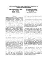
Fig. 1. Hypothetical model for the activation of pro-HGF ⁄ SF and pro-MSP. There may be diverse pathways for the activation of these multi-
functional growth factors. One pathway is mediated by HGFA that is activated in injured tissues and tumors. The second pathway is medi-
ated by membrane-bound serine proteases (cell surface activators), such as matriptase and hepsin. Pericellular activities of these proteases
are regulated by membrane-bound HAI-1 and HAI-2, and the serum activity of HGFA is regulated by PCI in humans. Although the activities
of other pro-HGF ⁄ SF activators, such as coagulation factors XII and XI, plasma kallikrein, and plasminogen activators, are very weak, they
may be stimulated by a certain microenvironment. The active forms of HGF ⁄ SF and MSP induce signaling through MET and RON, respec-
tively, expressed on the surface of tumor cells, epithelial cells, and ⁄ or endothelial cells, resulting in tumor progression, epithelial restitution
and regeneration, and angiogenesis.
HGFA: pathophysiological functions in vivo H. Kataoka and M. Kawaguchi
2234 FEBS Journal 277 (2010) 2230–2237 ª 2010 The Authors Journal compilation ª 2010 FEBS
Conclusions
Considering the multifunctional aspects of HGF ⁄ SF
and MSP [1,27], their processing and activation in the
extracellular milieu represent a tightly controlled phe-
nomenon in vivo, including both positive and negative
inputs. To date, the significance of the extracellular
proteolytic processing and activation of growth
factors ⁄ cytokines may have been underestimated. Both
HGF ⁄ SF and MSP are examples of growth
factors ⁄ cytokines whose activities are regulated by the
specific extracellular proteases. In this minireview, we
have summarized evidence regarding the roles of
HGFA in the activation of pro-HGF ⁄ SF and pro-
MSP in injured tissues and tumors, and discussed its
impact on subsequent tissue repair and tumor progres-
sion. The structural similarities of HGFA and its sub-
strates to coagulation factor XII and plasminogen,
respectively, suggest that this system has a similar evo-
lutionary nature to other plasma systems, such as
blood coagulation and fibrinolysis. Our hypothetical
model for the molecular interactions and cascades in
the activation of HGF ⁄ SF and MSP is shown in
Fig. 1. There is clearly a need for further studies on
the pathophysiological functions of HGFA in vivo. For
example, taking into account the substantial concentra-
tion of pro-HGFA in human plasma, it may be possi-
ble that other unknown unique substrates still exist in
pathological settings in vivo. Furthermore, the clinical
relevance of HGFA is an important matter to be eval-
uated in the future. Efficient activation of the pre-exist-
ing pericellular pro-HGF ⁄ SF and ⁄ or pro-MSP by the
administration of recombinant HGFA may be benefi-
cial for accelerated survival and regeneration of paren-
chymal cells, and thus may have relevance in therapies
for disorders with refractory wounds and ⁄ or persistent
inflammation. On the other hand, the deregulated
activities of HGFA and other HGF ⁄ SF-activating and
MSP-activating enzymes in tumor tissue may be thera-
peutic targets for some types of cancer.
References
1 Matsumoto K & Nakamura T (2006) Hepatocyte
growth factor and the Met system as a mediator of
tumor–stromal interactions. Int J Cancer 119, 477–483.
2 Kataoka H, Miyata S, Uchinokura S & Itoh H (2003)
Roles of hepatocyte growth factor (HGF) activator and
HGF activator inhibitor in the pericellular activation of
HGF ⁄ scatter factor. Cancer Metastasis Rev 22, 223–236.
3 Szabo R & Bugge TH (2008) Type II transmembrane
serine proteases in development and disease. Int J
Biochem Cell Biol 40, 1297–1316.
4 Shimomura T, Ochiai M, Kondo J & Morimoto Y
(1992) A novel protease obtained from FBS-containing
culture supernatant, that processes single chain form
hepatocyte growth factor to two chain form in serum-
free culture. Cytotechnology 8, 219–229.
5 Miyazawa K, Shimomura T, Kitamura A, Kondo J,
Morimoto Y & Kitamura N (1993) Molecular cloning
and sequence analysis of the cDNA for a human serine
protease reponsible for activation of hepatocyte growth
factor. Structural similarity of the protease precursor to
blood coagulation factor XII. J Biol Chem 268, 10024–
10028.
6 Kitajima Y, Ide T, Ohtsuka T & Miyazaki K (2008)
Induction of hepatocyte growth factor activator gene
expression under hypoxia activates the hepatocyte
growth factor ⁄ c-Met system via hypoxia inducible fac-
tor-1 in pancreatic cancer. Cancer Sci 99, 1341–1347.
7 Itoh H, Hamasuna R, Kataoka H, Yamauchi M,
Miyazawa K, Kitamura N & Koono M (2000) Mouse
hepatocyte growth factor activator gene: its expression
not only in the liver but also in the gastrointestinal
tract. Biochim Biophys Acta 1491, 295–302.
8 Kataoka H, Shimomura T, Kawaguchi T, Hamasuna
R, Itoh H, Kitamura N, Miyazawa K & Koono M
(2000) Hepatocyte growth factor activator inhibitor
type 1 is a specific cell surface binding protein of hepa-
tocyte growth factor activator (HGFA) and regulates
HGFA activity in the pericellular microenvironment.
J Biol Chem 275, 40453–40462.
9 Eigenbrot C, Ganesan R & Kirchhofer D (2010)
Hepatocyte growth factor activator (HGFA): molecular
structure and interactions with HAI-1. FEBS J 277,
2215–2222.
10 Suzuki K (2010) Hepatocyte growth factor activator
(HGFA): its regulation by protein C inhibitor. FEBS J
277, 2223–2229.
11 Shimomura T, Kondo J, Ochiai M, Naka D, Miyazawa
K, Morimoto Y & Kitamura N (1993) Activation of
the zymogen of hepatocyte growth factor activator by
thrombin. J Biol Chem 268, 22927–22932.
12 Nagakawa O, Yamagishi T, Fujiuchi Y, Junicho A,
Akashi T, Nagaike K & Fuse H (2005) Serum
hepatocyte growth factor activator (HGFA) in benign
prostatic hyperplasia and prostate cancer. Eur Urol 48,
686–690.
13 Wader KF, Fagerli UM, Holt RU, Stordal B, Borset
M, Sundan A & Waage A (2008) Elevated serum
concentrations of activated hepatocyte growth factor
activator in patients with multiple myeloma. Eur J
Haematol 81, 380–383.
14 van Adelsberg J, Sehgal S, Kukes A, Brady C, Barasch
J, Yang J & Huan Y (2001) Activation of hepatocyte
growth factor (HGF) by endogenous HGF activator is
required for metanephric kidney morphogenesis in vitro.
J Biol Chem 276, 15099–15106.
H. Kataoka and M. Kawaguchi HGFA: pathophysiological functions in vivo
FEBS Journal 277 (2010) 2230–2237 ª 2010 The Authors Journal compilation ª 2010 FEBS 2235
15 Hayashi T, Abe K, Sakurai M & Itoyama Y (1998)
Induction of hepatocyte growth factor and its activator
in rat brain with permanent middle cerebral artery
occlusion. Brain Res 799, 311–316.
16 Nagashima M, Hasegawa J, Kato K, Yamazaki J,
Nishigai K, Ishiwata T, Asano G & Yoshino S (2001)
Hepatocyte growth factor (HGF), HGF activator, and
c-Met in synovial tissues in rheumatoid arthritis and
osteoarthritis. J Rheumatol 28, 1772–1778.
17 Lee YR, Yamazaki M, Mitsui S, Tsuboi R & Ogawa H
(2001) Hepatocyte growth factor (HGF) activator
expressed in hair follicles is involved in in vitro HGF-
dependent hair follicle elongation. J Dermatol Sci 25,
156–163.
18 Moriyama T, Kataoka H, Tsubouchi H & Koono M
(1995) Concomitant expression of hepatocyte growth
factor (HGF), HGF activator and c-met genes in
human glioma cells in vitro. FEBS Lett 372, 78–82.
19 Kataoka H, Hamasuna R, Itoh H, Kitamura N &
Koono M (2000) Activation of hepatocyte growth fac-
tor ⁄ scatter factor in colorectal carcinoma. Cancer Res
60, 6148–6159.
20 Tjin EP, Derksen PW, Kataoka H, Spaargaren M &
Pals ST (2004) Multiple myeloma cells catalyze hepa-
tocyte growth factor (HGF) activation by secreting
the serine protease HGF-activator. Blood 104, 2172–
2175.
21 Tjin EP, Groen RW, Vogelzang I, Derksen PW, Klok
MD, Meijer HP, van Eeden S, Pals ST & Spaargaren
M (2006) Functional analysis of HGF ⁄ MET signaling
and aberrant HGF-activator expression in diffuse large
B-cell lymphoma. Blood 107, 760–768.
22 Su W, Gutmann DH, Perry A, Abounader R, Laterra J
& Sherman LS (2004) CD44-independent hepatocyte
growth factor ⁄ c-Met autocrine loop promotes malig-
nant peripheral nerve sheath tumor cell invasion
in vitro. Glia 45, 297–306.
23 Parr C & Jiang WG (2001) Expression of hepatocyte
growth factor ⁄ scatter factor, its activator, inhibitors
and the c-Met receptor in human cancer cells. Int J
Oncol 19, 857–863.
24 Miyazawa K, Shimomura T & Kitamura N (1996)
Activation of hepatocyte growth factor in the injured
tissues is mediated by hepatocyte growth factor activa-
tor. J Biol Chem 271, 3615–3618.
25 Mukai S, Fukushima T, Naka D, Tanaka H, Osada Y
& Kataoka H (2008) Activation of hepatocyte growth
factor activator zymogen (pro-HGFA) by human kallik-
rein 1-related peptidases. FEBS J 275, 1003–1017.
26 Kawaguchi M, Orikawa H, Baba T, Fukushima T &
Kataoka H (2009) Hepatocyte growth factor activator
is a serum activator of single-chain precursor macro-
phage-stimulating protein. FEBS J 276, 3481–3490.
27 Wang MH, Zhou YQ & Chen YQ (2002) Macrophage-
stimulating protein and RON receptor tyrosine kinase:
potential regulators of macrophage inflammatory
activities. Scand J Immunol 56, 545–553.
28 Matsubara Y, Ichinose M, Yahagi N, Tsukada S, Oka
M, Miki K, Kimura S, Omata M, Shiokawa K,
Kitamura N et al. (1998) Hepatocyte growth factor
activator: a possible regulator of morphogenesis during
fetal development of the rat gastrointestinal tract.
Biochem Biophys Res Commun 253, 477–484.
29 Tjin EP, Bende RJ, Derksen PW, van Huijstee AP,
Kataoka H, Spaargaren M & Pals ST (2005) Follicular
dendritic cells catalyze hepatocyte growth factor (HGF)
activation in the germinal center microenvironment by
secreting the serine protease HGF activator. J Immunol
175, 2807–2813.
30 Itoh H, Naganuma S, Takeda N, Miyata S, Uchinok-
ura S, Fukushima T, Uchiyama S, Tanaka H, Nagaike
K, Shimomura T et al. (2004) Regeneration of injured
intestinal mucosa is impaired in hepatocyte growth
factor activator-deficient mice. Gastroenterology 127,
1423–1435.
31 Miyazawa K (2010) Hepatocyte growth factor activator
(HGFA): a serine protease that links tissue injury to
activation of hepatocyte growth factor. FEBS J 277,
2208–2214.
32 Tang W, Miyazawa K & Kitamura N (1995) Hepato-
cyte growth factor remains as an inactive single chain
after partial hepatectomy or unilateral nephrectomy.
FEBS Lett 362, 220–224.
33 Kaibori M, Inoue T, Oda M, Naka D, Kawaguchi T,
Kitamura N, Miyazawa K, Kwon AH, Kamiyama Y &
Okumura T (2002) Exogenously administered HGF
activator augments liver regeneration through the
production of biologically active HGF. Biochem
Biophys Res Commun 290, 475–481.
34 Nagai M, Takahashi N, Miyazawa K, Kawaguchi M,
Chijiiwa K & Kataoka H (2008) Activation of MET
receptor tyrosine kinase in ulcer surface epithelial cells
undergoing restitution. Pathol Int 58, 462–464.
35 Marchand-Adam S, Fabre A, Mailleux AA, Marchal
J, Quesnel C, Kataoka H, Aubier M, Dehoux M,
Soler P & Crestani B (2006) Defect of pro-hepatocyte
growth factor activation by fibroblasts in idiopathic
pulmonary fibrosis. Am J Respir Crit Care Med 174,
58–66.
36 Phin S, Marchand-Adam S, Fabre A, Marchal-Somme
J, Bantsimba-Malanda C, Kataoka H, Soler P & Cre-
stani B (2010) Imbalance in the pro-HGF activation
system in bleomycin-induced lung fibrosis in mice. Am J
Respir Cell Mol Biol, 42, 288–293.
37 Olivero M, Rizzo M, Madeddu R, Casadio C, Pennac-
chietti S, Nicotra MR, Prat M, Maggi G, Arena N,
Natali PG et al. (1996) Overexpression and activation
of hepatocyte growth factor ⁄ scatter factor in human
non-small-cell lung carcinomas. Br J Cancer 74, 1862–
1868.
HGFA: pathophysiological functions in vivo H. Kataoka and M. Kawaguchi
2236 FEBS Journal 277 (2010) 2230–2237 ª 2010 The Authors Journal compilation ª 2010 FEBS
38 Uchinokura S, Miyata S, Fukushima T, Itoh H, Nak-
ano S, Wakisaka S & Kataoka H (2006) Role of hepa-
tocyte growth factor activator (HGF activator) in
invasive growth of human glioblastoma cells in vivo.
Int J Cancer 118, 583–592.
39 Parr C & Jiang WG (2006) Hepatocyte growth factor
activation inhibitors (HAI-1 and HAI-2) regulate HGF-
induced invasion of human breast cancer cells. Int J
Cancer 119, 1176–1183.
40 Morris MR, Gentle D, Abdulrahman M, Maina EN,
Gupta K, Banks RE, Wiesener MS, Kishida T, Yao M,
Teh B et al. (2005) Tumor suppressor activity and
epigenetic inactivation of hepatocyte growth factor acti-
vator inhibitor type 2 ⁄ SPINT2 in papillary and clear
cell renal cell carcinoma. Cancer Res 65, 4598–4606.
41 Nakamura K, Abarzua F, Hongo A, Kodama J, Nasu
Y, Kumon H & Hiramatsu Y (2009) Hepatocyte
growth factor activator inhibitor-2 (HAI-2) is a favor-
able prognosis marker and inhibits cell growth through
the apoptotic pathway in cervical cancer. Ann Oncol 20,
63–70.
42 Hamasuna R, Kataoka H, Meng JY, Itoh H, Moriyama
T, Wakisaka S & Koono M (2001) Reduced expression
of hepatocyte growth factor activator inhibitor type-
2 ⁄ placental bikunin (HAI-2 ⁄ PB) in human glioblasto-
mas: implication for anti-invasive role of HAI-2 ⁄ PB in
glioblastoma cells. Int J Cancer 93, 339–345.
43 Kongkham PN, Northcott PA, Ra YS, Nakahara Y,
Mainprize TG, Croul SE, Smith CA, Taylor MD &
Rutka JT (2008) An epigenetic genome-wide screen
identifies SPINT2 as a novel tumor suppressor gene in
pediatric medulloblastoma. Cancer Res 68, 9945–9953.
44 Fukai K, Yokosuka O, Chiba T, Hirasawa Y, Tada M,
Imazeki F, Kataoka H & Saisho H (2003) Hepatocyte
growth factor activator inhibitor 2 ⁄ placental bikunin
(HAI-2 ⁄ PB) gene is frequently hypermethylated in
human hepatocellular carcinoma. Cancer Res 63, 8674–
8679.
45 Yasuda K, Nagakawa O, Akashi T, Fujiuchi Y,
Koizumi K, Komiya A, Saiki I & Fuse H (2009) Serum
active hepatocyte growth factor (AHGF) in benign
prostatic disease and prostate cancer. Prostate 69, 346–
351.
46 Parr C, Watkins G, Mansel RE & Jiang WG (2004)
The hepatocyte growth factor regulatory factors in
human breast cancer. Clin Cancer Res 10, 202–211.
H. Kataoka and M. Kawaguchi HGFA: pathophysiological functions in vivo
FEBS Journal 277 (2010) 2230–2237 ª 2010 The Authors Journal compilation ª 2010 FEBS 2237