Báo cáo khoa học: Nuclear factor TDP-43 can affect selected microRNA levels pptx
Bạn đang xem bản rút gọn của tài liệu. Xem và tải ngay bản đầy đủ của tài liệu tại đây (823.19 KB, 14 trang )
Nuclear factor TDP-43 can affect selected microRNA
levels
Emanuele Buratti
1
, Laura De Conti
1
, Cristiana Stuani
1
, Maurizio Romano
2
, Marco Baralle
1
and
Francisco Baralle
1
1 International Centre for Genetic Engineering and Biotechnology (ICGEB), Trieste, Italy
2 Department of Life Sciences, University of Trieste, Italy
Introduction
TDP-43 is a protein belonging to the hnRNP class
of nuclear factors that has been described to play a
role in a variety of cellular processes, including gene
transcription, pre-mRNA splicing and mRNA stabil-
ity [1]. Recently, it has been identified as the major
protein component of neuronal inclusions in neurode-
generative diseases such as frontotemporal dementias
and amyotrophic lateral sclerosis [2]. The impact of
TDP-43 in the neurodegeneration field has been so
pervasive that disease nomenclature consensus is cur-
rently being modified to reflect the new clinical and
pathological findings originating from recent research
better [3,4]. This finding has promoted studies to
characterize better the functional role(s) played by
this protein inside the cell. As a result, apart from its
historical involvement in splicing and transcription
[5–7], several recent observations have successfully
highlighted new biological characteristics of this
protein, such as acting as a neuronal response
activity factor and an in vitro mRNA translational
repressor [8], an mRNA stability factor for neurofila-
ments [9,10] and as a regulator of Rho family
GTPase expression [11] and HDAC6 [12]. All of
these observations may be conducive to under-
standing the potentially pathogenic role of TDP-43 in
neurodegeneration.
Keywords
amyotrophic lateral sclerosis; let-7b;
microRNAs; miR-663; TDP-43
Correspondence
F. E. Baralle, Padriciano 99, 34012 Trieste,
Italy
Fax: +39 040 3757361
Tel: +39 040 3757337
E-mail:
(Received 2 September 2009, revised 26
February 2010, accepted 8 March 2010)
doi:10.1111/j.1742-4658.2010.07643.x
TDP-43 has recently been described as the major component of the inclu-
sions found in the brain of patients with a variety of neurodegenerative dis-
eases, such as frontotemporal lobar degeneration and amyotrophic lateral
sclerosis. TDP-43 is a ubiquitous protein whose specific functions are prob-
ably crucial to establishing its pathogenic role. Apart from its involvement
in transcription, splicing and mRNA stability, TDP-43 has also been
described as a Drosha-associated protein. However, our knowledge of the
role of TDP-43 in the microRNA (miRNA) synthesis pathway is limited to
the association mentioned above. Here we report for the first time which
changes occur in the total miRNA population following TDP-43 knock-
down in culture cells. In particular, we have observed that let-7b and
miR-663 expression levels are down- and upregulated, respectively. Interest-
ingly, both miRNAs are capable of binding directly to TDP-43 in different
positions: within the miRNA sequence itself (let-7b) or in the hairpin pre-
cursor (miR-663). Using microarray data and real-time PCR we have also
identified several candidate transcripts whose expression levels are selec-
tively affected by these TDP-43–miRNA interactions.
Abbreviations
DYRK-1A, dual-specificity tyrosine-(Y)-phosphorylation regulated kinase 1A; EPHX1, epoxide hydrolase; GAPDH, glyceraldehyde-3-phosphate
dehydrogenase; GST, glutathione S-transferase; LAMC1, laminin, gamma 1 (formerly LAMB2); miRNA, microRNA; siRNA, short inhibitory
RNA; STX3, syntaxin 3; VAMP3, vesicle-associated membrane protein 3.
2268 FEBS Journal 277 (2010) 2268–2281 ª ICGEB. Journal compilation ª 2010 FEBS
With regards to the wider biological properties of
TDP-43, a new indication has been provided by its
presence in both the human and the mouse micropro-
cessor complexes, suggesting a potential involvement
in microRNA (miRNA) biogenesis [13,14]. Further
support for a role in miRNA biogenesis for TDP-43
is its localization in perichromatin fibres [15], a
nuclear region specifically associated with this process
[16]. The Drosha nuclear complex is one of the key
enzymes involved in the biogenesis of miRNAs and
has the function of converting pri-miRNA molecules
to 70 nucleotide-long pre-miRNA molecules, which
are then exported to the cytoplasm and further pro-
cessed in mature miRNAs by Dicer [17]. These small
RNA molecules can then bind to their target
mRNAs through sequence complementarity and
affect gene expression by regulating either mRNA
levels or translation [18–21]. Recently, hnRNP pro-
teins were shown to be involved in miRNA process-
ing [22,23]. It was therefore of interest to investigate
the consequences on the cellular miRNA population
of removing TDP-43.
Results
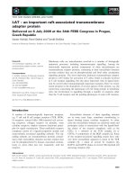
An analysis of Drosha levels by western blot in TDP-
43-depleted Hep-3B cells did not reveal any significant
changes in Drosha migration pattern or signal intensity
with respect to mock-treated cells (Fig. 1A), pointing
to specific miRNA targets for TDP-43. To investigate
this possibility, miRNA profiling in TDP-43-depleted
Hep-3B cells from three independent samples was per-
formed by Exiqon (Vedbaek, Denmark). The micro-
array experiment tested for 607 known and proprietary
miRNA sequences (438 and 169, respectively). In this
triplicate experiment, 146 miRNA sequences could be
detected in our samples and 90 of these miRNA signa-
tures could be quantitatively tested in all three short
interfering RNA (siRNA) and control experiments (a
list of the 67 registered ones can be found in Fig. S1).
The eight miRNAs that were either down- or upregu-
lated in a statistical significant manner following deple-
tion of TDP-43 in Hep-3B cells are shown in Fig. 1B.
For the three most statistically significant miRNAs
(let-7b, miR-663 and miR-744), the results were vali-
dated using the commercial miRvana kit, which is
based on a hybridization procedure with small radioac-
tive probes based on the miRNA of interest (Fig. 1C,
D). In this experiment, the changes in these miRNA
expression levels as detected by the microarray experi-
ment were confirmed in three cell lines: HeLa (adeno-
carcinoma), Hep-3B (hepatocarcinoma) and SH-S-5Y
(neuroblastoma).
As microarray experiments represent an indirect way
of measuring TDP-43 effects on the general miRNA
population, it was not possible, on the basis of these
data alone, to rule out the possibility that a lack of
TDP-43 may have affected the levels or activity of
another factor involved in miRNA processing (for
example, hnRNP A1 or other miRNA processing fac-
tors). Therefore, in order to establish a direct link
between TDP-43 and any of these miRNAs, we
focused on TDP-43 RNA binding properties that have
been previously characterized in our laboratory [24,25].
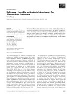
Looking at the miRNA sequences it was interesting
to note that let-7b contained in its sequence a discrete
number of (GU)
n
repeats, the preferred target sequence
of TDP-43 [24] (Fig. 1C). A band shift analysis per-
formed using recombinant GST–TDP-43 confirmed
that both the let-7b and the let-7b hairpin sequence
(Fig. 2A) could bind these sequences (Fig. 2B). Most
interestingly, variations in the levels of both let-7a and
let-7c did not appear to be statistically significant in
the microarray assay (Fig. 2C). By comparing the
let-7a, -7b and -7c sequences (Fig. 2D, upper panel) we
observed that a critical guanosine residue in the let-7b
sequence at position +17 had the effect of creating a
new GU repeat, suggesting that this miRNA could be
particularly sensitive to TDP-43 cellular levels as
opposed to the other let-7 family members. A band
shift experiment using labelled let-7a, -7b and -7c
sequences confirmed that recombinant GST–TDP-43
could only bind the let-7b sequence (Fig. 2D, lower
panel). The critical importance of the +17 residue is
highlighted by the observation that introducing
a + 17a > g substitution in the let-7a sequence can
promote TDP-43 binding (Fig. 2D, lower panel). It
should be noted that the importance of this critical res-
idue has also been confirmed using pulldown analysis
by immobilizing these miRNA sequences on adipic
acid dehydrazide beads and incubating with total
HeLa nuclear extracts. The results of this experiment
confirmed that introducing a + 17a > g nucleotide in
the let-7a sequence gave it the ability to bind TDP-43,
even in the presence of all other nuclear competing
proteins (Fig. S2).
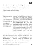
We also examined the sequences of all the other
miRNAs, and noted that within the sequence of the
miR-663 precursor (the second most statistically
affected miRNA after let-7b) there was an almost per-
fect GU repeated sequence localized in the apical por-
tion of the hairpin (Fig. 3A). A band shift analysis
with recombinant TDP-43 confirmed binding to the
precursor hairpin, but not to the miR-663 sequence
itself (Fig. 3B, left and central panels, respectively).
Deletion of the GU-rich sequence in the hairpin also
E. Buratti et al. TDP-43 and miRNA regulation
FEBS Journal 277 (2010) 2268–2281 ª ICGEB. Journal compilation ª 2010 FEBS 2269
abolished TDP-43 binding (Fig. 3B, right panel).
Finally, neither the miR-744 sequence and its hairpin
(Fig. S3) nor all the other identified miRNA sequences
could bind TDP-43 in band shift analyses (data not
shown). These data are consistent with the observation
that the sequence of this miRNA does not contain a
sufficient number of (ug)
n
repeats.
From a TDP-43–miRNA interaction point of view,
these results also suggest that there may be several
other potential miRNA targets of TDP-43 that could
not be detected in our analysis because they were not
expressed at sufficient levels (or at all) in Hep-3B cells.
In order to obtain some indication in this regard, we
examined the primary sequence of all known miRNAs
present in miRBase for GU-repeated regions. This
analysis identified two other miRNAs that could
potentially bind TDP-43: miR-574-5p in the miRNA
sequence itself (Fig. 3C) and miR-558 in the hairpin
element (Fig. 4A). Nothing is known regarding the
expression profile or importance of these miRNAs,
with the exception of miR-558, which has been
described to be transiently upregulated in fibroblasts
Drosha
Mock siRNA
kDa
175
TDP-43
siRNA
+ –
siRNA
+ –
siRNA
+ –
TDP-43
47.5
175
TDP 43
Tubulin
HeLa Hep-3B SH-SY-5Y
175
p
siRNA
siRNA
Let-7b
+–p
+–p
+–p
siRNA
siRNA
siRNA
siRNA
siRNA
siRNA
+–p
+–p
+–p
+–p
+–p
+–p
siRNA
Coomassie
47.5
miR-663
miR-744
HeLa Hep-3B SH-SY-5Y
HeLa Hep-3B SH-SY-5Y
HeLa Hep-3B SH-SY-5Y
Statistical
significance
(T-test)
let-7b 0.0039
0.0069
miR
-
629 0.019
0.017
0.0053
0.032
0.039
#3 #1 #2 #2 #1 #3
siRNA Mock
Down-regulated
following TDP-43
miR 629
miR-23a
miR-744
miR-373*
miR-663
miR-572
depletion
Up-regulated
following TDP-43
depletion
–2.0 –1.0 0 1.0 2.0
B
AC
D
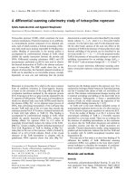
Fig. 1. Effect of TDP-43 depletion on Drosha and selected miRNA expression levels in Hep-3B cells. (A) Western blot assay of Hep-3B cells
treated with a control siRNA (mock) and a specific TDP-43 siRNA (siRNA). The protein extracts were normalized by Coomassie intensity
(lower panel) and hybridized with a polyclonal antibody against TDP-43 and a rabbit polyclonal antibody against Drosha. (B) Heat map
showing all the miRNAs (P < 0.05) differentially expressed in TDP-43-depleted Hep-3B cells with respect to mock-siRNA-treated cells. The
blue labels indicate downregulated miRNAs, the red labels indicate upregulated ones. The clustering is reported as log2(Hy3 ⁄ Hy5) ratios. (C)
TDP-43 knockdown levels achieved in three cell lines: HeLa, Hep-3B and SH-SY-5Y cells. (D) Quantification of let-7b, miR-663 and miR-744
expression levels in HeLa, Hep-3B and SH-SY-5Y cell lines using the commercial miRvana kit. Undigested probe (p).
TDP-43 and miRNA regulation E. Buratti et al.
2270 FEBS Journal 277 (2010) 2268–2281 ª ICGEB. Journal compilation ª 2010 FEBS
following high doses of radiation [26]. Band shift
assays confirmed that TDP-43 could bind efficiently to
the miR-574-5p sequence (Fig. 3D, left) but, unlike let-
7b, could not bind anymore to the miR-574-5p
sequence when it was embedded in the RNA second-
ary structure (compare Figs 2B and 3D, right). The
reason for this probably resides in the inability of
TDP-43 to compete for RNA secondary structure
formation in the miR-574-5p sequence. This structure,
in fact, is more extended and GC-rich than the
corresponding let-7b structure element. As expected,
TDP-43 could bind to the miR-558 hairpin sequence,
but not to the miR-558 miRNA (Fig. 4B).
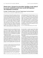
In order to confirm the functional significance of the
TDP-43 let-7b ⁄ miR-663 interactions we then used a
heterologous assay based on a luciferase reporter. Four
complementary target sequences for let-7b and
miR-663 were subcloned in the pGL3 vector, to obtain
pGL3-mir-let-7b and pGL3-mir-663 (Fig. 4C). Both
constructs, together with a pRL-TK Renilla luciferase
vector, were transiently transfected into Hep-3B cells
and assayed for luciferase activity in both the presence
(mock) or the absence (siRNA) of TDP-43 according
to the manufacturer’s instructions. The results were
normalized according to the firefly ⁄ Renilla luciferase
ratios obtained in each sample. As expected, no signifi-
cant difference could be detected in the firefly ⁄ Renilla
ratios of the pGL3 empty vector following knockdown
of TDP-43 in Hep-3B cells (Fig. 4D, left). However, a
significant increase in reporter gene activity was
observed following transfection of the pGL3-mir-let-7b
sequence following TDP-43 knockdown (Fig. 4D,
let-7a
let-7b
let-7c
let-7a+17a>g
let-7a
let-7b
let-7c
Statistical
significance
(T-test)
#1 #2 #3 #1 #2 #3
siRNA Mock
*let-7d-7e-7f-7g-7i - No detectable levels
let-7
family*
let-7b stem loop:
let-7b:
let-7b
let-7b
stem-loop
–1.0 –0.5 0 0.5 1.0
let-7b let-7a
let-7a
+17a>g let-7c
AC
BD
––––
Fig. 2. Specific interaction of TDP-43 with let-7b. (A) Schematic diagram of the let-7b miRNA sequence and of its precursor hairpin. (B) Band
shift assay with recombinant GST–TDP-43 using the labelled let-7b sequence itself (left) and the let-7b hairpin element (right). (C) Heat map
profile for all detected members of the let-7 family found in our assay, together with their statistical significance. (D) The upper panel shows
the sequence comparison (the GU dinucleotides are highlighted in bold), the lower panel shows a band shift analysis of labelled let-7a,
let-7a+17a>g, let-7b and -7c miRNA sequences incubated with recombinant GST–TDP-43.
E. Buratti et al. TDP-43 and miRNA regulation
FEBS Journal 277 (2010) 2268–2281 ª ICGEB. Journal compilation ª 2010 FEBS 2271
centre). This is the result that should have been
expected if depletion of TDP-43 was associated with
lower expression levels of let-7b (as this would have
meant lower translational inhibition on the pGL3-mir-
let-7b construct). Exactly the opposite effect was
observed when we transfected the pGL3-mir-663
construct in depleted or control cells (Fig. 4D, right).
Also, this result was completely consistent with
increased miR-663 expression following TDP-43
depletion, as such an outcome would have caused a
higher translational inhibition on the pGL3-mir-663
construct. One important issue that should be
mentioned is the fact that these two GU-rich regions
in the let-7b miRNA and miR-663 do not exactly
match the optimal TDP-43 binding consensus
represented by perfect GU-repeated sequences and this
may well explain why in both cases TDP-43 has only
modulating effects on their expression rather than an
all or nothing phenomena.
Most importantly, it was interesting to determine
the potential consequences of these changes in terms of
cellular transcript alterations. It was originally
thought, in fact, that miRNA-mediated regulation was
mainly at the level of translation and not at the level
of mRNA degradation. It is now clear, however, that
this view is only partially correct and that, depending
on a variety of factors still only partially understood,
many miRNA targets are regulated by degradation (as
recently reviewed by Nilsen [20]). This has enabled the
identification of miRNA targets by mRNA microarray
analysis but, of course, it still remains very difficult to
determine the proportion of mRNA targets affected in
this way as opposed to strictly translation regulatory
pathways (at least until large-scale proteomic
approaches reach the level of sensitivity now available
for mRNA microarray approaches).
Keeping in mind these limitations, we took advan-
tage of our previously determined microarray evalua-
tion of the cellular transcripts that were either down-
or upregulated following TDP-43 knockdown in HeLa
cells [27]. These transcripts (a total of 786) were com-
pared with a set of transcripts (numbering 838) that
have been observed to be downregulated following
let-7b overexpression in a culture of primary human
fibroblasts and which contained a let-7b seed target
region in their 3¢ UTRs [28]. The 23 common hits
miR-663:
miR-663 stem loop:
miR-663 miR-663
Stem loop
miR-663
Stem loop
delta-UG
miR-574-5p:
miR-574-5p stem-loop:
miR-574-5p miR-574-5p
Stem-loop
AC
B
D
Fig. 3. Interaction of TDP-43 with miR-663 and functional analysis. (A) Potential TDP-43 binding site to the miR-663 precursor hairpin
element (highlighted in bold). (B) Band shift assay with recombinant GST–TDP-43 using the labelled miR-663 sequence itself (left), the
miR-663 hairpin element (middle) and a miR-663 gucugugu-deleted sequence (right). (C) Potential TDP-43 binding site to the miR-574-5p
sequence and the sequence of its precursor hairpin element. (D) Band shift assay with recombinant GST–TDP-43 using the labelled
miR-574-5p sequence itself (left) and the miR-574-5p hairpin element (right).
TDP-43 and miRNA regulation E. Buratti et al.
2272 FEBS Journal 277 (2010) 2268–2281 ª ICGEB. Journal compilation ª 2010 FEBS
between the two lists are reported in Table 1. First of
all, it should be noted that in the microarray experi-
ment, 16 of the 23 hits were upregulated following
TDP-43 removal. This situation was therefore largely
consistent with the downregulatory effect on let-7b
expression levels following TDP-43 removal (Fig. 1B).
More interestingly, among the most upregulated tran-
scripts were several with a potentially important
function in neuronal and synapse development: the
dual-specificity tyrosine-(Y)-phosphorylation regulated
kinase 1A (DYRK-1A), syntaxin 3 (STX3), the vesicle-
associated membrane protein 3 (cellubrevin; VAMP3)
and laminin, gamma 1 (LAMC1, formerly LAMB2).
Interestingly, this list also contained the enzyme cyclin-
dependent kinase 6, which we previously found to be
upregulated following TDP-43 removal [27]. Upregula-
tion of these transcripts was confirmed by real-time
PCR (Figs 5A, 6A) using six independent siRNA
knockdown and siRNA control batches. The results
showed that all these transcripts were significantly up-
regulated from a minimum of 1.7- to 3-fold following
TDP-43 removal (Fig. 5A). In parallel to this analysis
we wanted to rule out the possibility that upregulation
of these transcripts could be due to changes in their
mRNA splicing profiles owing to the presence of sev-
eral putative TDP-43 binding sites in their intronic ele-
ments (Fig. 5B). Normal RT-PCR analysis of the
coding regions, however, also ruled out this possibility
by showing that the splicing profile of these transcripts
did not specifically change following TDP-43 removal
(Fig. 5C).
In the case of miR-663, no data are currently avail-
able regarding the variation in cellular transcripts fol-
lowing its overexpression ⁄ removal. In order to find an
alternative solution, our list of microarray targets fol-
lowing TDP-43 removal was compared with a list of
more than 1000 putative miR-663 targets obtained
using the miranda software and downloaded from
miRBase ( Only three
putative common transcripts were identified through
this comparison (Table 2). It can be seen that in this
reduced sample obtained by indirect methods we had
two cases that showed the expected decrease in tran-
script levels that could follow miR-633 increase due to
Fir.Luc.
pGL3AAAAA
SV40
promoter
SV40 polyA
Fir.Luc.
pGL3-mir-let-7b
AAAAA
XbaI
Fir.Luc.
pGL3-mir-663
AAAAA
pGL3 pGL3-mir-let-7b
0.2
0.4
0.6
0.8
1.0
pGL3-mir-663
1.2
0.2
0.4
0.6
0.8
1.0
1.2
0.5
1.0
1.5
2.0
2.5
3.0
miR-558:
miR-558 stem-loop:
miR-558
miR-558
Stem-loop
CA
DB
Fig. 4. Interaction of TDP-43 with miR-558 and miR-574-5p. (A) Potential TDP-43 binding site to the miR-558 sequence and the sequence
precursor hairpin element. (B) Band shift assay with recombinant GST–TDP-43 using the labelled miR-558 sequence itself (left) and the miR-
558 hairpin element (right). (C) Schematic diagrams of the constructs pGL3, pGL3-mir-let-7b and pGL3-mir-663. Each construct contained
four copies of the complementary target sequence of let-7b and miR-663, respectively. (D) Results of a luciferase assay performed on TDP-
43-depleted and mock-depleted Hep-3B cells following transfection of these constructs. In this type of experiment, the level of the interac-
tion between the endogenous let-7b and miR-663 and the expression vector determined the levels of luciferase expression. Transfection
efficiencies were normalized using the Renilla luciferase internal control. Standard deviation values from three independent experiments are
indicated.
E. Buratti et al. TDP-43 and miRNA regulation
FEBS Journal 277 (2010) 2268–2281 ª ICGEB. Journal compilation ª 2010 FEBS 2273
TDP-43 depletion. We have analysed in more detail
the enzyme epoxide hydrolase (EPHX1) because of its
putative role as an antagonist of oxidative stress [29].
The decrease in EPHX1 levels was confirmed by real-
time PCR (Fig. 6A) and RT-PCR ruled out any effect
of TDP-43 removal on the splicing process of this
enzyme (Fig. 6B–C).
Finally, we also began to investigate the regulatory
pathways that may be controlled by TDP-43. At least
for TDP-43, we decided to measure the pre-miRNA
levels in TDP-43-depleted and mock-depleted cells.
For this reason, we measured the levels of pri-let-7b
miRNAs according to established protocols [30]. As
shown in Fig. 6D, upper panel, following TDP-43
removal, the levels of pri-let-7b were significantly
increased to a level that was comparable with the loss
of mature let-7b miRNA within the cell. Moreover,
these changes were statistically significant. These
results demonstrate that TDP-43 actively participates
in the Drosha processing mechanisms and its absence
in the case of let-7b leads to a block in the maturation
of pri-let-7b miRNA. Finally, we also measured the
levels of pri-miR-663 using a similar procedure. In this
case, however, the difference in miR-663 precursor
levels did not reach statistical significance (Fig. 6D,
lower panel).
Discussion
The biological function of TDP-43 in the eukaryotic cell
is far from being fully understood. Even more obscure
is its role in the pathogenesis of amyotrophic lateral
sclerosis ⁄ frontotemporal lobar degeneration and other
neurodegenerative diseases. In particular, several gain-
or loss-of-function mechanisms have been put forward
in recent times. The gain-of-function mechanisms focus
on the generation of potentially toxic C-terminal frag-
ments [31–33], its toxicity in a yeast cellular model [34]
and increased aggregation properties in the presence of
missense mutations in the C-terminal region [35]. On
the other hand, loss-of-function mechanisms are sup-
ported by indications that TDP-43 may be playing a
fundamental role in a variety of nuclear processes, such
as splicing regulation [5], transcription [36], chromatin
organization [37] and a variety of other processes, such
as cell death and nuclear shape [27]. Loss-of-function
mechanisms are also supported by a recent Drosophila
animal model that has shown that removal of the fly
homologue of TDP-43 can recapitulate several features
of motoneuron disease [38]. These two different patho-
physiological mechanisms are not mutually exclusive
and may indeed take place at the same time, although
determining their relative importance may be especially
Table 1. List of altered cellular transcripts in TDP-43 knockdown experiments that have also been found to be downregulated following
let-7b overexpression.
Gene Accession number Full name Microarray variation
a
ADRB2 NM_000024 Adrenergic, beta-2-, receptor, surface +1.6
IGFBP3 NM_000598 Insulin-like growth factor binding protein 3 +2.3
IL6 NM_000600 Interleukin 6 (interferon, beta 2) )1.1
IGF1R NM_000875 Insulin-like growth factor 1 receptor +1.2
CDK6 NM_001259 Cyclin-dependent kinase 6 +10.0
DAB2 NM_001343 Disabled homolog 2, mitogen-responsive phosphoprotein (Dros.) +1.6
DYRK1A NM_001396 Dual-specificity tyrosine-(Y)-phosphorylation regulated kinase 1A +4.6
CSNK1E NM_001894 Casein kinase 1, epsilon )1.5
TNPO1 NM_002270 Transportin 1 +1.1
LAMC1 NM_002293 Laminin, gamma 1 (formerly LAMB2) +3.1
STX3 NM_004177 Syntaxin 3 +10.5
CALD1 NM_004342 Caldesmon 1 +1.1
VAMP3 NM_004781 Vesicle-associated membrane protein 3 (cellubrevin) +1.4
SMC1A NM_006306 Structural maintenance of chromosomes 1A )1.2
CAP2 NM_006366 CAP, adenylate cyclase-associated protein, 2 (yeast) +1.3
KIAA0152 NM_014730 KIAA0152 )1.4
PHF16 NM_014735 PHD finger protein 16 +1.1
RHOBTB3 NM_014899 Rho-related BTB domain containing 3 +1.5
HSD17B11 NM_016245 Hydroxysteroid (17-beta) dehydrogenase 11 )2.6
TOB2 NM_016272 Transducer of ERBB2, 2 +1.8
CDV3 NM_017548 CDV3 homolog (mouse) +1.5
SLC5A6 NM_021095 Solute carrier family 5 (sodium-dependent vitamin transp.), mem 6 )3.2
ZC3H12A NM_025079 Zinc finger CCCH-type containing 12A )1.2
a
Fold expression difference according to Ayala et al. [27].
TDP-43 and miRNA regulation E. Buratti et al.
2274 FEBS Journal 277 (2010) 2268–2281 ª ICGEB. Journal compilation ª 2010 FEBS
important with regards to planning and developing suc-
cessful therapeutic strategies.
To understand these pathological processes better, it
is of course important to define TDP-43 functional
properties as much as possible. In this regard, the
effects of TDP-43 on the miRNA population are par-
ticularly interesting, considering previous observation
that TDP-43 itself is a minor component of the
Drosha enzyme complex [13] and the increasing role
played by aberrant miRNA expression in a variety of
neurodegenerative diseases, as recently reviewed in sev-
eral publications [39–43].
However, to date no studies are yet available regard-
ing the potential role played by TDP-43 in miRNA
processing. In general, Drosha-associated factors are
required to help or inhibit the processing of particular
subsets of miRNA molecules. Indeed, this has been
shown to be the case for the p68 and p72 helicases [14]
and, more recently, for the KH-type splicing regula-
tory protein (KSRP) protein [44]. Of course, this regu-
latory role is not solely confined to Drosha-associated
proteins. Indeed, one of the best characterized example
of miRNA regulatory proteins is represented by Lin-
28, which can regulate let-7 processing [45–48] by
inducing uridylation of its precursor and cause its deg-
radation [49]. In a situation probably more similar to
TDP-43, miRNA regulating properties have also been
described for the well-known splicing factor hnRNP
A1. This protein has been shown to regulate the
expression of miR-18a by binding to the loop of pri-
miR-18a and inducing a relaxation at the stem, creat-
0
0.5
1
1.5
2
2.5
3
0
0.5
1
1.5
2
2.5
0
0.5
1
1.5
2
0
0.5
1
1.5
2
DYRK1A (P < 0.01) STX3 (P < 0.0001) VAMP3 (P < 0.001)
+Mock +siRNA
+Mock +siRNA +Mock +siRNA
Expression levels
SD = 0.07
SD = 0.06
SD = 0.03
SD = 0.1
SD = 0.09
SD = 0.1
DYRK1A (exons 1-13)
DYRK1A (150 kb)
STX3 (50 kb)
VAMP3 (10 kb)
** * * *
*
*
non-coding exons
coding exons
(ug)
6
repeats
****
LAMC1 (exons 1-14) LAMC1 (exons 14-28)
LAMC1 (120 kb)
**
STX3 (exons 1-9) VAMP3 (exons 2-5)
+Mock +siRNA
LAMC1 (P < 0.001)
SD = 0.05
SD = 0.1
A
B
C
Fig. 5. Real-time PCR levels of let-7b regulated transcripts. (A) Real-time PCR quantification analysis of the DYRK1A, LAMC1, STX3 and
VAMP3 transcript levels following TDP-43 knockdown in HeLa cells based on the results of Table 1. Six independent experiments were anal-
ysed and both standard deviations and P-values are shown for each transcript. (B) Schematic diagram of the intron ⁄ exon architecture of
these genes with the presence of potential TDP-43 binding motifs, (ug)
6
, indicated. (C) Standard RT-PCR of each transcript to rule out the
effects of TDP-43 on their RNA splicing process.
Table 2. List of altered cellular transcripts in TDP-43 knockdown
experiments that also represent putative miR-663 targets.
Gene
Accession
number Full name
Microarray
variation
a
EPHX1 NM_000120 Epoxide hydrolase 1 )2.1
CDA NM_001785 Cytidine deaminase +2.6
AAMP NM_001087 Angio-associated,
migratory cell protein
)2.3
a
Fold expression difference according to Ayala et al. [27].
E. Buratti et al. TDP-43 and miRNA regulation
FEBS Journal 277 (2010) 2268–2281 ª ICGEB. Journal compilation ª 2010 FEBS 2275
ing a more favourable cleavage site for Drosha
[22,23,50]. Our results have shown that TDP-43 has
the potential to affect the levels of four miRNAs, let-
7b, miR-663, miR-574-5p and miR-558, by potentially
binding to their sequence and ⁄ or precursor elements
(schematically summarized in Fig. 7). With regards to
the potential importance of the interaction between
TDP-43 and miRs 574-5p ⁄ 558 a cautionary note is
represented by the fact that, owing to the lack of cell
lines expressing these miRNAs, we were unable to
functionally validate them. Therefore, this is an issue
that will have to be addressed in future studies.
We then asked what kind of processing steps in the
biogenesis of these miRNAs may be affected. In the
case of the let-7b family, the data that let-7a, which
originates from the same precursor as let-7b, is not
affected by TDP-43 support that the regulation is
post-transcriptional. In particular, the observation that
TDP-43 depletion leads to an increase in pri-let-7b lev-
els suggests that for this miRNA, TDP-43 helps to
keep ⁄ recruit the pri-miRNA sequences in place during
Drosha processing. In the case of miR-663, we should
consider the fact that for several miRNAs, such as
miR-30 and miR-21, efficient processing is dependent
on the presence of a terminal loop more than 10
nucleotides long [51]. However, the measurement of
miR-663 precursor levels in TDP-43 minus and mock-
depleted cells has failed to find a statistically significant
difference. This suggests that miR-663 regulation by
TDP-43 may take place in steps subsequent to Drosha
cleavage, an observation that may be consistent with
the opposite effect of TDP-43 on miR-663 levels
(upregulated) as opposed to let-7b (downregulated).
The function of these different up- or downregulatory
mechanisms is, of course, still an open question. The
most probable explanation is that there might be
two sets of transcripts whose expression has to be
upregulated (in the case of let-7b) and downregulated
(in the case of miR-663) at the same time to achieve a
functionally specific effect. At the moment, identifying
these hypothetical effects is hampered by our incom-
plete knowledge of TDP-43 general functions and its
expression regulation within the cell (especially in nor-
mal, nonpathological conditions).
With regards to the miRNA we have identified,
nothing is known about the functions of miR-663,
miR-558 and miR-574-5p. On the other hand, the
let-7b family is an abundant, highly conserved family
0
0.2
0.4
0.6
0.8
1
1.2
EPHX1 (P < 0.01)
+Mock +siRNA
Expression levels
SD = 0.08
SD = 0.09
EPHX (20 kb)
non-coding exons
coding exons
(ug)
6
repeats
EPHX (exons 2-9)
0.0
0.5
1.0
1.5
2.0
0.0
0.5
1.0
1.5
2.0
+Mock +siRNA
hsa-let7b precursor levels (P < 0.05)
SD = 0.05
SD = 0.03
+Mock +siRNA
Expression levels
Expression levels
miR-663 precursor levels (P > 0.05)
SD = 0.42
SD = 0.22
A
B
C
D
Fig. 6. Real-time PCR levels of let-7b and
miR-663 regulated transcripts. (A) Real-time
PCR quantification analysis of the EPHX1
transcript levels following TDP-43
knockdown in HeLa cells based on the
results of Table 2. Six independent
experiments were analysed and both
standard deviations and P-values are shown
for each transcript. (B) Schematic diagram
of the intron ⁄ exon architecture of these
genes with the presence of potential
TDP-43 binding motifs indicated. (C)
Standard RT-PCR of each transcript to rule
out the effects of TDP-43 on their RNA
splicing process. (D) Measurement by
real-time PCR of the let-7b and miR-663
precursor levels following TDP-43 depletion
and mock depletion in HeLa cells. Standard
deviations are shown above each bar and
P-values are indicated.
TDP-43 and miRNA regulation E. Buratti et al.
2276 FEBS Journal 277 (2010) 2268–2281 ª ICGEB. Journal compilation ª 2010 FEBS
of miRNAs that are important in cellular differentia-
tion processes and their misregulation may lead to can-
cer formation, as recently reviewed by Roush and
Slack [52]. However, Drosophila let-7 has been
described as being essential for correct neuromuscular
development in the transition from larva to adult [53],
suggesting that members of this family may also par-
ticipate in neuronal and developmental processes.
In keeping with this hypothesis, we provide evidence
that the removal of TDP-43 from the cell nucleus
causes specific downregulation of let-7b, and this can
in turn influence the expression levels of several poten-
tially important transcripts involved in neurodegenera-
tion and synapse formation (Fig. 7). These transcripts
include DYRK1A, a kinase that has been found to be
upregulated in patients affected by Down syndrome
and whose increased expression correlates with the
neuronal defects [54,55]. They also include components
of synapse formation, such as STX3, which is impor-
tant for the growth of neurite processes [56], and
VAMP3, which can functionally substitute for syna-
ptobrevin in synaptic exocytosis [57]. The upregulation
of LAMC1, on the other hand, is particularly interest-
ing in light of previous observations that dysmorphic
nuclear shape phenotypes are produced upon removal
of TDP-43 [27]. Finally, another interesting transcript
that is downregulated following TDP-43 knockdown
(but this time due to miR-663 upregulation) is repre-
sented by the EPHX1 enzyme, a detoxifying enzyme
that functions to regulate oxidative stress and has been
previously shown to be significantly elevated in the
hippocampal region of patients suffering from Alzhei-
mer’s disease [29].
Taken together, these results provide an experimen-
tal basis suggesting that TDP-43 can play a role in
miRNA expression pathways. Of course, how these
changes relate to TDP-43¢s other normal biological
properties (splicing, transcription, mRNA export ⁄
translation) and, most importantly, to an eventual dis-
ease context, will require future analyses. Finally, as
TDP-43 is also a splicing factor, it will also be interest-
ing to explore the potential role of TDP-43 in Drosha-
free miRNA synthesis (miRtrons) [58]. At the moment,
going through the list of miRtron genes recently com-
piled by Berezikov et al. [59], the consensus sequences
of the small introns responsible for miRtron formation
in vertebrates display a G-rich sequence at the 5¢ end
and a U ⁄ C-rich sequence at the 3¢ end. None of these
two sequences contains a number of GU repeats that
may resemble (at least visually) potentially strong
TDP-43 binding sites. However, it is a possibility that
warrants experimental testing in the future.
let-7b
RNA
Pol II
miR-663
RNA
Pol II
miR-574-5p
RNA
Pol II
miR-558
RNA
Pol II
m7G
AAAAA
m7G
AAAAA
m7G
AAAAA
m7G
AAAAA
pri-let-7b
pri-miR-663
pri-miR-574-5p
pri-miR-558
pre-miRNA
miRNA
TDP
-43
Gene(s) potentially
affected in neuro
degeneration:
DYRK1A
STX3
VAMP3
LAMC1
Effect of TDP-43
removal on cellular
concentration of the
miRNA
TDP-43 binding to:
TDP
-43
TDP
-43
TDP
-43
TDP
-43
EPHX1
Fig. 7. Schematic diagram of TDP-43–miRNA interactions. This figure shows a summary of TDP-43 interactions with the various miRNA
sequences and precursors identified in the present study. Moreover, it summarizes the effects of its removal on miRNA expression levels
and on potentially important transcripts for neuronal development or degeneration.
E. Buratti et al. TDP-43 and miRNA regulation
FEBS Journal 277 (2010) 2268–2281 ª ICGEB. Journal compilation ª 2010 FEBS 2277
Materials and methods
Cell culture and siRNA transfection
Hep-3B cells (ATCC, Manassas, VA, USA) were grown in
Dulbecco’s modified Eagle’s medium (Gibco, Rockville,
MD, USA) supplemented with 10% fetal bovine serum (Gib-
co), glutamine, 5% glucose and antibiotic antimytotic
(Sigma, St Louis, MO, USA) in 5% CO
2
at 37 °C. Two
transfections using 0.1 nmol siRNA were carried out at
intervals of 24–48 h and cells were collected after 24 or
48 h from the second transfection. Western blot against
Drosha was performed using a commercial rabbit poly-
clonal antibody (Abcam, Cambridge, MA, USA).
Microarray and direct miRNA analysis
Total RNA from TDP-43 siRNA, control siRNA-treated
(siCONTROL nontargeting siRNA #2) and untreated
Hep-3B cells were obtained using Trizol (Invitrogen, Carls-
bad, CA, USA) and cleaned up using the miRNAeasy Kit
(Qiagen, Valencia, CA, USA) according to the manufac-
turer’s instructions. Three independent RNA batches from
each category of treated and untreated cells were prepared.
The RNA samples were then sent for microarray analysis
to Exiqon (Denmark) [60]. The results are reported as a
heat map diagram according to Eisen et al. [61]. The false
discovery rate method was used for the interpretation
of microarray results (607 miRNAs were analysed, at a
P-value <0.05). A direct miRNA expression level analysis
was carried out using the miRvana kit (Ambion, Austin,
TX, USA) according to the manufacturer’s instructions.
Real-time expression profiling of miRNA
precursors
In order to analyse the expression levels of hsa-mir-663, a
specific TaqMan
Ò
pri-miRNA assay (Applied Biosystems,
Foster City, CA, USA) was used according to the manufac-
turer’s instructions. Primers were designed to amplify spe-
cifically the primary precursor molecule for hsa-mir-let-7b,
as described previously [30]. Sequences of primers to the
hairpin-containing precursor were let-7b_for, 5¢-tgaggtagta
ggttgtgtggtt-3¢ and let-7b_rev, 5¢-gggaaggcagtaggttgtatag-3¢.
The TaqMan minor groove binder (MGB) probe, let-7b
5¢-S-carboxyfluorescein (FAM)-agtgatgttgcccc-MGB 3¢, was
designed to have a 5¢ FAM and an MGB at the 3¢ end.
The TaqMan MGB probe was synthesized by Applied Bio-
systems. To normalize the results, the housekeeping gene
glyceraldehyde-3-phosphate dehydrogenase (GAPDH) was
used. Real-time PCR was performed on a CFX96TM real-
time PCR detection system (Bio-Rad, Hercules, CA, USA).
PCR was performed for 15 s at 95 °C and 1 min at 60 °C
for 45 cycles followed by the thermal denaturation proto-
col, as described previously [30]. The expression levels of
hsa-mir-let-7b relative to GAPDH RNA were determined
using the 2
)DDCT
method [62].
Band shift analysis
Each miRNA sequence obtained from the miRBase
resource [63] was cloned in the SacI-BamH1 restriction sites
of Bls KS+ sites using sense and antisense oligonucleotides
(sequences available upon request from E.B., ICGEB). The
BamH1 linearized plasmids were in vitro transcribed accord-
ing to standard protocols in the presence a-
32
P-UTP (Per-
kin-Elmer, Boston, MA, USA). Binding reactions with
300 ng purified GST–TDP-43 were performed in 1 · bind
shift binding buffer (20 mm Hepes pH 7.9, 72 mm KCl,
1.5 mm MgCl
2
, 0.78 mm magnesium acetate, 0.52 mm dith-
iothreitol, 3.8% glycerol, 0.75 mm ATP and 1 mm GTP)
and electrophoresed on a 5% polyacrylamide gel at 100 V
for 1 h in 0.5 · Tris borate EDTA (TBE) buffer at 4 °C.
The gel was then dried and exposed with X-OMAT autora-
diographic film (Kodak, Rochester, NY, USA) for 24 h at
)80 °C.
pGL3 luciferase gene reporter constructs and
assays
Four complementary target sequences for the let-7b and
663 miRNAs were cloned in the XbaI site of the pGL3.1-
basic vector (Promega, Madison, WI, USA) (to obtain plas-
mids pGL3-mir-let-7b and pGL3-mir-663, respectively).
Hep-3B cells were plated in 24-well culture plates 24 h prior
to TDP-43 siRNA or control siRNA treatment. Cells were
cotransfected with 120 ng each reporter construct and
80 ng pRL-TK Renilla luciferase vector (Promega) using
oligofectamine (Invitrogen) for each transfection. pRL-TK
Renilla luciferase activity was used to control for transfection
efficiency. Twelve hours post-transfection the cells were
washed twice with phosphate-buffered saline and harvested
using passive lysis buffer, as described by the manufacturer.
Samples were analysed for both firefly and Renilla luciferase
activity by luminometry (Turner Biosystems, Sunnyvale, CA,
USA, 20 ⁄ 20
n
luminometer) using dual-luciferase reporter
assay reagents according to the manufacturer’s protocol
(Promega) and normalized to Renilla luciferase expression.
For each construct, three independent transfection experi-
ments were performed (using triplicate samples for each
experiment).
Quantitative real-time PCR analysis
Total RNA was extracted from luciferase and TDP-43
siRNAs-treated HeLa cells using Trizol reagent (Invitrogen)
according to the manufacturer’s instructions. The cDNA
synthesis was performed starting from 1 lg of each RNA
TDP-43 and miRNA regulation E. Buratti et al.
2278 FEBS Journal 277 (2010) 2268–2281 ª ICGEB. Journal compilation ª 2010 FEBS
sample using Moloney murine leukaemia virus reverse
transcriptase (Invitrogen) and exameric random primers. In
order to detect any genomic DNA contamination, parallel
reactions for each RNA sample were performed in the
absence of reverse transcriptase. The quantification of gene
expression levels was performed by real-time PCR using
SYBR green technology. Specific primers for DYRK1A,
STX3, VAMP3, EPHX1, LAMC1 and GAPDH genes were
designed using beacon designer software (Bio-Rad)
(sequence available upon request from E.B., ICGEB). The
housekeeping gene GAPDH was amplified and used to nor-
malize the results. All amplifications were performed on a
CFX96Ô real-time PCR detection system (Bio-Rad). The
relative expression levels were calculated according to the
following equations: D C
T
= C
T(target)
) C
T(normilizer)
. Com-
parative expression level (i.e. difference between luciferase
and TDP-43 siRNA-treated HeLa cells) = 2
)DDCT
.
Acknowledgements
The authors wish to thank Samdhutta Dhir for help
with the bioinformatics analysis. This work was sup-
ported by the Telethon Onlus Foundation (Italy) and
by a European community grant (EURASNET-
LSHG-CT-2005-518238).
References
1 Buratti E & Baralle FE (2008) Multiple roles of
TDP-43 in gene expression, splicing regulation, and
human disease. Front Biosci 13, 867–878.
2 Neumann M, Sampathu DM, Kwong LK, Truax AC,
Micsenyi MC, Chou TT, Bruce J, Schuck T, Grossman
M, Clark CM et al. (2006) Ubiquitinated TDP-43 in
frontotemporal lobar degeneration and amyotrophic
lateral sclerosis. Science 314, 130–133.
3 Mackenzie IR, Neumann M, Bigio EH, Cairns NJ,
Alafuzoff I, Kril J, Kovacs GG, Ghetti B, Halliday G,
Holm IE et al. (2009) Nomenclature for neuropatho-
logic subtypes of frontotemporal lobar degeneration:
consensus recommendations. Acta Neuropathol 117,
15–18.
4 Geser F, Martinez-Lage M, Kwong LK, Lee VM &
Trojanowski JQ (2009) Amyotrophic lateral sclerosis,
frontotemporal dementia and beyond: the TDP-43 dis-
eases. J Neurol 256, 1205–1214.
5 Buratti E, Dork T, Zuccato E, Pagani F, Romano M &
Baralle FE (2001) Nuclear factor TDP-43 and SR pro-
teins promote in vitro and in vivo CFTR exon 9 skip-
ping. EMBO J 20, 1774–1784.
6 Bose JK, Wang IF, Hung L, Tarn WY & Shen CK
(2008) TDP-43 overexpression enhances exon 7 inclu-
sion during the survival of motor neuron pre-mRNA
splicing. J Biol Chem 283, 28852–28859.
7 Ou SH, Wu F, Harrich D, Garcia-Martinez LF &
Gaynor RB (1995) Cloning and characterization of a
novel cellular protein, TDP-43, that binds to human
immunodeficiency virus type 1 TAR DNA sequence
motifs. J Virol 69, 3584–3596.
8 Wang IF, Wu LS, Chang HY & Shen CK (2008) TDP-
43, the signature protein of FTLD-U, is a neuronal
activity-responsive factor. J Neurochem 105, 797–806.
9 Strong MJ, Volkening K, Hammond R, Yang W, Strong
W, Leystra-Lantz C & Shoesmith C (2007) TDP43 is a
human low molecular weight neurofilament (hNFL)
mRNA-binding protein. Mol Cell Neurosci 35, 320–327.
10 Moisse K, Mepham J, Volkening K, Welch I, Hill T &
Strong MJ (2009) Cytosolic TDP-43 expression follow-
ing axotomy is associated with caspase 3 activation in
NFL() ⁄ )) mice: support for a role for TDP-43 in the
physiological response to neuronal injury. Brain Res
1296, 176–186.
11 Iguchi Y, Katsuno M, Niwa J, Yamada S, Sone J,
Waza M, Adachi H, Tanaka F, Nagata K, Arimura N
et al. (2009) TDP-43 depletion induces neuronal cell
damage through dysregulation of Rho family GTPases.
J Biol Chem 284 , 22059–22066.
12 Fiesel FC, Voigt A, Weber SS, Van den Haute C, Wald-
enmaier A, Gorner K, Walter M, Anderson ML, Kern
JV, Rasse TM et al. (2009) Knockdown of transactive
response DNA-binding protein (TDP-43) downregulates
histone deacetylase 6. EMBO J 29, 209–221.
13 Gregory RI, Yan KP, Amuthan G, Chendrimada T,
Doratotaj B, Cooch N & Shiekhattar R (2004) The
microprocessor complex mediates the genesis of
microRNAs. Nature 432, 235–240.
14 Fukuda T, Yamagata K, Fujiyama S, Matsumoto T,
Koshida I, Yoshimura K, Mihara M, Naitou M, Endoh
H, Nakamura T et al. (2007) DEAD-box RNA helicase
subunits of the Drosha complex are required for pro-
cessing of rRNA and a subset of microRNAs. Nat Cell
Biol 9, 604–611.
15 Casafont I, Bengoechea R, Tapia O, Berciano MT &
Lafarga M (2009) TDP-43 localizes in mRNA
transcription and processing sites in mammalian
neurons. J Struct Biol
167, 235–241.
16 Lin SL, Chang DC & Ying SY (2006) Isolation and
identification of gene-specific microRNAs. Methods Mol
Biol 342, 313–320.
17 Rana TM (2007) Illuminating the silence: understanding
the structure and function of small RNAs. Nat Rev Mol
Cell Biol 8, 23–36.
18 Pillai RS, Bhattacharyya SN & Filipowicz W (2007)
Repression of protein synthesis by miRNAs: how many
mechanisms? Trends Cell Biol 17, 118–126.
19 Hutvagner G & Simard MJ (2008) Argonaute proteins:
key players in RNA silencing. Nat Rev Mol Cell Biol 9,
22–32.
E. Buratti et al. TDP-43 and miRNA regulation
FEBS Journal 277 (2010) 2268–2281 ª ICGEB. Journal compilation ª 2010 FEBS 2279
20 Nilsen TW (2007) Mechanisms of microRNA-mediated
gene regulation in animal cells. Trends Genet 23, 243–
249.
21 Standart N & Jackson RJ (2007) MicroRNAs repress
translation of m7Gppp-capped target mRNAs in vitro
by inhibiting initiation and promoting deadenylation.
Genes Dev 21, 1975–1982.
22 Guil S & Caceres JF (2007) The multifunctional RNA-
binding protein hnRNP A1 is required for processing of
miR-18a. Nat Struct Mol Biol 14, 591–596.
23 Michlewski G, Guil S, Semple CA & Caceres JF (2008)
Posttranscriptional regulation of miRNAs harboring
conserved terminal loops. Mol Cell 32, 383–393.
24 Buratti E & Baralle FE (2001) Characterization and
functional implications of the RNA binding properties
of nuclear factor TDP-43, a novel splicing regulator of
CFTR exon 9. J Biol Chem 276, 36337–36343.
25 Ayala YM, Pantano S, D’Ambrogio A, Buratti E,
Brindisi A, Marchetti C, Romano M & Baralle FE
(2005) Human, Drosophila, and C. elegans TDP43:
nucleic acid binding properties and splicing regulatory
function. J Mol Biol 348, 575–588.
26 Maes OC, An J, Sarojini H, Wu H & Wang E (2008)
Changes in MicroRNA expression patterns in human
fibroblasts after low-LET radiation. J Cell Biochem 105,
824–834.
27 Ayala YM, Misteli T & Baralle FE (2008) TDP-43
regulates retinoblastoma protein phosphorylation
through the repression of cyclin-dependent kinase 6
expression. Proc Natl Acad Sci USA 105, 3785–3789.
28 Legesse-Miller A, Elemento O, Pfau SJ, Forman JJ,
Tavazoie S & Coller HA (2009) let-7 overexpression
leads to an increased fraction of cells in G2 ⁄ M, direct
down-regulation of Cdc34, and stabilization of Wee1
kinase in primary fibroblasts. J Biol Chem 284,
6605–6609.
29 Liu M, Sun A, Shin EJ, Liu X, Kim SG, Runyons CR,
Markesbery W, Kim HC & Bing G (2006) Expression
of microsomal epoxide hydrolase is elevated in
Alzheimer’s hippocampus and induced by exogenous
beta-amyloid and trimethyl-tin. Eur J Neurosci 23,
2027–2034.
30 Jiang J, Lee EJ, Gusev Y & Schmittgen TD (2005)
Real-time expression profiling of microRNA precursors
in human cancer cell lines. Nucleic Acids Res 33,
5394–5403.
31 Zhang YJ, Xu YF, Cook C, Gendron TF, Roettges P,
Link CD, Lin WL, Tong J, Castanedes-Casey M, Ash
P et al. (2009) Aberrant cleavage of TDP-43 enhances
aggregation and cellular toxicity. Proc Natl Acad Sci
USA 106, 7607–7612.
32 Igaz LM, Kwong LK, Chen-Plotkin A, Winton MJ,
Unger TL, Xu Y, Neumann M, Trojanowski JQ & Lee
VM (2009) Expression of TDP-43 C-terminal fragments
in vitro recapitulates pathological features of TDP-43
proteinopathies. J Biol Chem 284, 8516–8524.
33 Dormann D, Capell A, Carlson AM, Shankaran SS,
Rodde R, Neumann M, Kremmer E, Matsuwaki T,
Yamanouchi K, Nishihara M et al. (2009) Proteolytic
processing of TAR DNA binding protein-43 by caspas-
es produces C-terminal fragments with disease defining
properties independent of progranulin. J Neurochem
110, 1082–1094.
34 Johnson BS, McCaffery JM, Lindquist S & Gitler AD
(2008) A yeast TDP-43 proteinopathy model: exploring
the molecular determinants of TDP-43 aggregation and
cellular toxicity. Proc Natl Acad Sci USA 105, 6439–
6444.
35 Johnson BS, Snead D, Lee JJ, McCaffery JM, Shorter J
& Gitler AD (2009) TDP-43 is intrinsically aggregation-
prone, and amyotrophic lateral sclerosis-linked muta-
tions accelerate aggregation and increase toxicity. J Biol
Chem 284, 20329–20339.
36 Abhyankar MM, Urekar C & Reddi PP (2007) A novel
CpG-free vertebrate insulator silences the testis-specific
SP-10 gene in somatic tissues: role for TDP-43 in insu-
lator function. J Biol Chem 282, 36143–36154.
37 Ayala YM, Zago P, D’Ambrogio A, Xu YF, Petrucelli
L, Buratti E & Baralle FE (2008) Structural determi-
nants of the cellular localization and shuttling of
TDP-43. J Cell Sci 121, 3778–3785.
38 Feiguin F, Godena VK, Romano G, D’Ambrogio A,
Klima R & Baralle FE (2009) Depletion of TDP-43
affects Drosophila motoneurons terminal synapsis and
locomotive behavior. FEBS Lett 583, 1586–1592.
39 Christensen M & Schratt GM (2009) microRNA
involvement in developmental and functional aspects of
the nervous system and in neurological diseases.
Neurosci Lett 466, 55–62.
40 Chang S, Wen S, Chen D & Jin P (2009) Small regula-
tory RNAs in neurodevelopmental disorders. Hum Mol
Genet 18, R18–R26.
41 Bushati N & Cohen SM (2008) MicroRNAs in neu-
rodegeneration. Curr Opin Neurobiol 18, 292–296.
42 Hebert SS & De Strooper B (2007) Molecular biology.
miRNAs in neurodegeneration. Science 317, 1179–1180.
43 Nelson PT, Wang WX & Rajeev BW (2008)
MicroRNAs (miRNAs) in neurodegenerative diseases.
Brain Pathol 18, 130–138.
44 Trabucchi M, Briata P, Garcia-Mayoral M, Haase AD,
Filipowicz W, Ramos A, Gherzi R & Rosenfeld MG
(2009) The RNA-binding protein KSRP promotes the
biogenesis of a subset of microRNAs. Nature 459,
1010–1014.
45 Viswanathan SR, Daley GQ & Gregory RI (2008)
Selective blockade of microRNA processing by Lin28.
Science 320, 97–100.
46 Newman MA, Thomson JM & Hammond SM (2008)
Lin-28 interaction with the Let-7 precursor loop
TDP-43 and miRNA regulation E. Buratti et al.
2280 FEBS Journal 277 (2010) 2268–2281 ª ICGEB. Journal compilation ª 2010 FEBS
mediates regulated microRNA processing. RNA 14,
1539–1549.
47 Rybak A, Fuchs H, Smirnova L, Brandt C, Pohl EE,
Nitsch R & Wulczyn FG (2008) A feedback loop com-
prising lin-28 and let-7 controls pre-let-7 maturation
during neural stem-cell commitment. Nat Cell Biol 10,
987–993.
48 Piskounova E, Viswanathan SR, Janas M, LaPierre RJ,
Daley GQ, Sliz P & Gregory RI (2008) Determinants of
microRNA processing inhibition by the developmentally
regulated RNA-binding protein Lin28. J Biol Chem
283, 21310–21314.
49 Heo I, Joo C, Cho J, Ha M, Han J & Kim VN (2008)
Lin28 mediates the terminal uridylation of let-7 precur-
sor MicroRNA. Mol Cell 32, 276–284.
50 Nielsen AF, Leuschner PJ & Martinez J (2007) Not
miR-ly a splicing factor: hnRNP A1 succumbs to micr-
oRNA temptation. Nat Struct Mol Biol 14, 572–573.
51 Zeng Y & Cullen BR (2005) Efficient processing of pri-
mary microRNA hairpins by Drosha requires flanking
nonstructured RNA sequences. J Biol Chem 280,
27595–27603.
52 Roush S & Slack FJ (2008) The let-7 family of
microRNAs. Trends Cell Biol 18, 505–516.
53 Sokol NS, Xu P, Jan YN & Ambros V (2008)
Drosophila let-7 microRNA is required for remodeling
of the neuromusculature during metamorphosis. Genes
Dev 22, 1591–1596.
54 Lepagnol-Bestel AM, Zvara A, Maussion G, Quignon
F, Ngimbous B, Ramoz N, Imbeaud S, Loe-Mie Y,
Benihoud K, Agier N et al. (2009) DYRK1A interacts
with the REST ⁄ NRSF-SWI ⁄ SNF chromatin remodel-
ling complex to deregulate gene clusters involved in the
neuronal phenotypic traits of Down syndrome. Hum
Mol Genet 18, 1405–1414.
55 Canzonetta C, Mulligan C, Deutsch S, Ruf S,
O’Doherty A, Lyle R, Borel C, Lin-Marq N, Delom F,
Groet J et al. (2008) DYRK1A-dosage imbalance
perturbs NRSF ⁄ REST levels, deregulating pluripotency
and embryonic stem cell fate in Down syndrome. Am J
Hum Genet 83, 388–400.
56 Darios F & Davletov B (2006) Omega-3 and omega-6
fatty acids stimulate cell membrane expansion by acting
on syntaxin 3. Nature 440, 813–817.
57 Deak F, Shin OH, Kavalali ET & Sudhof TC (2006)
Structural determinants of synaptobrevin 2 function in
synaptic vesicle fusion. J Neurosci 26, 6668–6676.
58 Chan SP & Slack FJ (2007) And now introducing mam-
malian mirtrons. Dev Cell 13, 605–607.
59 Berezikov E, Chung WJ, Willis J, Cuppen E & Lai EC
(2007) Mammalian mirtron genes. Mol Cell 28, 328–
336.
60 Castoldi M, Schmidt S, Benes V, Noerholm M, Kulozik
AE, Hentze MW & Muckenthaler MU (2006) A sensitive
array for microRNA expression profiling (miChip) based
on locked nucleic acids (LNA). RNA 12, 913–920.
61 Eisen MB, Spellman PT, Brown PO & Botstein D
(1998) Cluster analysis and display of genome-wide
expression patterns. Proc Natl Acad Sci USA 95
,
14863–14868.
62 Livak KJ & Schmittgen TD (2001) Analysis of relative
gene expression data using real-time quantitative PCR
and the 2(-Delta Delta C(T)) method. Methods 25,
402–408.
63 Griffiths-Jones S, Saini HK, van Dongen S & Enright
AJ (2008) miRBase: tools for microRNA genomics.
Nucleic Acids Res 36, D154–D158.
Supporting information
The following supplementary material is available:
Fig. S1. A full list of the known miRNAs identified in
the microarray screening of Hep-3B TDP-43-depleted
cells.
Fig. S2. Affinity pull down analysis of various miRNA
sequences.
Fig. S3. Lack of interaction between TDP-43 and
miR-744 sequences.
This supplementary material can be found in the
online version of this article.
Please note: As a service to our authors and readers,
this journal provides supporting information supplied
by the authors. Such materials are peer-reviewed and
may be re-organized for online delivery, but are not
copy-edited or typeset. Technical support issues arising
from supporting information (other than missing files)
should be addressed to the authors.
E. Buratti et al. TDP-43 and miRNA regulation
FEBS Journal 277 (2010) 2268–2281 ª ICGEB. Journal compilation ª 2010 FEBS 2281