A3 Adenosine Receptors from Cell Biology to Pharmacology and Therapeutics pdf
Bạn đang xem bản rút gọn của tài liệu. Xem và tải ngay bản đầy đủ của tài liệu tại đây (7.89 MB, 324 trang )
A
3
Adenosine Receptors from Cell Biology
to Pharmacology and Therapeutics
Pier Andrea Borea
Editor
A
3
Adenosine Receptors
from Cell Biology
to Pharmacology
and Therapeutics
Editor
Pier Andrea Borea
Universita di Ferrara
Sezione di Farmacologia
Ferrara
Italy
ISBN 978-90-481-3143-3 e-ISBN 978-90-481-3144-0
DOI 10.1007/978-90-481-3144-0
Springer Dordrecht Heidelberg London New York
Library of Congress Control Number: 2009930635
© Springer Science+Business Media B.V. 2010
No part of this work may be reproduced, stored in a retrieval system, or transmitted in any form or by any
means, electronic, mechanical, photocopying, microfilming, recording or otherwise, without written
permission from the Publisher, with the exception of any material supplied specifically for the purpose
of being entered and executed on a computer system, for exclusive use by the purchaser of the work.
Cover illustration: “MECA” (5¢-N-methylcarboxamidoadenosine) docked to the human A
3
receptor.
Cover image was kindly provided by Dr. Andrei Ivanov, National Institutes of Health, Bethesda, MD
Printed on acid-free paper
Springer is part of Springer Science+Business Media (www.springer.com)
v
Preface
This book, with its 16 chapters, documents the present state of knowledge of the
adenosine A
3
receptor. It covers a wide range of information, including data from
studies of theoretical, molecular and cellular pharmacology, signal transduction,
integrative physiology, new drug discoveries and clinical applications. It fills an
important gap in the literature since no alternative source of such information is
currently available. Although the A
3
receptor is increasingly being recognized for
its increasing number of biological roles throughout the body and many A
3
receptor
ligands have proven useful in elucidating peripheral and central pathologies, many
issues remain unresolved. Moreover, research activity in this field continues to
grow exponentially, resulting in a constant flow of new information. The chapters
in this book cover both basic science and the relevant applications and provide an
authoritative account of the current status of the field. They have enabled my goal
as editor to make “A
3
Adenosine Receptors from Cell Biology to Pharmacology and
Therapeutics” an up to date, scientifically excellent, reference source, attractive to
basic and clinical scientists alike, a reality.
Detailed understanding of the physico-chemical aspects and molecular biology
of the A
3
receptor provides a solid basis for its future development as a target for
adenosine-based pharmacotherapies (Chapters 2 and 3). Recognition and charac-
terization of intracellular pathways modulated by A
3
receptor activation supports
the belief that modulating these signaling routes is likely to lead to considerable
advances in the management of many diseases (Chapters 4 and 5). The identifica-
tion of new potent and selective A
3
receptor ligands opens new frontiers for the
elucidation of the therapeutic potential arising from stimulating or blocking the A
3
receptor (Chapters 1, 6, 7 and 8). The A
3
receptor appears to play a prominent role
under ischemic conditions and remains a promising target for promoting angio-
genesis and treating neurodegenerative diseases associated with acute ischemia
(Chapter 9). In terms of clinical utility, it will be critical to explore in greater detail
the efficacy of the A
3
receptor-mediated protective response in diseased hearts,
particularly with respect to diabetes, hypertension, hypertrophy, and dyslipidemias
(Chapters 10 and 14). The important protective role of the adenosine A
3
receptor,
originally discovered in studies of ischemia-reperfusion injury in the heart, has
now been extended to skeletal muscle (Chapter 13). The importance of eosino-
phils in allergy and asthma is well recognized and targeting the A
3
receptor for the
vi Preface
treatment of eosinophil-dependent pulmonary diseases such as asthma, chronic
obstructive pulmonary disease and rhinitis offers realistic hope of novel therapies
(Chapters 1 and 11). A
3
receptors are present in many immune cells and are
involved in the regulation of inflammatory and immune processes, suggesting new
therapeutic strategies may emerge for inflammatory conditions such as sepsis,
asthma and autoimmune disorders including rheumatoid arthritis, Crohn’s disease
and psoriasis (Chapter 12). The oral bioavailability of certain A
3
agonists and
encouraging data from early clinical studies support the development of these
agents as anti-rheumatic drugs (Chapter 15). The effectiveness of the A
3
receptor
agonist, Cl-IB-MECA, in several animal tumor models led to the introduction of
this molecule into a program of pre-clinical and clinical studies. The excellent
safety profile led to the initiation of clinical studies in patients with hepatocellular
carcinoma which are currently ongoing. Paradoxically, it appears that A
3
receptor
antagonists can also be considered promising in the treatment of human cancer
(Chapter 16). These exciting results arising from the hard work of the people
associated with this book hold promise for a future generation of new drugs for
the treatment of important diseases.
I would like to express my gratitude to the distinguished contributors who have
expressed their confidence in this book by contributing to it and who will be key
players in the success of the research on A
3
receptors in the future. 2011–2012 will
mark the 20th anniversary of the cloning of the A
3
receptor. It would give me enor-
mous pleasure if new molecules targeting the A
3
receptor could become drugs at
this time with the help and participation of the eminent scientists who have authored
this book.
I would also like to thank very sincerely the Publishing Editor of Springer
Biomedicine, Dr. Max Haring. It has been a pleasure working with him in this
project. I am also extremely grateful for the critical contributions by Dr. John
Fozard who has played a key role in the entire project. Last, but certainly not least,
I wish to thank the members of my Research Group for their scientific work in the
field of A
3
adenosine receptors.
The first edition of “A
3
Adenosine Receptors from Cell Biology to Pharmacology
and Therapeutics” volume is dedicated to my wife Cristina and to all the friends
and colleagues who contributed to this book.
Ferrara, Italy Prof. Pier Andrea Borea
vii
Contents
Part I Introduction
1 From Hypertension (+) to Asthma: Interactions
with the Adenosine A
3
Receptor from a Personal Perspective 3
John R. Fozard
Part II Physico-chemical Properties and Molecular Biology
2 Thermodynamic Analysis in Drug–Receptor Binding:
The A
3
Adenosine Receptor 29
Pier Andrea Borea, Stefania Gessi, Stefania Merighi,
and Katia Varani
3 Pharmacology and Molecular Biology of A
3
Adenosine Receptors 49
Karl-Norbert Klotz
Part III Signal Transduction
4 Regulation of Second Messenger Systems
and Intracellular Pathways 61
Stefania Merighi, Carolina Simioni, Rob Lane,
and Adriaan P. Ijzerman
5 The Desensitisation as A
3
Adenosine Receptor Regulation:
Physiopathological Implications 75
Maria Letizia Trincavelli, Osele Ciampi, and Claudia Martini
viii Contents
Part IV Medicinal Chemistry
6 A
3
Adenosine Receptor Agonists: History
and Future Perspectives 93
Kenneth A. Jacobson, Zhan-Guo Gao, Dilip K. Tosh,
Gangadhar J. Sanjayan, and Sonia de Castro
7 A
3
Adenosine Receptor Antagonists: History and Future
Perspectives 121
Pier Giovanni Baraldi, Romeo Romagnoli, Giulia Saponaro,
Stefania Baraldi, Mojgan Aghazadeh Tabrizi, and Delia Preti
8 Molecular Modeling and Reengineering of A
3
Adenosine Receptors 149
Stefano Moro, Erika Morizzo, and Kenneth A. Jacobson
Part V Effects on Tissues and Organs and Therapeutic Applications
9 Adenosine A
3
Receptor Signaling in the Central
Nervous System 165
Felicita Pedata, Anna Maria Pugliese, Ana M. Sebastião,
and Joaquim A. Ribeiro
10 Cardiovascular Biology of the A
3
Adenosine Receptor 189
John P. Headrick, Jason N. Peart, Tina C. Wan, Wai-Meng Kwok,
and John A. Auchampach
11 A
3
Adenosine Receptor in the Pulmonary System 209
Yifat Klein and Idit Matot
12 A
3
Adenosine Receptor Regulation of Cells of the Immune
System and Modulation of Inflammation 235
Stefania Gessi, Valerio Sacchetto, Eleonora Fogli, and John Fozard
13 Adenosine A
3
Receptors in Muscle Protection 257
Bruce T. Liang, Maria Urso, Edward Zambraski,
and Kenneth A. Jacobson
14 A
3
Adenosine Receptors, HIF-1 Modulation
and Atherosclerosis 281
Stefania Gessi, Stephen MacLennan, Edward Leung,
and Pier Andrea Borea
ixContents
Part VI Inflammatory and Auto-Immune Diseases
15 Rheumatoid Arthritis: History, Molecular Mechanisms
and Therapeutic Applications 291
Pnina Fishman and Sara Bar-Yehuda
Part VII Cancer
16 Agonists and Antagonists: Molecular Mechanisms
and Therapeutic Applications 301
Pnina Fishman, Sara Bar-Yehuda, Katia Varani, Stefania Gessi,
Stefania Merighi, and Pier Andrea Borea
Index 319
xi
Contributors
John A. Auchampach
Department of Pharmacology, Medical College of Wisconsin,
Milwaukee, WI 53226
Pier Giovanni Baraldi
Department of Pharmaceutical Sciences, University of Ferrara,
Via Fossato di Mortara 17/19b, 44100 Ferrara, Italy
Stefania Baraldi
Department of Pharmaceutical Sciences, University of Ferrara,
Via Fossato di Mortara 17/19b, 44100 Ferrara, Italy
Sara Bar-Yehuda
Can-Fite BioPharma, 10 Bareket st., Kiryat Matalon, Petach-Tikva,
49170, Israel,
Pier Andrea Borea
Department of Clinical and Experimental Medicine, Pharmacology Unit,
University of Ferrara, via Fossato di Mortara 17-19, 44100 Ferrara, Italy
Osele Ciampi
Department of Psychiatry, Neurobiology, Pharmacology and Biotechnology,
University of Pisa, Via Bonanno, 6, 56126 Pisa, Italy
Sonia de Castro
Molecular Recognition Section, Laboratory of Bioorganic Chemistry,
National Institute of Diabetes, Digestive and Kidney Diseases,
National Institutes of Health, Bethesda, MD 20892-0810, USA
xii Contributors
Pnina Fishman
Can-Fite BioPharma, 10 Bareket st., Kiryat Matalon, Petach-Tikva,
49170, Israel,
Eleonora Fogli
Department of Clinical and Experimental Medicine,
Pharmacology Unit and Interdisciplinary Center for the Study
of Inflammation, University of Ferrara, Italy
John R. Fozard
Novartis Institutes for Biomedical Research, Basel, Switzerland
Zhan-Guo Gao
Molecular Recognition Section, Laboratory of Bioorganic Chemistry,
National Institute of Diabetes, Digestive and Kidney Diseases,
National Institutes of Health, Bethesda, MD 20892-0810, USA
Stefania Gessi
Department of Clinical and Experimental Medicine,
Pharmacology Unit, University of Ferrara,
via Fossato di Mortara 17-19, 44100 Ferrara, Italy
John P. Headrick
Heart Foundation Research Center, Griffith University, Parklands Drive,
QLD 4222, Gold Coast, Australia
Adriaan P. Ijzerman
Leiden/Amsterdam Center for Drug Research, Leiden University,
Division of Medicinal Chemistry, PO Box 9502, 2300RA Leiden,
The Netherlands,
Kenneth A. Jacobson
Molecular Recognition Section, Laboratory of Bioorganic Chemistry,
National Institute of Diabetes, Digestive and Kidney Diseases,
National Institutes of Health, Bethesda, MD 20892-0810
Yifat Klein
Department of Anesthesiology and Intensive Care, Tel-Aviv Sourasky
Medical Center, 6 Weizmann St. Tel Aviv 64239, Israel
xiiiContributors
Karl-Norbert Klotz
Institut für Pharmakologie und Toxikologie, Universität Würzburg,
Versbacher Str. 9, D-97078 Würzburg, Germany
Rob Lane
Leiden/Amsterdam Center for Drug Research, Leiden University,
Division of Medicinal Chemistry, PO Box 9502, 2300RA Leiden,
The Netherlands,
Edward Leung
King Pharmaceuticals Research and Development, Inc., 4000 Centre Green Way,
Cary, NC 27513, USA
Bruce T. Liang
Pat and Jim Calhoun Cardiovascular Center, Division of Cardiology,
University of Connecticut Health Center, 263 Farmington Avenue,
Farmington, CT 06030-3946,
Stephen MacLennan
King Pharmaceuticals Research and Development, Inc., 4000 Centre Green Way,
Cary, NC 27513, USA
Claudia Martini
Department of Psychiatry, Neurobiology, Pharmacology and Biotechnology,
University of Pisa, Via Bonanno, 6, 56126 Pisa, Italy,
Idit Matot
Department of Anesthsiology & Intensive Care, Tel-Aviv Sourasky
Medical Center, 6 Weitzman Street, Tel Aviv 64239, Israel,
Wai-Meng Kwok
Departments of Pharmacology and Anesthesiology, Medical College
of Wisconsin, Milwaukee, WI
Stefania Merighi
Department of Clinical and Experimental Medicine, Pharmacology Unit,
University of Ferrara, via Fossato di Mortara 17-19, 44100 Ferrara, Italy,
Erika Morizzo
Molecular Modeling Section (MMS), Department of Pharmaceutical Sciences,
University of Padova, Via Marzolo 5, 35131 Padova, Italy
xiv Contributors
Stefano Moro
Molecular Modeling Section (MMS), Department of Pharmaceutical Sciences,
University of Padova, Via Marzolo 5, 35131 Padova, Italy,
Jason N. Peart
Heart Foundation Research Center, Griffith University, Parklands Drive,
QLD 4222, Gold Coast, Australia
Felicita Pedata
Department of Preclinical and Clinical Pharmacology, University of Florence,
Viale Pieraccini 6, 50139 Florence, Italy,
Delia Preti
Department of Pharmaceutical Sciences,University of Ferrara,
Via Fossato di Mortara 17/19b, 44100 Ferrara, Italy
Anna Maria Pugliese
Department of Preclinical and Clinical Pharmacology, University of Florence,
Viale Pieraccini 6, 50139 Florence, Italy,
Joaquim A. Ribeiro
Institute of Pharmacology and Neurosciences, Faculty of Medicine
and Unit of Neurosciences, Institute of Molecular Medicine,
University of Lisbon, Libson, Portugal,
Romeo Romagnoli
Department of Pharmaceutical Sciences,University of Ferrara,
Via Fossato di Mortara 17/19b, 44100 Ferrara, Italy
Valeria Sacchetto
Department of Clinical and Experimental Medicine, Pharmacology
Unit and Interdisciplinary Center for the Study of Inflammation,
University of Ferrara, Italy
Gangadhar J. Sanjayan
Molecular Recognition Section, Laboratory of Bioorganic Chemistry,
National Institute of Diabetes, Digestive and Kidney Diseases,
National Institutes of Health, Bethesda, MD 20892-0810, USA
xvContributors
Giulia Saponaro
Department of Pharmaceutical Sciences,University of Ferrara,
Via Fossato di Mortara 17/19b, 44100 Ferrara, Italy
Ana M. Sebastião
Institute of Pharmacology and Neurosciences, Faculty of Medicine
and Unit of Neurosciences, Institute of Molecular Medicine,
University of Lisbon, Libson, Portugal,
Carolina Simioni
University of Ferrara, Department of Clinical and Experimental Medicine,
Pharmacology Section, Via Fossato di Mortara 17/19, 44100 Ferrara,
Mojgan Aghazadeh Tabrizi
Department of Pharmaceutical Sciences,University of Ferrara,
Via Fossato di Mortara 17/19b, 44100 Ferrara, Italy
Maria Letizia Trincavelli
Department of Psychiatry, Neurobiology, Pharmacology and Biotechnology,
University of Pisa, Via Bonanno, 6, 56126 Pisa, Italy,
Dilip K. Tosh
Molecular Recognition Section, Laboratory of Bioorganic Chemistry,
National Institute of Diabetes, Digestive and Kidney Diseases,
National Institutes of Health, Bethesda, MD 20892-0810, USA
Maria Urso
Military Performance Division, USARIEM, Kansas St, Building 42,
Natick, MA 01760
Katia Varani
Department of Clinical and Experimental Medicine, Pharmacology Unit,
University of Ferrara, via Fossato di Mortara 17-19, 44100 Ferrara,
Italy,
Tina C. Wan
Department of Pharmacology, Medical College of Wisconsin, Milwaukee, WI
Edward Zambraski
Division Chief, Military Performance Division US Army Research Institute
of Environmental Medicine, 42 Kansas Street, Natick, MA 01760-5007
Part I
Introduction
3
P.A. Borea (ed.), A
3
Adenosine Receptors from Cell Biology to Pharmacology
and Therapeutics, DOI 10.1007/978-90-481-3144-0_1,
© Springer Science+Business Media B.V. 2010
1.1 Introduction
There have been several detailed accounts of the structure, biological functions and
ligands of the A
3
receptor in the recent literature (Fredholm et al. 2001a; Jacobson
and Gao 2006; Press et al. 2007; Gessi et al. 2008; Hasko et al. 2008) and the con-
tents of this book will certainly add significantly to bringing our knowledge of this
site up to date. There seems little point, therefore, in following tradition in an intro-
ductory chapter such as this and writing a ‘Past, Present and Future of the field’
type of article. I have, therefore, decided to risk the charge of self-promotion and
give an account of my own personal ‘interaction’ with the A
3
receptor. This started
before the A
3
receptor was discovered as a offshoot of an interest in adenosine A
1
receptor agonists as a novel approach to the treatment of hypertension and emerged,
chameleon-like, almost a decade later as part of a concept for the treatment of aller-
gic asthma. Those of my readers who have in the past, or are currently, working to
define the biological relevance of the A
3
receptor will surely recognise in what fol-
lows some of the unique challenges posed by this intriguing site.
1.2 Homage to the Discoverers of the A
3
Receptor
The A
3
receptor was identified during the G-protein receptor cloning frenzy of the
early 1990s. Although I had no part in its discovery, in recognition of those who set
the scientific ball rolling for so many of us, I summarise the two seminal papers in
which the discovery was described. In 1991, Meyerhof and colleagues reported the
isolation of a cDNA clone encoding a novel putative G-protein coupled receptor
J.R. Fozard (*)
Novartis Distinguished Scientist, Novartis Institutes for Biomedical Research,
Lichtstrasse 35, CH-4056 Basel, Switzerland
e-mail:
2, rue du Sundgau, F-68220 Hegenheim, France
Chapter 1
From Hypertension (+) to Asthma:
Interactions with the Adenosine A
3
Receptor
from a Personal Perspective
John R. Fozard
4 J.R. Fozard
from a rat testis cDNA library. A corresponding 1.5 kb mRNA was expressed
exclusively in the testis localized in spermatocytes and spermatids but not in sper-
matogonia, Leydig or Sertoli cells. Although the ligand for this receptor was not
identified, the authors, understandably, speculated that the receptor, designated
tgpcr1, may have a role in male reproduction (Meyerhof et al. 1991). In 1992, Zhou
and colleagues described several cDNA sequences from rat striatum that encoded
G-protein coupled receptors, one of which, designated R226, was identical to
tgpcr1 (Zhou et al. 1992). On the basis of the sequence homology in its transmem-
brane domains with the adenosine A
1
(58%) and A
2A
(57%) receptors and its capac-
ity to bind adenosine receptor agonist ligands, Zhou and colleagues concluded that
R226 encoded an adenosine receptor which they designated the A
3
receptor. They
confirmed the high expression of the receptor in the testis but, importantly, also
showed low-level mRNAs to be present in the lung, kidney, heart and parts of the
central nervous system implying that the A
3
receptor could have more widespread
biological significance than simply to modulate testicular function. It seems
remarkable that the biological significance of the A
3
receptor present in the testis
has never been established. Despite the fact that biochemical readouts show a num-
ber of cell types in the rat testis to respond functionally to A
3
receptor stimulation
(Rivkees 1994), mice lacking the A
3
receptor breed with no difficulty (Salvatore
et al. 2000). Despite this reassuring finding the presence and significance of the A
3
receptor in human testis remains unknown and it cannot be assumed that a selective
A
3
receptor ligand put forward for clinical development would be free of effects on
male reproductive function.
1.3 Hypertension (+)
1.3.1 A Cardiovascular Response to Adenosine Receptor Ligands
in the Rat That Is Not Mediated by A
1
or A
2
Receptors
In the late 1980s, whilst at the Preclinical Research Department of Sandoz in Basel,
I had an interest in adenosine and its receptors based on the belief that selective A
1
receptor agonists could be exploited as novel antihypertensive drugs. The project,
known as hypertension (+), was aimed at identifying compounds which not only
lowered the elevated blood pressure but had beneficial effects on other aspects of the
condition. In this context, A
1
receptor agonists were considered attractive since such
agents would be expected to lower blood pressure without causing reflex tachycardia,
to suppress plasma renin, to reduce plasma free fatty acid and triglyceride concentra-
tions and to increase insulin sensitivity, all of which could bring significant benefits
in the treatment of hypertension. A highly selective A
1
receptor agonist, SDZ WAG-
994, arose from this work which was used to confirm the concept both in preclinical
studies (Wagner et al. 1995) and in early clinical development. For pharmacokinetic
reasons, however, SDZ WAG-994 did not progress in development.
51 From Hypertension (+) to Asthma
At the outset of our A
1
receptor project, the fall in blood pressure induced by
A
1
receptor agonists in the rat was assumed to be primarily a consequence of the
intense bradycardia associated with the hypotensive response (see Webb et al.
1990 and references therein). As part of the support studies during the develop-
ment of SDZ WAG-994, we felt it important to explore in further detail the
mechanism of the blood pressure fall resulting from A
1
receptor activation. To
this end, we analysed the cardiovascular effects in the rat of N
6
-cyclopentyladenosine
(CPA), a commercially available, reasonably selective, A
1
receptor agonist. In
some experiments, in order to eliminate reflex cardiovascular effects and thus
simplify the interpretation of the data, we used the pithed preparation with blood
pressure raised to normal with an infusion of angiotensin II. As expected, in such
preparations, intravenously administered CPA powerfully reduced heart rate and
there was an associated fall in blood pressure. To our surprise, however, whilst
the bradycardia could be blocked by the broad spectrum adenosine receptor
antagonist, 8-(p-sulphophenyl)theophylline (8-SPT) the blood pressure fall
induced by CPA was resistant to blockade with a maximal dose of this agent. Two
obvious conclusions followed from this: First, the bradycardia could not be the
explanation for the fall in blood pressure and second, as we concluded at the time,
the blood pressure fall was ‘unlikely to be mediated by A
1
or A
2
receptors’
(Fozard and Carruthers 1993a).
In late 1992, the paper of Zhou and colleagues describing the discovery and
biological properties of the rat A
3
receptor appeared (Zhou et al. 1992). Their
description of a new adenosine receptor at which alkylxanthine-type adenosine
receptor antagonists were at best weakly active, provided an obvious possible
explanation for the 8-SPT-resistant fall in blood pressure induced by CPA.
A key finding of Zhou et al. was that that the A
3
receptor could be labeled
with high affinity by the agonist radioligand, I
125
APNEA (N
6
-2-(4-amino-3-
iodophenyl)ethyladenosine). In our pithed rat preparation, we found that low
doses of the non-iodinated derivative, APNEA, induced hypotensive responses
which were unaffected by high doses of 8-SPT. Similar responses were seen
with NECA (5¢-N-ethylcarboxamidoadenosine) and the R and S enantiomers of
PIA (N
6
-phenylisopropyladenosine) (Fozard and Carruthers 1993b). Xanthine
insensitivity, high potencies of APNEA, NECA and R-PIA and an enantiomeric
selectivity favouring R- over S-PIA were the distinguishing features of the A
3
receptor described by Zhou et al. (1992). Further analysis disclosed that the
8-SPT-resistant fall in blood pressure induced by APNEA was suppressed by
pertussis toxin (Carruthers and Fozard 1993a), which implicates inhibitory Gi/Go
G-proteins in the response as is the case for the coupling mechanism of the
cloned receptor. The response was also blocked, by BW-A522 (3-(3-iodo-4-
aminobenzyl)-8-(4-oxyacetate)-1-propylxanthine (Fozard and Hannon 1994),
which exceptionally for a xanthine derivative, shows nM affinity and high
selectivity for the sheep and human equivalent of the rat A
3
receptor (Linden
et al. 1993). Thus, we felt confident in concluding that activation of the A
3
receptor initiates a fall in blood pressure in the rat. However, we did not know
the target cell(s) involved in the response.
6 J.R. Fozard
1.3.2 The Hypotensive Response to A
3
Receptor Ligands
in the Rat Is Mast Cell Dependent
Although we were able to show that a fall in systemic vascular resistance and a
decrease in cardiac output was the basis of the 8-SPT-resistant fall in blood pressure
induced in the rat by APNEA (Salzmann and Fozard 1994), we did not know whether
APNEA acted directly on elements of the cardiovascular system and/or indirectly by
modulating transmitter or mediator release. Possibly favouring the latter option was
the latency of onset of action of APNEA of several seconds (Fozard and Carruthers
1993a) which was not seen with a variety of other vasodepressor agents in this prepa-
ration (Carruthers and Fozard 1993b) and would be entirely consistent with a delay
due to activation of an intermediary mechanism. Moreover, a plausible candidate for
such a role had recently been identified; cells of the cultured rat mast cell line,
RBL-2H3, contained the A
3
receptor activation of which facilitated the release of
allergic mediators induced by allergen (Ramkumar et al. 1993). We therefore set out
to test the hypothesis that the fall in blood pressure induced by A
3
receptor activation
in the rat involved the mast cell. The key results from a comprehensive analysis
(Hannon et al. 1995, 2002a; Fozard et al. 1996) are summarised below. They pro-
vided convincing evidence that mast cells throughout the body are the target cell
involved in adenosine A
3
-receptor mediated hypotension in the rat.
Hypotension induced by APNEA could be mimicked by the mast cell degranu-•
lating agent, compound 48/80.
Neither APNEA nor compound 48/80 induced cardiovascular effects in animals •
depleted of their mast cell mediators by repeated dosing with compound 48/80.
Hypotension induced by APNEA could be blocked by the mast cell stabilizing •
agents, disodium cromoglycate and lodoxamide.
Plasma and serum histamine concentrations were markedly increased associated •
with the hypotensive effects of APNEA.
APNEA induced rapid and widespread mast cell degranulation in (e.g.) connec-•
tive tissue, thymus, mesenteric lymph node, kidney, skin and diaphragm.
1.3.3 Comments on the Significance of Adenosine A
3
Receptor-Induced, Mast Cell Degranulation In Vivo
Naturally, the above data had repercussions for our selective A
1
receptor agonist
approach to the treatment of hypertension. When 8-SPT-resistant hypotension was
used as an indicator of A
3
receptor activation, the selectivity of the available, nomi-
nally selective A
1
receptor agonists was substantially less with respect to the A
3
receptor than the A
2A
or A
2B
receptors (Carruthers and Fozard 1993b; Fozard and
Carruthers 1993a, b). Although at the time it was not known whether the human
mast cell responded to A
3
receptor stimulation with degranulation, it was recognized
that a highly selective A
1
receptor agonist may be needed to avoid a potentially
71 From Hypertension (+) to Asthma
dangerous activation and degranulation of mast cells. SDZ WAG 994 was therefore
designed to be sufficiently selective that complications arising from A
3
receptor
activation would have been unlikely (Wagner et al. 1995).
Second, our data had (and retain) significance for the interpretation of results
obtained with A
3
receptor adenosine receptor agonists in in vivo studies in rodents
and possibly other species. For example, studies with the selective A
3
receptor
agonist, IB-MECA (N
6
-(3-iodobenzyl)adenosine)-5¢-N-methyl carboxamide),
implicate the A
3
receptor in behavioural depression in mice (Jacobson et al. 1993)
and both post ischaemic brain damage (Von Lubitz et al. 1994) and seizure suscep-
tibility (Von Lubitz et al. 1995) in gerbils. However, scratching in mice which could
be blocked by the 5-hydroxytryptamine/histamine receptor antagonist, cyprohepta-
dine, and long lasting hypotension in gerbils suggests that extensive mast cell
degranulation is occurring under the conditions of these experiments. More recently,
cytokine modulation induced by 2-Cl-IB-MECA (2-chloro-((N
6
-(3-iodobenzyl)
adenosine)-5¢-N-methyl carboxamide) in mice treated with endotoxin has been
shown to be mediated by histamine released from mast cells (Smith et al. 2002).
Histamine was also released by 2-CI-IB-MECA in studies on myocardial ischae-
mia/reperfusion injury in mice although this was not the basis of the cardioprotec-
tion (Ge et al. 2006). In general, however, the effects observed in rodents with A
3
receptor agonists are likely to reflect the polypharmacology of A
3
receptor activa-
tion plus the effects of the mediators released from mast cells. Importantly, such
data would be of limited relevance, if any, to the human where the A
3
receptor
appears to play no role in mast cell degranulation (Gessi et al. 2008; Hasko et al.
2008; Wilson 2008).
Finally, we showed that NECA (a non-selective adenosine receptor agonist), CGS
21680 (2-[p-2-(carboxyethyl)phenylethylamino]-5¢-N-ethylcarboxamidoadenosine
– a selective A
2A
adenosine receptor agonist) and several nominally selective A
1
receptor agonists induced hypotensive responses in the pithed rat in the presence of
a high dose of 8-SPT which fully blocks the A
1
receptor mediated bradycardia
(Carruthers and Fozard 1993b; Fozard and Carruthers (1993a, b). These data indi-
cate that significant activity at the (rat) A
3
receptor is a widespread property
amongst adenosine receptor ligands traditionally used to discriminate between
adenosine A
1
and A
2
receptor subtypes and suggest prudence in the use of these
agents as pharmacological tools.
1.4 Antagonists of the A
3
Receptor for the Treatment
of Asthma
1.4.1 Background and Concept
In 1994, I took over the leadership of the asthma group in preclinical research in
Sandoz, Basel. Perhaps because of our previous focus on the link between adenosine
8 J.R. Fozard
A
3
receptor activation and mast cell degranulation, I was intrigued by the fact that
the airways of allergic asthmatics were much more sensitive to inhaled adenosine
(or more usually adenosine monophosphate – AMP, used for convenience because
of its superior solubility) than the airways of non-asthmatics and that the resulting
bronchoconstriction appeared to be mast cell mediated (for a comprehensive and
balanced review see Holgate 2005). As mentioned above, the facilitation of mast
cell mediator release induced by allergen by activation of A
3
receptors had been
demonstrated in rat RBL-2H3 cells (Ali et al. 1990; Ramkumar et al. 1993).
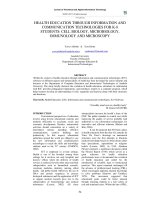
In in-house experiments using guinea-pig lung or in vivo in the guinea pig we
identified a remarkable potentiation of the bronchoconstrictor response to allergen
by activation of A
3
receptors (Fig. 1.1).
Together these findings spawned a concept, ‘The response of the airway mast
cells to allergen is determined by adenosine acting through A
3
receptors’ and, in
1995, a programme, ‘Antagonists of the A
3
receptor for the treatment of asthma’,
was initiated based on the following considerations:
14
1000
800
600
400
200
0
0246
HISTAMINE RELEASE (REL. TO CONTROL-1)
RESISTANCE (cm water /(1/sec))
12
10
8
6
4
2
0
control
Allergen
8
7
6
5
NECA 10-5M
[NECA] (–log molar)
ADMINISTRATION CYCLES OF OVALBUM IN
(IN HALED)
Guinea-pig chopped lung
(histamine release)
Guinea pig in vivo
(bronchoconstriction)
APNEA 30 µg/kg –15 min
vehicle
Fig. 1.1 Interaction between allergen and adenosine receptor agonists with respect to histamine
release from guinea-pig chopped lung (left) or bronchoconstriction in the guinea pig in vivo
(right). Tissues and animals were passively sensitized to ovalbumin. In the histamine release
assay, a threshold response to allergen is augmented concentration-dependently by NECA. In the
whole animal, a single intravenous injection of APNEA markedly enhances the bronchoconstric-
tor response to allergen (J.R. Fozard and H.J. Pfannkuche, unpublished observations 1994)
91 From Hypertension (+) to Asthma
Adenosine has a pivotal role in the pathophysiology of asthma.•
Adenosine facilitates preformed mediator and possibly cytokine release from •
mast cells by activating A
3
receptors.
Antagonists at the A•
3
receptor would prevent periodic exacerbations of asthma
and lead over the longer term to a reduction in airways inflammation and bron-
chial hyperresponsiveness.
With this brief, our chemistry colleagues set out to design antagonists with potency
and selectivity at the A
3
receptor and we biologists went off to devise mechanistic
and/or disease models in which their molecules could be evaluated.
1.4.2 The Design and Synthesis of Novel Potent and Selective
A
3
Receptor Antagonists
To rapidly identify a compound class with potential affinity at the human A
3
ade-
nosine receptor, a diverse library of compounds was obtained and high throughput
screening initiated. N-[4-(4-methoxyphenyl)-thiazol-2-yl]-acetamide came out as a
hit and structure-activity relationship studies led rapidly to the synthesis of a num-
ber of aminothiazole derivatives, exemplified by N-[5-pyridin-4-yl-4-(3,4,5-
methoxyphenyl)-thiazol-2-yl]-acetamide, with subnanomolar antagonist activity at
the human A
3
receptor and greater than 1,000-fold selectivity over the other adenos-
ine receptor subtypes (For full details see Press et al. 2004). Moreover, this com-
pound was a selective antagonist of the hypotensive response to the prototype A
3
receptor agonist, 2-Cl-IB-MECA, in the rat indicating that blockade of the A
3
receptor
could be obtained in vivo.
1.4.3 An Example of the Species Selectivity of the A
3
Receptor:
The Receptor Responsible for Adenosine Augmentation of
Mediator Release from Human Mast Cells Is Not the A
3
Receptor
At about the time that the efforts of the chemists to synthesise selective A
3
antago-
nists had begun to bear fruit (1995/1996), the assumption implicit in our concept
that human mast cells would behave like those of the rodent was called into serious
question. The first (and key) observations came from Feoktistov and Biaggioni
(1995) who provided evidence that in the human mast cell line, HMC-1 (which
although derived from a patient with mast cell leukaemia shows some biochemical
characteristics similar to the mast cells of the lung (Feoktistov et al. 1998)), the A
2B
receptor and not the A
3
receptor is responsible for the potentiation of phorbol
10 J.R. Fozard
12-myristate 13-acetate (PMA)-induced augmentation of IL-8 release. With hindsight,
there were already at least two observations in the literature which did not accord
with a facilitatory role for the A
3
receptor in human mast cells. First, Hughes et al.
(1984) showed that responses of mechanically dispersed human lung mast cells to
adenosine following immunological challenge with anti-IgE had the features of
an A
2
receptor with respect to agonist relative potencies and blockade by low con-
centrations of 8-phenyltheophylline. Second, Peachell et al. (1991) showed that
adenosine and its analogues potentiated mediator release induced by anti-IgE from
passively sensitised human lung mast cells and here too the response was suscep-
tible to blockade by 8-phenyltheophylline.
In May 1997, based on an increasing awareness of the importance of the A
2B
receptor on human mast cells (see Feoktistov and Biaggioni 1997a), we decided to
refocus our programme and set potent and selective blockade of the human A
2B
receptor as the major criterion for identifying a compound for possible clinical
development. The A
3
receptor was retained as a target for several reasons (based on
the information available at the time). First, A
3
receptors had been shown to be
present on human eosinophils and to couple to signalling pathways that lead to cell
activation (Kohno et al. 1996; observations subsequently supported by Reeves et al.
2000). Since asthmatic inflammation is characterised by extensive infiltration of the
airways by activated eosinophils, it is possible that the elevated adenosine concen-
trations associated with asthma would contribute to eosinophil activation through
stimulation of A
3
receptors. Second, activation of A
3
receptors mediates inhibition
of eosinophil chemotaxis (Knight et al. 1997). Since adenosine levels are highest at
the site of inflammation, A
3
receptor activation could be pro-inflammatory by
inhibiting eosinophil migration away from the sites of inflammation. Last, but not
least, the chemists had built up considerable expertise in designing A
3
receptor
antagonists. It was decided that either a dual A
2B
/A
3
antagonist or a selective A
2B
antagonist would be considered relevant for clinical evaluation.
1.4.4 The Design of Mixed A
2B
/A
3
Receptor Antagonists
and Their Biological Evaluation In Vitro
As described above, a series of 5-pyridylaminothiazoles had been designed and syn-
thesised as highly potent and selective antagonists at the adenosine A
3
receptor (Press
et al. 2004). It was essentially an extension of the structure-activity relationship to
involve 5-imidazo and 5-triazolo substituted aminothiazoles which enabled the rapid
identification of several dual A
2B
/A
3
receptor antagonists with acceptable selectivity
over the A
1
and A
2A
receptors. Of the lead compounds, the mesylate salt of 3-[5-(meth-
ylimidazol-1-yl)-2-(pyrazin-2-ylamino)-thiazol-4-yl benzonitrile (QAF805) was con-
sidered to be the superior compound (Compound 5f – Press et al. 2005).
QAF805 has high affinity for the human recombinant adenosine A
2B
receptors and
shows selectivity for these sites over the human A
1
and A
2A
receptors (55- and 522-fold,
111 From Hypertension (+) to Asthma
respectively). QAF805 also shows high affinity for the human A
3
receptor and is
18- and 174-fold selective for this site relative to the human A
1
and A
2A
receptors, respec-
tively (Press et al. 2005; Table 1.1). In functional models of the rat, dog and guinea pig
A
2B
receptors (Fozard et al. 2003a), QAF805 was a silent, surmountable antagonist
yielding K
B
values close to the Ki values from the human receptor binding assay (8 ±
1; n = 5), 1 ± 0.2 (4) and 7 ± 1 (4) nM, respectively). It bears emphasis that QAF805
was somewhat more potent as an antagonist of the rat A
1
receptor mediating contraction
of the rat spleen (Fozard and Milavec-Krizman 1993) (K
B
42 ± 7 nM, n = 4) than at the
human A
1
receptor (Ki from radioligand binding assay 186 nM – Table 1.1). QAF805
was inactive in a broad screen against other receptor and enzyme targets and had a good
in vivo pharmacokinetic profile when given orally in the rat (Press et al. 2005).
1.4.5 A Second Example of the Species Selectivity
of the A
3
Receptor: The In Vivo Evaluation of QAF805
Whilst the in vitro evaluation of QAF805 had been relatively straightforward, the
in vivo evaluation was more of a challenge since at that time (1999) there were no
disease-relevant animal models available to detect antagonist activity at human or
indeed rodent A
2B
or A
3
receptors. Moreover, there was no in vitro assay available
Table 1.1 Affinities of QAF805 for human adenosine receptor subtypes
A
1
a
A
2A
b
A
2B
c
A
3
d
K
i
(nM) K
i
(nM) K
B
(nM) K
i
(nM)
186 ± 38 (5) 1775 ± 548 (5) 3.4 ± 0.2 (3) 10.2 ± 0.4 (5)
Values represent means ± s.e. mean of the number of experiments indicated
in parentheses.
a
Radioligand binding assay with
3
H-DPCPX.
b
Radioligand
binding assay with
3
H-ZM 241385.
c
Reporter gene assay.
d
Scintillation
proximity assay with
125
I-AB-MECA (Press et al. 2005).
N
S
N
N
N
H
N
N
N
12 J.R. Fozard
to us to measure activity at the rat A
3
receptor. Thus, our initial strategy was to
confirm that we could show A
3
receptor blockade in vivo and for this we returned
to our rat model where activation of A
3
receptors results in mast cell-mediated
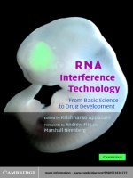
hypotensive responses. The result was convincing although disappointing. At an
oral dose of 10 mg kg
−1
, QAF805 completely blocked the cardiovascular response
to the selective A
1
receptor agonist, CPA (Fig. 1.2 ), but had no effect on the fall in
blood pressure induced by the selective A
3
agonist, 2-Cl-IB-MECA (Fig. 1.3).
Thus, unlike the A
2B
receptor which manifests no species selectivity with respect to
the antagonist potency of QAF805, QAF805 shows high affinity for the human A
3
receptor (Table 1.1) but is at best a very weak antagonist at the rat A
3
receptor.
1.5 Modelling the Airways Response to Adenosine:
An Atypical Receptor Mechanism Mediates
the Bronchoconstrictor Response to Adenosine
Augmented Following Allergen Challenge
During the time that QAF805 was being identified and profiled as a potential
candidate for clinical development, we had been working to design a disease model
which would be predictive for clinical activity. Our conceptual starting point was
0.250.5 124
–80
–60
–40
–20
–80
–60
–40
–20
0
0.25 0.5 124
*
*
***
**
**
**
Decrease in BP (%)
*
*
*
***
**
***
***
CPA(µg kg
–1
i.v.)
Decrease in HR (%)
Vehicle (2ml kg
–1
p.o. –1h n=4)
QAF805 (5mg kg
–1
p.o. –1h n=4)
QAF805 (20mg kg
–1
p.o. –1h n=4)
Fig. 1.2 The effect of QAF805 on the cardiovascular response to CPA in the rat. Shown are the
decreases in blood pressure (BP) and heart rate (HR) induced by CPA (0.25, 0.5, 1, 2, and 4 µg kg
−1
;
cumulative doses i.v.). Vehicle or NVP-QAF805 was given orally 1 h prior to CPA. Results are
expressed as means ± s.e.mean. **p < 0.01, ***p < 0.001; indicates significant difference by com-
parison with vehicle-treated animals (J.R. Fozard and L. Mazzoni, unpublished observations 1999)