Báo cáo khoa học: Curcumin suppresses the dynamic instability of microtubules, activates the mitotic checkpoint and induces apoptosis in MCF-7 cells ppt
Bạn đang xem bản rút gọn của tài liệu. Xem và tải ngay bản đầy đủ của tài liệu tại đây (749.33 KB, 12 trang )
Curcumin suppresses the dynamic instability of
microtubules, activates the mitotic checkpoint and
induces apoptosis in MCF-7 cells
Mithu Banerjee, Parminder Singh and Dulal Panda
Department of Biosciences & Bioengineering, Indian Institute of Technology Bombay, Mumbai, India
Introduction
Curcumin, a natural product found in the rhizome of
Curcuma longa, is emerging as an important anticancer
agent on account of its manifold clinical applications
[1–5]. Although the phase I clinical trial of curcumin
for the prevention of colon cancer has already been
completed (clinicaltrials.gov Identifier: NCT00027495),
clinical trials to determine its efficacy in the treat-
ment of rectal cancer (clinicaltrials.gov Identifier:
NCT00745134), advanced pancreatic cancer (clinicaltri-
als.gov Identifier: NCT00094445), colorectal can-
cer (clinicaltrials.gov Identifier: NCT00973869) and
multiple myeloma (clinicaltrials.gov Identifier: NCT-
00113841) are currently in progress. In addition, the
potential of curcumin to reduce the symptomatic side
effects of chemoradiation in patients suffering from
non-small cell lung cancer (clinicaltrials.gov Identifier:
NCT01048983) is under clinical investigation. Curcu-
min has also entered into a phase II clinical trial for
Keywords
apoptosis; BubR1; combination study;
delayed mitosis; dynamic instability
Correspondence
D. Panda, Department of Biosciences &
Bioengineering, Indian Institute of
Technology Bombay, Powai,
Mumbai-400076, India
Fax: +91 222 572 3480
Tel: +91 222 576 7838 ⁄ 7770
E-mail:
(Received 14 April 2010, revised 21 May
2010, accepted 24 June 2010)
doi:10.1111/j.1742-4658.2010.07750.x
In this study, curcumin, a potential anticancer agent, was found to dampen
the dynamic instability of individual microtubules in living MCF-7 cells. It
strongly reduced the rate and extent of shortening states, and modestly
reduced the rate and extent of growing states. In addition, curcumin
decreased the fraction of time microtubules spent in the growing state and
strongly increased the time microtubules spent in the pause state. Brief
treatment with curcumin depolymerized mitotic microtubules, perturbed
microtubule–kinetochore attachment and disturbed the mitotic spindle
structure. Curcumin also perturbed the localization of the kinesin protein
Eg5 and induced monopolar spindle formation. Further, curcumin
increased the accumulation of Mad2 and BubR1 at the kinetochores, indi-
cating that it activated the mitotic checkpoint. In addition, curcumin treat-
ment increased the metaphase ⁄ anaphase ratio, indicating that it can delay
mitotic progression from the metaphase to anaphase. We provide evidence
suggesting that the affected cells underwent apoptosis via the p53-depen-
dent apoptotic pathway. The results support the idea that kinetic stabiliza-
tion of microtubule dynamics assists in the nuclear translocation of p53.
Curcumin exerted additive effects when combined with vinblastine,
a microtubule depolymerizing drug, whereas the combination of curcumin
with paclitaxel, a microtubule-stabilizing drug, produced an antagonistic
effect on the inhibition of MCF-7 cell proliferation. The results together
suggested that curcumin inhibited MCF-7 cell proliferation by inhibiting
the assembly dynamics of microtubules.
Abbreviations
CI, combination index; FITC, fluorescein isothiocyanate; PI, propidium iodide.
FEBS Journal 277 (2010) 3437–3448 ª 2010 The Authors Journal compilation ª 2010 FEBS 3437
advanced pancreatic cancer [2] and a phase III clinical
trial in combination with gemcitabine and celebrex for
the treatment of metastatic colon cancer [2]. Curcumin
inhibits tumor growth in animal models [3]. Further,
the uptake of high doses of curcumin in both animals
and humans has been found to be nonhazardous and
relatively nontoxic [4,5]. Curcumin has also been found
to be an effective stress reliever and neuroprotective
agent [6].
Curcumin has been shown to inhibit the prolifera-
tion of several types of cancer cells in culture, includ-
ing pancreatic, cervical, colon and breast cancer [7–15].
It arrests the cell-cycle progression of human pancre-
atic cancer cells (BxPC-3) and glioma cells (U251) at
the G
2
⁄ M phase of the cell cycle [7,8] and has been
shown to affect the progression of MCF-7 cells
through the G
2
⁄ M phase [9]. Curcumin treatment
caused an increase in the G
0
⁄ G
1
phase of the cell pop-
ulation implying apoptosis in MCF-7 cells [10]. Curcu-
min is found to induce apoptosis in several cell lines
[1,7,10]. It stimulated Bax-mediated p53-dependent
apoptosis in MCF-7 cells [10]. Curcumin promotes the
action of certain drugs by overcoming chemoresistance
[11]. It overcomes P-glycoprotein-mediated multidrug
resistance in multiple cell lines [1]. The migration and
invasion of human lung cancer cells are also inhibited
by curcumin [1].
Curcumin has been suggested to inhibit cell prolifer-
ation by diverse mechanisms [1,12–16]; however, the
primary mechanism by which inhibition occurs
remains obscure. Recently, curcumin has been found
to bind to purified tubulin, to inhibit tubulin polymeri-
zation in vitro and to depolymerize microtubules in
HeLa and MCF-7 cells in culture [12]. In addition,
curcumin has been shown to perturb the microtubule
spindle structure [12,13] and to stimulate micronucle-
ation in MCF-7 cells [13]. In 32D cells, curcumin has
also been shown to affect the activity of the chromo-
somal passenger complex, resulting in multipolar chro-
mosome segregation promoting mitotic catastrophe
[14]. Moreover, curcumin induced mitotic catastrophe
in Ishikawa and HepG2 cancer cells, indicating that it
might perturb microtubule assembly dynamics [15].
Dynamic microtubules are the key structural ele-
ments in mitotic spindle formation and they orches-
trate chromosome distribution during the cell division
[17,18]. In this study, we found that curcumin strongly
suppressed the dynamic instability of individual micro-
tubules in live MCF-7 cells. At low effective prolifera-
tion inhibitory concentrations, curcumin inhibited
microtubule dynamics in MCF-7 cells without causing
a significant depolymerization of microtubules. How-
ever, high concentrations (‡ 2 · IC
50
) of curcumin
were found to depolymerize both the interphase and
mitotic microtubules in MCF-7 cells. Curcumin treat-
ment perturbed the mitotic spindle network in MCF-7
cells, activated the mitotic checkpoint and delayed
mitotic progression. We present several lines of evi-
dence indicating that curcumin inhibits cell prolifera-
tion by inhibiting microtubule dynamics. The results
suggest that tubulin is one of the major targets for the
antiproliferative activity of curcumin.
Results
Curcumin inhibited the proliferation of MCF-7
cells and induced apoptosis
Consistent with previous studies [9,10,12], curcumin
was found to inhibit the proliferation of MCF-7 cells
in a concentration-dependent manner (Fig. 1A). For
example, 20 and 40 lm curcumin inhibited the prolifer-
ation of MCF-7 cells by 70% and 93%, respectively,
and the half-maximal inhibition of proliferation (IC
50
)
was determined to be 16 ± 0.3 lm. MCF-7 cells were
either treated with the vehicle or different concentra-
tions of curcumin for 48 h. Vehicle-treated MCF-7
cells did not display Annexin V and propidium iodide
(PI) staining, although a population (46%) of cells
treated with 12 lm curcumin stained positive for Ann-
exin V alone, showing that the cells were at the early
stage of apoptosis (Fig. 1B). Cells treated with 24 lm
curcumin showed greater numbers (71%) stained with
Annexin V (Fig. 1B). At a still higher curcumin con-
centration (36 lm), cells were stained with both Ann-
exin V and PI, indicating late apoptosis (Fig. 1B).
Further, curcumin treatment strongly increased the
nuclear localization of p53 and p21 in MCF-7 cells
(Fig. 1C,D). For example, 4 and 24% of MCF-7 cells
showed nuclear localization of p53 (Fig. 1C), whereas
7 and 33% of cells showed nuclear localization of p21
in the absence and presence of 24 lm curcumin,
respectively (Fig. 1D).
Curcumin disrupted the mitotic spindle network
inducing formation of monopolar spindles and
depolymerized microtubules in MCF-7 cells
MCF-7 cells were incubated without or with 24 and
36 lm curcumin for 6 h. Curcumin treatment strongly
depolymerized mitotic spindle microtubules (Fig. 2A).
However, it did not induce significant depolymeriza-
tion of the interphase microtubules after 6 h of incuba-
tion, suggesting that curcumin exerted a stronger
depolymerizing effect on the microtubules of the mito-
tic cells than those of the interphase cells (data not
Curcumin suppresses microtubule dynamics M. Banerjee et al.
3438 FEBS Journal 277 (2010) 3437–3448 ª 2010 The Authors Journal compilation ª 2010 FEBS
A
CD
B
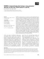
Fig. 1. Curcumin inhibited the proliferation of MCF-7 cells and induced cell death. (A) MCF-7 cells were treated with different concentrations
of curcumin for one cell cycle and the inhibition of cell proliferation was determined by the sulforhodamine B assay. (B) Curcumin induced
apoptosis in MCF-7 cells. MCF-7 cells were incubated with 0.1% dimethylsulfoxide (control) and different concentrations (12–36 l
M) of curc-
umin for 48 h and then stained with Annexin V ⁄ PI. Scale bar, 10 lm. Curcumin (24 l
M) treatment increased the nuclear accumulation of p53
(C) and p21 (D) in MCF-7 cells. Scale bar, 10 lm.
Tubulin DNA
Merge
Control
Curcumin 24 μ
M
Curcumin 36 μM
Control
Curcumin
0 min
15 min
15 min
25 min
25 min0 min
AB
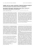
Fig. 2. Curcumin-perturbed mitotic spindle structures of MCF-7 cells. (A) MCF-7 cells were incubated without or with 24 and 36 lM of curcu-
min for 6 h. Microtubules are shown in red and the nucleus in blue. Scale bar, 10 lm. (B) Curcumin suppressed the reassembly of the cold-
depolymerized mitotic spindle microtubules. The upper and lower panels show growth kinetics of spindle microtubules in the absence or the
presence of 36 l
M curcumin. Scale bar, 10 lm.
M. Banerjee et al. Curcumin suppresses microtubule dynamics
FEBS Journal 277 (2010) 3437–3448 ª 2010 The Authors Journal compilation ª 2010 FEBS 3439
shown). Consistent with a previous study [12], curcu-
min was found to induce significant depolymerization
of both the interphase and mitotic microtubules of
MCF-7 cells after 24 h of incubation (Fig. S1A,B). In
the control population, 70% and 30% of the mitotic
cells were found to be bipolar and monopolar, respec-
tively, whereas 85% of the mitotic cells were mono-
polar in the presence of 24 lm curcumin, suggesting
that curcumin induced the formation of monopolar
spindles.
The effect of curcumin on the polymerized mass of
microtubules in MCF-7 cells was analysed by western
blotting. The ratio of polymeric ⁄ soluble tubulin was
found to be 2.44 ± 0.77 in the absence of curcumin,
and 1.97 ± 0.20, 1.56 ± 0.17 (P < 0.03) and
1.32 ± 0.15 (P < 0.01) in the presence of 12, 24 and
36 lm curcumin, respectively, indicating that curcumin
depolymerized microtubules in MCF-7 cells
(Fig. S1C).
Curcumin inhibited the reassembly of mitotic
microtubules in MCF-7 cells
MCF-7 cells were synchronized in the M phase of the
cell cycle by treating with 1.3 lm nocodazole for 20 h.
Nocodazole treatment completely depolymerized the
spindle microtubules. Nocodazole was removed and
the cells were incubated with fresh media in the
absence or presence of 36 lm curcumin on ice for
30 min. Subsequently, cells were incubated at 37 °C.
Spindle microtubules in control cells reassembled
within 25 min to form normal mitotic spindle; the
spindle microtubules of curcumin-treated cells did not
reassemble (Fig. 2B). The results showed that cur-
cumin inhibited reassembly of the mitotic spindle
microtubules.
Curcumin suppressed the dynamic instability of
individual microtubules in live MCF-7 cells
Consistent with previous reports [19,20], microtubules
in control MCF-7 cells were found to be highly
dynamic (Fig. 3A). Low concentrations of curcumin (5
and 12 lm) noticeably dampened the dynamic instabil-
ity of the individual microtubules in live MCF-7 cells
(Fig. 3B,C). Curcumin treatment reduced the rate and
extent of both growing and shortening events
(Table 1). For example, 12 lm curcumin reduced the
rates of shortening and growing phases by 39% and
19%, respectively, and reduced the extent of the grow-
ing and shortening phases by 60% and 65%, respec-
tively. Like several other tubulin-targeted agents such
as benomyl, estramustine, epothilone B and paclitaxel
[19–22], curcumin also strongly increased the time that
microtubules spent in the pause state, neither growing
nor shortening detectably, and decreased the time
microtubules spent in the growing or shortening
phases. Curcumin (12 lm) increased the time spent in
the pause state from 28.9% (control) to 71.6%. Fur-
ther, curcumin (12 lm) altered both the time- and
length-based transition frequencies of the interphase
microtubules in MCF-7 cells. The dynamicity (dimer
exchange per unit time from the ends of microtubules)
was reduced by 50% and 72% in the presence of
5 and 12 lm curcumin, respectively.
A
B
C
Fig. 3. Curcumin suppressed dynamic instability of individual micro-
tubules in live MCF-7 cells. Life-history traces of individual microtu-
bules in MCF-7 cells in the absence (A) and presence of (B) 5 l
M
curcumin and (C) 12 lM curcumin, respectively.
Curcumin suppresses microtubule dynamics M. Banerjee et al.
3440 FEBS Journal 277 (2010) 3437–3448 ª 2010 The Authors Journal compilation ª 2010 FEBS
Effects of curcumin on cell-cycle progression
It was previously reported that curcumin treatment
markedly increased the number of MCF-7 cells in
metaphase [13]. Because curcumin suppressed microtu-
bule dynamic instability (Fig. 3 and Table 1), we
examined whether it could inhibit mitosis. The number
of cells in mitosis in the absence or presence of differ-
ent concentrations of curcumin was determined by
Hoechst 33258 staining of the chromosomes. Only
2.6 ± 1.6% of the control (vehicle-treated) cells were
found to be in mitosis, whereas 3.2 ± 0.2%,
5.0 ± 0.1% and 6.2 ± 1% ( P < 0.0001) of the cells
were found to be in mitosis in the presence of 12, 24
and 36 lm curcumin, respectively. If curcumin caused
a delay in mitosis, it might lead to an increase in the
metaphase ⁄ anaphase ratio. The metaphase ⁄ anaphase
ratio was calculated to be 0.43 ± 0.06 and
1.88 ± 0.40 (P < 0.0001) in the absence and presence
of 24 lm curcumin, supporting the idea that curcumin
could prolong the duration of metaphase.
Further, MCF-7 cells were synchronized in the M
phase of the cell cycle by nocodazole treatment for
20 h. Nocodazole-blocked cells were washed with fresh
medium and subsequently incubated in medium with-
out and with curcumin. Flow cytometry analysis dem-
onstrated that nocodazole-induced mitotic arrest was
gradually released over time for control cells. For
example, the percentage of cells in mitosis was 87%,
53% and 16% in nocodazole-treated control flask
immediately, and 4 and 8 h after release of the noco-
dazole block. However, in the presence of curcumin,
the percentage of cells in the mitotic phase was 83%
and 81% after 4 and 8 h of block release. Thus, treat-
ment of cells with curcumin significantly delayed
release of the mitotic block (Fig. 4A). However, flow
cytometric analysis of the cell cycle using PI staining
showed that there was no significant cell-cycle block
after 24 h of curcumin treatment (Fig. S2).
Microtubule inhibitors are known to induce mitotic
block by activating the spindle assembly checkpoint
proteins [20,23,24]. It has been suggested that a com-
pound may drive the cells towards delayed mitosis
through activation of spindle checkpoint proteins such
as BubR1 [23] and Mad2 [24]. Nocodazole, a well-
known inhibitor of mitosis, led to the accumulation of
Mad2 and BubR1 at the kinetochores (Fig. 4B,C).
Similar to the action of nocodazole, curcumin treat-
ment also activated Mad2 and BubR1 in MCF-7 cells
(Fig. 4B,C).
Curcumin exhibited antagonism with paclitaxel,
but an additive effect with vinblastine for
inhibition of MCF-7 cell proliferation
Curcumin, paclitaxel and vinblastine inhibited MCF-7
cell proliferation with median inhibitory doses of
15±4lm,40±6nm and 17 ± 10 nm (Fig. S3A–C).
Curcumin (8 lm) and paclitaxel (2 nm) inhibited prolif-
eration of MCF-7 cells by 26% and 13%, respectively,
when used alone, whereas their combination inhibited
proliferation by 9%. The combination index (CI) for
the combination of 8 lm curcumin and 2 nm paclitaxel
was found to be 3.1 ± 1.5. The proliferation of MCF-
7 cells was inhibited by 22% and 24% in the presence
of 2 and 3 nm vinblastine, respectively, whereas in
Table 1. Effects of curcumin on the dynamic instability parameters of the interphase microtubules in MCF-7 cells. Twenty-five microtubules
were measured for each condition. Data are given as mean ± SD.
Control 5 l
M curcumin 12 lM curcumin
Growth rate (lmÆmin
)1
) 14.7 ± 2.9 12.1 ± 2.6
a
11.9 ± 2.9
a
Growth length (lm) 2.4 ± 1.1 1.12 ± 0.5
b
0.97 ± 0.3
a
Growth time (min) 1.2 ± 0.3 0.55 ± 0.23
a
0.33 ± 0.15
a
Shortening rate (lmÆmin
)1
) 23.5 ± 10.4 18.2 ± 5.7
a
14.4 ± 4.95
a
Shortening length (lm) 3.3 ± 1.9 2.1 ± 1.1
a
1.14 ± 0.51
a
Shortening time (min) 0.53 ± 0.18 0.43 ± 0.19 0.28 ± 0.11
a
Pause time (min) 0.72 ± 0.28 1.43 ± 0.35
a
1.82 ± 0.36
a
% Time spent in growing 47.5 ± 9.8 22.7 ± 8.8
a
13.5 ± 7.3
a
% Time spent in shortening 22.4 ± 8.3 18.7 ± 7.9
a
11.6 ± 4.7
a
% Time spent in pause 28.9 ± 11.6 59.5 ± 14.0
a
71.6 ± 13.8
a
Dynamicity (lmÆmin
)1
) 12.6 ± 4.6 6.3 ± 3.0
a
3.50 ± 1.99
a
Rescue frequency (eventsÆmin
)1
) 7.7 ± 3.4 9.9 ± 3.5
a
12.74 ± 2.50
a
Catastrophe frequency (eventsÆmin
)1
) 2.1 ± 0.70 2 ± 1.1
a
1.50 ± 0.96
a
Rescue frequency (eventsÆlm
)1
) 0.35 ± 0.20 0.59 ± 0.35
a
0.99 ± 0.42
a
Catastrophe frequency (eventsÆlm
)1
) 0.24 ± 0.12 0.67 ± 0.27
a
1.08 ± 0.55
a
a
P < 0.0001;
b
P < 0.001.
M. Banerjee et al. Curcumin suppresses microtubule dynamics
FEBS Journal 277 (2010) 3437–3448 ª 2010 The Authors Journal compilation ª 2010 FEBS 3441
combination with 8 lm curcumin, these concentrations
of vinblastine inhibited proliferation by 44% and 51%,
respectively. The CI values for the combination of
8 lm curcumin with 2 and 3 nm of vinblastine were
estimated to be 0.92 ± 0.23 and 0.97 ± 0.19, respec-
tively. A CI value < 1 indicates a synergistic effect,
1 indicates an additive effect and > 1 indicates an
antagonistic effect [25,26]. The results suggested that
curcumin was antagonistic to paclitaxel, whereas it dis-
played an additive effect with vinblastine in inhibiting
MCF-7 cell proliferation.
Curcumin affected the localization of the kinesin
protein Eg5
Because curcumin produced monopolar spindles in
MCF-7 cells, we examined the effect of curcumin on
the localization of Eg5, a motor protein that plays an
essential role in bipolar spindle formation [27,28]. In
control cells, Eg5 was localized throughout the bipolar
spindle and remained concentrated at the spindle poles
(Fig. 5A). Consistent with a previous study [27], mon-
astrol (50 lm) was found to induce monopolar spindle
formation (Fig. 5B). In monastrol-treated cells, Eg5
mainly localized to the pole of the monoastral spindle
and also diffused all along the monoastral microtu-
bules (Fig. 5B). In the presence of 24 lm curcumin,
Eg5 primarily remained confined to the pole of the
monopolar spindles. Some Eg5 also delocalized along
the microtubules of the monopolar spindles (Fig. 5C).
Discussion
In this study, we have provided several lines of evi-
dence indicating that the antiproliferative mechanism
of action of curcumin involves the perturbation of
microtubule dynamics. Brief incubation of curcumin
with MCF-7 cells produced a noticeable depolymeriz-
ing effect on the mitotic microtubules of MCF-7 cells
and also inhibited the assembly of cold-depolymerized
spindle microtubules indicating that curcumin perturbs
microtubule assembly in cells. Further, similar to the
effects of several other microtubule-targeted drugs such
as benomyl [19], estramustine [20], epothilone [21] and
paclitaxel [22] on microtubule dynamics, curcumin was
also found to reduce the dynamic instability of individ-
ual microtubules in live MCF-7 cells. Curcumin
treatment caused defective chromosome alignment in
the mitotic spindles and the cells eventually died via
the p53-dependent apoptotic pathway. Curcumin was
ABC
0 200 400 600 80010000 100 200 300 400 500 0 80 160 240 320
0
80 160 240 320
0
80 160 240 320
0 100 200 300 400 500
0 20 40 60 80 100 120
0 20 40 60 80 100 120
0 30 60 90 120 150
0 30 60 90 120 150
0 30 60 90 120 150
0 20 40 60 80 100 120
Fig. 4. Curcumin treatment delayed mitotic progression in MCF-7 cells. (A) MCF-7 cells were incubated with 1.3 lM nocodazole. Nocodazole
was washed off with fresh medium. Cells were incubated in the absence or presence of 35 l
M curcumin for 4 and 8 h and then stained
with PI. DNA content of the cells was quantified by flow cytometry. Nocodazole and curcumin treatment activated Mad2 (B) and BubR1 (C)
in MCF-7 cells. MCF-7 cells were incubated with nocodazole (500 n
M) and curcumin (36 lM) for 24 h and cells were then stained with Mad2
and BubR1 antibodies. Scale bar, 10 lm.
Curcumin suppresses microtubule dynamics M. Banerjee et al.
3442 FEBS Journal 277 (2010) 3437–3448 ª 2010 The Authors Journal compilation ª 2010 FEBS
found to bind to purified tubulin and to perturb
microtubule assembly in vitro [12]. The results together
indicated that curcumin inhibits MCF-7 cell prolifera-
tion by targeting microtubules.
The plus-end-directed motor Eg5 (kinesin spindle
protein) plays an important role in proper chromo-
some separation and the formation of a proper bipolar
spindle [27,28]. Similar to the action of monastrol [27],
curcumin also induced monopolar spindle formation in
association with the perturbation of Eg5 localization
in MCF-7 cells, indicating that curcumin may inhibit
Eg5 function and thereby induce monopolar spindle
formation. Curcumin might inhibit the binding of Eg5
to microtubules and perturb the movement of Eg5
over the microtubules leading to abnormal spindle for-
mation. Alternatively, curcumin might directly interact
with Eg5 and inhibit its function.
Effects of curcumin on the progression of the
cell cycle
Curcumin increased the metaphase ⁄ anaphase ratio and
slowed the release of mitotic block in nocodazole-
synchronized MCF-7 cells, indicating that it can delay
cell-cycle progression at mitosis. However, it failed to
induce substantial mitotic block in MCF-7 cells. In
several cases, higher concentrations of microtubule-
targeted agents are required to inhibit cell-cycle
progression at mitosis than are required to inhibit the
proliferation [29–32]. In a KB ⁄ HeLa (human cervical
epitheloid carcinoma) cell line, a derivative of benzylid-
ene-9(10H)-anthracenone gave an IC
50
value of
0.09 lm for the inhibition of cell proliferation, whereas
50% arrest in the G
2
⁄ M phase occurred in the pres-
ence of 0.205 lm of compound [29]. The anthracenone
derivative caused cell-cycle arrest in a K-562 cell line
at 0.3 lm, whereas its IC
50
in the same cell line was
0.02 lm. In smooth muscle cells, 68.6% of the cells
were arrested in the G
2
⁄ M phase at 100 nm concentra-
tion of LY290181 (IC
50
of inhibition of cell prolifera-
tion being 20 nm) [30]. In human non-small cell lung
carcinoma cells A549, low concentrations of paclitaxel
(3-6 nm) inhibited cell proliferation without causing
mitotic arrest [31]. Moreover, treatment with a low
concentration of paclitaxel induced abnormal cell for-
mation without the G
2
⁄ M block [32]. A 50% inhibi-
tion of cell growth after 72 h incubation required
3.4 nm paclitaxel and 9.5 nm discodermolide [32].
These concentrations were closer to that required for
aneuploidy induction rather than mitotic arrest [32].
Tubulin
Eg5
DNA Tubulin + Eg5
Tubulin + Eg5 + DNA
A
B
C
Fig. 5. Localization of Eg5 in control and curcumin-treated MCF-7 cells. Cells were treated without and with curcumin for 24 h, fixed, and
co-immunostained with a-tubulin (green), Eg5 antibody (red) and DNA was stained with Hoechst 33258. (A) In control mitotic cells, Eg5
remained mainly concentrated at the poles of the bipolar spindle and to some extent delocalized along the spindle microtubules. (B) In the
presence of 50 l
M monastrol, monopolar spindles were formed. Eg5 localized mainly at the pole of the monopolar spindle and remained dif-
fused along the microtubules in the overlayed image. (C) Curcumin at a concentration of 24 l
M induced monopolar spindle formation. In the
overlain image the Eg5 localized to the centre of the monopolar spindle and also remained dispersed over the microtubules. Scale bar,
10 lm.
M. Banerjee et al. Curcumin suppresses microtubule dynamics
FEBS Journal 277 (2010) 3437–3448 ª 2010 The Authors Journal compilation ª 2010 FEBS 3443
Thus, the induction of abnormal mitosis and aneu-
ploidy is dependent on the drug mechanism and the
concentration of the drug used [31,32].
Several microtubule-targeted agents are known to
activate checkpoint proteins and to arrest cells in mito-
sis [20,33–35]. The checkpoint proteins accumulate in
the kinetochoric region after detecting a flaw in kineto-
chore–microtubule attachment or reduced tension at the
kinetochores [24]. For example, nocodazole enhances
the accumulation of Mad2 and BubR1 to the kinetoch-
ores and induces mitotic arrest (Fig. 4B,C) [36].
Several inhibitors of microtubule dynamics were
found to delay G
2
⁄ M transition [37]. The ability of a
compound to activate spindle checkpoint proteins may
sometimes lead to delayed mitosis [38]. Conditions that
perturb proper kinetochore–microtubule attachment
may cause checkpoint protein translocation and the
affected cells may be held back from progressing fur-
ther in the cell cycle, leading to a delay in mitosis [38].
Curcumin was found to perturb microtubule–kineto-
chore attachment and also activated the mitotic check-
point, resulting in delayed mitosis. A delay in mitosis
has been shown to induce apoptosis in cancer cells
[39].
Curcumin treatment enhanced the nuclear
accumulation of p53 in MCF-7 cells
An alteration in expression of the tumor suppressor
gene p53 is known to induce apoptosis in several types
of cells [40–42]. It has been suggested that p53 is trans-
ported into the nucleus through the microtubule net-
work [40,41]. Compounds that stabilize microtubule
dynamics have been suggested to promote p53 translo-
cation to the nucleus [19,41]. Several antimitotic drugs
have been found to induce apoptosis by inhibiting
microtubule assembly dynamics [43]. Curcumin
suppresses the dynamic instability of microtubules,
therefore, it may enhance nuclear translocation of p53
through the stabilized microtubule track.
Curcumin in combination with vinblastine, a micro-
tubule depolymerizing agent, inhibited cell prolifera-
tion in an additive fashion. However, it antagonized
the action of paclitaxel, a compound that promotes
microtubule assembly; supporting the idea that curcu-
min inhibits cell proliferation by targeting micro-
tubules. The results also indicated that curcumin may be
used in combination with microtubule depolymerizing
agents such as vinblastine to improve the efficacy and
reduce the toxic dose of the drug. It has been found
that an oral intake of curcumin is not toxic to humans
up to 8000 mgÆday
)1
for 3 months [44]. Moreover,
curcumin (C
3
ComplexÔ, Sabinsa Corp., East Wind-
sor, NJ, USA) in single oral doses up to 12 000 mg
was found to be well tolerated in healthy volunteers
[45]. Therefore, the concentrations of curcumin used in
this study are expected to be within tolerable doses. It
has been suggested that less potent dietary compounds
can enhance the effect of a more potent and toxic drug
by lowering its toxicity level [46,47]. Therefore, combi-
nation between two such drugs can provide superior
clinical efficacy than a single drug alone [46].
Materials and methods
Reagents
Curcumin, sulforhodamine B, fetal bovine serum, BSA and
G418 were purchased from Sigma (St Louis, MO, USA).
Annexin V and PI were purchased from Santa Cruz Bio-
technology (Santa Cruz, CA, USA). All other reagents were
of analytical grade.
Cell culture
MCF-7 cells, human breast carcinoma cells, were grown in
minimum essential medium (HiMedia, Mumbai, India) sup-
plemented with 10% fetal bovine serum, 2.2 g Æ L
)1
sodium
bicarbonate, along with 1% antibacterial and antimycotic
solution containing streptomycin, amphotericin B and peni-
cillin and 10 lgÆ mL
)1
of human insulin at 37 °Cina
humidified atmosphere of 5% CO
2
[48]. Curcumin stock
solution was prepared in 100% dimethylsulfoxide and dif-
ferent concentrations of curcumin were added to the culture
medium (dimethylsulfoxide was £ 0.1% v ⁄ v) 24 h after
seeding. Dimethylsulfoxide (0.1%) was used as a vehicle
control.
Cell proliferation assay and mitotic index
calculation
The effect of curcumin on the proliferation of MCF-7 cells
was determined by sulforhodamine B assay [49]. For mito-
tic index calculation, MCF-7 cells were seeded at a density
of 1.0 · 10
5
cellsÆmL
)1
on poly(l-lysine)-coated glass cover-
slips followed by treatment with curcumin for 24 h [20].
The coverslips were centrifuged in a Labofuge 400R cyto-
spin (Heraeus, Hanau, Germany) for 10 min (1200 g at
30 °C) and fixed with 3.7% formaldehyde for 30 min at
37 °C. The cells were permeabilized with methanol and
stained with Hoechst 33258. The number of cells in mitosis
and interphase were counted using the Eclipse TE2000-U
microscope (Nikon, Tokyo, Japan). At least 800 cells were
counted for each set and the experiment was repeated three
times. The numbers of cells at the metaphase and anaphase
stages of the cell cycle were calculated for both the control
and curcumin-treated cells.
Curcumin suppresses microtubule dynamics M. Banerjee et al.
3444 FEBS Journal 277 (2010) 3437–3448 ª 2010 The Authors Journal compilation ª 2010 FEBS
Immunofluorescence microscopy and
transfection
MCF-7 cells (0.6 · 10
5
ÆmL
)1
) were seeded on glass cover-
slips in 24-well plates for 24 h and incubated with different
concentrations of curcumin for another 24 h. The cells were
fixed with 3.7% formaldehyde at 37 °C, and immuno-
stained as reported earlier [48]. Cells were stained with the
following primary antibodies: mouse monoclonal anti-
(a-tubulin IgG) (1 : 300) from Sigma, rabbit polyclonal
anti-(a-tubulin IgG) (1 : 300) from Abcam (Cambridge,
MA, USA), mouse monoclonal anti-p53 IgG (1 : 300),
mouse monoclonal anti-p21 IgG (1 : 300) purchased from
Santa Cruz (Santa Cruz, CA, USA), mouse anti-BubR1
IgG (1 : 500) from BD Biosciences (San Jose, CA, USA)
rabbit anti-Mad2 IgG (1 : 300), mouse monoclonal anti-
Eg5 IgG (1 : 800) from Abcam (Cambridge, MA, USA).
The secondary antibodies used were Alexa 568-conjugated
sheep anti-(mouse IgG) (1 : 400) purchased from Molecular
Probes (Eugene, OR, USA), fluorescein isothiocyanate
(FITC)-conjugated anti-(mouse IgG) (1 : 400) and FITC-
conjugated anti-(rabbit IgG) (1 : 400) from Sigma. The
nucleus was stained using 1 lgÆmL
)1
of Hoechst 33258
(Sigma). The slides were observed under an Eclipse
TE2000-U microscope (Nikon, Tokyo, Japan) using a 40 ·
objective. The images were captured using CoolSNAP-Pro
camera. image-pro plus software 4.0 (Media Cybernetics,
Bethesda, MD, USA) was used for image acquisition and
processing. MCF-7 cells were transfected with EGFP–
a-tubulin plasmid, as described previously [20] and the
stably transfected MCF-7 cells were maintained in the pres-
ence of the antibiotic G418.
Annexin V
⁄
propidium iodide staining
MCF-7 cells were grown in the absence and presence of dif-
ferent concentrations of curcumin for 48 h and were stained
with Annexin V ⁄ PI, as reported previously [20,48]. The
manufacturer’s protocol was used for staining the cells
using an Annexin V apoptosis detection kit (Santa Cruz
Biotechnology) and processed for microscopy [20,48]. The
cells exhibiting positive Annexin V and PI staining were
seen under microscope using the FITC and PI fluorescence,
differential interference contrast microscopy was used for
visualizing total number of cells.
Cell-cycle analysis
MCF-7 cells were grown in the absence and presence of 25
and 35 lm curcumin for 24 h. The cells were first fixed in
70% ethanol, washed with NaCl ⁄ P
i
and then incubated
with 50 lgÆmL
)1
PI containing 8 lgÆmL
)1
RNase for 2 h at
4 °C. The DNA content of the cells was quantified using a
flow cytometer (FACS Aria; Becton Dickinson, San Jose,
CA, USA).
MCF-7 cells were treated without and with 1.3 lm noco-
dazole for 20 h. Nocodazole was washed off with fresh
media. The cells were incubated without or with curcumin
for 4 and 8 h, and then stained with PI. The effect of curc-
umin on the kinetics of the release of the mitotic block was
examined in a flow cytometer and the data were analysed
using the modfit lt program (Verity Software, Topsham,
ME, USA).
Effect of curcumin on the reassembly of
cold-depolymerized mitotic microtubules
MCF-7 cells (0.5 · 10
5
ÆmL
)1
) were seeded on glass cover-
slips for 24 h and then incubated with 1.3 lm nocodazole
for 20 h. Nocodazole was removed by washing with fresh
medium. Cells were then incubated without or with 36 lm
curcumin on ice for 30 min. Subsequently, cells were trans-
ferred to 37 °C and the assembly of microtubules was fol-
lowed by fixing the cells after every 5 min. The
microtubule network was visualized by staining the fixed
cells with anti-a-tubulin Ig. The DNA was stained with
Hoechst 33258.
Western blot analysis
The effect of curcumin on the polymeric mass of microtu-
bules in the cells was analysed by western blot, as described
previously [20]. The protein concentrations of the polymeric
and the soluble fraction were determined by the Bradford
method [50]. The polymeric and the soluble tubulin frac-
tions were run on SDS ⁄ PAGE and electroblotted on
poly(vinylidene difluoride) membranes. The membranes
were probed with mouse monoclonal anti-(a-tubulin IgG)
(1 : 1000) and alkaline phosphatase-conjugated secondary
anti-(mouse IgG) (1 : 5000) (Sigma). The band intensities
were calculated using image j software.
Effects of curcumin on the dynamic instability of
individual microtubules in MCF-7 cells
The effects of curcumin on the dynamic instability of the
interphase microtubules in MCF-7 cells were determined as
described previously [20,51]. Briefly, MCF-7 cells having
stably transfected green fluorescent protein–a-tubulin were
grown on glass coverslips for 24 h. Cells were then incu-
bated in the absence or presence of 5 and 12 lm curcumin
for an additional 24 h. The coverslips were transferred to
glass-bottomed dishes (Prime BioScience, Pandan Loop,
Singapore) containing media without phenol red and were
maintained at 37 °C on a warm stage. Time-lapse imaging
of microtubules was carried out using an FV-500 laser
scanning confocal microscope (Olympus, Tokyo, Japan)
with a 60 · water immersion objective. The images were
acquired at 4 s intervals for a maximum duration of 3 min
using fluoview software (Olympus, Tokyo, Japan). The
M. Banerjee et al. Curcumin suppresses microtubule dynamics
FEBS Journal 277 (2010) 3437–3448 ª 2010 The Authors Journal compilation ª 2010 FEBS 3445
plus end of microtubules was tracked using image j soft-
ware. Life-history traces were obtained by plotting the
length of individual microtubules against time. Length
changes of ‡ 0.5 lm for a minimum of two data points
were considered as growth or shortening excursions and a
change < 0.5 lm in length was considered as a pause state.
A transition from a shortening to a growth or pause state
is called a rescue, whereas the transition from a growth or
pause state to a shortening state is defined as a catastrophe
[51]. Twenty-five microtubules were analysed for each
experimental condition. Statistical significance was calcu-
lated using the Student’s t-test.
CI determination
MCF-7 cells were incubated either separately with curcu-
min, paclitaxel and vinblastine or in combination with curc-
umin and vinblastine or paclitaxel for one cell cycle. The
combination index was calculated using the Chou and Tala-
lay method [26,52], with the help of the following equation
CI ¼ðDÞ1=ðDxÞ1 þðDÞ2=ðDxÞ2
where (D)1 and (D)2 are the concentrations of drug 1 and
drug 2 used in combination, which produce a particular
effect, (Dx)1 and (Dx)2 are the concentrations of the drugs
that produce similar effect when used alone. The concentra-
tion of curcumin which produced a particular effect was
calculated from the median effect equation
Dx ¼ Dm½f
a
=f
u
1=m
where, Dm, f
a
and f
u
represent the median dose, fraction
affected and fraction unaffected, respectively [27]. Dm was
estimated from the antilog of the X-intercept of the median
effect plot, where X = log (D) versus Y = log (f
a
⁄ f
u
);
which means Dm =10
)(Y-intercept) ⁄ m
, m being the slope of
the median effect plot.
Acknowledgement
The work was partly supported by Swarnajayanti Fel-
lowship (to DP) from the Department of Science and
Technology and partly by a grant from the Council of
Scientific and Industrial Research, Government of
India.
References
1 Shishodia S, Chaturvedi MM & Aggarwal BB (2007)
Role of curcumin in cancer therapy. Curr Probl Cancer
31, 243–305.
2 Hatcher H, Planalp R, Cho J, Torti FM & Torti SV
(2008) Curcumin: from ancient medicine to current clin-
ical trials. Cell Mol Life Sci 65, 1631–1652.
3 Chun KS, Sohn Y, Kim HS, Kim OH, Park KK, Lee
JM, Moon A, Lee SS & Surh YJ (1999) Anti-tumor
promoting potential of naturally occurring diarylhepta-
noids structurally related to curcumin. Mutat Res 428,
49–57.
4 Shankar TN, Shantha NV, Ramesh HP, Murthy IA &
Murthy VS (1980) Toxicity studies on turmeric
(Curcuma longa): acute toxicity studies in rats, guinea
pigs and monkeys. Indian J Exp Biol 18, 73–75.
5 Aggarwal BB, Kumar A & Bharti AC (2003) Antican-
cer potential of curcumin: preclinical and clinical stud-
ies. Anticancer Res 23, 363–398.
6 Dohare P, Garg P, Jain V, Nath C & Ray M (2008)
Dose dependence and therapeutic window for the neu-
roprotective effects of curcumin in thromboembolic
model of rat. Behav Brain Res 193, 289–297.
7 Sahu RP, Batra S & Srivastava SK (2009) Activation of
ATM ⁄ Chk1 by curcumin causes cell cycle arrest and
apoptosis in human pancreatic cancer cells. Br J Cancer
100, 1425–1433.
8 Liu E, Wu J, Cao W, Zhang J, Liu W, Jiang X & Zhang
X (2007) Curcumin induces G2 ⁄ M cell cycle arrest in a
p53-dependent manner and upregulates ING4 expression
in human glioma. J Neurooncol 85, 263–270.
9 Simon A, Allais DP, Duroux JL, Basly JP, Durand-
Fontanier S & Delage C (1998) Inhibitory effect of
curcuminoids on MCF-7 cell proliferation and
structure–activity relationships. Cancer Lett 129,
111–116.
10 Choudhuri T, Pal S, Agwarwal ML, Das T & Sa G
(2002) Curcumin induces apoptosis in human breast
cancer cells through p53-dependent Bax induction.
FEBS Lett 512, 334–340.
11 Kawamori T, Lubet R, Steele VE, Kelloff GJ, Kaskey
RB, Rao CV & Reddy BS (1999) Chemopreventive
effect of curcumin, a naturally occurring anti-inflamma-
tory agent, during the promotion ⁄ progression stages of
colon cancer. Cancer Res 59, 597–601.
12 Gupta KK, Bharne SS, Rathinasamy K, Naik NR &
Panda D (2006) Dietary antioxidant curcumin inhibits
microtubule assembly through tubulin binding. FEBS J
273, 5320–5332.
13 Holy JM (2002) Curcumin disrupts mitotic spindle
structure and induces micronucleation in MCF-7 breast
cancer cells. Mutat Res 518, 71–84.
14 Wolanin K, Magalska A, Mosieniak G, Klinger R,
McKenna S, Vejda S, Sikora E & Piwocka K (2006)
Curcumin affects components of the chromosomal pas-
senger complex and induces mitotic catastrophe in
apoptosis-resistant Bcr-Abl-expressing cells. Mol Cancer
Res 4, 457–469.
15 Dempe JS, Pfeiffer E, Grimm AS & Metzler M (2008)
Metabolism of curcumin and induction of mitotic catas-
trophe in human cancer cells. Mol Nutr Food Res 52,
1074–1081.
Curcumin suppresses microtubule dynamics M. Banerjee et al.
3446 FEBS Journal 277 (2010) 3437–3448 ª 2010 The Authors Journal compilation ª 2010 FEBS
16 Thomas SL, Zhong D, Zhou W, Malik S, Liotta D,
Snyder JP, Hamel E & Giannakakou P (2008) EF24,
a novel curcumin analog, disrupts the microtubule
cytoskeleton and inhibits HIF-1. Cell Cycle 7,
2409–2417.
17 Desai A & Mitchison TJ (1997) Microtubule
polymerization dynamics. Annu Rev Cell Dev Biol 13,
83–117.
18 Walczak CE & Heald R (2008) Mechanism of mitotic
spindle and function. Int Rev Cytol 265, 111–158.
19 Rathinasamy K & Panda D (2008) Kinetic stabilization
of microtubule dynamic instability by benomyl increases
the nuclear transport of p53. Biochem Pharmacol 76,
1669–1680.
20 Mohan R & Panda D (2008) Kinetic stabilization of
microtubule dynamics by estramustine is associated with
tubulin acetylation, spindle abnormalities, and mitotic
arrest. Cancer Res 68, 6181–6189.
21 Kamath K & Jordan MA (2003) Suppression of micro-
tubule dynamics by epothilone B is associated with
mitotic arrest. Cancer Res 63, 6026–6031.
22 Yvon AM, Wadsworth P & Jordan MA (1999)
Paclitaxel suppresses dynamics of individual microtu-
bules in living human tumor cells. Mol Biol Cell 10,
947–959.
23 Chen RH (2002) BubR1 is essential for kinetochore
localization of other spindle checkpoint proteins and its
phosphorylation requires Mad1. J Cell Biol 158, 487–
496.
24 Skoufias DA, Andreassen PR, Lacroix FB, Wilson L &
Margolis RL (2001) Mammalian Mad2 and bub1 ⁄
bubR1 recognize distinct spindle-attachment and
kinetochore-tension checkpoints. Proc Natl Acad Sci
USA 98, 4492–4497.
25 Tyagi AK, Singh RP, Agarwal C, Chan DC & Agarwal
R (2002) Silibinin strongly synergizes human prostate
carcinoma DU145 cells to doxorubicin-induced growth
inhibition, G2–M arrest, and apoptosis. Clin Cancer
Res 8, 3512–3519.
26 Chou TC & Talalay P (1984) Quantitative analysis of
dose–effect relationships: the combined effects of multi-
ple drugs or enzyme inhibitors. Adv Enzyme Regul 22,
27–55.
27 Mayer TU, Kapoor TM, Haggarty SJ, King RW,
Schreiber SL & Mitchison TJ (1999) Small molecule
inhibitor of mitotic spindle bipolarity identified in a
phenotype-based screen. Science 286, 971–974.
28 Kwok BH, Yang JG & Kapoor TM (2004) The rate of
bipolar spindle assembly depends on the microtubule-
gliding velocity of the mitotic kinesin Eg5. Curr Biol 14,
1783–1788.
29 Prinz H, Ishii Y, Hirano T, Stoiber T, Camacho Gomez
JA, Schmidt P, Du
¨
ssmann H, Burger AM, Prehn JH,
Gu
¨
nther EG et al. (2003) Novel benzylidene-9(10H)-
HMBAs as highly active antimicrotubule agents.
Synthesis, antiproliferative activity, and inhibition
of tubulin polymerization. J Med Chem 46, 3382–
3394.
30 Wood DL, Panda D, Wiernicki TR, Wilson L, Jordan
MA & Singh JP (1997) Inhibition of mitosis and micro-
tubule function through direct tubulin binding by a
novel antiproliferative naphthopyran LY290181. Mol
Pharmacol 52
, 437–444.
31 Giannakakou P, Robey R, Fojo T & Blagosklonny MV
(2001) Low concentrations of paclitaxel induce cell
type-dependent p53, p21 and G1 ⁄ G2 arrest instead of
mitotic arrest: molecular determinants of paclitaxel-
induced cytotoxicity. Oncogene 20, 3806–3813.
32 Torres K & Horwitz SB (1998) Mechanisms of Paclit-
axel-induced cell death are concentration dependent.
Cancer Res 58, 3620–3626.
33 Meraldi P, Draviam VM & Sorger PK (2004) Timing
and checkpoints in the regulation of mitotic progres-
sion. Dev Cell 7, 45–60.
34 Sudo T, Nitta M, Saya H & Ueno NT (2004) Depen-
dence of paclitaxel sensitivity on a functional spindle
assembly checkpoint. Cancer Res 64, 2502–2508.
35 Srivastava P & Panda D (2007) Rotenone inhibits mam-
malian cell proliferation by inhibiting microtubule
assembly through tubulin binding. FEBS J 274, 4788–
4801.
36 Chen RH, Waters JC, Salmon ED & Murray AW
(1996) Association of spindle assembly checkpoint com-
ponent XMAD2 with unattached kinetochores. Science
274, 242–246.
37 Rieder CL & Cole R (2000) Microtubule disassembly
delays the G2–M transition in vertebrates. Curr Biol 10,
1067–1070.
38 Rieder CL & Maiato H (2004) Stuck in division or
passing through: what happens when cells cannot satisfy
the spindle assembly checkpoint. Dev Cell 7, 637–651.
39 DeLuca JG, Moree B, Hickey JM, Kilmartin JV &
Salmon ED (2002) hNuf2 inhibition blocks stable
kinetochore–microtubule attachment and induces mito-
tic cell death in HeLa cells. J Cell Biol 159, 549–555.
40 Chari NS, Pinaire NL, Thorpe L, Medeiros LJ,
Routbort MJ & McDonnell TJ (2009) The p53 tumor
suppressor network in cancer and the therapeutic
modulation of cell death. Apoptosis 14, 336–347.
41 Giannakakou P, Nakano M, Nicolaou KC, O’Brate A,
Yu J, Blagosklonny MV, Greber UF & Fojo T (2002)
Enhanced microtubule-dependent trafficking and p53
nuclear accumulation by suppression of microtubule
dynamics. Proc Natl Acad Sci USA 99, 10855–10860.
42 Kastan MB, Canman CE & Leonard CJ (1995) P53,
cell cycle control and apoptosis: implications for cancer.
Cancer Metastasis Rev 14, 3–15.
43 Este
`
ve MA, Carre
´
M & Braguer D (2007) Microtubules
in apoptosis induction: are they necessary? Curr Cancer
Drug Targets 7, 713–729.
M. Banerjee et al. Curcumin suppresses microtubule dynamics
FEBS Journal 277 (2010) 3437–3448 ª 2010 The Authors Journal compilation ª 2010 FEBS 3447
44 Cheng AL, Hsu CH, Lin JK, Hsu MM, Ho YF,
Shen TS, Ko JY, Lin JT, Lin BR, Ming-Shiang W
et al. (2001) Phase I clinical trial of curcumin, a
chemopreventive agent, in patients with high-risk
or pre-malignant lesions. Anticancer Res 21, 2895–
2900.
45 Lao CD, Ruffin MT IV, Normolle D, Heath DD,
Murray SI, Bailey JM, Boggs ME, Crowell J, Rock CL
& Brenner DE (2006) Dose escalation of a curcuminoid
formulation. BMC Complement Altern Med 6, 10.
46 Sarkar FH & Li Y (2006) Using chemopreventive
agents to enhance the efficacy of cancer therapy. Cancer
Res 66, 3347–3350.
47 Lev-Ari S, Strier L, Kazanov D, Madar-Shapiro L,
Dvory-Sobol H, Pinchuk I, Marian B, Lichtenberg D &
Arber N (2005) Celecoxib and curcumin synergistically
inhibit the growth of colorectal cancer cells. Clin Cancer
Res 11, 6738–6744.
48 Rathinasamy K & Panda D (2006) Suppression of
microtubule dynamics by benomyl decreases tension
across kinetochore pairs and induces apoptosis in
cancer cells. FEBS J 273, 4114–4128.
49 Voigt W (2005) Sulforhodamine B assay and chemosen-
sitivity. Methods Mol Med 110, 39–48.
50 Bradford MM (1976) A rapid and sensitive method for
the quantitation of microgram quantities of protein
utilizing the principle of protein–dye binding. Anal
Biochem 72, 248–254.
51 Walker RA, O’Brien ET, Pryer NK, Soboeiro MF,
Voter WA, Erickson HP & Salmon ED (1988) Dynamic
instability of individual microtubules analyzed by video
light microscopy: rate constants and transition frequen-
cies. J Cell Biol 107, 1437–1448.
52 Chou TC & Talalay P (1983) Analysis of combined
drug effects: a new look at a very old problem. Trends
Pharmacol Sci 4, 450–454.
Supporting information
The following supplementary material is available:
Fig. S1. Effect of curcumin on cellular microtubules.
Fig. S2. Effect of curcumin on the progression of
MCF-7 cell cycle.
Fig. S3. Median effect plots for the inhibition of
MCF-7 cell proliferation by (A) curcumin, (B) paclit-
axel and (C) vinblastine.
This supplementary material can be found in the
online version of this article.
Please note: As a service to our authors and readers,
this journal provides supporting information supplied
by the authors. Such materials are peer-reviewed and
may be re-organized for online delivery, but are not
copy-edited or typeset. Technical support issues arising
from supporting information (other than missing files)
should be addressed to the authors.
Curcumin suppresses microtubule dynamics M. Banerjee et al.
3448 FEBS Journal 277 (2010) 3437–3448 ª 2010 The Authors Journal compilation ª 2010 FEBS