Báo cáo khoa học: Natural and amyloid self-assembly of S100 proteins: structural basis of functional diversity doc
Bạn đang xem bản rút gọn của tài liệu. Xem và tải ngay bản đầy đủ của tài liệu tại đây (594.91 KB, 13 trang )
MINIREVIEW
Natural and amyloid self-assembly of S100 proteins:
structural basis of functional diversity
Gu
¨
nter Fritz
1
, Hugo M. Botelho
2
, Ludmilla A. Morozova-Roche
3
and Cla
´
udio M. Gomes
2
1 Department of Neuropathology, University of Freiburg, Germany
2 Instituto de Tecnologia Quı
´
mica e Biolo
´
gica, Universidade Nova de Lisboa, Oeiras, Portugal
3 Department of Medical Biochemistry and Biophysics, Umea
˚
University, Sweden
Introduction
The S100 protein family represents the largest sub-
group within the Ca
2+
-binding EF-hand superfamily.
The name of the protein family has derived from the
fact that the first identified S100 proteins were
obtained from the soluble (S) bovine brain fraction
upon fractionation with saturated (100%) ammonium
sulfate [1]. The genes encoding the large majority of
human S100 proteins are organized in a gene cluster
located in chromosomal region 1q21 [2,3]. This region
harbours the genes of S100A1 to S100A16, which are
the result of several gene duplication events. The genes
of other S100 proteins, such as S100B, S100P or
S100Z, are located in humans in chromosomes 21, 4
and 5, respectively.
Keywords
amyloid; fibril; function; metal ions;
misfolding; oligomer; self-assembly;
structure; S100 proteins
Correspondence
C. M. Gomes, Instituto de Tecnologia
Quı
´
mica e Biolo
´
gica, Universidade Nova de
Lisboa, Oeiras, Portugal
Fax: +351 214 411 277
Tel: +351 214 469 332
E-mail:
L. A. Morozova-Roche, Department of
Medical Biochemistry and Biophysics, Umea
˚
University, Umea
˚
, Sweden
Fax: +46 90 786 9795
Tel: +46 90 786 5283
E-mail: ludmilla.morozova-roche@medchem.
umu.se
(Received 27 May 2010, revised 2 August
2010, accepted 18 August 2010)
doi:10.1111/j.1742-4658.2010.07887.x
The S100 proteins are 10–12 kDa EF-hand proteins that act as central reg-
ulators in a multitude of cellular processes including cell survival, prolifera-
tion, differentiation and motility. Consequently, many S100 proteins are
implicated and display marked changes in their expression levels in many
types of cancer, neurodegenerative disorders, inflammatory and autoim-
mune diseases. The structure and function of S100 proteins are modulated
by metal ions via Ca
2+
binding through EF-hand motifs and binding of
Zn
2+
and Cu
2+
at additional sites, usually at the homodimer interfaces.
Ca
2+
binding modulates S100 conformational opening and thus promotes
and affects the interaction with p53, the receptor for advanced glycation
endproducts and Toll-like receptor 4, among many others. Structural plas-
ticity also occurs at the quaternary level, where several S100 proteins self-
assemble into multiple oligomeric states, many being functionally relevant.
Recently, we have found that the S100A8 ⁄ A9 proteins are involved in amy-
loidogenic processes in corpora amylacea of prostate cancer patients, and
undergo metal-mediated amyloid oligomerization and fibrillation in vitro.
Here we review the unique chemical and structural properties of S100 pro-
teins that underlie the conformational changes resulting in their oligomeri-
zation upon metal ion binding and ultimately in functional control. The
possibility that S100 proteins have intrinsic amyloid-forming capacity is
also addressed, as well as the hypothesis that amyloid self-assemblies may,
under particular physiological conditions, affect the S100 functions within
the cellular milieu.
Abbreviations
RAGE, receptor for advanced glycation endproducts; ThT, thioflavin-T.
4578 FEBS Journal 277 (2010) 4578–4590 ª 2010 The Authors Journal compilation ª 2010 FEBS
In humans, 21 different S100 proteins have been
identified to date and similar numbers have been found
in other mammalia based on genomic analysis. Further
diverse branches of S100 proteins were found in other
vertebrates. The level of sequence identity among the
S100 proteins within one species varies considerably,
e.g. for human proteins the identity ranges between
22% and 57%. Many S100 proteins exhibit very dis-
tinctive expression patterns in different tissues and cell
types, as well as specific subcellular localization, under-
lining the high degree of specialization among them.
Corresponding to their diversity in primary structure
and localization, the S100 proteins are involved in the
regulation of a multitude of cellular processes, such as
cell cycle control, cell growth, differentiation and
motility. Considering the diverse S100 protein func-
tions, it is no surprise to find that these proteins are
implicated in numerous human diseases, such as differ-
ent types of cancer characterized by altered expression
levels of S100 proteins [4], neurodegenerative disorders
such as Alzheimer’s disease [5,6], inflammatory and
autoimmune diseases [4].
The conformational properties and function of S100
proteins are modulated by metal ion binding. The
binding of Ca
2+
to EF-hand type domains triggers
conformational changes allowing interactions with
other proteins. In many S100 proteins, additional bind-
ing of Zn
2+
fine tunes protein folding and function
[7,8]. Intracellularly, S100 proteins act as Ca
2+
sen-
sors, translating intracellular Ca
2+
level increases into
a cellular response. An increasing number of S100 pro-
teins is also reported to occur extracellularly, binding
to the receptor for advanced glycation endproducts
(RAGE) [9–12] or Toll-like receptor 4 [13]. Recently, a
new property among S100 proteins was unveiled: we
have found that the S100A8 ⁄ A9 proteins can form
amyloids in a metal ion-mediated fibrillation process
in the ageing prostate [14]. In the following sections
these aspects and the possible functional and biological
implications of physiological amyloid formation by
S100 proteins will be addressed.
Structural properties of S100 proteins
Monomers, dimers and multimers
Most S100 protein family members form homo- and
heterodimers, but with largely different preferences.
Larger multimeric assemblies, such as tetramers
[11,15,16], hexamers [11,17,18] and octamers [11], also
form spontaneously. The exception is S100G, which
functions as a monomer. Other S100 proteins might
exist as monomers at very low concentrations in the
cell [19]. The monomer–dimer equilibrium may facili-
tate heterodimer formation in the cell [19,20]. Several
heterodimeric S100 proteins have been reported, but
only the S100A8 ⁄ A9 heterodimer is well characterized
[13,16,21–23]. The list of S100 heterodimers is steadily
growing: S100B forms heterodimers with S100A1 [24],
S100A6 [25,26] and S100A11 [26]; S100A1 with
S100A4 [27] and S100P [28]; and S100A7 with
S100A10 [29]. Noncovalent multimers were observed
for S100A12 [18], S100A8 ⁄ A9 [16,30], S100B [11],
S100A4 [31] and a Zn
2+
-dependent tetramer for
S100A2 [15]. Comparison of the structure of
S100A8 ⁄ A9 with those of the corresponding homodi-
mers revealed that the solvent exposed area is reduced
in the heterodimer, which might represent the driving
force of heterodimer formation [16]. It is proposed that
heterodimer formation apart from homodimeric assem-
bly might lead to further diversification of S100 pro-
tein functions [20,32].
EF-hand Ca
2+
binding
All S100 proteins exhibit the same key structural fea-
tures. Each S100 monomer is 10–12 kDa and com-
posed of two EF-hand helix-loop-helix structural
motifs arranged in a back to back manner and con-
nected by a flexible linker. The C-terminal EF-hand
contains the classical Ca
2+
-binding motif, common to
all EF-hand proteins. The loop has a typical sequence
signature of 12 amino acids flanked by helices H
III
and
H
IV
(Fig. 1B). The N-terminal EF-hand exhibits a
slightly different architecture and contains a specific 14
amino acid motif flanked by helices H
I
and H
II
(Fig. 1A). This motif is characteristic for S100 pro-
teins and therefore it is often called ‘S100-specific’or
‘pseudo EF-hand’. Generally, the dimeric S100 pro-
teins bind four Ca
2+
ions per dimer with micromolar
to hundreds micromolar binding constants and strong
cooperativity. The S100 protein dimer interface is
formed by helices H
I
and H
IV
from both monomers,
building a compact four helix bundle (Fig. 1C,D).
Zn
2+
-binding sites
Many S100 proteins are reported to bind Zn
2+
with
high affinity. The Zn
2+
-binding S100 proteins can be
subdivided into two subgroups: one, where Cys resi-
dues are involved in Zn
2+
coordination, and a second
group, where Zn
2+
binds exclusively via the side
chains of His, Glu and Asp residues. The first group
has been characterized by spectroscopic analysis in
combination with molecular modelling, showing, for
example for S100A2 that Zn
2+
is coordinated by
G. Fritz et al. Natural and amyloid self-assembly of S100 proteins
FEBS Journal 277 (2010) 4578–4590 ª 2010 The Authors Journal compilation ª 2010 FEBS 4579
residues from different monomers [15]. For the second
group, encompassing S100A7, S100A8 ⁄ A9, S100A12
and S100B, detailed structural information mainly by
X-ray crystallography is available. S100A7, S100A12
and S100B bind two Zn
2+
ions per homodimer at
the subunit interface that further stabilize the dimer
[17,33,34].
Metal ions as modulators of S100
conformation and stability
The metal-binding properties of S100 proteins have a
pivotal influence as modulators of their conformation,
folding, oligomerization state and, ultimately, function.
As outlined above, S100 proteins are able to bind
different metal ions, including Ca
2+
,Zn
2+
and Cu
2+
.
In the Ca
2+
-free state, the helices of both EF-hands in
each monomer adopt an antiparallel conformation
masking the target protein interaction site. Upon Ca
2+
binding, the C-terminus undergoes a major conforma-
tional change (Fig. 1B). Helix H
III
makes a 90° move-
ment, opening the structure, whereas the N-terminal
EF-hand exhibits only minor structural changes
(Fig. 1A,B). This leads to the exposure of a wide
hydrophobic cleft, which mediates target recognition.
This surface is formed by residues of the hinge region,
helix H
III
and the C-terminus, the regions exhibiting
the largest variation in amino acid sequence through-
out the S100 family. Helices H
I
and H
IV
barely move
during Ca
2+
binding, maintaining the dimeric state of
the S100 proteins. The residue invariability and the
conserved spatial arrangement of the helices at the
dimer interface are the basis for heterodimer forma-
tion. In the absence of Ca
2+
, the EF-hands can
accommodate Na
+
(as in S100A2 [35]) or Mg
2+
ions.
The reported affinities for Mg
2+
ions are rather low,
having only a minor effect on Ca
2+
binding.
In addition to Ca
2+
, many S100 proteins (S100B,
S100A2, S100A3, S100A6, S100A7, S100A8 ⁄ 9,
S100A12) bind Zn
2+
in specific sites, whose metalla-
tion state also influences protein conformation, folding
and presumably function. One of these proteins is
S100A7, which is upregulated in the keratinocytes of
patients suffering from the chronic skin disease psoria-
sis, and which has been hypothesized to account for
the microbial resistance of skin [36]. The structure of
this protein has elicited two identical high-affinity
Zn
2+
-binding sites formed by His ⁄ Asp residues from
different monomers that ‘clip’ together the two subun-
its. A substantial stabilization of the dimer is expected
to arise from Zn
2+
binding, as it promotes head-to-tail
interactions between the two monomers, although in
Ca
2+
Ca
2+
H
I
H
II
H
III
H
IV
S-100
EF-hand
EF-hand
S100B
S100A8/A9
S100A12
90°
A
CE
F
G
D
B
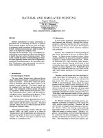
Fig. 1. Structure of S100 proteins. (A,B) Calcium-driven conformational changes at the EF-hands in S100 proteins. Structure of the N-termi-
nal, S100-specific EF-hand (A) and the C-terminal, canonical EF-hand (B) in the metal-free (lighter) and Ca
2+
-bound (darker) form of S100A6.
The EF-hand flanking helices (H
I
–H
IV
) are identified. (C,D) Structure of the human S100B homodimer loaded with Ca
2+
and Zn
2+
(T. Osten-
dorp, J. Diez, C.W. Heizmann, G. Fritz, unpublished results, 3D10). (C) Side view; (D) top view. The monomers are shown in blue and green.
The N-terminal S100-specific EF-hand (EF-hand 1) is shown in a dark colour, the C-terminal canonical EF-hand in a brighter colour (EF-hand
2). The hinges connecting both EF-hands are shown in magenta and orange. The four bound Ca
2+
ions are shown as red spheres. The two
Zn
2+
bound at the dimer interface of S100B are shown as yellow spheres. (E–G) Multimeric states of S100 proteins. S100B octamer, 2H61
(E), S100A12 hexamer, 1GQM (F) and S100A8 ⁄ A9 tetramer, 1XK4 (G). Each dimer in S100B or S100A12 is shown in an individual colour.
S100A8 is shown in red, S100A9 in blue. Bound Ca
2+
ions are shown as spheres; intersubunit Ca
2+
ions are shown as magenta spheres.
Natural and amyloid self-assembly of S100 proteins G. Fritz et al.
4580 FEBS Journal 277 (2010) 4578–4590 ª 2010 The Authors Journal compilation ª 2010 FEBS
this particular case Zn
2+
does not seem to be essential
for protein stability [33].
There is evidence for an interesting cross-talk
between Ca
2+
and Zn
2+
binding to S100 proteins,
illustrating how binding of different metal ions results
in conformational adjustments and modulation of pro-
tein folding and function. In S100B and S100A12,
Zn
2+
binding leads to an increase in Ca
2+
affinity
[37,38], whereas in S100A2 the opposite effect was
observed, i.e. Zn
2+
decreased Ca
2+
affinity, pointing
to an interplay of the metal ions in the activation of
S100 proteins [15]. For S100A12 and S100B, the
molecular mechanism of the increase in Ca
2+
affinity
by Zn
2+
can be deduced from the structural informa-
tion available (T. Ostendorp, J. Diez, C.W. Heizmann,
G. Fritz, unpublished results) [17]. In both proteins
there is one Zn
2+
coordinating His residue located in
the Ca
2+
-binding loop, which might help to stabilize
the Ca
2+
-bound conformation, thereby increasing
Ca
2+
affinity. The structure of S100A12 with only
bound Zn
2+
also shows that Zn
2+
alone can already
induce structural changes similar to those induced by
Ca
2+
, which will also lead to an increase in Ca
2+
affinity. Other Zn
2+
coordinating residues are located
in the C-terminus of the S100 proteins. Zn
2+
coordi-
nation leads to a stabilization and extension of the
C-terminal helix, changing the orientation of residues
involved in target binding. As expected from these
structural changes, Zn
2+
binding modulates target
binding properties of different S100 proteins. For
example, Zn
2+
prevents S100A8 ⁄ A9 binding to arachi-
donic acid [39]. On the other hand, Zn
2+
and Ca
2+
binding to S100A9 are both required for interaction
with receptors such as RAGE or Toll-like receptor 4
[9,13]. Similarly, Zn
2+
increased the Ca
2+
-dependent
interaction of S100A12 with RAGE [40]. In the case of
S100B, Zn
2+
alone could trigger binding to tau [41,42]
or IQGAP1 [43]. Moreover, Zn
2+
binding enhanced
the Ca
2+
-dependent interaction with AHNAK [44]
and the target protein-derived peptide TRTK-12 [45].
Recent work on the S100A2 protein, a cell cycle reg-
ulator that binds and activates p53 in a Ca
2+
-depen-
dent manner, has shown that metal ion binding
influences protein conformation and stability [7].
S100A2 binds two Ca
2+
and two Zn
2+
ions per sub-
unit, known to be associated with activation (Ca
2+
)or
inhibition (Zn
2+
) of downstream signalling. Zn
2+
binds at distinct sites that have different metal-binding
affinities, and physiologically relevant Zn
2+
concentra-
tions decrease the affinity for Ca
2+
binding, resulting
in a blockage of p53 activation. It has been recently
elicited that the S100A2 conformation is sensitive to
the metallation state, although rearrangements result-
ing from metal binding preserve the overall fold of the
protein: S100A2 is destabilized by Zn
2+
and stabilized
by Ca
2+
, suggesting a synergistic effect between the
binding of different metals. Thus, the decrease in Ca
2+
affinity through Zn
2+
is presumably a result of the
general destabilization of the protein. Further contri-
butions might come from the exposure of a hydropho-
bic surface upon Zn
2+
binding, making additional
exposure of the hydrophobic surface induced by Ca
2+
less favourable. The antagonistic effect of Zn
2+
and
Ca
2+
in the control of S100A2 stability provides a
molecular rationale for the action of both metal ions:
hypothetically, in tissues expressing S100A2, the Zn
2+
imbalance, which may arise in some types of cancer as
a result of the upregulation of Zn
2+
transporters
[46,47], may contribute to enhanced cell proliferation
through destabilization of S100A2. This would impair
the interaction with p53 and disrupt subsequent down-
stream cell cycle regulation. This further illustrates how
the binding of different metal ions to S100 proteins has
the potential to result in conformational adjustments
and modulation of protein folding and functions.
A number of S100 proteins also bind Cu
2+
(S100B
[48], S100A5 [49], S100A12 [50] and S100A13 [51]) and
this frequently occurs at the same sites to which Zn
2+
binds. That is, for example, the case in S100A12, an
important protein in the inflammatory response and a
factor in host ⁄ parasite defences, which binds Cu
2+
and Zn
2+
at the same site and corresponds to the
Zn
2+
-binding site in S100A7, evoking a possibly simi-
lar structural and functional role. S100B, one of the
most abundant proteins in the human brain, also binds
Cu
2+
, and in this case a putative neuroprotective role
was suggested.
S100 functional oligomers
Metal ions also play a crucial role in the formation of
larger oligomeric species of S100 proteins, namely tet-
ramers, hexamers and octamers. These are, in many
cases, essential for biological function and signalling:
tetrameric S100B [11] and hexameric S100A12 [52]
bind RAGE with higher affinity than the dimeric
counterparts, only multimeric S100A4 promotes neu-
rite outgrowth [53], and microtubule formation is only
promoted by the Ca
2+
-induced S100A8 ⁄ A9 tetramer
[16]. Ca
2+
-loaded S100A12 forms a functional hex-
amer whose quaternary structure is maintained by
additional interdimer bridging Ca
2+
ions, which are
coordinated by residues from the C-terminal EF-hand
and helix H
III
from two adjacent dimers. This arrange-
ment of ligands for the interdimer Ca
2+
‘cross-linker’
is only possible when the C-terminal EF-hand is in the
G. Fritz et al. Natural and amyloid self-assembly of S100 proteins
FEBS Journal 277 (2010) 4578–4590 ª 2010 The Authors Journal compilation ª 2010 FEBS 4581
Ca
2+
-bound state [50]. Similarly, two S100A8 ⁄ A9
heterodimers can assemble into a heterotetramer in a
strictly Ca
2+
-dependent manner [16]. However, the ini-
tial S100A8 ⁄ A9 heterodimer can be formed in the pres-
ence or absence of Ca
2+
. By contrast, the formation
of S100B tetramers is not dependent on Ca
2+
and the
tetramer remains stable in the absence of the metal ion
[11]. This difference may result from the additional
hydrophobic moieties found in interfaces of S100B,
which are essentially polar in S100A12 and
S100A8 ⁄ A9 [11]. Nevertheless, the presence of Ca
2+
enhances the oligomerization of S100B into hexamers
and octamers, and the octameric crystal structure
reveals intersubunit Ca
2+
ions. The oligomerization
role is not restricted to Ca
2+
, as in S100A2 binding
of Zn
2+
to the low affinity site triggers the forma-
tion of a tetramer via the assembly of two S100A2
dimers [15]. Together, these results point to a very
clear role of metal ions in the formation of func-
tional S100 oligomers. However, novel roles for non-
functional S100 oligomers are emerging with the
recent finding of metal-dependent amyloid formation
by S100A8 ⁄ A9, which will be addressed further.
Functional diversity of S100 proteins
To date a great number of distinct functions have been
attributed to S100 proteins in both the intra- and
extracellular milieu. Although S100 proteins appear to
lack enzymatic activity themselves, they play biological
roles through binding to other proteins and changing
the activity of their targets. As discussed above, the
conformation and even oligomerization state of S100s
are responsive to Ca
2+
and consequently they mediate
Ca
2+
signals by binding to other intracellular target
proteins and modulating their conformation and activ-
ity in a Ca
2+
- and possibly also in a Zn
2+
- and Cu
2+
-
dependent manner. Indeed, the assembly into multiple
complexes is considered in general as a significant gen-
eric mechanism of protein functional diversification via
varying their conformational states and associated
ligands [54]. Several S100 proteins exhibit Ca
2+
-depen-
dent interactions with metabolic enzymes (S100A1 and
S100B with aldolase C) [55], with kinases (S100B with
Ndr or Src kinases) [56,57], with cytoskeletal proteins
(S100A1 with tubulin, S100B with CapZ and S100P
with ezrin) [58–63] or with DNA-binding proteins
(S100A2, S100A4 and S100B interact with p53) [64–
66]. As a result, intracellularly S100 proteins are
involved in the regulation of the cell cycle, cell growth
and differentiation, apoptosis, migration, calcium
homeostasis, protein phosphorylation, cellular motility
and other important processes.
Some S100 proteins, including S100A4, S100A7,
S100A8 ⁄ A9, S100A11, S100A12, S100B and others,
can be secreted, exhibiting cytokine-like and chemotac-
tic activity. When S100A7, S100A8, S100A9, S100A12
or S100B are secreted in response to cell damage or
activation, they become danger signals, activating
other immune and endothelial cells. Accordingly, they
were defined as damage-associated molecular pattern
molecules in innate immunity [67,68]. The S100A8 ⁄ A9
complex accounts for up to 40% of total cytosolic pro-
teins in neutrophils and secreted S100A8 ⁄ A9 as well as
S100A12 are found at high concentrations in inflamed
tissues, producing strong proinflammatory effects.
S100A8 and S100A9 activate Toll-like receptor 4, act-
ing as innate amplifiers of inflammation and cancer
[69,70], with direct implication in metastasization [70].
Recently it was demonstrated in a mouse model that
via activation of Toll-like receptor 4, S100A8 and
S100A9 induce the development of systemic autoim-
munity [69].
S100B is highly expressed in the human brain and
actively secreted by astrocytes, neurons, microglia,
glioblastoma or Schwann cells [71]. Its extracellular
concentration reaches micromolar levels after trau-
matic brain injury and in neurodegenerative disorders
such as Alzheimer’s disease or Down’s syndrome. The
action of S100B is strongly dependent on its concen-
tration: at nanomolar levels it is neuroprotective,
whereas in the micromolar concentration range it pro-
motes apoptosis [72]. Both trophic and toxic effects
of extracellular S100B are mediated by RAGE [23]. A
large number of S100 proteins have been shown to
interact with RAGE, including S100A1, S100A2,
S100A4, S100A5, S100A6, 100A7, S100A8 ⁄ A9,
S100A11, S100A12 and S100B [22]. However, S100-
associated cell signalling may be promiscuous. This
can be best exemplified through S100A8 ⁄ A9, which
promotes RAGE-dependent cell survival [73] as well
as multiple RAGE-independent cell death pathways
[74–76].
Because of their deregulated expression, response to
stress and association with neoplastic, degenerative
and autoimmune disorders, S100 proteins gain signifi-
cant interest as potential therapeutic targets. In view
of the large number of tertiary and quaternary struc-
tures adopted by S100s and the complex structure–
functional relationship affecting their interactions with
target proteins, it is tempting to speculate that this
variability may account for the promiscuity of S100
proteins. Therefore, systematic studies of the confor-
mational changes and oligomerization of S100 proteins
will be of critical importance in the development of
potential therapeutics.
Natural and amyloid self-assembly of S100 proteins G. Fritz et al.
4582 FEBS Journal 277 (2010) 4578–4590 ª 2010 The Authors Journal compilation ª 2010 FEBS
Amyloid formation by S100A8
⁄
A9
proteins
Recently, we have found a new amyloidogenic prop-
erty of S100A8 ⁄ A9 proteins, implicating them in
another degenerative process in the ageing prostate,
specifically in amyloid deposition and tissue remodel-
ling [14]. The conversion of functional proteins and
peptides into insoluble amyloid structures and their
deposition in a variety of tissues and organs is a hall-
mark of a growing number of age-related degenerative
disorders, including Alzheimer’s and Parkinson’s dis-
eases, type II diabetes and systemic amyloidoses. Pros-
tate amyloid deposits known as corpora amylacea
belong to a type of localized amyloidoses, they are
associated with age-related prostate tissue remodelling
and occur frequently in middle-aged and elderly men.
These inclusions can vary in size from submillimetre
to a few millimetres in diameter (Fig. 2A) and can, in
some instances, constitute up to a third of the prostate
gland bulk weight. Despite their high prevalence in
later life [77], their role in prostate benign and malig-
nant changes is still disputed. The fact that proinflam-
matory S100 proteins contribute to corpora amylacea
formation elevates their role as potential cancer risk
factors. There is a growing body of evidence indicating
that inflammation is a crucial prerequisite in prostate
pathogenesis, as it is found to be associated with
40)90% of benign prostatic hyperplasia and with 20%
of all human cancers [78]. Prostate cancer is the most
common noncutaneous malignant neoplasm in men in
Western countries, affecting several million men in the
Western world, and its incidence is rising rapidly with
population ageing. Therefore, cancer risk assessment is
of critical significance in its preventing strategies.
By using mass spectrometry, gel electrophoresis and
western blot analyses, we have found that proinflam-
matory S100A8⁄ A9 proteins are persistently present in
all specimens obtained as a result of prostatectomy in
prostate cancer patients [14]. Immunohistochemical
ABC
DEF
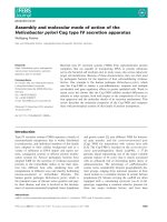
Fig. 2. Amyloid formation by S100A8 ⁄ A9 proteins in the ageing prostate. (A) Corpora amylacea deposits extracted as a result of prostatecto-
my (ruler is shown in centimetres). (B) Co-immunostaining of corpora amylacea with anti-S100A8 (shown in purple) and anti-S100A9 IgG
(shown in brown). (C) Immunostaining of corpora amylacea by antibodies towards amyloid fibrils (shown in purple). Atomic force microscopy
images of (D) ex vivo amyloid oligomers; (E) ex vivo amyloid fibrillar network and (F) amyloid fibrils produced in vitro at pH 7.4, 37 °C with
agitation. The fibril height analysis corresponds to the cross-section marked as a red line. Scale bars represent 250 nm.
G. Fritz et al. Natural and amyloid self-assembly of S100 proteins
FEBS Journal 277 (2010) 4578–4590 ª 2010 The Authors Journal compilation ª 2010 FEBS 4583
analysis of corpora amylacea revealed that they are
stained positively with both anti-S100A8 and anti-
S100A9 IgGs (Fig. 2B). Positive foci of S100A8 and
S100A9, including glandular epithelial cells and tissue
macrophages, were observed in the tissues adjacent to
corpora amylacea inclusions, indicating that the latter
infiltrate inflamed glands and ultimately lead to raising
local concentrations of S100A8 ⁄ A9. Proteinaceous
compounds constitute up to 30–40% of corpora amyl-
acea deposits, as revealed by X-ray photoelectron spec-
troscopy and FTIR, whereas the rest correspond to
inorganic components consisting of hydroxylapatite
[Ca
5
(PO4)
3
OH] and whitlockite [Ca
2
(PO4)
3
], contain-
ing high concentrations of Zn
2+
ions. The calcification
of protein deposits leads effectively to their further sta-
bilization in the protease-rich prostate fluid. The min-
eral content of corpora amylacea was rather uniform
in all seven studied patients, indicating that calcifica-
tion can be a regulated process. A recently reported
function of S100A9 is associated with promoting calci-
fication [79], suggesting that dystrophic calcification of
corpora amylacea deposits could be influenced by the
activities of S100A8⁄ A9.
Remarkably, all corpora amylacea specimens were
also stained with anti-amyloid fibril IgGs [80] (Fig. 2C)
and Congo Red dye, used as a marker for the presence
of the amyloid form of proteins, demonstrating that
the amyloid material constitutes a significant mass of
these specimens. Indeed, atomic force and transmission
electron microscopy analyses revealed a variety of
highly heterogeneous aggregates in the corpora amyla-
cea extracts (Fig. 2D, E), ranging from oligomeric spe-
cies to extensive networks of mature fibrils, which is
typical for the amyloid assemblies [81], as well as
larger-scale supramolecular assemblies, reaching a
few microns in length. Similar amyloid forms of
S100A8 ⁄ A9 were produced in vitro, providing further
insight into their amyloidogenic properties. The
S100A8 ⁄ A9 complexes, extracted from granulocytes
and produced recombinantly from Escherichia coli,
were each incubated under the native conditions of pH
7.4 and 37 °C with agitation and at pH 2.0 and 57 °C
without agitation. Under both conditions, the proteins
were assembled into heterogeneous fibrillar species. At
pH 7.4, species resembling ex vivo oligomers and short
protofilaments were formed after 2 weeks and thick
bundles of fibrils with heights of 15)20 nm and a few
microns in length constituted the major population of
fibrillar aggregates after 8 weeks of incubation
(Fig. 2F). In the S100A8 ⁄ A9 samples incubated at pH
2.0, oligomeric species and protofilaments also
emerged in 2 weeks, while after 4 weeks of incubation
flexible fibrils with a height of 4)5 nm and microns
in length together with straight and rigid fibrillar struc-
tures a few hundred nanometres long were observed,
all closely resembling the ex vivo species.
It is important to note that Ca
2+
and Zn
2+
play a
critical role in promoting amyloid assembly of
S100A8 ⁄ A9 proteins. As ex vivo corpora amylacea
deposits are calcified and contain zinc salts, these ions
can play a critical role in S100A8 ⁄ A9 amyloid forma-
tion in vivo. Indeed, after 2 weeks of incubation, the
S100A8 ⁄ A9 amyloid protofilaments of 2 nm height
were assembled in the presence of 10 mm ZnCl
2
and in
a suspension of Ca
3
(PO
4
)
2
[14], but not when EDTA
was added in solution. These species were converted
into the fibrillar assemblies after 4 weeks of incuba-
tion, and again no filamentous structures developed in
the presence of EDTA.
The bundles of amyloid fibrils of S100A8 ⁄ A9 pro-
teins, formed both in vivo and in vitro (Fig. 2F), are
among the largest reported amyloid supramolecular
species. The lateral association and thickening of the
fibrils is probably a contributing factor to their stabil-
ity in the prostate gland. It has been suggested that the
various functions of the S100A8 ⁄ A9 hetero- and
homo-oligomers may be regulated by their differential
protease sensitivity [22]. The hetero-oligomeric com-
plexes of S100A8 ⁄ A9 are characterized by significant
stability and protease resistance comparable with that
of prions. In the protease-rich environment of the
prostate gland, and especially at sites of inflammation,
where proteases are present at even higher levels, pro-
tease resistance of the S100A8 ⁄ A9 proteins could
favour their accumulation and conversion into amyloid
structures. If so, the amyloid structures formed by
S100A8 ⁄ A9 can be at the extreme end of the scale of
resistance to proteolysis.
As prostatic fluid is very rich in protein content,
small quantities of other proteins were also found in
the corpora amylacea inclusions, presumably being
trapped in the aggregating and growing deposits.
Among them, the finding of E. coli DNA and E. coli
proteins indicates that corpora amylacea formation
may be associated with bacterial infection, conse-
quently causing inflammation in surrounding tissues
during the course of corpora amylacea establishment
and growth. The identification of the highly amyloido-
genic bacterial co-chaperonin GroES can be related
not only to the fact that bacterial infection is a con-
tributory factor to inflammation, but also suggests the
potential role of bacterial infection in the initiating of
the amyloid depositions via seeding [82].
As a result, a self-perpetuating cycle can be triggered
in the ageing prostate, leading ultimately to amyloid
growth. The increasing concentration of aggregation-prone
Natural and amyloid self-assembly of S100 proteins G. Fritz et al.
4584 FEBS Journal 277 (2010) 4578–4590 ª 2010 The Authors Journal compilation ª 2010 FEBS
proteins in the sites of inflammation would favour
their amyloid assembly and deposition, as amyloid
formation is a concentration-dependent process. This
can be further promoted by the presence of calcium
and zinc salts abundant in corpora amylacea and
S100A8 ⁄ A9 in turn can themselves regulate their own
calcification. In the course of corpora amylacea
growth, neighbouring acini are obstructed, exacerbat-
ing inflammation and enhancing the risk of neoplastic
transformation. Thus, the direct involvement of proin-
flammatory S100A8 ⁄ A9 proteins in corpora amylacea
biogenesis emphasizes their role in the age-dependent
prostate remodelling and accompanied ailments.
Amyloidogenic potential of S100
proteins
The amyloidogenic potential of a protein can be esti-
mated using different algorithms that compute the
aggregation and fibrillation propensity of a particular
sequence. This approach was carried out using the
zyggregator algorithm to calculate the intrinsic
aggregation propensity scores of monomeric S100A8
and S100A9 at pH 7.0 and 2.0, the conditions of their
in vitro amyloid formation [14] (for a recent review, see
[83]). The results evidenced a rather high propensity,
comparable with that of Ab peptides, forming amyloid
deposits in Alzheimer’s disease. The overall aggrega-
tion scores for S100A8 are 0.76 at pH 7.0 and 0.77 at
pH 2.0; for S100A9, 1.04 and 0.65, and for Ab
(1–40)
and Ab
(1–42)
peptides at pH 7.0, 0.97 and 0.94, respec-
tively. In both proteins, the Ca
2+
-binding sites with
low affinity (amino acid residues 20–33 for S100A8
and 23–36 for S100A9) and high affinity (amino acid
residues 59–70 for S100A8 and 67–78 for S100A9) are
located in close proximity to the segments that are
highly aggregation prone. In the S100A8 ⁄ A9 oligo-
meric complex, however, the amyloid scores for
S100A8 and S100A9 are significantly reduced and
equal to 0.18 and 0.32, respectively, indicating that
most of the aggregation-prone sequences are involved
in native complex formation. Therefore, we surmise
that calcium-dependent native complex formation can
effectively compete under physiological conditions with
the calcium-dependent amyloid assembly, the latter
possibly being prevalent in a destabilizing environ-
ment, leading to protein partial unfolding and native
complex dissociation.
Building on these initial observations, and consider-
ing the fact that S100 proteins share a rather high
chemical and structural identity, we further addressed
the hypothesis that amyloid formation could be a gen-
eralized property among the members of the S100 pro-
tein family. For this purpose, we have carried out a
series of preliminary experiments in conditions identi-
cal to those assayed for S100A8 ⁄ A9 (pH 2 and 57 °C
[14]), to test if other S100 proteins (S100A3, S100A6,
S100A12 and S100B) would form thioflavin-T (ThT)-
reactive amyloid species (H.M. Botelho, K. Yanaman-
dra, G. Fritz, L.A. Morozova-Roche, C.M. Gomes,
manuscript in preparation). Upon incubation for 50 h,
three of the tested S100 proteins formed ThT-binding
amyloid structures that resulted in an increase in fluo-
rescence intensity of ThT, comparable with that
observed upon dye interaction with the lysozyme amy-
loids used as a positive control (Fig. 3A). Only
S100A12 did not yield ThT-reactive species under the
tested conditions. The presence of amyloid and other
precursor structures (fibres, protofibrils and disordered
aggregates) was indentified using atomic force micros-
80
120
S100 EF-hand
EF-hand
2
3
4
Z
agg
score
S100A3
S100A6
S100A12
S100B
0
Lysozyme
S100A3
S100A6
S100A12
S100B
40
ThT fluorescence
intensity (a. u.)
0 20 40 60 80 100
0
1
Amino acid position
AB
H1
H4
H2
H3
Fig. 3. Amyloidogenic potential of S100 proteins. (A) ThT fluorescence (482 nm) of S100 proteins (3 mgÆmL
)1
) after 2 days incubation at
pH 2.5, 57 °C without agitation and the positive control of lysozyme amyloid (10 mgÆmL
)1
) after 8 days. Values are mean ± standard
deviation. (B) In silico analysis of aggregation and amyloid formation propensities of selected S100 proteins. The top picture illustrates the
location of consensus S100 motifs, the thick horizontal lines indicate the regions with high (> 95%)
WALTZ score and the plot represents the
position-dependent
ZYGGREGATOR score. The horizontal dashed line indicates the significance threshold, the higher scores being significant.
The amino acid position numbering is obtained after sequence alignment.
G. Fritz et al. Natural and amyloid self-assembly of S100 proteins
FEBS Journal 277 (2010) 4578–4590 ª 2010 The Authors Journal compilation ª 2010 FEBS 4585
copy (data not shown). A complementary in silico
analysis of the aggregation propensities and amyloid-
forming sequences at pH 7 was also carried out using
zyggregator [83,84] and waltz [85] prediction tools,
respectively. The results obtained with waltz (Fig. 3B,
top) indicate that S100 proteins always contain amyloi-
dogenic segments within helices H
I
or H
IV
, or in both
helices. The aggregation propensity analysis using the
zyggregator algorithm allowed the propensity for
the formation of b-rich oligomers (Z
tox
) to be discrimi-
nated from the formation of fibrillar aggregates (Z
agg
).
The results of this analysis applied to the assayed S100
proteins revealed a similar high propensity clustering
at helices H
I
and H
IV
, although with somewhat lower
absolute values.
Together, these findings suggest that amyloid-like
conformations (b-rich oligomers, protofibrils and
fibres) might be accessible to S100 proteins under par-
ticular physiological conditions, and clearly metal ions
play a determinant role in the process (Fig. 4). It is
already established that Ca
2+
,Zn
2+
and Cu
2+
pro-
mote conformational changes within the S100 fold that
have an impact on protein stability (as in S100A2), on
the formation of functional oligomers (as in S100B)
and on the formation of amyloid fibres (as in
S100A8 ⁄ A9). Considering the latent propensity
encoded in the primary sequence of S100 proteins to
form b-rich oligomers and fibres, it is reasonable to
envisage that factors such as an imbalance in metal
homeostasis and anomalous protein–metal interactions,
inflammation, oxidative stress or⁄ and genetic muta-
tions may provide conditions in the cellular milieu that
affect any of the functional states of S100 pro-
teins (Fig. 4) and result in the formation of amyloid
structures or of its precursor oligomers in a physiologi-
cal context. One interesting aspect that remains to be
addressed and may even suggest a toxic gain of func-
tion characteristic to amyloid oligomers in general [86],
is if S100 amyloids exacerbate the apoptoptic activity
of the S100A8 ⁄ A9 complex [74–76] or interact with the
RAGE receptors, further contributing or abrogating
the toxic effects. The latter are already known to be
involved in Ab peptide amyloid transport and recogni-
tion processes in the context of Alzheimer’s disease. A
contrasting perspective can also be hypothesized:
considering that most of the S100 proteins have upregu-
lated expression patterns in inflammatory, neurodegen-
erative and malignant proliferation processes, could
amyloid formation serve as a sink for dangerous or
somehow harmful proteins promoting inflammation or
involved in cancer? Now that even Ab plaques are
viewed from a positive side [87], is it possible that the
amyloid formation of S100 proteins may potentially
play some ‘positive’ role? Future research in the com-
ing years will certainly contribute to clarify some of
these and other questions and will ultimately bring us
to a higher level of understanding the biology of
tumour and degeneration and enable to use our
acquired knowledge of S100 structure and functions in
developing strategies to modulate their activity for
therapeutic purposes.
Acknowledgements
The work described in this review was supported by
grants POCTI ⁄ QUI ⁄ 45758 and PTDC ⁄ QUI ⁄ 70101 (to
CMG) from the Fundac¸ a
˜
o para a Cieˆ ncia e a Tecnolo-
gia (FCT ⁄ MCTES, Portugal), by grants FR 1488 ⁄ 3-1
and FR 1488 ⁄ 3-1 from the Deutsche Forschungsgeme-
inschaft (DFG) (to GF). CMG and GF are recipients
of a CRUP ⁄ DAAD collaborative grant A-15 ⁄ 08.
HMB is a recipient of a PhD fellowship (SFRH ⁄ BD ⁄
31126 ⁄ 2006) from Fundac¸ a
˜
o para a Cieˆ ncia e a Tecno-
logia (FCT ⁄ MCTES, Portugal). LMR research is sup-
ported by the Swedish Medical Research Council,
Kempe Foundation, Brain Foundation and Insam-
lingsstiftelsen Sweden.
References
1 Moore BW (1965) A soluble protein characteristic of
the nervous system. Biochem Biophys Res Commun 19,
739–744.
2 Engelkamp D, Scha
¨
fer BW, Mattei MG, Erne P &
Heizmann CW (1993) Six S100 genes are clustered on
human chromosome 1q21: identification of two
genes coding for the two previously unreported
Ca
Ca
Ca
Ca
+ Ca
2+
+ M
2+
Functional
oligomers
Amyloidogenic
oligomers
Apo S100 Ca
2+
loaded Ca
2+
, M
2+
+
Zn
2+
+
Zn
2+
Ca
2+
Ca
Ca
Ca
Ca
M
Ca
Ca
Ca
Amyloid fibres
(S100A8/A9)
Protofibrils
Oligomers interacting
with target proteins
(RAGE, Toll-like receptor 4, p53)
?
Fig. 4. Native states and oligomerization pathways in S100
proteins. A scheme outlining interconversion pathways of S100
proteins, evidencing Ca
2+
and other metal (M
2+
) binding sites, and
possible routes for oligomerization pathways.
Natural and amyloid self-assembly of S100 proteins G. Fritz et al.
4586 FEBS Journal 277 (2010) 4578–4590 ª 2010 The Authors Journal compilation ª 2010 FEBS
calcium-binding proteins S100D and S100E. Proc Natl
Acad Sci USA 90, 6547–6551.
3 Scha
¨
fer BW, Wicki R, Engelkamp D, Mattei MG &
Heizmann CW (1995) Isolation of a YAC clone cover-
ing a cluster of nine S100 genes on human chromo-
some 1q21: rationale for a new nomenclature of the
S100 calcium-binding protein family. Genomics 25,
638–643.
4 Salama I, Malone PS, Mihaimeed F & Jones JL (2008)
A review of the S100 proteins in cancer. Eur J Surg
Oncol 34, 357–364.
5 Boom A, Pochet R, Authelet M, Pradier L,
Borghgraef P, Van Leuven F, Heizmann CW & Brion
JP (2004) Astrocytic calcium ⁄ zinc binding protein
S100A6 over expression in Alzheimer’s disease and in
PS1 ⁄ APP transgenic mice models. Biochim Biophys Acta
1742, 161–168.
6 Mrak RE & Griffinbc WS (2001) The role of activated
astrocytes and of the neurotrophic cytokine S100B in
the pathogenesis of Alzheimer’s disease. Neurobiol
Aging 22, 915–922.
7 Botelho HM, Koch M, Fritz G & Gomes CM (2009)
Metal ions modulate the folding and stability of the
tumor suppressor protein S100A2. FEBS J 276, 1776–
1786.
8 Heizmann CW & Cox JA (1998) New perspectives on
S100 proteins: a multi-functional Ca(2+)-, Zn(2+)-
and Cu(2+)-binding protein family. Biometals 11,
383–397.
9 Bjork P, Bjork A, Vogl T, Stenstrom M, Liberg D,
Olsson A, Roth J, Ivars F & Leanderson T (2009)
Identification of human S100A9 as a novel target for
treatment of autoimmune disease via binding to
quinoline-3-carboxamides. PLoS Biol 7, e97.
10 Leclerc E, Fritz G, Vetter SW & Heizmann CW (2009)
Binding of S100 proteins to RAGE: an update. Biochim
Biophys Acta 1793, 993–1007.
11 Ostendorp T, Leclerc E, Galichet A, Koch M, Demling
N, Weigle B, Heizmann CW, Kroneck PM & Fritz G
(2007) Structural and functional insights into RAGE
activation by multimeric S100B. EMBO J 26, 3868–
3878.
12 Donato R (2007) RAGE: a single receptor for several
ligands and different cellular responses: the case of cer-
tain S100 proteins. Curr Mol Med 7, 711–724.
13 Vogl T, Tenbrock K, Ludwig S, Leukert N, Ehrhardt
C, van Zoelen MA, Nacken W, Foell D, van der Poll
T, Sorg C et al. (2007) Mrp8 and Mrp14 are endoge-
nous activators of Toll-like receptor 4, promoting lethal,
endotoxin-induced shock. Nat Med 13, 1042–1049.
14 Yanamandra K, Alexeyev O, Zamotin V, Srivastava V,
Shchukarev A, Brorsson AC, Tartaglia GG, Vogl T,
Kayed R, Wingsle G et al. (2009) Amyloid formation
by the pro-inflammatory S100A8 ⁄ A9 proteins in the
ageing prostate. PLoS ONE 4, e5562.
15 Koch M, Bhattacharya S, Kehl T, Gimona M, Vasak
M, Chazin W, Heizmann CW, Kroneck PM & Fritz G
(2007) Implications on zinc binding to S100A2. Biochim
Biophys Acta 1773, 457–470.
16 Korndo
¨
rfer IP, Brueckner F & Skerra A (2007) The
crystal structure of the human (S100A8 ⁄ S100A9)2 het-
erotetramer, calprotectin, illustrates how conforma-
tional changes of interacting alpha-helices can
determine specific association of two EF-hand proteins.
J Mol Biol 370, 887–898.
17 Moroz OV, Blagova EV, Wilkinson AJ, Wilson KS &
Bronstein IB (2009) The crystal structures of human
S100A12 in apo form and in complex with zinc: new
insights into S100A12 oligomerisation. J Mol Biol 391,
536–551.
18 Moroz OV, Antson AA, Dodson EJ, Burrell HJ, Grist
SJ, Lloyd RM, Maitland NJ, Dodson GG, Wilson KS,
Lukanidin E et al. (2002) The structure of S100A12 in a
hexameric form and its proposed role in receptor signal-
ling. Acta Crystallogr D Biol Crystallogr 58, 407–413.
19 Marlatt NM, Boys BL, Konermann L & Shaw GS
(2009) Formation of monomeric S100B and S100A11
proteins at low ionic strength. Biochemistry 48, 1954–
1963.
20 Santamaria-Kisiel L, Rintala-Dempsey AC & Shaw GS
(2006) Calcium-dependent and -independent interactions
of the S100 protein family. Biochem J 396, 201–214.
21 Hunter MJ & Chazin WJ (1998) High level expression
and dimer characterization of the S100 EF-hand pro-
teins, migration inhibitory factor-related proteins 8 and
14. J Biol Chem 273, 12427–12435.
22 Nacken W & Kerkhoff C (2007) The hetero-oligomeric
complex of the S100A8 ⁄ S100A9 protein is extremely
protease resistant. FEBS Lett 581, 5127–5130.
23 Vogl T, Leukert N, Barczyk K, Strupat K & Roth J
(2006) Biophysical characterization of S100A8 and
S100A9 in the absence and presence of bivalent cations.
Biochim Biophys Acta 1763, 1298–1306.
24 Baudier J, Mandel P & Gerard D (1983) Bovine brain
S100 proteins: separation and characterization of a new
S100 protein species. J Neurochem 40, 145–152.
25 Yang Q, O’Hanlon D, Heizmann CW & Marks A
(1999) Demonstration of heterodimer formation
between S100B and S100A6 in the yeast two-hybrid sys-
tem and human melanoma. Exp Cell Res 246, 501–509.
26 Deloulme JC, Assard N, Mbele GO, Mangin C, Kuw-
ano R & Baudier J (2000) S100A6 and S100A11 are
specific targets of the calcium- and zinc-binding S100B
protein in vivo. J Biol Chem 275, 35302–35310.
27 Wang G, Rudland PS, White MR & Barraclough R
(2000) Interaction in vivo and in vitro of the metastasis-
inducing S100 protein, S100A4 (p9Ka) with S100A1.
J Biol Chem 275, 11141–11146.
28 Wang G, Zhang S, Fernig DG, Spiller D, Martin-
Fernandez M, Zhang H, Ding Y, Rao Z, Rudland PS
G. Fritz et al. Natural and amyloid self-assembly of S100 proteins
FEBS Journal 277 (2010) 4578–4590 ª 2010 The Authors Journal compilation ª 2010 FEBS 4587
& Barraclough R (2004) Heterodimeric interaction and
interfaces of S100A1 and S100P. Biochem J 382, 375–
383.
29 Lehmann R, Melle C, Escher N & von Eggeling F
(2005) Detection and identification of protein interac-
tions of S100 proteins by ProteinChip technology.
J Proteome Res 4, 1717–1721.
30 Vogl T, Roth J, Sorg C, Hillenkamp F & Strupat K
(1999) Calcium-induced noncovalently linked tetramers
of MRP8 and MRP14 detected by ultraviolet matrix-
assisted laser desorption ⁄ ionization mass spectrometry.
J Am Soc Mass Spectrom 10, 1124–1130. doi: S1044-
0305(99)00085-9 [pii].
31 Novitskaya V, Grigorian M, Kriajevska M,
Tarabykina S, Bronstein I, Berezin V, Bock E &
Lukanidin E (2000) Oligomeric forms of the metastasis-
related Mts1 (S100A4) protein stimulate neuronal differ-
entiation in cultures of rat hippocampal neurons. J Biol
Chem 275, 41278–41286. doi: 10.1074/jbc.M007058200
[pii].
32 Rezvanpour A, Phillips JM & Shaw GS (2009) Design
of high-affinity S100-target hybrid proteins. Protein Sci
18, 2528–2536.
33 Brodersen DE, Nyborg J & Kjeldgaard M (1999) Zinc-
binding site of an S100 protein revealed. Two crystal
structures of Ca
2+
-bound human psoriasin (S100A7) in
the Zn
2+
-loaded and Zn
2+
-free states. Biochemistry 38,
1695–1704.
34 Charpentier TH, Wilder PT, Liriano MA, Varney KM,
Pozharski E, MacKerell AD Jr, Coop A, Toth EA &
Weber DJ (2008) Divalent metal ion complexes of
S100B in the absence and presence of pentamidine.
J Mol Biol 382, 56–73. doi: S0022-2836(08)00763-8 [pii]
10.1016/j.jmb.2008.06.047.
35 Koch M, Diez J & Fritz G (2008) Crystal structure of
Ca
2+
-free S100A2 at 1.6 A resolution. J Mol Biol 378,
933–942.
36 Glaser R, Harder J, Lange H, Bartels J, Christophers E
& Schroder JM (2005) Antimicrobial psoriasin
(S100A7) protects human skin from Escherichia coli
infection. Nat Immunol 6 , 57–64.
37 Baudier J, Glasser N & Gerard D (1986) Ions binding
to S100 proteins. I. Calcium- and zinc-binding proper-
ties of bovine brain S100 alpha alpha, S100a (alpha
beta), and S100b (beta beta) protein: Zn
2+
regulates
Ca
2+
binding on S100b protein. J Biol Chem 261,
8192–8203.
38 Dell’Angelica EC, Schleicher CH & Santome JA (1994)
Primary structure and binding properties of calgranulin
C, a novel S100-like calcium-binding protein from pig
granulocytes. J Biol Chem 269, 28929–28936.
39 Kerkhoff C, Vogl T, Nacken W, Sopalla C & Sorg C
(1999) Zinc binding reverses the calcium-induced arachi-
donic acid-binding capacity of the S100A8 ⁄ A9 protein
complex. FEBS Lett 460, 134–138.
40 Moroz OV, Burkitt W, Wittkowski H, He W, Ianoul A,
Novitskaya V, Xie J, Polyakova O, Lednev IK, Shekht-
man A et al. (2009) Both Ca
2+
and Zn
2+
are essential
for S100A12 protein oligomerization and function.
BMC Biochem 10, 11.
41 Baudier J & Cole RD (1988) Interactions between the
microtubule-associated tau proteins and S100b regulate
tau phosphorylation by the Ca2+ ⁄ calmodulin-depen-
dent protein kinase II. J Biol Chem 263, 5876–5883.
42 Yu WH & Fraser PE (2001) S100beta interaction with
tau is promoted by zinc and inhibited by hyperphosph-
orylation in Alzheimer’s disease. J Neurosci 21, 2240–
2246.
43 Mbele GO, Deloulme JC, Gentil BJ, Delphin C, Ferro
M, Garin J, Takahashi M & Baudier J (2002) The zinc-
and calcium-binding S100B interacts and co-localizes
with IQGAP1 during dynamic rearrangement of cell
membranes. J Biol Chem 277, 49998–50007.
44 Gentil BJ, Delphin C, Mbele GO, Deloulme JC, Ferro
M, Garin J & Baudier J (2001) The giant protein AH-
NAK is a specific target for the calcium- and zinc-bind-
ing S100B protein: potential implications for Ca2+
homeostasis regulation by S100B. J Biol Chem 276,
23253–23261.
45 Barber KR, McClintock KA, Jamieson GA Jr, Dimlich
RV & Shaw GS (1999) Specificity and Zn
2+
enhance-
ment of the S100B binding epitope TRTK-12. J Biol
Chem 274, 1502–1508.
46 Ionescu JG, Novotny J, Stejskal V, Latsch A, Blau-
rock-Busch E & Eisenmann-Klein M (2006) Increased
levels of transition metals in breast cancer tissue. Neuro
Endocrinol Lett 27(Suppl. 1), 36–39.
47 Li M, Zhang Y, Liu Z, Bharadwaj U, Wang H, Wang
X, Zhang S, Liuzzi JP, Chang SM, Cousins RJ et al.
(2007) Aberrant expression of zinc transporter ZIP4
(SLC39A4) significantly contributes to human pancre-
atic cancer pathogenesis and progression. Proc Natl
Acad Sci USA 104, 18636–18641.
48 Nishikawa T, Lee IS, Shiraishi N, Ishikawa T, Ohta Y
& Nishikimi M (1997) Identification of S100b protein
as copper-binding protein and its suppression of cop-
per-induced cell damage. J Biol Chem 272, 23037–
23041.
49 Schafer BW, Fritschy JM, Murmann P, Troxler H,
Durussel I, Heizmann CW & Cox JA (2000) Brain
S100A5 is a novel calcium-, zinc-, and copper ion-binding
protein of the EF-hand superfamily. J Biol Chem 275,
30623–30630. doi: 10.1074/jbc.M002260200 [pii].
50 Moroz OV, Antson AA, Grist SJ, Maitland NJ,
Dodson GG, Wilson KS, Lukanidin E & Bronstein IB
(2003) Structure of the human S100A12–copper
complex: implications for host-parasite defence. Acta
Crystallogr D Biol Crystallogr 59, 859–867.
51 Landriscina M, Bagala C, Mandinova A, Soldi R,
Micucci I, Bellum S, Prudovsky I & Maciag T (2001)
Natural and amyloid self-assembly of S100 proteins G. Fritz et al.
4588 FEBS Journal 277 (2010) 4578–4590 ª 2010 The Authors Journal compilation ª 2010 FEBS
Copper induces the assembly of a multiprotein aggre-
gate implicated in the release of fibroblast growth factor
1 in response to stress. J Biol Chem 276, 25549–25557.
doi: 10.1074/jbc.M102925200 [pii].
52 Xie J, Burz DS, He W, Bronstein IB, Lednev I &
Shekhtman A (2007) Hexameric calgranulin C
(S100A12) binds to the receptor for advanced glycated
end products (RAGE) using symmetric hydrophobic
target-binding patches. J Biol Chem 282, 4218–4231.
53 Kiryushko D, Novitskaya V, Soroka V, Klingelhofer J,
Lukanidin E, Berezin V & Bock E (2006) Molecular
mechanisms of Ca(2+) signaling in neurons induced by
the S100A4 protein. Mol Cell Biol 26, 3625–3638.
54 Mossberg A, Mok KH, Morozova-Roche LA &
Svanborg C (2010) Structure and function of human
a-lactalbumin made lethal to tumor cells (HAMLET)-
type complexes. FEBS J 277, 4614–4625.
55 Zimmer DB & Van Eldik LJ (1986) Identification of a
molecular target for the calcium-modulated protein
S100. Fructose-1,6-bisphosphate aldolase. J Biol Chem
261, 11424–11428.
56 Brozzi F, Arcuri C, Giambanco I & Donato R (2009)
S100B protein regulates astrocyte shape and migration
via interaction with Src kinase: implications for astro-
cyte development, activation, and tumor growth. J Biol
Chem 284, 8797–8811.
57 Millward TA, Heizmann CW, Schafer BW &
Hemmings BA (1998) Calcium regulation of Ndr
protein kinase mediated by S100 calcium-binding
proteins. EMBO J 17, 5913–5922.
58 Garbuglia M, Verzini M, Rustandi RR, Osterloh D,
Weber DJ, Gerke V & Donato R (1999) Role of the
C-terminal extension in the interaction of S100A1 with
GFAP, tubulin, the S100A1- and S100B-inhibitory pep-
tide, TRTK-12, and a peptide derived from p53, and
the S100A1 inhibitory effect on GFAP polymerization.
Biochem Biophys Res Commun 254, 36–41.
59 Donato R (1987) Quantitative analysis of the interac-
tion between S-100 proteins and brain tubulin. Cell Cal-
cium 8, 283–297.
60 Kilby PM, Van Eldik LJ & Roberts GC (1997) Identifi-
cation of the binding site on S100B protein for the actin
capping protein CapZ. Protein Sci 6, 2494–2503.
61 Wright NT, Cannon BR, Wilder PT, Morgan MT,
Varney KM, Zimmer DB & Weber DJ (2009) Solution
structure of S100A1 bound to the CapZ peptide
(TRTK12). J Mol Biol 386, 1265–1277.
62 Austermann J, Nazmi AR, Heil A, Fritz G, Kolinski
M, Filipek S & Gerke V (2009) Generation and charac-
terization of a novel, permanently active S100P mutant.
Biochim Biophys Acta 1793, 1078–1085.
63 Koltzscher M, Neumann C, Konig S & Gerke V
(2003) Ca2+-dependent binding and activation of
dormant ezrin by dimeric S100P. Mol Biol Cell 14,
2372–2384.
64 Baudier J, Delphin C, Grunwald D, Khochbin S &
Lawrence JJ (1992) Characterization of the tumor sup-
pressor protein p53 as a protein kinase C substrate and
a S100b-binding protein. Proc Natl Acad Sci USA 89,
11627–11631.
65 Mueller A, Schafer BW, Ferrari S, Weibel M, Makek M,
Hochli M & Heizmann CW (2005) The calcium-binding
protein S100A2 interacts with p53 and modulates its
transcriptional activity. J Biol Chem 280, 29186–29193.
66 Grigorian M, Andresen S, Tulchinsky E, Kriajevska M,
Carlberg C, Kruse C, Cohn M, Ambartsumian N,
Christensen A, Selivanova G et al. (2001) Tumor
suppressor p53 protein is a new target for the
metastasis-associated Mts1 ⁄ S100A4 protein: functional
consequences of their interaction. J Biol Chem 276,
22699–22708.
67 Ehrchen JM, Sunderkotter C, Foell D, Vogl T & Roth
J (2009) The endogenous Toll-like receptor 4 agonist
S100A8 ⁄ S100A9 (calprotectin) as innate amplifier of
infection, autoimmunity, and cancer.
J Leukoc Biol 86,
557–566. doi: jlb.1008647 [pii] 10.1189/jlb.1008647.
68 Foell D, Wittkowski H, Vogl T & Roth J (2007) S100
proteins expressed in phagocytes: a novel group of
damage-associated molecular pattern molecules.
J Leukoc Biol 81, 28–37. doi: jlb.0306170 [pii] 10.1189/
jlb.0306170.
69 Loser K, Vogl T, Voskort M, Lueken A, Kupas V,
Nacken W, Klenner L, Kuhn A, Foell D, Sorokin L et al.
(2010) The Toll-like receptor 4 ligands Mrp8 and Mrp14
are crucial in the development of autoreactive CD8+ T
cells. Nat Med 16, 713–717. doi:10.1038/nm.2150.
70 Hiratsuka S, Watanabe A, Sakurai Y, Akashi-Takamura
S, Ishibashi S, Miyake K, Shibuya M, Akira S, Aburatani
H & Maru Y (2008) The S100A8-serum amyloid
A3-TLR4 paracrine cascade establishes a pre-metastatic
phase. Nat Cell Biol 10, 1349–1355. doi: ncb1794 [pii]
10.1038/ncb1794.
71 Donato R, Sorci G, Riuzzi F, Arcuri C, Bianchi R,
Brozzi F, Tubaro C & Giambanco I (2009) S100B’s
double life: intracellular regulator and extracellular sig-
nal. Biochim Biophys Acta 1793, 1008–1022. doi: S0167-
4889(08)00409-6 [pii] 10.1016/j.bbamcr.2008.11.009.
72 Businaro R, Leone S, Fabrizi C, Sorci G, Donato R,
Lauro GM & Fumagalli L (2006) S100B protects
LAN-5 neuroblastoma cells against Abeta amyloid-
induced neurotoxicity via RAGE engagement at low
doses but increases Abeta amyloid neurotoxicity at high
doses. J Neurosci Res 83, 897–906. doi: 10.1002/
jnr.20785.
73 Turovskaya O, Foell D, Sinha P, Vogl T, Newlin R,
Nayak J, Nguyen M, Olsson A, Nawroth PP, Bierhaus
A et al. (2008) RAGE, carboxylated glycans and
S100A8 ⁄ A9 play essential roles in colitis-associated car-
cinogenesis. carcinogenesis 29, 2035–2043. doi: bgn188
[pii] 10.1093/carcin/bgn188.
G. Fritz et al. Natural and amyloid self-assembly of S100 proteins
FEBS Journal 277 (2010) 4578–4590 ª 2010 The Authors Journal compilation ª 2010 FEBS 4589
74 Ghavami S, Kerkhoff C, Chazin WJ, Kadkhoda K,
Xiao W, Zuse A, Hashemi M, Eshraghi M, Schulze-
Osthoff K, Klonisch T et al. (2008) S100A8 ⁄ 9 induces
cell death via a novel, RAGE-independent pathway that
involves selective release of Smac ⁄ DIABLO and
Omi ⁄ HtrA2. Biochim Biophys Acta 1783, 297–311.
doi: S0167-4889(07)00248-0 [pii] 10.1016/j.bbamcr.2007.
10.015.
75 Viemann D, Barczyk K, Vogl T, Fischer U,
Sunderkotter C, Schulze-Osthoff K & Roth J (2007)
MRP8 ⁄ MRP14 impairs endothelial integrity and
induces a caspase-dependent and -independent cell
death program. Blood 109, 2453–2460. doi: blood-2006-
08-040444 [pii] 10.1182/blood-2006-08-040444.
76 Ghavami S, Eshragi M, Ande SR, Chazin WJ, Klonisch
T, Halayko AJ, McNeill KD, Hashemi M, Kerkhoff C
& Los M (2010) S100A8 ⁄ A9 induces autophagy and
apoptosis via ROS-mediated cross-talk between mito-
chondria and lysosomes that involves BNIP3. Cell Res
20, 314–331. doi: cr2009129 [pii] 10.1038/cr.2009.129.
77 Cross PA, Bartley CJ & McClure J (1992) Amyloid in
prostatic corpora amylacea. J Clin Pathol 45, 894–897.
78 De Marzo AM, Platz EA, Sutcliffe S, Xu J, Gronberg
H, Drake CG, Nakai Y, Isaacs WB & Nelson WG
(2007) Inflammation in prostate carcinogenesis. Nat Rev
Cancer 7, 256–269.
79 McCormick MM, Rahimi F, Bobryshev YV, Gaus K,
Zreiqat H, Cai H, Lord RS & Geczy CL (2005) S100A8
and S100A9 in human arterial wall. Implications for
atherogenesis. J Biol Chem 280, 41521–41529.
80 Kayed R, Head E, Sarsoza F, Saing T, Cotman CW,
Necula M, Margol L, Wu J, Breydo L, Thompson JL
et al. (2007) Fibril specific, conformation dependent
antibodies recognize a generic epitope common to
amyloid fibrils and fibrillar oligomers that is absent in
prefibrillar oligomers. Mol Neurodegener 2, 18.
81 Pedersen JS, Andersen CB & Otzen DE (2010) Amyloid
structure – one but not the same: the many levels of
fibrillar polymorphism. FEBS J 277, 4591–4601.
82 Otzen D & Nielsen PH (2008) We find them here, we
find them there: functional bacterial amyloid. Cell Mol
Life Sci 65, 910–927.
83 Tartaglia GG & Vendruscolo M (2008) The Zyggrega-
tor method for predicting protein aggregation propensi-
ties. Chem Soc Rev 37, 1395–1401.
84 Tartaglia GG, Pawar AP, Campioni S, Dobson CM,
Chiti F & Vendruscolo M (2008) Prediction of aggrega-
tion-prone regions in structured proteins. J Mol Biol
380, 425–436.
85 Maurer-Stroh S, Debulpaep M, Kuemmerer N, de la
Paz ML, Martins IC, Reumers J, Morris KL, Copland
A, Serpell L, Serrano L et al. (2010) Exploring the
sequence determinants of amyloid structure using posi-
tion-specific scoring matrices. Nat Methods 7, 237–242.
86 Stefani M (2010) Biochemical and biophysical features
of both oligomer ⁄ fibril and cell membrane in amyloid
cytotoxicity. FEBS J 277, 4602–4613.
87 Heo C, Chang KA, Choi HS, Kim HS, Kim S, Liew H,
Kim JA, Yu E, Ma J & Suh YH (2007) Effects of the
monomeric, oligomeric, and fibrillar Abeta42 peptides
on the proliferation and differentiation of adult neural
stem cells from subventricular zone. J Neurochem 102,
493–500. doi: JNC4499 [pii] 10.1111/j.1471-4159.2007.
04499.x.
Natural and amyloid self-assembly of S100 proteins G. Fritz et al.
4590 FEBS Journal 277 (2010) 4578–4590 ª 2010 The Authors Journal compilation ª 2010 FEBS